Electrospun Antimicrobial Films of Poly(3-hydroxybutyrate-co-3-hydroxyvalerate) Containing Eugenol Essential Oil Encapsulated in Mesoporous Silica Nanoparticles

 , , , ,
  and
and 
Abstract
:
1. Introduction
2. Materials and Methods
2.1. Materials
2.2. Synthesis and Complexation of MCM-41
2.2.1. Synthesis of MCM-41
2.2.2. Eugenol Complexation on MCM-41
2.3. Electrospinning Process
2.4. Film Preparation
2.5. Characterization
2.5.1. Electron Microscopy
2.5.2. Thermal Analysis
2.5.3. Mechanical Tests
2.5.4. Permeability Tests
2.6. Antimicrobial Assays
3. Results
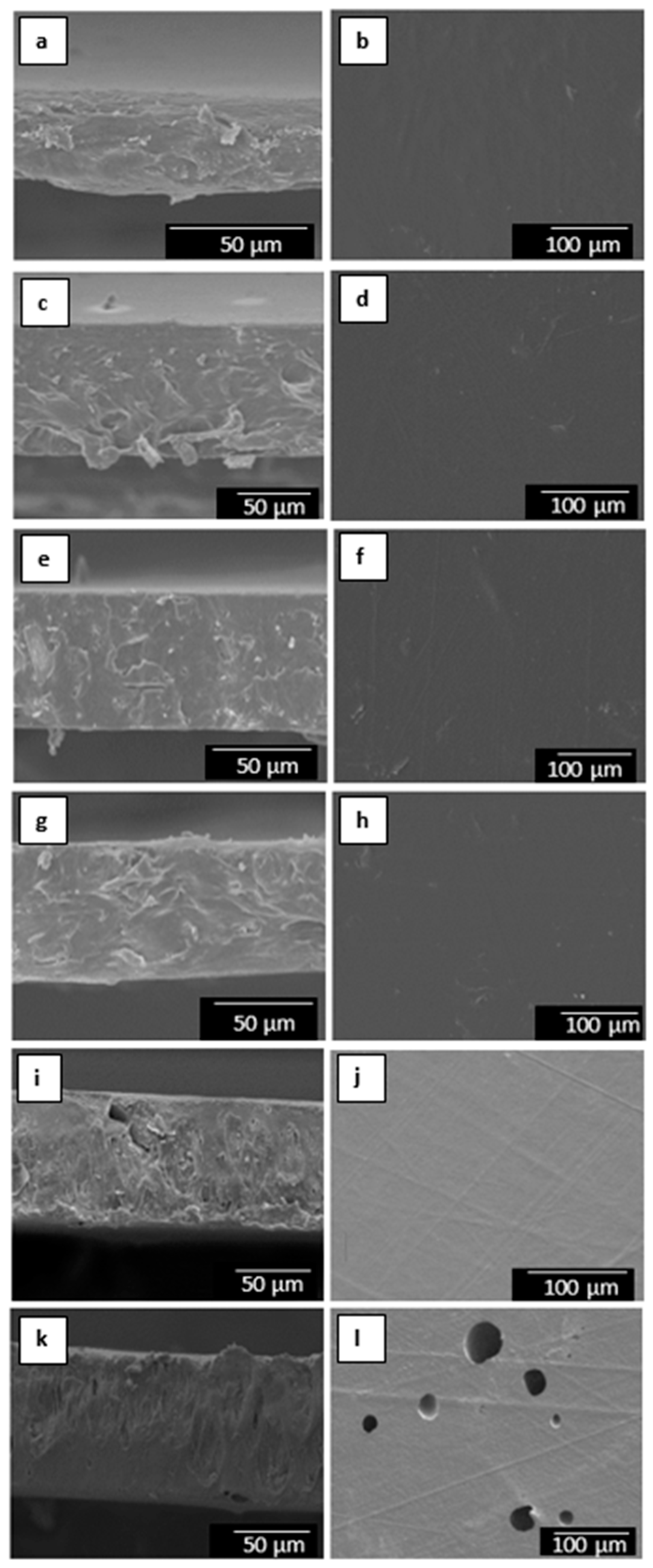
3.1. Morphology
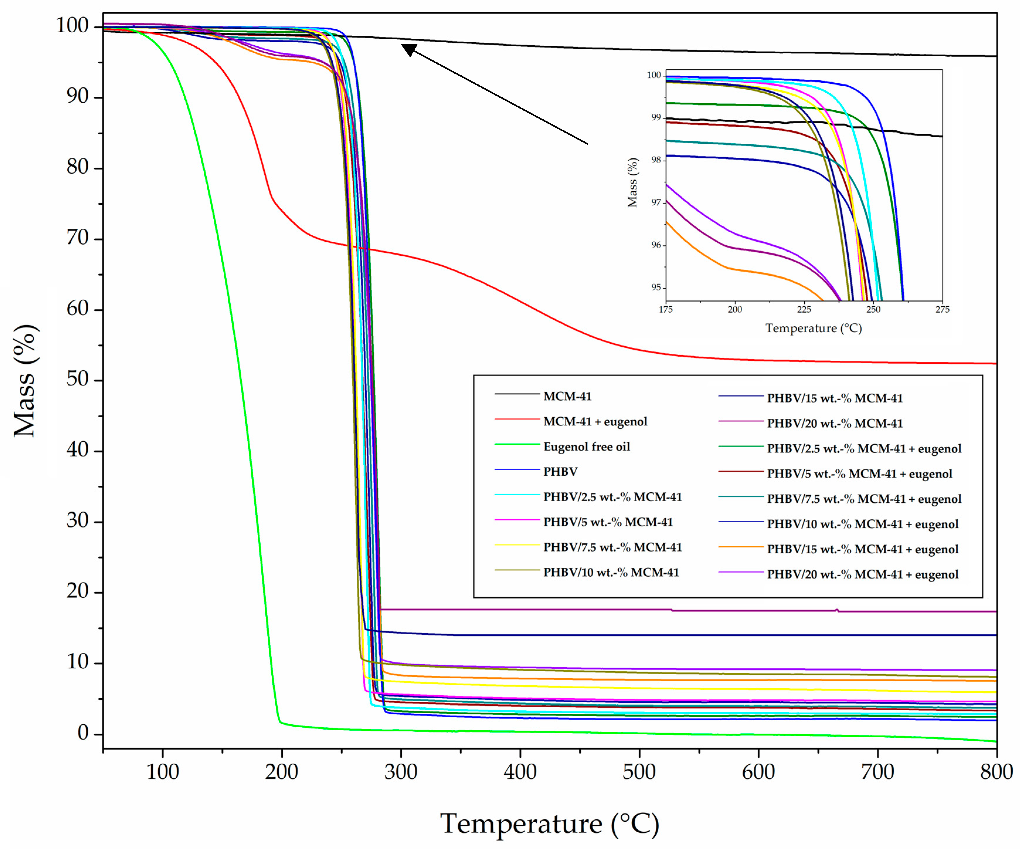
3.2. Thermal Properties
3.3. Mechanical Properties
3.4. Barrier properties
3.5. Antimicrobial activity
4. Conclusions
Author Contributions
Funding
Acknowledgments
Conflicts of Interest
References
- Torres-Giner, S.; Montanes, N.; Fombuena, V.; Boronat, T.; Sanchez-Nacher, L. Preparation and characterization of compression-molded green composite sheets made of poly(3-hydroxybutyrate) reinforced with long pita fibers. Adv. Polym. Technol. 2018, 37, 1305–1315. [Google Scholar] [CrossRef]
- Reddy, C.S.K.; Ghai, R.; Rashmi; Kalia, V.C. Polyhydroxyalkanoates: An overview. Bioresour. Technol. 2003, 87, 137–146. [Google Scholar] [CrossRef]
- Keshavarz, T.; Roy, I. Polyhydroxyalkanoates: Bioplastics with a green agenda. Curr. Opin. Microbiol. 2010, 13, 321–326. [Google Scholar] [CrossRef] [PubMed]
- Lee, S.Y. Plastic bacteria? Progress and prospects for polyhydroxyalkanoate production in bacteria. Trends Biotechnol. 1996, 14, 431–438. [Google Scholar] [CrossRef]
- Choi, J.I.; Lee, S.Y. Process analysis and economic evaluation for poly(3-hydroxybutyrate) production by fermentation. Bioprocess Eng. 1997, 17, 335–342. [Google Scholar] [CrossRef]
- Díez-Pascual, A.M.; Díez-Vicente, A.L. ZnO-reinforced poly(3-hydroxybutyrate-co-3-hydroxyvalerate) bionanocomposites with antimicrobial function for food packaging. ACS Appl. Mater. Interfaces 2014, 6, 9822–9834. [Google Scholar] [CrossRef] [PubMed]
- Torres-Giner, S.; Montanes, N.; Boronat, T.; Quiles-Carrillo, L.; Balart, R. Melt grafting of sepiolite nanoclay onto poly(3-hydroxybutyrate-co-4-hydroxybutyrate) by reactive extrusion with multi-functional epoxy-based styrene-acrylic oligomer. Eur. Polym. J. 2016, 84, 693–707. [Google Scholar] [CrossRef]
- Khosravi-Darani, K.; Bucci, D.Z. Application of poly(hydroxyalkanoate) in food packaging: Improvements by nanotechnology. Chem. Biochem. Eng. Q. 2015, 29, 275–285. [Google Scholar] [CrossRef]
- Kulkarni, S.O.; Kanekar, P.P.; Jog, J.P.; Patil, P.A.; Nilegaonkar, S.S.; Sarnaik, S.S.; Kshirsagar, P.R. Characterisation of copolymer, poly (hydroxybutyrate-co-hydroxyvalerate) (PHB-co-PHV) produced by halomonas campisalis (MCM B-1027), its biodegradability and potential application. Bioresour. Technol. 2011, 102, 6625–6628. [Google Scholar] [CrossRef]
- Keskin, G.; Kızıl, G.; Bechelany, M.; Pochat-Bohatier, C.; Öner, M. Potential of polyhydroxyalkanoate (PHA) polymers family as substitutes of petroleum based polymers for packaging applications and solutions brought by their composites to form barrier materials. Pure Appl. Chem. 2017, 89, 1841–1848. [Google Scholar] [CrossRef]
- Philip, S.; Keshavarz, T.; Roy, I. Polyhydroxyalkanoates: Biodegradable polymers with a range of applications. J. Chem. Technol. Biotechnol. 2007, 82, 233–247. [Google Scholar] [CrossRef]
- Robertson, G.L. Food Packaging: Principles and Practice, 3rd ed.; Taylor & Francis: Boca Raton, FL, USA, 2012. [Google Scholar]
- Requena, R.; Vargas, M.; Chiralt, A. Release kinetics of carvacrol and eugenol from poly(hydroxybutyrate-co-hydroxyvalerate) (PHBV) films for food packaging applications. Eur. Polym. J. 2017, 92, 185–193. [Google Scholar] [CrossRef]
- Torres-Giner, S.; Hilliou, L.; Melendez-Rodriguez, B.; Figueroa-Lopez, K.J.; Madalena, D.; Cabedo, L.; Covas, J.A.; Vicente, A.A.; Lagaron, J.M. Melt processability, characterization, and antibacterial activity of compression-molded green composite sheets made of poly(3-hydroxybutyrate-co-3-hydroxyvalerate) reinforced with coconut fibers impregnated with oregano essential oil. Food Packag. Shelf Life 2018, 17, 39–49. [Google Scholar] [CrossRef]
- Li, D.; Xia, Y. Electrospinning of Nanofibers: Reinventing the Wheel? Adv. Mater. 2004, 16, 1151–1170. [Google Scholar] [CrossRef]
- Torres-Giner, S. Electrospun nanofibers for food packaging applications. In Multifunctional and Nanoreinforced Polymers for Food Packaging; Lagaron, J.M., Ed.; Woodhead Publishing Ltd.: Cambridge, UK, 2011; pp. 108–125. [Google Scholar]
- Torres-Giner, S.; Busolo, M.; Cherpinski, A.; Lagaron, J.M. Electrospinning in the packaging industry. In Electrospinning: From Basic Research to Commercialization; Kny, E., Ghosal, K., Thomas, S., Eds.; The Royal Society of Chemistry: Cambridge, UK, 2018; pp. 238–260. [Google Scholar]
- Torres-Giner, S.; Pérez-Masiá, R.; Lagaron Jose, M. A review on electrospun polymer nanostructures as advanced bioactive platforms. Polym. Eng. Sci. 2016, 56, 500–527. [Google Scholar] [CrossRef]
- Torres-Giner, S.; Wilkanowicz, S.; Melendez-Rodriguez, B.; Lagaron, J.M. Nanoencapsulation of aloe vera in synthetic and naturally occurring polymers by electrohydrodynamic processing of interest in food technology and bioactive packaging. J. Agric. Food Chem. 2017, 65, 4439–4448. [Google Scholar] [CrossRef] [PubMed]
- Torres-Giner, S. Novel antimicrobials obtained by electrospinning methods. In Antimicrobial Polymers; John Wiley & Sons, Inc.: Hoboken, NJ, USA, 2011; pp. 261–285. [Google Scholar]
- Spagnol, C.; Fragal, E.H.; Pereira, A.G.B.; Nakamura, C.V.; Muniz, E.C.; Follmann, H.D.M.; Silva, R.; Rubira, A.F. Cellulose nanowhiskers decorated with silver nanoparticles as an additive to antibacterial polymers membranes fabricated by electrospinning. J. Colloid Interface Sci. 2018, 531, 705–715. [Google Scholar] [CrossRef] [PubMed]
- Hu, M.; Li, C.; Li, X.; Zhou, M.; Sun, J.; Sheng, F.; Shi, S.; Lu, L. Zinc oxide/silver bimetallic nanoencapsulated in PVP/PCL nanofibres for improved antibacterial activity. Artif. Cells Nanomed. Biotechnol. 2018, 46, 1248–1257. [Google Scholar] [CrossRef] [PubMed]
- An, J.; Zhang, H.; Zhang, J.; Zhao, Y.; Yuan, X. Preparation and antibacterial activity of electrospun chitosan/poly(ethylene oxide) membranes containing silver nanoparticles. Colloid Polym. Sci. 2009, 287, 1425–1434. [Google Scholar] [CrossRef]
- Castro-Mayorga, J.L.; Fabra, M.J.; Cabedo, L.; Lagaron, J.M. On the use of the electrospinning coating technique to produce antimicrobial polyhydroxyalkanoate materials containing in situ-stabilized silver nanoparticles. Nanomaterials 2017, 7, 4. [Google Scholar] [CrossRef]
- Castro Mayorga, J.L.; Fabra Rovira, M.J.; Cabedo Mas, L.; Sánchez Moragas, G.; Lagarón Cabello, J.M. Antimicrobial nanocomposites and electrospun coatings based on poly(3-hydroxybutyrate-co-3-hydroxyvalerate) and copper oxide nanoparticles for active packaging and coating applications. J. Appl. Polym. Sci. 2018, 135, 45673. [Google Scholar] [CrossRef]
- Burt, S. Essential oils: Their antibacterial properties and potential applications in foods—A review. Int. J. Food Microbiol. 2004, 94, 223–253. [Google Scholar] [CrossRef] [PubMed]
- Lang, G.; Buchbauer, G. A review on recent research results (2008–2010) on essential oils as antimicrobials and antifungals. A review. Flavour Fragr. J. 2012, 27, 13–39. [Google Scholar] [CrossRef]
- Da Silva, F.F.M.; Monte, F.J.Q.; de Lemos, T.L.G.; do Nascimento, P.G.G.; de Medeiros Costa, A.K.; de Paiva, L.M.M. Eugenol derivatives: Synthesis, characterization, and evaluation of antibacterial and antioxidant activities. Chem. Cent. J. 2018, 12, 34. [Google Scholar] [CrossRef] [PubMed]
- Wieczyńska, J.; Cavoski, I. Antimicrobial, antioxidant and sensory features of eugenol, carvacrol and trans-anethole in active packaging for organic ready-to-eat iceberg lettuce. Food Chem. 2018, 259, 251–260. [Google Scholar] [CrossRef] [PubMed]
- Majeed, H.; Bian, Y.-Y.; Ali, B.; Jamil, A.; Majeed, U.; Khan, Q.F.; Iqbal, K.J.; Shoemaker, C.F.; Fang, Z. Essential oil encapsulations: Uses, procedures, and trends. RSC Adv. 2015, 5, 58449–58463. [Google Scholar] [CrossRef]
- Kailasapathy, K. Encapsulation technologies for functional foods and nutraceutical product development. CAB Rev. Perspect. Agric. Vet. Sci. Nutr. Nat. Resour. 2009, 4, 1–19. [Google Scholar] [CrossRef]
- Kresge, C.T.; Leonowicz, M.E.; Roth, W.J.; Vartuli, J.C.; Beck, J.S. Ordered mesoporous molecular sieves synthesized by a liquid-crystal template mechanism. Nature 1992, 359, 710–712. [Google Scholar] [CrossRef]
- Vallet-Regi, M.; Rámila, A.; Del Real, R.P.; Pérez-Pariente, J. A new property of MCM-41: Drug delivery system. Chem. Mater. 2001, 13, 308–311. [Google Scholar] [CrossRef]
- He, D.; He, X.; Wang, K.; Zou, Z.; Yang, X.; Li, X. Remote-controlled drug release from graphene oxide-capped mesoporous silica to cancer cells by photoinduced pH-jump activation. Langmuir 2014, 30, 7182–7189. [Google Scholar] [CrossRef]
- Muñoz, B.; Rámila, A.; Pérez-Pariente, J.; Díaz, I.; Vallet-Regí, M. MCM-41 organic modification as drug delivery rate regulator. Chem. Mater. 2003, 15, 500–503. [Google Scholar] [CrossRef]
- Bernardos, A.; Marina, T.; Žáček, P.; Pérez-Esteve, É.; Martínez-Mañez, R.; Lhotka, M.; Kouřimská, L.; Pulkrábek, J.; Klouček, P. Antifungal effect of essential oil components against aspergillus niger when loaded into silica mesoporous supports. J. Sci. Food Agric. 2015, 95, 2824–2831. [Google Scholar] [CrossRef] [PubMed]
- Fan, J.; Yu, C.; Gao, F.; Lei, J.; Tian, B.; Wang, L.; Luo, Q.; Tu, B.; Zhou, W.; Zhao, D. Cubic mesoporous silica with large controllable entrance sizes and advanced adsorption properties. Angew. Chem. Int. Ed. 2003, 42, 3146–3150. [Google Scholar] [CrossRef]
- Ruiz-Rico, M.; Fuentes, C.; Pérez-Esteve, É.; Jiménez-Belenguer, A.I.; Quiles, A.; Marcos, M.D.; Martínez-Máñez, R.; Barat, J.M. Bactericidal activity of caprylic acid entrapped in mesoporous silica nanoparticles. Food Control 2015, 56, 77–85. [Google Scholar] [CrossRef]
- Janatova, A.; Bernardos, A.; Smid, J.; Frankova, A.; Lhotka, M.; Kourimská, L.; Pulkrabek, J.; Kloucek, P. Long-term antifungal activity of volatile essential oil components released from mesoporous silica materials. Ind. Crop. Prod. 2015, 67, 216–220. [Google Scholar] [CrossRef]
- Ribes, S.; Ruiz-Rico, M.; Pérez-Esteve, É.; Fuentes, A.; Talens, P.; Martínez-Máñez, R.; Barat, J.M. Eugenol and thymol immobilised on mesoporous silica-based material as an innovative antifungal system: Application in strawberry jam. Food Control 2017, 81, 181–188. [Google Scholar] [CrossRef]
- Ruiz-Rico, M.; Pérez-Esteve, É.; Bernardos, A.; Sancenón, F.; Martínez-Máñez, R.; Marcos, M.D.; Barat, J.M. Enhanced antimicrobial activity of essential oil components immobilized on silica particles. Food Chem. 2017, 233, 228–236. [Google Scholar] [CrossRef] [PubMed]
- Popova, M.; Lazarova, H.; Trusheva, B.; Popova, M.; Bankova, V.; Mihály, J.; Najdenski, H.; Tsvetkova, I.; Szegedi, Á. Nanostructured silver silica materials as potential propolis carriers. Microporous Mesoporous Mater. 2018, 263, 28–33. [Google Scholar] [CrossRef]
- Chatterjee, D.; Bhattacharjee, P. Comparative evaluation of the antioxidant efficacy of encapsulated and un-encapsulated eugenol-rich clove extracts in soybean oil: Shelf-life and frying stability of soybean oil. J. Food Eng. 2013, 117, 545–550. [Google Scholar] [CrossRef]
- Estela, C.; Pilar, C.; Dolores, M.M.; Ramón, M.M.; Félix, S.; Juan, S. Selective chromofluorogenic sensing of heparin by using functionalised silica nanoparticles containing binding sites and a signalling reporter. Chem. Eur. J. 2009, 15, 1816–1820. [Google Scholar]
- Cherpinski, A.; Torres-Giner, S.; Vartiainen, J.; Peresin, M.S.; Lahtinen, P.; Lagaron, J.M. Improving the water resistance of nanocellulose-based films with polyhydroxyalkanoates processed by the electrospinning coating technique. Cellulose 2018, 25, 1291–1307. [Google Scholar] [CrossRef]
- Torres-Giner, S.; Torres, A.; Ferrándiz, M.; Fombuena, V.; Balart, R. Antimicrobial activity of metal cation-exchanged zeolites and their evaluation on injection-molded pieces of bio-based high-density polyethylene. J. Food Saf. 2017, 37, 1–12. [Google Scholar] [CrossRef]
- Beck, J.S.; Vartuli, J.C.; Roth, W.J.; Leonowicz, M.E.; Kresge, C.T.; Schmitt, K.D.; Chu, C.T.W.; Olson, D.H.; Sheppard, E.W.; McCullen, S.B.; et al. A new family of mesoporous molecular sieves prepared with liquid crystal templates. J. Am. Chem. Soc. 1992, 114, 10834–10843. [Google Scholar] [CrossRef]
- Alfredsson, V.; Keung, M.; Monnier, A.; Stucky, G.D.; Unger, K.K.; Schüth, F. High-resolution transmission electron microscopy of mesoporous MCM-41 type materials. J. Chem. Soc. Chem. Commun. 1994, 921–922. [Google Scholar] [CrossRef]
- Ravikovitch, P.I.; O’Domhnaill, S.C.; Neimark, A.V.; Schiith, F.; Unger, K.K. Capillary hysteresis in nanopores: Theoretical and experimental studies of nitrogen adsorption on MCM-41. Langmuir 1995, 11, 4765–4772. [Google Scholar] [CrossRef]
- Sayed, E.; Karavasili, C.; Ruparelia, K.; Haj-Ahmad, R.; Charalambopoulou, G.; Steriotis, T.; Giasafaki, D.; Cox, P.; Singh, N.; Giassafaki, L.-P.N.; et al. Electrosprayed mesoporous particles for improved aqueous solubility of a poorly water soluble anticancer agent: In vitro and ex vivo evaluation. J. Control. Release 2018, 278, 142–155. [Google Scholar] [CrossRef] [PubMed]
- Torres-Giner, S.; Lagaron, J.M. Zein-based ultrathin fibers containing ceramic nanofillers obtained by electrospinning. I. Morphology and thermal properties. J. Appl. Polym. Sci. 2010, 118, 778–789. [Google Scholar] [CrossRef]
- Torres-Giner, S.; Martinez-Abad, A.; Lagaron, J.M. Zein-based ultrathin fibers containing ceramic nanofillers obtained by electrospinning. II. Mechanical properties, gas barrier, and sustained release capacity of biocide thymol in multilayer polylactide films. J. Appl. Polym. Sci. 2014, 131, 9270–9276. [Google Scholar] [CrossRef]
- Cherpinski, A.; Gozutok, M.; Sasmazel, H.; Torres-Giner, S.; Lagaron, J. Electrospun oxygen scavenging films of poly(3-hydroxybutyrate) containing palladium nanoparticles for active packaging applications. Nanomaterials 2018, 8, 469. [Google Scholar] [CrossRef]
- Cherpinski, A.; Torres-Giner, S.; Cabedo, L.; Lagaron, J.M. Post-processing optimization of electrospun submicron poly(3-hydroxybutyrate) fibers to obtain continuous films of interest in food packaging applications. Food Addit. Contam. Part A 2017, 34, 1817–1830. [Google Scholar] [CrossRef]
- Melendez-Rodriguez, B.; Castro-Mayorga, J.L.; Reis, M.A.M.; Sammon, C.; Cabedo, L.; Torres-Giner, S.; Lagaron, J.M. Preparation and characterization of electrospun food biopackaging films of poly(3-hydroxybutyrate-co-3-hydroxyvalerate) derived from fruit pulp biowaste. Front. Sustain. Food Syst. 2018, 2, 38. [Google Scholar] [CrossRef]
- Muratore, F.; Martini, R.E.; Barbosa, S.E. Bioactive paper by eugenol grafting onto cellulose. Effect of reaction variables. Food Packag. Shelf Life 2018, 15, 159–168. [Google Scholar] [CrossRef]
- Torres-Giner, S.; Montanes, N.; Fenollar, O.; García-Sanoguera, D.; Balart, R. Development and optimization of renewable vinyl plastisol/wood flour composites exposed to ultraviolet radiation. Mater. Des. 2016, 108, 648–658. [Google Scholar] [CrossRef]
- Fernandes Nassar, S.; Dombre, C.; Gastaldi, E.; Touchaleaume, F.; Chalier, P. Soy protein isolate nanocomposite film enriched with eugenol, an antimicrobial agent: Interactions and properties. J. Appl. Polym. Sci. 2017, 135, 45941. [Google Scholar] [CrossRef]
- Narayanan, A.; Neera; Mallesha; Ramana, K.V. Synergized antimicrobial activity of eugenol incorporated polyhydroxybutyrate films against food spoilage microorganisms in conjunction with pediocin. Appl. Biochem. Biotechnol. 2013, 170, 1379–1388. [Google Scholar] [CrossRef] [PubMed]
- Ju, C.; Kim, T.; Kang, H. Renewable, eugenol—Modified polystyrene layer for liquid crystal orientation. Polymers 2018, 10, 201. [Google Scholar] [CrossRef]
- Loganathan, S.; Jacob, J.; Valapa, R.B.; Thomas, S. Influence of linear and branched amine functionalization in mesoporous silica on the thermal, mechanical and barrier properties of sustainable poly(lactic acid) biocomposite films. Polymer 2018, 148, 149–157. [Google Scholar] [CrossRef]
- Garrido-Miranda, K.A.; Rivas, B.L.; Pérez-Rivera, M.A.; Sanfuentes, E.A.; Peña-Farfal, C. Antioxidant and antifungal effects of eugenol incorporated in bionanocomposites of poly(3-hydroxybutyrate)-thermoplastic starch. LWT 2018, 98, 260–267. [Google Scholar] [CrossRef]
- Woranuch, S.; Yoksan, R. Eugenol-loaded chitosan nanoparticles: II. Application in bio-based plastics for active packaging. Carbohydr. Polym. 2013, 96, 586–592. [Google Scholar] [CrossRef] [PubMed]
- Fang, Z.; Bhandari, B. Encapsulation of polyphenols—A review. Trends Food Sci. Technol. 2010, 21, 510–523. [Google Scholar] [CrossRef]
- Requena, R.; Jiménez, A.; Vargas, M.; Chiralt, A. Poly[(3-hydroxybutyrate)-co-(3-hydroxyvalerate)] active bilayer films obtained by compression moulding and applying essential oils at the interface. Polym. Int. 2016, 65, 883–891. [Google Scholar] [CrossRef]
- Rivero, S.; García, M.A.; Pinotti, A. Composite and bi-layer films based on gelatin and chitosan. J. Food Eng. 2009, 90, 531–539. [Google Scholar] [CrossRef]
- Voon, H.C.; Bhat, R.; Easa, A.M.; Liong, M.T.; Karim, A.A. Effect of addition of halloysite nanoclay and SiO2 nanoparticles on barrier and mechanical properties of bovine gelatin films. Food Bioprocess Technol. 2012, 5, 1766–1774. [Google Scholar] [CrossRef]
- Jia, X.; Li, Y.; Cheng, Q.; Zhang, S.; Zhang, B. Preparation and properties of poly(vinyl alcohol)/silica nanocomposites derived from copolymerization of vinyl silica nanoparticles and vinyl acetate. Eur. Polym. J. 2007, 43, 1123–1131. [Google Scholar] [CrossRef]
- Tang, S.; Zou, P.; Xiong, H.; Tang, H. Effect of nano-SiO2 on the performance of starch/polyvinyl alcohol blend films. Carbohydr. Polym. 2008, 72, 521–526. [Google Scholar] [CrossRef]
- Quiles-Carrillo, L.; Montanes, N.; Lagaron, J.M.; Balart, R.; Torres-Giner, S. In situ compatibilization of biopolymer ternary blends by reactive extrusion with low-functionality epoxy-based styrene–acrylic oligomer. J. Polym. Environ. 2019, 27, 84–96. [Google Scholar] [CrossRef]
- Nielsen, L.E. Models for the permeability of filled polymer systems. J. Macromol. Sci. Part A Chem. 1967, 1, 929–942. [Google Scholar] [CrossRef]
- Sanchez-Garcia, M.D.; Gimenez, E.; Lagaron, J.M. Morphology and barrier properties of solvent cast composites of thermoplastic biopolymers and purified cellulose fibers. Carbohydr. Polym. 2008, 71, 235–244. [Google Scholar] [CrossRef]
- Sanchez-Garcia, M.D.; Gimenez, E.; Lagaron, J.M. Novel pet nanocomposites of interest in food packaging applications and comparative barrier performance with biopolyester nanocomposites. J. Plast. Film Sheeting 2007, 23, 133–148. [Google Scholar] [CrossRef]
- Hashemi Tabatabaei, R.; Jafari, S.M.; Mirzaei, H.; Mohammadi Nafchi, A.; Dehnad, D. Preparation and characterization of nano-SiO2 reinforced gelatin-k-carrageenan biocomposites. Int. J. Biol. Macromol. 2018, 111, 1091–1099. [Google Scholar] [CrossRef] [PubMed]
- Hassannia-Kolaee, M.; Khodaiyan, F.; Pourahmad, R.; Shahabi-Ghahfarrokhi, I. Development of ecofriendly bionanocomposite: Whey protein isolate/pullulan films with nano-SiO2. Int. J. Biol. Macromol. 2016, 86, 139–144. [Google Scholar] [CrossRef] [PubMed]
- Tongnuanchan, P.; Benjakul, S.; Prodpran, T. Properties and antioxidant activity of fish skin gelatin film incorporated with citrus essential oils. Food Chem. 2012, 134, 1571–1579. [Google Scholar] [CrossRef] [PubMed]
- Aguirre, A.; Borneo, R.; León, A.E. Antimicrobial, mechanical and barrier properties of triticale protein films incorporated with oregano essential oil. Food Biosci. 2013, 1, 2–9. [Google Scholar] [CrossRef]
- Atarés, L.; De Jesús, C.; Talens, P.; Chiralt, A. Characterization of SPI-based edible films incorporated with cinnamon or ginger essential oils. J. Food Eng. 2010, 99, 384–391. [Google Scholar] [CrossRef]
- Atarés, L.; Chiralt, A. Essential oils as additives in biodegradable films and coatings for active food packaging. Trends Food Sci. Technol. 2016, 48, 51–62. [Google Scholar] [CrossRef]
- Torres-Giner, S.; Gil, L.; Pascual-Ramírez, L.; Garde-Belza, J. Packaging: Food waste reduction. In Encyclopedia of Polymer Applications; Mishra, M., Ed.; CRC Press: Boca Raton, FL, USA, 2018; Volume 3, pp. 1990–2009. [Google Scholar]
- Exner, M.; Bhattacharya, S.; Christiansen, B.; Gebel, J.; Goroncy-Bermes, P.; Hartemann, P.; Heeg, P.; Ilschner, C.; Kramer, A.; Larson, E.; et al. Antibiotic resistance: What is so special about multidrug-resistant gram-negative bacteria? GMS Hyg. Infect. Control 2017, 12, Doc05. [Google Scholar] [PubMed]
- Castro-Mayorga, J.L.; Fabra, M.J.; Pourrahimi, A.M.; Olsson, R.T.; Lagaron, J.M. The impact of zinc oxide particle morphology as an antimicrobial and when incorporated in poly(3-hydroxybutyrate-co-3-hydroxyvalerate) films for food packaging and food contact surfaces applications. Food Bioprod. Process. 2017, 101, 32–44. [Google Scholar] [CrossRef]
- Figueroa-Lopez, K.J.; Vicente, A.A.; Reis, M.A.M.; Torres-Giner, S.; Lagaron, J.M. Antimicrobial and antioxidant performance of various essential oils and natural extracts and their incorporation into biowaste derived poly(3-hydroxybutyrate-co-3-hydroxyvalerate) layers made from electrospun ultrathin fibers. Nanomaterials 2019, 9, 144. [Google Scholar] [CrossRef]
- Li, Z.; Zhou, P.; Zhou, F.; Zhao, Y.; Ren, L.; Yuan, X. Antimicrobial eugenol-loaded electrospun membranes of poly(ε-caprolactone)/gelatin incorporated with REDV for vascular graft applications. Colloids Surf. B Biointerfaces 2018, 162, 335–344. [Google Scholar] [CrossRef] [PubMed]
- Park, S.-Y.; Barton, M.; Pendleton, P. Mesoporous silica as a natural antimicrobial carrier. Colloids Surf. A Physicochem. Eng. Asp. 2011, 385, 256–261. [Google Scholar] [CrossRef]


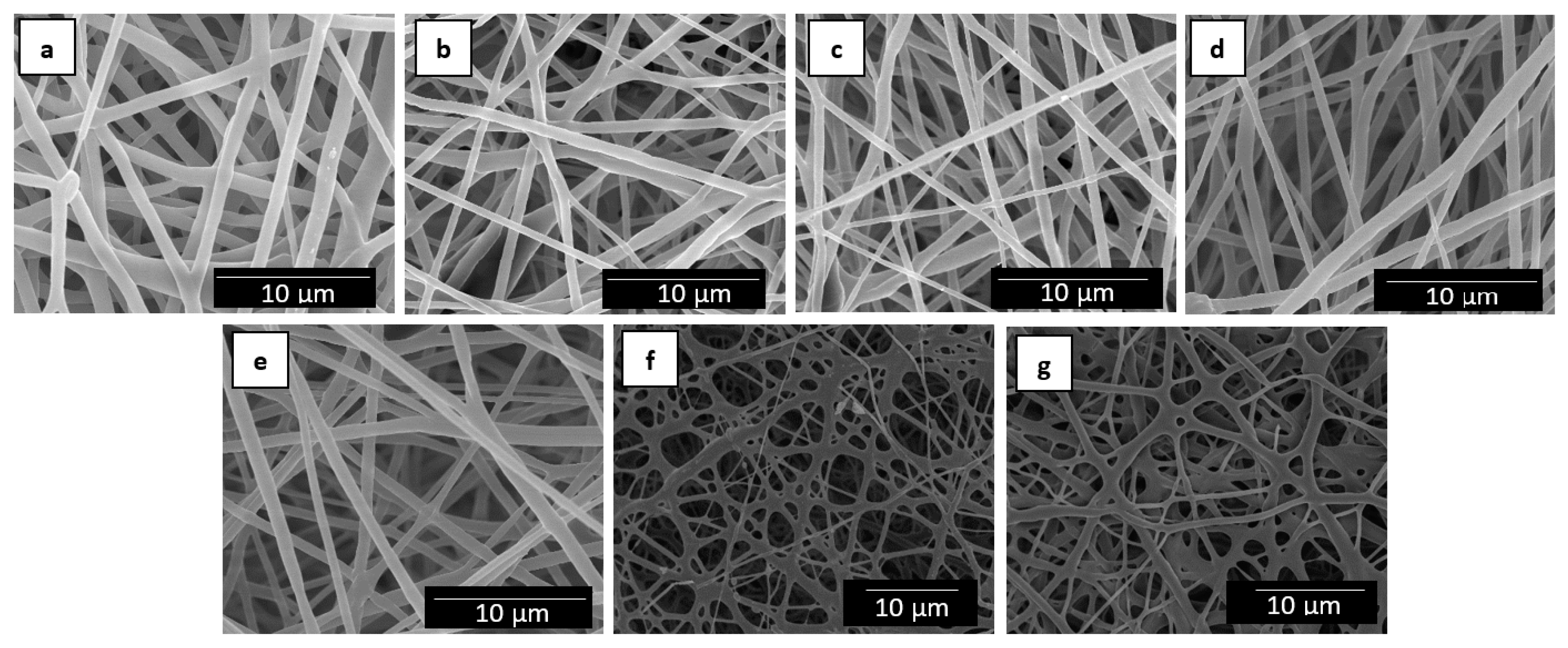
| Fibers | Diameter (µm) |
|---|---|
| PHBV | 0.89 ± 0.30 |
| PHBV/2.5 wt.-% MCM-41 + eugenol | 0.65 ± 0.19 |
| PHBV/5 wt.-% MCM-41 + eugenol | 0.66 ± 0.16 |
| PHBV/7.5 wt.-% MCM-41 + eugenol | 0.63 ± 0.18 |
| PHBV/10 wt.-% MCM-41 + eugenol | 0.64 ± 0.19 |
| PHBV/15 wt.-% MCM-41 + eugenol | 0.65 ± 0.19 |
| PHBV/20 wt.-% MCM-41 + eugenol | 0.67 ± 0.24 |
| Film | Tg (°C) | Tc (°C) | Tm (°C) | ΔHm (J/g) |
|---|---|---|---|---|
| PHBV | 2.6 ± 0.4 | 116.8 ± 0.5 | 170.4 ± 0.2 | 83.2 ± 3.0 |
| PHBV/2.5 wt.-% MCM-41 | 2.6 ± 0.2 | 117.8 ± 0.6 | 169.9 ± 0.1 | 85.7 ± 0.5 |
| PHBV/5 wt.-% MCM-41 | 2.3 ± 0.5 | 118.1 ± 0.4 | 170.1 ± 0.6 | 86.2 ± 4.0 |
| PHBV/7.5 wt.-% MCM-41 | 2.4 ± 0.4 | 118.2 ± 0.2 | 171.7 ± 2.1 | 87.4 ± 7.3 |
| PHBV/10 wt.-% MCM-41 | 2.3 ± 0.3 | 118.9 ± 0.1 | 170.2 ± 0.6 | 89.7 ± 6.3 |
| PHBV/15 wt.-% MCM-41 | 2.2 ± 0.2 | 120.5 ± 0.3 | 170.1 ± 1.0 | 100.6 ± 7.4 |
| PHBV/20 wt.-% MCM-41 | 2.5 ± 0.1 | 116.9 ± 0.1 | 169.0 ± 0.1 | 67.9 ± 5.9 |
| PHBV/2.5 wt.-% MCM-41 + eugenol | 1.8 ± 0.6 | 116.7 ± 0.1 | 169.0 ± 1.6 | 77.4 ± 3.4 |
| PHBV/5 wt.-% MCM-41 + eugenol | 1.4 ± 0.8 | 116.4 ± 0.6 | 167.4 ± 0.3 | 74.9 ± 6.2 |
| PHBV/7.5 wt.-% MCM-41 + eugenol | 1.5 ± 0.3 | 118.5 ± 0.3 | 168.8 ± 3.6 | 72.3 ± 6.9 |
| PHBV/10 wt.-% MCM-41 + eugenol | 1.3 ± 0.4 | 114.9 ± 0.7 | 165.4 ± 0.4 | 66.8 ± 4.8 |
| PHBV/15 wt.-% MCM-41 + eugenol | 0.9 ± 0.5 | 113.9 ± 0.2 | 163.4 ± 0.3 | 50.3 ± 4.5 |
| PHBV/20 wt.-% MCM-41 + eugenol | 0.6 ± 0.2 | 113.8 ± 0.6 | 160.5 ± 1.5/168.6 ± 0.1 | 47.4 ± 2.1 |
| Sample | T5% (°C) | Tdeg (°C) | Mass Loss (%) | Residual Mass (%) |
|---|---|---|---|---|
| MCM-41 powder | - | - | - | 95.0 ± 2.3 |
| MCM-41 with eugenol powder | 143.7 ± 2.4 | 178.3 ± 0.7 | 16.3 ± 0.5 | 52.4 ± 1.9 |
| Eugenol free oil | 105.9 ± 3.2 | 185.5 ± 0.6 | 80.6 ± 1.1 | 0.9 ± 0.1 |
| PHBV | 259.9 ± 1.2 | 277.3 ± 0.6 | 62.0 ± 0.8 | 2.0 ± 0.2 |
| PHBV/2.5 wt.-% MCM-41 | 250.8 ± 2.3 | 270.9 ± 0.2 | 74.4 ± 1.2 | 3.3 ± 0.4 |
| PHBV/5 wt.-% MCM-41 | 245.3 ± 2.7 | 265.4 ± 1.0 | 72.1 ± 0.6 | 5.4 ± 0.8 |
| PHBV/7.5 wt.-% MCM-41 | 245.3 ± 2.2 | 265.4 ± 0.8 | 70.3 ± 0.3 | 8.0 ± 0.4 |
| PHBV/10 wt.-% MCM-41 | 240.7 ± 1.8 | 262.7 ± 0.5 | 70.8 ± 0.2 | 9.1 ± 1.0 |
| PHBV/15 wt.-% MCM-41 | 230.0 ± 2.0 | 262.0 ± 0.2 | 70.2 ± 0.7 | 14.6 ± 0.9 |
| PHBV/20 wt.-% MCM-41 | 215.2 ± 1.7 | 256.4 ± 0.6 | 80.5 ± 0.9 | 17.2 ± 0.2 |
| PHBV/2.5 wt.-% MCM-41 + eugenol | 259.8 ± 2.6 | 281.0 ± 2.5 | 71.3 ± 0.3 | 2.5 ± 0.4 |
| PHBV/5 wt.-% MCM-41 + eugenol | 251.7 ± 1.4 | 276.4 ± 1.6 | 71.4 ± 0.3 | 3.4 ± 0.7 |
| PHBV/7.5 wt.-% MCM-41 + eugenol | 248.0 ± 2.7 | 273.7 ± 0.9 | 72.8 ± 0.4 | 3.8 ± 0.8 |
| PHBV/10 wt.-% MCM-41 + eugenol | 247.1 ± 3.2 | 271.8 ± 0.7 | 73.9 ± 0.7 | 4.3 ± 1.0 |
| PHBV/15 wt.-% MCM-41 + eugenol | 215.2 ± 7.3 | 261.0 ± 4.2 | 74.6 ± 1.4 | 7.5 ± 3.6 |
| PHBV/20 wt.-% MCM-41 + eugenol | 205.1 ± 5.1 | 259.2 ± 4.4 | 75.6 ± 1.2 | 9.1 ± 3.7 |
| Film | (MPa) | (%) | T (mJ/m3) | |
|---|---|---|---|---|
| PHBV | 1252 ± 79 | 18.1 ± 2.1 | 2.4 ± 0.3 | 0.3 ± 0.1 |
| PHBV/2.5 wt.-% MCM-41 + eugenol | 1735 ± 60 | 27.0 ± 2.4 | 2.4 ± 0.1 | 0.4 ± 0.1 |
| PHBV/5 wt.-% MCM-41 + eugenol | 1976 ± 162 | 25.1 ± 7.8 | 1.7 ± 0.4 | 0.2 ± 0.1 |
| PHBV/7.5 wt.-% MCM-41 + eugenol | 2000 ± 365 | 27.7 ± 5.4 | 2.2 ± 0.2 | 0.4 ± 0.1 |
| PHBV/10 wt.-% MCM-41 + eugenol | 1802 ± 288 | 21.7 ± 5.8 | 1.8 ± 0.2 | 0.2 ± 0.1 |
| PHBV/15 wt.-% MCM-41 + eugenol | 1702 ± 140 | 29.4 ± 3.4 | 2.0 ± 0.3 | 0.3 ± 0.1 |
| PHBV/20 wt.-% MCM-41 + eugenol | 1462 ± 358 | 25.1 ± 5.4 | 2.1 ± 0.5 | 0.3 ± 0.2 |
| Sample | WVP × 10−14 (kg·m·m−2·Pa−1·s−1) | LP × 10−14 (kg·m·m−2·Pa−1·s−1) |
|---|---|---|
| PHBV | 5.34 ± 1.79 | 2.68 ± 1.82 |
| PHBV/2.5 wt.-% MCM-41 + eugenol | 8.68 ± 3.57 | 3.41 ± 0.97 |
| PHBV/5 wt.-% MCM-41 + eugenol | 8.84 ± 4.36 | 3.49 ± 1.17 |
| PHBV/7.5 wt.-% MCM-41 + eugenol | 4.25 ± 4.04 | 3.51 ± 0.54 |
| PHBV/10 wt.-% MCM-41 + eugenol | 2.99 ± 0.95 | 2.32 ± 0.68 |
| PHBV/15 wt.-% MCM-41 + eugenol | 0.25 ± 0.19 | 0.38 ± 0.20 |
| PHBV/20 wt.-% MCM-41 + eugenol | 4.08 ± 1.98 | 4.66 ± 2.91 |
| Films | Initial | After 15 Days | ||
|---|---|---|---|---|
| Bacterial Counts [log (CFU/mL)] | R | Bacterial Counts [log (CFU/mL)] | R | |
| Control day 0 | 5.75 ± 0.09 | - | 5.75 ± 0.09 | - |
| Control 24 h | 5.67 ± 0.07 | - | 5.68 ± 0.03 | - |
| PHBV | 5.39 ± 0.56 | 0.28 ± 0.52 | 5.29 ± 0.41 | 0.38 ± 0.38 |
| PHBV/2.5 wt.-% MCM-41 | 4.86 ± 0.54 | 0.81 ± 0.58 | 5.06 ± 0.48 | 0.61 ± 0.46 |
| PHBV/2.5 wt.-% MCM-41 + eugenol | 4.33 ± 0.35 | 1.04 ± 0.39 | 4.75 ± 0.09 | 0.92 ± 0.12 |
| PHBV/5 wt.-% MCM-41 | 5.47 ± 0.58 | 0.20 ± 0.65 | 5.51 ± 0.09 | 0.16 ± 0.06 |
| PHBV/5 wt.-% MCM-41 + eugenol | 4.60 ± 0.23 | 1.07 ± 0.23 | 4.69 ± 0.14 | 0.99 ± 0.14 |
| PHBV/7.5 wt.-% MCM-41 | 5.77 ± 0.07 | 0.10 ± 0.01 | 5.44 ± 0.55 | 0.24 ± 0.57 |
| PHBV/7.5 wt.-% MCM-41 + eugenol | 4.55 ± 0.06 | 1.12 ± 0.11 | 4.55 ± 0.12 | 1.12 ± 0.15 |
| PHBV/10 wt.-% MCM-41 | 5.98 ± 0.57 | 0.31 ± 0.06 | 4.70 ± 0.06 | 0.97 ± 0.09 |
| PHBV/10 wt.-% MCM-41 + eugenol | 4.43 ± 0.24 | 1.23 ± 0.20 | 4.55 ± 0.07 | 1.24 ± 0.10 |
| Films | Initial | After 15 Days | ||
|---|---|---|---|---|
| Bacterial Counts [log (CFU/mL)] | R | Bacterial Counts [log (CFU/mL)] | R | |
| Control day 0 | 5.76 ± 0.01 | - | 5.71 ± 0.02 | - |
| Control 24 h | 6.81 ± 0.01 | - | 6.80 ± 0.02 | - |
| PHBV | 5.99 ± 0.07 | 0.82 ± 0.01 | 6.08 ± 0.03 | 0.72 ± 0.05 |
| PHBV/15 wt.-% MCM-41 | 6.41 ± 0.01 | 0.40 ± 0.03 | 6.15 ± 0.04 | 0.65 ± 0.06 |
| PHBV/15 wt.-% MCM-41 + eugenol | 5.51 ± 0.02 | 1.30 ± 0.02 | 5.40 ± 0.01 | 1.40 ± 0.06 |
| Films | Initial | After 15 Days | ||
|---|---|---|---|---|
| Bacterial Counts [log (CFU/mL)] | R | Bacterial Counts [log (CFU/mL)] | R | |
| Control day 0 | 5.61 ± 0.03 | - | 5.65 ± 0.01 | - |
| Control 24 h | 6.82 ± 0.06 | - | 6.85 ± 0.01 | - |
| PHBV | 6.23 ± 0.08 | 0.59 ± 0.01 | 6.11 ± 0.03 | 0.74 ± 0.05 |
| PHBV/10 wt.-% MCM-41 | 6.30 ± 0.01 | 0.52 ± 0.03 | 6.09 ± 0.04 | 0.76 ± 0.06 |
| PHBV/10 wt.-% MCM-41 + eugenol | 5.47 ± 0.01 | 1.35 ± 0.15 | 5.21 ± 0.01 | 1.64 ± 0.09 |
| Films | Initial | After 15 Days | ||
|---|---|---|---|---|
| Bacterial Counts [log (CFU/mL)] | R | Bacterial Counts [log (CFU/mL)] | R | |
| Control day 0 | 5.68 ± 0.03 | - | 5.66 ± 0.06 | - |
| Control 24 h | 6.83 ± 0.01 | - | 6.60 ± 0.01 | - |
| PHBV | 6.10 ± 0.01 | 0.73 ± 0.01 | 6.11 ± 0.03 | 0.49 ± 0.04 |
| PHBV/15 wt.-% MCM-41 | 6.24 ± 0.01 | 0.59 ± 0.03 | 6.26 ± 0.06 | 0.34 ± 0.01 |
| PHBV/15 wt.-% MCM-41 + eugenol | 5.49 ± 0.03 | 1.34 ± 0.03 | 5.02 ± 0.07 | 1.58 ± 0.01 |
© 2019 by the authors. Licensee MDPI, Basel, Switzerland. This article is an open access article distributed under the terms and conditions of the Creative Commons Attribution (CC BY) license (http://creativecommons.org/licenses/by/4.0/).
Share and Cite
Melendez-Rodriguez, B.; Figueroa-Lopez, K.J.; Bernardos, A.; Martínez-Máñez, R.; Cabedo, L.; Torres-Giner, S.; M. Lagaron, J. Electrospun Antimicrobial Films of Poly(3-hydroxybutyrate-co-3-hydroxyvalerate) Containing Eugenol Essential Oil Encapsulated in Mesoporous Silica Nanoparticles. Nanomaterials 2019, 9, 227. https://0-doi-org.brum.beds.ac.uk/10.3390/nano9020227
Melendez-Rodriguez B, Figueroa-Lopez KJ, Bernardos A, Martínez-Máñez R, Cabedo L, Torres-Giner S, M. Lagaron J. Electrospun Antimicrobial Films of Poly(3-hydroxybutyrate-co-3-hydroxyvalerate) Containing Eugenol Essential Oil Encapsulated in Mesoporous Silica Nanoparticles. Nanomaterials. 2019; 9(2):227. https://0-doi-org.brum.beds.ac.uk/10.3390/nano9020227
Chicago/Turabian StyleMelendez-Rodriguez, Beatriz, Kelly J. Figueroa-Lopez, Andrea Bernardos, Ramón Martínez-Máñez, Luis Cabedo, Sergio Torres-Giner, and Jose M. Lagaron. 2019. "Electrospun Antimicrobial Films of Poly(3-hydroxybutyrate-co-3-hydroxyvalerate) Containing Eugenol Essential Oil Encapsulated in Mesoporous Silica Nanoparticles" Nanomaterials 9, no. 2: 227. https://0-doi-org.brum.beds.ac.uk/10.3390/nano9020227