
Neurocognitive Profiles of Caucasian Moyamoya Disease Patients in Greece: A Case Series

 , , , ,
Abstract
:1. Introduction
2. Materials and Methods
2.1. Sample
2.2. Neuropsychological Assessment
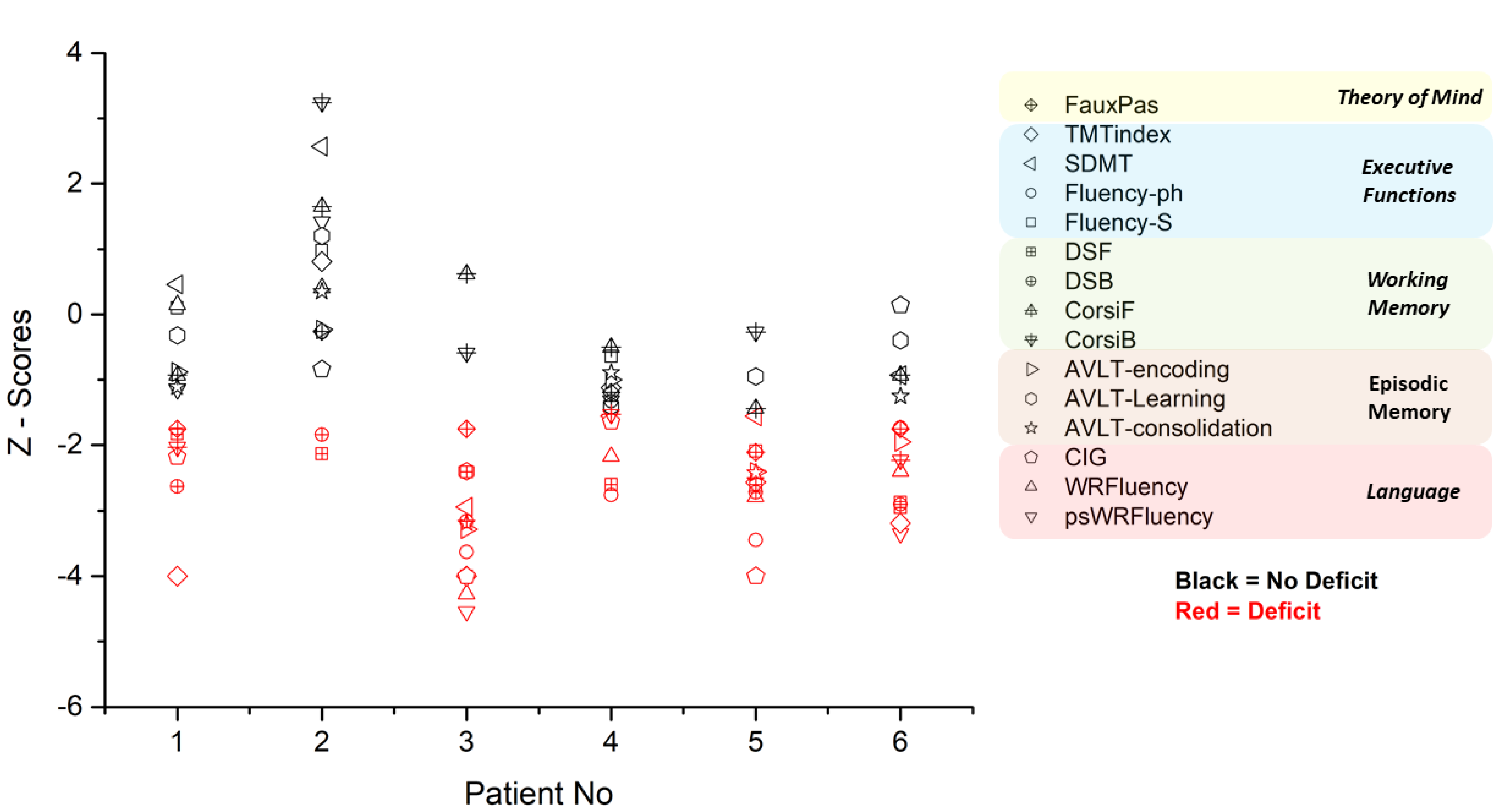
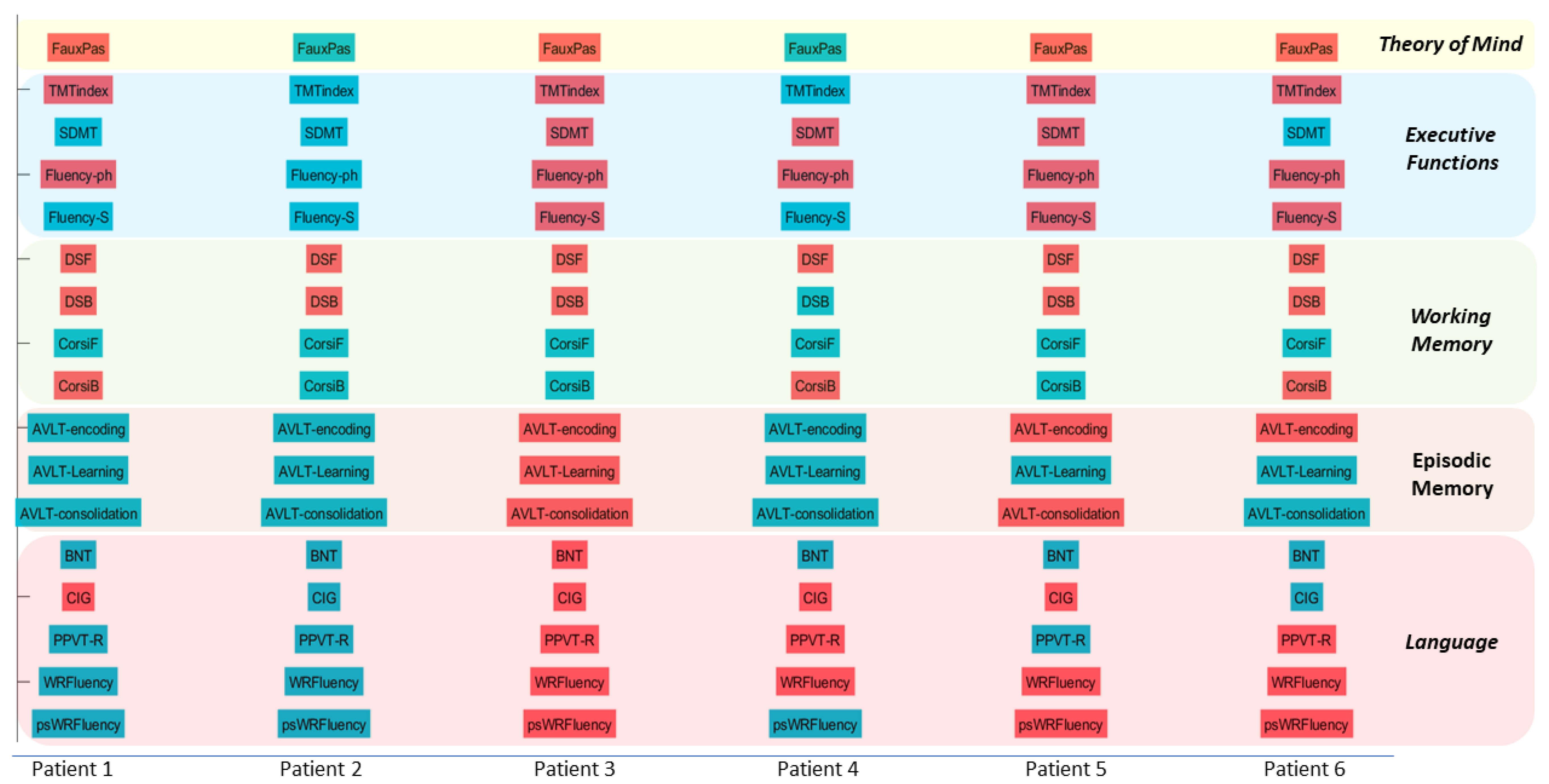
3. Results
3.1. Patient 1 (P2)
3.2. Patient 2 (P2)
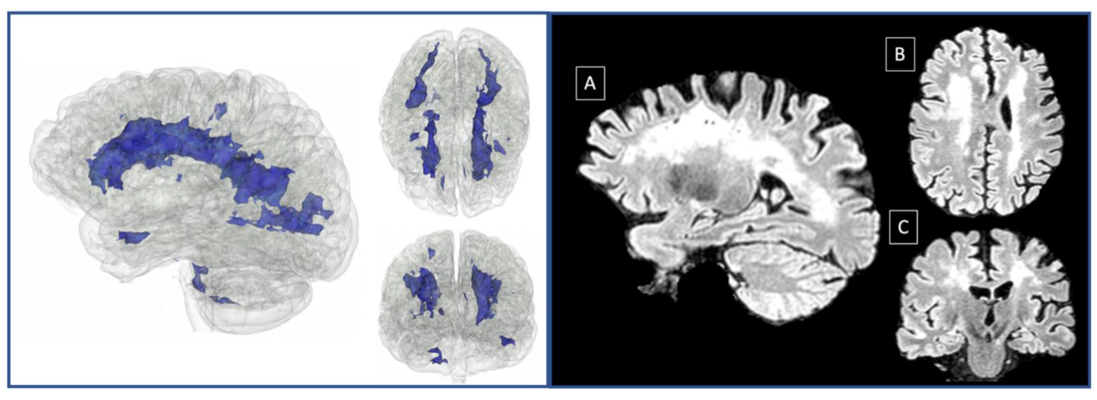
3.3. Patient 3 (P3)
3.4. Patient 4 (P4)
3.5. Patient 5 (P5)
3.6. Patient 6 (P6)
3.7. Patient 7 (P7)
3.8. Patient 8 (P8)
4. Discussion
Author Contributions
Funding
Institutional Review Board Statement
Informed Consent Statement
Data Availability Statement
Conflicts of Interest
References
- Kim, J.S. Moyamoya Disease: Epidemiology, Clinical Features, and Diagnosis. J. Stroke 2016, 18, 2–11. [Google Scholar] [CrossRef] [PubMed] [Green Version]
- Bang, O.Y.; Fujimura, M.; Kim, S.K. The Pathophysiology of Moyamoya Disease: An Update. J. Stroke 2016, 18, 12–20. [Google Scholar] [CrossRef] [PubMed] [Green Version]
- Fujimura, M.; Sonobe, S.; Nishijima, Y.; Niizuma, K.; Sakata, H.; Kure, S.; Tominaga, T. Genetics and Biomarkers of Moyamoya Disease: Significance of RNF213 as a Susceptibility Gene. J. Stroke 2014, 16, 65–72. [Google Scholar] [CrossRef] [PubMed] [Green Version]
- Matsushima, Y.; Aoyagi, M.; Masaoka, H.; Suzuki, R.; Ohno, K. Mental outcome following encephaloduroarteriosynangiosis in children with moyamoya disease with the onset earlier than 5 years of age. Child’s Nerv. Syst. 1990, 6, 440–443. [Google Scholar] [CrossRef]
- Scott, R.M.; Smith, E.R. Moyamoya disease and moyamoya syndrome. N. Engl. J. Med. 2009, 360, 1226–1237. [Google Scholar] [CrossRef] [Green Version]
- Bornstein, R.A. Neuropsychological performance in Moya Moya disease: A case study. Int. J. Neurosci. 1985, 26, 39–46. [Google Scholar] [CrossRef]
- Hoare, A.M.; Keogh, A.J. Cerebrovascular Moyamoya disease. Br. Med. J. 1974, 1, 430–432. [Google Scholar] [CrossRef] [Green Version]
- Jefferson, A.L.; Glosser, G.; Detre, J.A.; Sinson, G.; Liebeskind, D.S. Neuropsychological and perfusion MR imaging correlates of revascularization in a case of moyamoya syndrome. Am. J. Neuroradiol. 2006, 27, 98–100. [Google Scholar]
- Lubman, D.I.; Pantelis, C.; Desmond, P.; Proffitt, T.M.; Velakoulis, D. Moyamoya disease in a patient with schizophrenia. J. Int. Neuropsychol. Soc. 2003, 9, 806–810. [Google Scholar] [CrossRef]
- Ogasawara, K.; Komoribayashi, N.; Kobayashi, M.; Fukuda, T.; Inoue, T.; Yamadate, K.; Ogawa, A. Neural damage caused by cerebral hyperperfusion after arterial bypass surgery in a patient with moyamoya disease: Case report. Neurosurgery 2005, 56, E1380. [Google Scholar] [CrossRef]
- Karzmark, P.; Zeifert, P.D.; Tan, S.; Dorfman, L.J.; Bell-Stephens, T.E.; Steinberg, G.K. Effect of moyamoya disease on neuropsychological functioning in adults. Neurosurgery 2008, 62, 1048–1052. [Google Scholar] [CrossRef] [Green Version]
- Karzmark, P.; Zeifert, P.D.; Bell-Stephens, T.E.; Steinberg, G.K.; Dorfman, L.J. Neurocognitive impairment in adults with moyamoya disease without stroke. Neurosurgery 2012, 70, 634–638. [Google Scholar] [CrossRef]
- Ahmed, F.S.; Miller, L.S. Executive function mechanisms of theory of mind. J. Autism Dev. Disord. 2011, 41, 667–678. [Google Scholar] [CrossRef]
- Jones, C.; Simonoff, E.; Baird, G.; Pickles, A.; Marsden, A.; Tregay, J.; Happé, F.; Charman, T. The association between theory of mind, executive function, and the symptoms of autism spectrum disorder. Autism Res. 2018, 11, 95–109. [Google Scholar] [CrossRef]
- Hughes, C.; Graham, A. Measuring executive functions in childhood: Problems and solutions? Child Adolesc. Ment. Health 2002, 7, 131–142. [Google Scholar] [CrossRef]
- Perner, J.; Lang, B. Theory of mind and executive function: Is there a developmental relationship? In Understanding Other Minds: Perspectives from Developmental Cognitive Neuroscience; Baron-Cohen, S., Tager-Flusberg, H., Cohen, D.J., Eds.; Oxford University Press: Oxford, UK, 2000; pp. 150–181. [Google Scholar]
- Moore, D.P.; Lee, M.Y.; Macciocchi, S.N. Neurorehabilitation outcome in moyamoya disease. Arch. Phys. Med. Rehabil. 1997, 78, 672–675. [Google Scholar] [CrossRef]
- Araki, Y.; Takagi, Y.; Ueda, K.; Ubukata, S.; Ishida, J.; Funaki, T.; Kikuchi, T.; Takahashi, J.C.; Murai, T.; Miyamoto, S. Cognitive function of patients with adult moyamoya disease. J. Stroke Cerebrovasc. Dis. 2014, 23, 1789–1794. [Google Scholar] [CrossRef] [Green Version]
- Zalonis, I.; Kararizou, E.; Triantafyllou, N.I.; Kapaki, E.; Papageorgiou, S.; Sgouropoulos, P.; Vassilopoulos, D. A normative study of the trail making test A and B in greek adults. Clin. Neuropsychol. 2007, 22, 842–850. [Google Scholar] [CrossRef]
- Constantinidou, F.; Christodoulou, M.; Prokopiou, J. The effects of age and education on executive functioning and oral naming performance in Greek Cypriot adults: The neurocognitive study for the aging. Folia Phoniatr. Logop. 2012, 64, 187–198. [Google Scholar] [CrossRef]
- Kosmidis, M.H.; Vlahou, C.H.; Panagiotaki, P.; Kiosseoglou, G. The verbal fluency task in the Greek population: Normative data, and clustering and switching strategies. J. Int. Neuropsychol. Soc. 2004, 10, 164–172. [Google Scholar] [CrossRef]
- Christidi, F.; Kararizou, E.; Triantafyllou, N.; Anagnostouli, M.; Zalonis, I. Derived Trail Making Test indices: Demographics and cognitive background variables across the adult life span. Aging Neuropsychol. Cogn. 2015, 22, 667–678. [Google Scholar] [CrossRef]
- Constantinidou, F.; Zaganas, I.; Papastefanakis, E.; Kasselimis, D.; Nidos, A.; Simos, P.G. Age-related decline in verbal learning is moderated by demographic factors, working memory capacity, and presence of amnestic mild cognitive impairment. J. Int. Neuropsychol. Soc. 2014, 20, 822–835. [Google Scholar] [CrossRef]
- Simos, P.G.; Papastefanakis, E.; Panou, T.; Kasselimis, D. The Greek Memory Scale. 2011; (Unpublished neuropsychological battery). [Google Scholar]
- Corsi, P.M. Human Memory and the Medial Temporal Region of the Brain. Doctoral Dissertation, McGill University, Montreal, Canada, 1972. [Google Scholar]
- Kessels, R.P.C.; Van Den Berg, E.; Ruis, C.; Brands, A.M.A. The backward span of the corsi block-tapping task and its association with the WAIS-III digit span. Assessment 2008, 15, 426–434. [Google Scholar] [CrossRef]
- Kaplan, E.F.; Goodglass, H.; Weintraub, S. The Boston Naming Test, 2nd ed.; Lea & Febiger: Philadelphia, PA, USA, 1983. [Google Scholar]
- Dunn, L.M.; Dunn, E.S. Peabody Picture Vocabulary Test—Revised; American Guidance Service: Circle Pines, MN, USA, 1981. [Google Scholar]
- Simos, P.G.; Kasselimis, D.; Mouzaki, A. Age, gender, and education effects on vocabulary measures in Greek. Aphasiology 2011, 25, 475–491. [Google Scholar] [CrossRef]
- Simos, P.G.; Kasselimis, D.; Mouzaki, A. Effects of demographic variables and health status on brief vocabulary measures in Greek. Aphasiology 2011, 25, 492–504. [Google Scholar] [CrossRef]
- Simos, P.G.; Kasselimis, D.; Potagas, C.; Evdokimidis, I. Verbal comprehension ability in aphasia: Demographic and lexical knowledge effects. Behav. Neurol. 2014, 258303. [Google Scholar] [CrossRef]
- Simos, P.G.; Sideridis, G.D.; Kasselimis, D.; Mouzaki, A. Reading fluency estimates of current intellectual function: Demographic factors and effects of type of stimuli. J. Int. Neuropsychol. Soc. 2013, 19, 355–361. [Google Scholar] [CrossRef]
- Gregory, C.; Lough, S.; Stone, V.E.; Erzinclioglu, S.; Martin, L.; Baron-Cohen, S.; Hodges, J. Theory of mind in frontotemporal dementia and Alzheimer’s disease: Theoretical and practical implications. Brain 2002, 125, 752–764. [Google Scholar] [CrossRef]
- Stone, V.E.; Baron-Cohen, S.; Knight, R.T. Frontal lobe contributions to theory of mind. J. Cogn. Neurosci. 1998, 10, 640–656. [Google Scholar] [CrossRef]
- Konstantakopoulos, G.; Ploumpidis, D.; Oulis, P.; Patrikelis, P.; Soumani, A.; Papadimitriou, G.N.; Politis, A.M. Apathy, cognitive deficits and functional impairment in schizophrenia. Schizophr. Res. 2011, 133, 193–198. [Google Scholar] [CrossRef]
- Konstantakopoulos, G.; Ploumpidis, D.; Oulis, P.; Patrikelis, P.; Nikitopoulou, S.; Papadimitriou, G.N.; David, A.S. The relationship between insight and theory of mind in schizophrenia. Schizophr. Res. 2014, 152, 217–222. [Google Scholar] [CrossRef] [PubMed]
- Weinberg, D.G.; Rahme, R.J.; Aoun, S.G.; Batjer, H.H.; Bendok, B.R. Moyamoya Disease: Functional and Neurocognitive Outcomes in the Pediatric and Adult Populations. Neurosurg. Focus 2011, 30, 21. [Google Scholar] [CrossRef] [PubMed]
- Festa, J.R.; Schwarz, L.R.; Pliskin, N.; Cullum, C.M.; Lacritz, L.; Charbel, F.T.; Mathews, D.; Starke, R.M.; Connolly, E.S.; Marshall, R.S.; et al. Neurocognitive dysfunction in adult moyamoya disease. J. Neurol. 2010, 257, 806–815. [Google Scholar] [CrossRef] [PubMed]
- Calviere, L.; Catalaa, I.; Marlats, F.; Viguier, A.; Bonneville, F.; Cognard, C.; Larrue, V. Correlation between cognitive impairment and cerebral hemodynamic disturbances on perfusion magnetic resonance imaging in European adults with moyamoya disease. J. Neurosurg. 2010, 113, 753–759. [Google Scholar] [CrossRef] [Green Version]
- Chan, E.; MacPherson, S.E.; Robinson, G.; Turner, M.; Lecce, F.; Shallice, T.; Cipolotti, L. Limitations of the trail making test part-B in assessing frontal executive dysfunction. J. Int. Neuropsychol. Soc. 2015, 21, 169–174. [Google Scholar] [CrossRef] [Green Version]
- Zakzanis, K.K.; Mraz, R.; Graham, S.J. An fMRI study of the trail making test. Neuropsychologia 2005, 43, 1878–1886. [Google Scholar] [CrossRef]
- Jurado, M.B.; Rosselli, M. The elusive nature of executive functions: A review of our current understanding. Neuropsychol. Rev. 2007, 17, 213–233. [Google Scholar] [CrossRef]
- Rottschy, C.; Langner, R.; Dogan, I.; Reetz, K.; Laird, A.R.; Schulz, J.B.; Fox, P.T.; Eickhoff, S.B. Modelling neural correlates of working memory: A coordinate-based meta-analysis. Neuroimage 2012, 60, 830–846. [Google Scholar] [CrossRef] [Green Version]
- Wager, T.D.; Smith, E.E. Neuroimaging studies of working memory. Cogn. Affect. Behav. Neurosci. 2003, 3, 255–274. [Google Scholar] [CrossRef] [Green Version]
- Champod, A.S.; Petrides, M. Dissociable roles of the posterior parietal and the prefrontal cortex in manipulation and monitoring processes. Proc. Natl. Acad. Sci. USA 2007, 104, 14837–14842. [Google Scholar] [CrossRef] [Green Version]
- Champod, A.S.; Petrides, M. Dissociation within the frontoparietal network in verbal working memory: A parametric functional magnetic resonance imaging study. J. Neurosci. 2010, 30, 3849–3856. [Google Scholar] [CrossRef] [Green Version]
- Chapados, C.; Petrides, M. Ventrolateral and dorsomedial frontal cortex lesions impair mnemonic context retrieval. Proc. R. Soc. B Biol. Sci. 2015, 282, 20142555. [Google Scholar] [CrossRef] [Green Version]
- Amodio, D.M.; Frith, C.D. Meeting of minds: The medial frontal cortex and social cognition. Nat. Rev. Neurosci. 2006, 7, 268–277. [Google Scholar] [CrossRef]
- Frith, U.; Frith, C.D. Development and neurophysiology of mentalizing. Philos. Trans. R. Soc. Lond. Ser. B Biol. Sci. 2003, 358, 459–473. [Google Scholar] [CrossRef] [Green Version]
- Gallagher, H.L.; Happé, F.; Brunswick, N.; Fletcher, P.C.; Frith, U.; Frith, C.D. Reading the mind in cartoons and stories: An fMRI study of ‘theory of mind’ in verbal and nonverbal tasks. Neuropsychologia 2000, 38, 11–21. [Google Scholar] [CrossRef]
- Mason, R.A.; Just, M.A. The Role of the Theory-of-Mind Cortical Network in the Comprehension of Narratives. Lang. Linguist. Compass 2009, 3, 157–174. [Google Scholar] [CrossRef] [Green Version]
- Samson, D.; Apperly, I.A.; Chiavarino, C.; Humphreys, G.W. Left temporoparietal junction is necessary for representing someone else’s belief. Nature Neurosci. 2004, 7, 499–500. [Google Scholar] [CrossRef]
- Vogeley, K.; Bussfeld, P.; Newen, A.; Herrmann, S.; Happé, F.; Falkai, P.; Maier, W.; Shah, N.J.; Fink, G.R.; Zilles, K. Mind reading: Neural mechanisms of theory of mind and self-perspective. NeuroImage 2001, 14 Pt 1, 170–181. [Google Scholar] [CrossRef] [Green Version]
- Ffytche, D.H.; Catani, M. Beyond localization: From hodology to function. Philos. Trans. R. Soc. B Biol. Sci. 2005, 360, 767–779. [Google Scholar] [CrossRef] [Green Version]
- Catani, M.; Ffytche, D.H. The rises and falls of disconnection syndromes. Brain 2005, 128, 2224–2239. [Google Scholar] [CrossRef] [Green Version]
- Fedorov, A.; Beichel, R.; Kalpathy-Cramer, J.; Finet, J.; Fillion-Robin, J.-C.; Pujol, S.; Kikinis, R. 3D Slicer as an image computing platform for the Quantitative Imaging Network. Magn. Reson. Imaging 2012, 30, 1323–1341. [Google Scholar] [CrossRef] [PubMed] [Green Version]
- Cabezas, M.; Oliver, A.; Roura, E.; Freixenet, J.; Vilanova, J.C.; Ramió-Torrentà, L.; Rovira, À.; Lladó, X. Automatic multiple sclerosis lesion detection in brain MRI by FLAIR thresholding. Comput. Methods Programs Biomed. 2014, 115, 147–161. [Google Scholar] [CrossRef] [PubMed]
- Da Silva Senra Filho, A.C. A hybrid approach based on logistic classification and iterative contrast enhancement algorithm for hyperintense multiple sclerosis lesion segmentation. Med. Biol. Eng. Comput. 2018, 56, 1063–1076. [Google Scholar] [CrossRef] [PubMed]


Publisher’s Note: MDPI stays neutral with regard to jurisdictional claims in published maps and institutional affiliations. |
© 2022 by the authors. Licensee MDPI, Basel, Switzerland. This article is an open access article distributed under the terms and conditions of the Creative Commons Attribution (CC BY) license (https://creativecommons.org/licenses/by/4.0/).
Share and Cite
Papageorgiou, G.; Kasselimis, D.; Angelopoulou, G.; Tsolakopoulos, D.; Laskaris, N.; Tountopoulou, A.; Korompoki, E.; Velonakis, G.; Chatziioannou, A.; Spengos, K.; et al. Neurocognitive Profiles of Caucasian Moyamoya Disease Patients in Greece: A Case Series. NeuroSci 2022, 3, 119-129. https://0-doi-org.brum.beds.ac.uk/10.3390/neurosci3010010
Papageorgiou G, Kasselimis D, Angelopoulou G, Tsolakopoulos D, Laskaris N, Tountopoulou A, Korompoki E, Velonakis G, Chatziioannou A, Spengos K, et al. Neurocognitive Profiles of Caucasian Moyamoya Disease Patients in Greece: A Case Series. NeuroSci. 2022; 3(1):119-129. https://0-doi-org.brum.beds.ac.uk/10.3390/neurosci3010010
Chicago/Turabian StylePapageorgiou, Georgios, Dimitrios Kasselimis, Georgia Angelopoulou, Dimitrios Tsolakopoulos, Nikolaos Laskaris, Argyro Tountopoulou, Eleni Korompoki, Georgios Velonakis, Achilles Chatziioannou, Konstantinos Spengos, and et al. 2022. "Neurocognitive Profiles of Caucasian Moyamoya Disease Patients in Greece: A Case Series" NeuroSci 3, no. 1: 119-129. https://0-doi-org.brum.beds.ac.uk/10.3390/neurosci3010010