Plant Fortification of the Diet for Anti-Ageing Effects: A Review

 ,
,  ,
,  ,
,  ,
,  ,
, 
Abstract
:1. Introduction
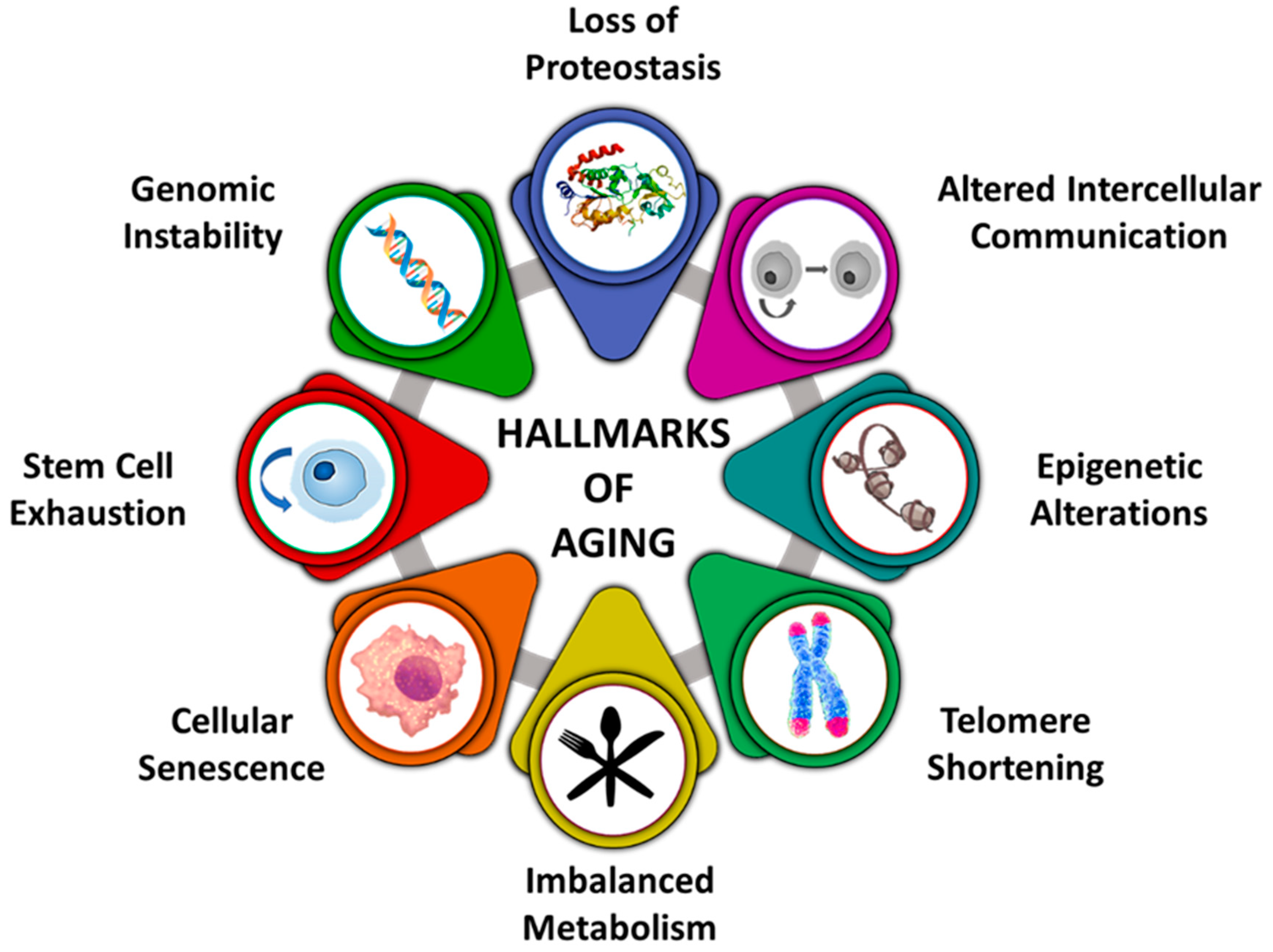
2. Manifestation of Ageing
3. Theories of Ageing
4. Plant-Based Supplements with Anti-Ageing Potential
4.1. Adaptogens
4.2. Bacopa monnieri
4.3. Curcuma longa
4.4. Emblica officinalis
4.5. Ginkgo biloba
4.6. Glycyrrhiza glabra
4.7. Panax ginseng
5. Plant-Based Metabolites with Anti-Ageing and Medicinal Properties
5.1. Polyphenols
5.2. Carotenoids
5.3. Vitamins
6. Concluding Remarks
Author Contributions
Funding
Conflicts of Interest
References
- Jin, K.; Rose, M.R. Modern Biological Theories of Aging. Aging Dis. 1988, 1, 220–221. [Google Scholar] [CrossRef]
- Zhang, S.; Duan, E. Fighting against Skin Aging: The Way from Bench to Bedside. Cell Transplant. 2018, 27, 729–738. [Google Scholar] [CrossRef] [PubMed]
- Bocheva, G.; Slominski, R.M.; Slominski, A.T. Neuroendocrine aspects of skin aging. Int. J. Mol. Sci. 2019, 20, 2798. [Google Scholar] [CrossRef] [PubMed] [Green Version]
- Schagen, S.K.; Zampeli, V.A.; Makrantonaki, E.; Zouboulis, C.C. Discovering the link between nutrition and skin aging. Dermatoendocrinology 2012, 4, 298–307. [Google Scholar] [CrossRef] [PubMed] [Green Version]
- Lobo, V.; Patil, A.; Phatak, A.; Chandra, N. Free radicals, antioxidants and functional foods: Impact on human health. Pharmacogn. Rev. 2010, 4, 118–126. [Google Scholar] [CrossRef] [Green Version]
- Alam, I.; Almajwal, A.M.; Alam, W.; Alam, I.; Ullah, N.; Abulmeaaty, M.; Razak, S.; Khan, S.; Pawelec, G.; Paracha, P.I. The immune-nutrition interplay in aging-Facts and controversies. Nutr. Healthy Aging 2019, 5, 73–95. [Google Scholar] [CrossRef] [Green Version]
- Rathore, H.; Prasad, S.; Sharma, S. Mushroom nutraceuticals for improved nutrition and better human health: A review. PharmaNutrition 2017, 5, 35–46. [Google Scholar] [CrossRef]
- Da Costa, J.P. A current look at nutraceuticals–Key concepts and future prospects. Trends Food Sci. Technol. 2017, 62, 68–78. [Google Scholar] [CrossRef]
- Chauhan, B.; Kumar, G.; Kalam, N.; Ansari, S.H. Current concepts and prospects of herbal nutraceutical: A review. J. Adv. Pharm. Technol. Res. 2013, 4, 4–8. [Google Scholar]
- Liu, Z.; Ren, Z.; Zhang, J.; Chuang, C.C.; Kandaswamy, E.; Zhou, T.; Zuo, L. Role of ROS and nutritional antioxidants in human diseases. Front. Physiol. 2018, 9, 477. [Google Scholar] [CrossRef] [Green Version]
- Conlon, M.A.; Bird, A.R. The impact of diet and lifestyle on gut microbiota and human health. Nutrients 2015, 7, 17–44. [Google Scholar] [CrossRef] [PubMed]
- Eming, S.A.; Martin, P.; Tomic-Canic, M. Wound repair and regeneration: Mechanisms, signaling, and translation. Sci. Transl. Med. 2014, 6, 265sr6. [Google Scholar] [CrossRef] [PubMed] [Green Version]
- Salam, N.; Rane, S.; Das, R.; Faulkner, M.; Gund, R.; Kandpal, U.; Lewis, V.; Mattoo, H.; Prabhu, S.; Ranganathan, V.; et al. T cell ageing: Effects of age on development, survival & function. Indian J. Med. Res. 2013, 138, 595–608. [Google Scholar] [PubMed]
- Amarya, S.; Singh, K.; Sabharwal, M. Ageing Process and Physiological Changes. In Gerontology; InTech: London, UK, 2018. [Google Scholar]
- Prohaska, T.R.; Keller, M.L.; Leventhal, E.A.; Leventhal, H. Impact of symptoms and aging attribution on emotions and coping. Health Psychol. 1987, 6, 495–514. [Google Scholar] [CrossRef]
- Heinemann, L.A.J.; Zimmermann, T.; Vermeulen, A.; Thiel, C.; Hummel, W. A new «aging males» symptoms’ rating scale. Aging Male 1999, 2, 105–114. [Google Scholar] [CrossRef]
- Maddy, A.J.; Tosti, A. Hair and nail diseases in the mature patient. Clin. Dermatol. 2018, 36, 159–166. [Google Scholar] [CrossRef]
- Sarbacher, C.A.; Halper, J.T. Connective tissue and age-related diseases. In Subcellular Biochemistry; Springer: New York, NY, USA, 2019; pp. 281–310. [Google Scholar]
- Patel, I.; West, S.K. Presbyopia: Prevalence, impact, and interventions. Community Eye Health J. 2007, 20, 40–41. [Google Scholar]
- Boostani, R.; Karimzadeh, F.; Nami, M. A comparative review on sleep stage classification methods in patients and healthy individuals. Comput. Methods Progr. Biomed. 2017, 140, 77–91. [Google Scholar] [CrossRef]
- Locantore, P.; Del Gatto, V.; Gelli, S.; Paragliola, R.M.; Pontecorvi, A. The Interplay between Immune System and Microbiota in Osteoporosis. Mediat. Inflamm. 2020, 2020, 3686749. [Google Scholar] [CrossRef]
- Trexler, E.T.; Smith-Ryan, A.E.; Norton, L.E. Metabolic adaptation to weight loss: Implications for the athlete. J. Int. Soc. Sports Nutr. 2014, 11, 7. [Google Scholar] [CrossRef] [Green Version]
- Wood, R.L. Accelerated cognitive aging following severe traumatic brain injury: A review. Brain Inj. 2017, 31, 1270–1278. [Google Scholar] [CrossRef] [PubMed]
- Sarnak, M.J. A patient with heart failure and worsening kidney function. Clin. J. Am. Soc. Nephrol. 2014, 9, 1790–1798. [Google Scholar] [CrossRef] [Green Version]
- De Martinis, M.; Sirufo, M.M.; Ginaldi, L. Allergy and aging: An Old/new emerging health issue. Aging Dis. 2017, 8, 162–175. [Google Scholar] [CrossRef] [Green Version]
- Santoro, N.; Epperson, C.N.; Mathews, S.B. Menopausal Symptoms and Their Management. Endocrinol. Metab. Clin. N. Am. 2015, 44, 497–515. [Google Scholar] [CrossRef] [PubMed] [Green Version]
- Booth, F.W.; Roberts, C.K.; Laye, M.J. Lack of exercise is a major cause of chronic diseases. Compr. Physiol. 2012, 2, 1143–1211. [Google Scholar] [CrossRef] [Green Version]
- Da Costa, J.P.; Vitorino, R.; Silva, G.M.; Vogel, C.; Duarte, A.C.; Rocha-Santos, T. A synopsis on aging—Theories, mechanisms and future prospects. Ageing Res. Rev. 2016, 29, 90–112. [Google Scholar] [CrossRef] [PubMed]
- De, A.; Ghosh, C. Basics of aging theories and disease related aging-an overview. PharmaTutor 2017, 5, 16–23. [Google Scholar]
- Weinert, B.T.; Timiras, P.S. Invited review: Theories of aging. J. Appl. Physiol. 2003, 95, 1706–1716. [Google Scholar] [CrossRef]
- Gladyshev, V.N. The free radical theory of aging is dead. Long live the damage theory! Antioxid. Redox Signal. 2014, 20, 727–731. [Google Scholar] [CrossRef]
- Sailaja Rao, P.; Kalva, S.; Yerramilli, A.; Mamidi, S. Free Radicals and Tissue Damage: Role of Antioxidants. Free Radic. Antioxid. 2011, 1, 2–7. [Google Scholar] [CrossRef]
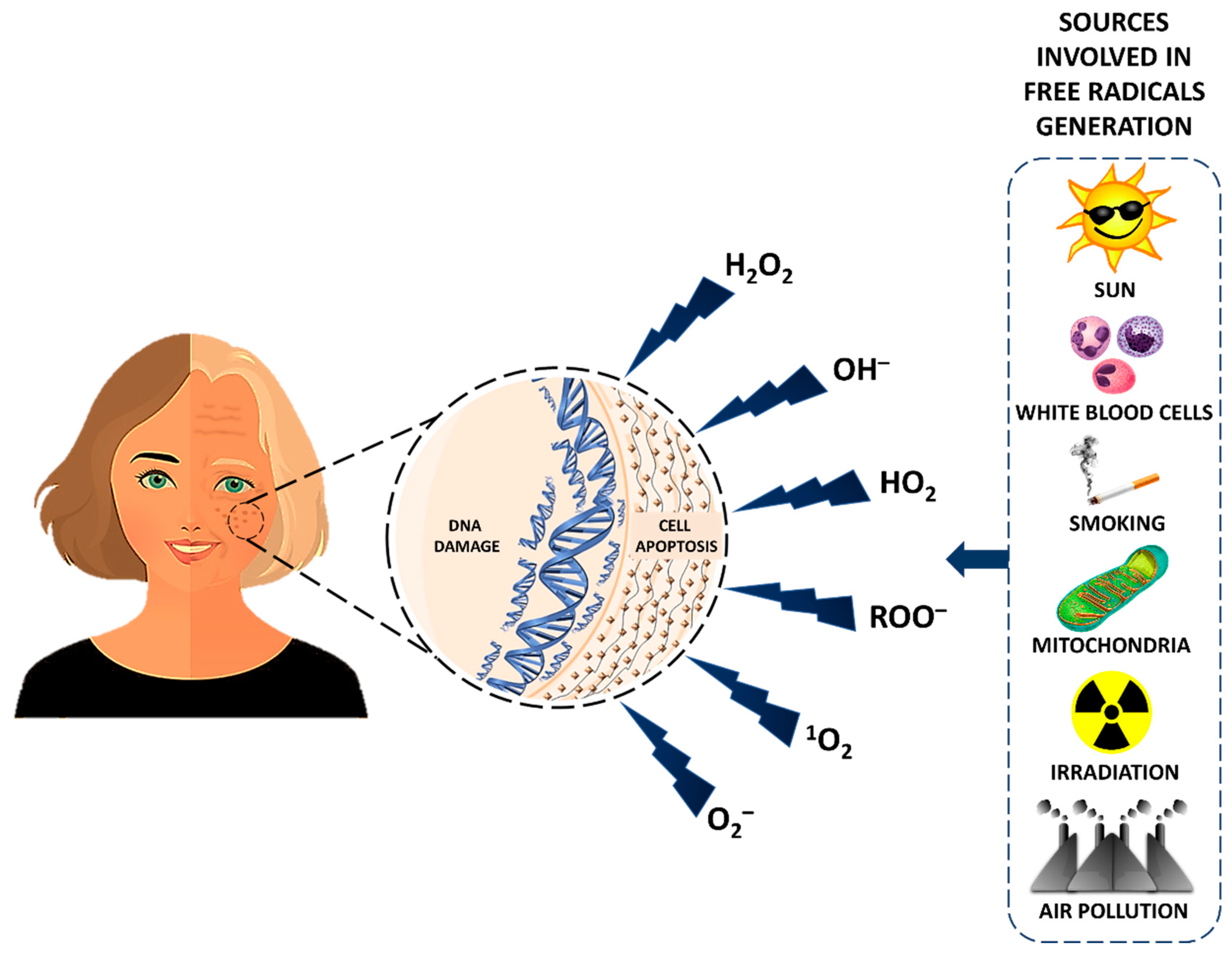
- Phaniendra, A.; Jestadi, D.B.; Periyasamy, L. Free Radicals: Properties, Sources, Targets, and Their Implication in Various Diseases. Indian J. Clin. Biochem. 2015, 30, 11–26. [Google Scholar] [CrossRef] [PubMed] [Green Version]
- Di Meo, S.; Venditti, P. Evolution of the Knowledge of Free Radicals and Other Oxidants. Oxid. Med. Cell. Longev. 2020, 2020, 9829176. [Google Scholar] [CrossRef] [PubMed]
- Pham-Huy, L.A.; He, H.; Pham-Huy, C. Free radicals, antioxidants in disease and health. Int. J. Biomed. Sci. 2008, 4, 89–96. [Google Scholar] [PubMed]
- Aruoma, O.I. Nutrition and health aspects of free radicals and antioxidants. Food Chem. Toxicol. 1994, 32, 671–683. [Google Scholar] [CrossRef]
- Dröge, W. Free radicals in the physiological control of cell function. Physiol. Rev. 2002, 82, 47–95. [Google Scholar] [CrossRef]
- Floyd, R.A.; Carney, J.M. Free radical damage to protein and DNA: Mechanisms involved and relevant observations on brain undergoing oxidative stress. Ann. Neurol. 1992, 32, S22–S27. [Google Scholar] [CrossRef]
- Kumar, H.; Bhardwaj, K.; Nepovimova, E.; Kuča, K.; Singh Dhanjal, D.; Bhardwaj, S.; Bhatia, S.K.; Verma, R.; Kumar, D. Antioxidant Functionalized Nanoparticles: A Combat against Oxidative Stress. Nanomaterials 2020, 10, 1334. [Google Scholar] [CrossRef]
- Thanan, R.; Oikawa, S.; Hiraku, Y.; Ohnishi, S.; Ma, N.; Pinlaor, S.; Yongvanit, P.; Kawanishi, S.; Murata, M. Oxidative stress and its significant roles in neurodegenerative diseases and cancer. Int. J. Mol. Sci. 2014, 16, 193–217. [Google Scholar] [CrossRef] [Green Version]
- Sharma, P.; Jha, A.B.; Dubey, R.S.; Pessarakli, M. Reactive Oxygen Species, Oxidative Damage, and Antioxidative Defense Mechanism in Plants under Stressful Conditions. J. Bot. 2012, 2012, 217037. [Google Scholar] [CrossRef] [Green Version]
- Ozcan, A.; Ogun, M. Biochemistry of Reactive Oxygen and Nitrogen Species. In Basic Principles and Clinical Significance of Oxidative Stress; InTech: London, UK, 2015. [Google Scholar]
- Pourahmad, J.; Salimi, A.; Seydi, E. Role of Oxygen Free Radicals in Cancer Development and Treatment. In Free Radicals and Diseases; InTech: London, UK, 2016. [Google Scholar]
- Jamshidi-kia, F.; Wibowo, J.P.; Elachouri, M.; Masumi, R.; Salehifard-Jouneghani, A.; Abolhasanzadeh, Z.; Lorigooini, Z. Battle between plants as antioxidants with free radicals in human body. J. Herbmed Pharmacol. 2020, 9, 191–199. [Google Scholar] [CrossRef]
- Santo, A.; Zhu, H.; Li, Y.R. Free radicals: From health to disease. React. Oxyg. Species 2016, 2, 245–263. [Google Scholar] [CrossRef]
- Liguori, I.; Russo, G.; Curcio, F.; Bulli, G.; Aran, L.; Della-Morte, D.; Gargiulo, G.; Testa, G.; Cacciatore, F.; Bonaduce, D.; et al. Oxidative stress, aging, and diseases. Clin. Interv. Aging 2018, 13, 757–772. [Google Scholar] [CrossRef] [PubMed] [Green Version]
- Nissanka, N.; Moraes, C.T. Mitochondrial DNA damage and reactive oxygen species in neurodegenerative disease. FEBS Lett. 2018, 592, 728–742. [Google Scholar] [CrossRef] [PubMed]
- Cantó, C.; Auwerx, J. Caloric restriction, SIRT1 and longevity. Trends Endocrinol. Metab. 2009, 20, 325–331. [Google Scholar] [CrossRef] [Green Version]
- Armandola, E. Caloric Restriction and Life Expectancy: Highlights of the 5th European Molecular Biology Organization Interdisciplinary Conference on Science and Society—Time & Aging: Mechanisms and Meanings; November 5–6, 2004; Heidelberg, Germany. Med. Gen. Med. 2004, 6, 16. [Google Scholar]
- Gutierrez, J.; Ballinger, S.W.; Darley-Usmar, V.M.; Landar, A. Free radicals, mitochondria, and oxidized lipids: The emerging role in signal transduction in vascular cells. Circ. Res. 2006, 99, 924–932. [Google Scholar] [CrossRef]
- Peng, C.; Wang, X.; Chen, J.; Jiao, R.; Wang, L.; Li, Y.M.; Zuo, Y.; Liu, Y.; Lei, L.; Ma, K.Y.; et al. Biology of ageing and role of dietary antioxidants. BioMed Res. Int. 2014, 2014, 831841. [Google Scholar] [CrossRef] [Green Version]
- Hornsby, P.J. Telomerase and the aging process. Exp. Gerontol. 2007, 42, 575–581. [Google Scholar] [CrossRef] [Green Version]
- Schmidt, J.C.; Cech, T.R. Human telomerase: Biogenesis, trafficking, recruitment, and activation. Genes Dev. 2015, 29, 1095–1105. [Google Scholar] [CrossRef] [PubMed] [Green Version]
- Kalmbach, K.H.; Fontes Antunes, D.M.; Dracxler, R.C.; Knier, T.W.; Seth-Smith, M.L.; Wang, F.; Liu, L.; Keefe, D.L. Telomeres and human reproduction. Fertil. Steril. 2013, 99, 23–29. [Google Scholar] [CrossRef] [Green Version]
- Kasote, D.M.; Katyare, S.S.; Hegde, M.V.; Bae, H. Significance of antioxidant potential of plants and its relevance to therapeutic applications. Int. J. Biol. Sci. 2015, 11, 982–991. [Google Scholar] [CrossRef] [PubMed] [Green Version]
- Chen, L.; Deng, H.; Cui, H.; Fang, J.; Zuo, Z.; Deng, J.; Li, Y.; Wang, X.; Zhao, L. Inflammatory responses and inflammation-associated diseases in organs. Oncotarget 2018, 9, 7204–7218. [Google Scholar] [CrossRef] [PubMed] [Green Version]
- Liao, L.Y.; He, Y.F.; Li, L.; Meng, H.; Dong, Y.M.; Yi, F.; Xiao, P.G. A preliminary review of studies on adaptogens: Comparison of their bioactivity in TCM with that of ginseng-like herbs used worldwide Milen Georgiev, Ruibing Wang. Chin. Med. 2018, 13, 57. [Google Scholar] [CrossRef] [PubMed] [Green Version]
- Bhatia, N.; Jaggi, A.S.; Singh, N.; Anand, P.; Dhawan, R. Adaptogenic potential of curcumin in experimental chronic stress and chronic unpredictable stress-induced memory deficits and alterations in functional homeostasis. J. Nat. Med. 2011, 65, 532–543. [Google Scholar] [CrossRef] [PubMed]
- Singh, M.K.; Jain, G.; Das, B.K.; Patil, U.K. Biomolecules from Plants as an Adaptogen. Med. Aromat. Plants 2017, 6, 307. [Google Scholar] [CrossRef]
- Aguiar, S.; Borowski, T. Neuropharmacological review of the nootropic herb Bacopa monnieri. Rejuvenation Res. 2013, 16, 313–326. [Google Scholar] [CrossRef] [Green Version]
- Jain, P.K.; Das, D.; Kumar Jain, P. Pharmacognostic Comparison of Bacopa Monnieri, Cyperus Rotundus and Emblica Officinalis. Innovare J. Ayurvedic Sci. 2016, 4, 16–26. [Google Scholar]
- Tewari, I.; Sharma, L.; Lal Gupta, G. Synergistic antioxidant activity of three medicinal plants Hypericum perforatum, Bacopa monnieri, and Camellia Sinensis. Indo Am. J. Pharm. Res. 2014, 4, 2563–2568. [Google Scholar]
- Vollala, V.R.; Upadhya, S.; Nayak, S. Effect of Bacopa monniera Linn. (brahmi) extract on learning and memory in rats: A behavioral study. J. Vet. Behav. Clin. Appl. Res. 2010, 5, 69–74. [Google Scholar] [CrossRef]
- Simpson, T.; Pase, M.; Stough, C. Bacopa monnieri as an Antioxidant Therapy to Reduce Oxidative Stress in the Aging Brain. Evid. Based Complement. Altern. Med. 2015, 2015, 615384. [Google Scholar] [CrossRef] [Green Version]
- Bhattacharya, S.K.; Bhattacharya, A.; Kumar, A.; Ghosal, S. Antioxidant activity of Bacopa monniera in rat frontal cortex, striatum and hippocampus. Phyther. Res. 2000, 14, 174–179. [Google Scholar] [CrossRef]
- Shinomol, G.K.; Srinivas Bharath, M.M. Muralidhara Neuromodulatory propensity of bacopa monnieri leaf extract against 3-nitropropionic acid-induced oxidative stress: In vitro and in vivo evidences. Neurotox. Res. 2012, 22, 102–114. [Google Scholar] [CrossRef]
- Kumar, N.; Abichandani, L.G.; Thawani, V.; Gharpure, K.J.; Naidu, M.U.R.; Venkat Ramana, G. Efficacy of Standardized Extract of Bacopa monnieri (Bacognize®) on Cognitive Functions of Medical Students: A Six-Week, Randomized Placebo-Controlled Trial. Evid. Based Complement. Altern. Med. 2016, 2016. [Google Scholar] [CrossRef] [Green Version]
- Kocaadam, B.; Şanlier, N. Curcumin, an active component of turmeric (Curcuma longa), and its effects on health. Crit. Rev. Food Sci. Nutr. 2017, 57, 2889–2895. [Google Scholar] [CrossRef] [PubMed]
- Prasad, S.; Aggarwal, B.B. Turmeric, the golden spice: From traditional medicine to modern medicine. In Herbal Medicine: Biomolecular and Clinical Aspects: Second Edition; CRC Press: Boca Raton, FL, USA, 2011; pp. 263–288. ISBN 9781439807163. [Google Scholar]
- Tomeh, M.A.; Hadianamrei, R.; Zhao, X. A review of curcumin and its derivatives as anticancer agents. Int. J. Mol. Sci. 2019, 20, 1033. [Google Scholar] [CrossRef] [PubMed] [Green Version]
- Desai, S.J.; Prickril, B.; Rasooly, A. Mechanisms of Phytonutrient Modulation of Cyclooxygenase-2 (COX-2) and Inflammation Related to Cancer. Nutr. Cancer 2018, 70, 350–375. [Google Scholar] [CrossRef] [PubMed]
- Engwa, G.A. Free Radicals and the Role of Plant Phytochemicals as Antioxidants Against Oxidative Stress-Related Diseases. In Phytochemicals-Source of Antioxidants and Role in Disease Prevention; InTech: London, UK, 2018. [Google Scholar]
- Lima, C.F.; Pereira-Wilson, C.; Rattan, S.I.S. Curcumin induces heme oxygenase-1 in normal human skin fibroblasts through redox signaling: Relevance for anti-aging intervention. Mol. Nutr. Food Res. 2011, 55, 430–442. [Google Scholar] [CrossRef] [Green Version]
- Soh, J.W.; Marowsky, N.; Nichols, T.J.; Rahman, A.M.; Miah, T.; Sarao, P.; Khasawneh, R.; Unnikrishnan, A.; Heydari, A.R.; Silver, R.B.; et al. Curcumin is an early-acting stage-specific inducer of extended functional longevity in Drosophila. Exp. Gerontol. 2013, 48, 229–239. [Google Scholar] [CrossRef]
- Shen, L.-R.; Parnell, L.D.; Ordovas, J.M.; Lai, C.-Q. Curcumin and aging. BioFactors 2013, 39, 133–140. [Google Scholar] [CrossRef]
- Lee, K.S.; Lee, B.S.; Semnani, S.; Avanesian, A.; Um, C.Y.; Jeon, H.J.; Seong, K.M.; Yu, K.; Min, K.J.; Jafari, M. Curcumin extends life span, improves health span, and modulates the expression of age-associated aging genes in drosophila melanogaster. Rejuvenation Res. 2010, 13, 561–570. [Google Scholar] [CrossRef]
- He, Y.; Yue, Y.; Zheng, X.; Zhang, K.; Chen, S.; Du, Z. Curcumin, inflammation, and chronic diseases: How are they linked? Molecules 2015, 20, 9183–9213. [Google Scholar] [CrossRef] [PubMed]
- Olszanecki, R.; Jawien, J.; Gajda, M.; Mateuszuk, L.; Gebska, A.; Korabiowska, M.; Chlopicki, S.; Korbut, R. Effect of curcumin on atherosclerosis in apoE-LDLR-double knockout mice. J. Physiol. Pharmacol. 2005, 4, 627–635. [Google Scholar]
- Swamy, A.V.; Gulliaya, S.; Thippeswamy, A.; Koti, B.C.; Manjula, D.V. Cardioprotective effect of curcumin against doxorubicin-induced myocardial toxicity in albino rats. Indian J. Pharmacol. 2012, 44, 73–77. [Google Scholar] [CrossRef] [PubMed] [Green Version]
- Ryan, J.L.; Heckler, C.E.; Ling, M.; Katz, A.; Williams, J.P.; Pentland, A.P.; Morrow, G.R. Curcumin for radiation dermatitis: A randomized, double-blind, placebo-controlled clinical trial of thirty breast cancer patients. Radiat. Res. 2013, 180, 34–43. [Google Scholar] [CrossRef] [PubMed] [Green Version]
- Ray Hamidie, R.D.; Yamada, T.; Ishizawa, R.; Saito, Y.; Masuda, K. Curcumin treatment enhances the effect of exercise on mitochondrial biogenesis in skeletal muscle by increasing cAMP levels. Metabolism 2015, 64, 1334–1347. [Google Scholar] [CrossRef] [PubMed]
- Cox, K.H.M.; Pipingas, A.; Scholey, A.B. Investigation of the effects of solid lipid curcumin on cognition and mood in a healthy older population. J. Psychopharmacol. 2015, 29, 642–651. [Google Scholar] [CrossRef]
- Yadav, S.S.; Singh, M.K.; Singh, P.K.; Kumar, V. Traditional knowledge to clinical trials: A review on therapeutic actions of Emblica officinalis. Biomed. Pharmacother. 2017, 93, 1292–1302. [Google Scholar] [CrossRef]
- Kapoor, M.P.; Suzuki, K.; Derek, T.; Ozeki, M.; Okubo, T. Clinical evaluation of Emblica Officinalis Gatertn (Amla) in healthy human subjects: Health benefits and safety results from a randomized, double-blind, crossover placebo-controlled study. Contemp. Clin. Trials Commun. 2020, 17, 100499. [Google Scholar] [CrossRef]
- Wilson, D.W.; Nash, P.; Singh, H.; Griffiths, K.; Singh, R.; De Meester, F.; Horiuchi, R.; Takahashi, T. The role of food antioxidants, benefits of functional foods, and influence of feeding habits on the health of the older person: An overview. Antioxidants 2017, 6, 81. [Google Scholar] [CrossRef] [Green Version]
- Hasan, M.R.; Islam, M.N.; Islam, M.R. Phytochemistry, pharmacological activities and traditional uses of Emblica officinalis: A review. Int. Curr. Pharm. J. 2016, 5, 14–21. [Google Scholar] [CrossRef] [Green Version]
- Lauer, A.C.; Groth, N.; Haag, S.F.; Darvin, M.E.; Lademann, J.; Meinke, M.C. Dose-dependent vitamin C uptake and radical scavenging activity in human skin measured with in vivo electron paramagnetic resonance spectroscopy. Skin Pharmacol. Physiol. 2013, 26, 147–154. [Google Scholar] [CrossRef] [PubMed]
- Bhattacharya, A.; Ghosal, S.; Bhattacharya, S.K. Antioxidant activity of tannoid principles of Emblica officinalis (amla) in chronic stress induced changes in rat brain. Indian J. Exp. Biol. 2000, 38, 877–880. [Google Scholar] [PubMed]
- Bhattachary, S.K.; Bhattacharya, D.; Muruganandam, A.V. Effect of Emblica officinalis tannoids on a rat model of tardive dyskinesia. Indian J. Exp. Biol. 2000, 38, 945–947. [Google Scholar] [PubMed]
- Dhingra, D.; Joshi, P.; Gupta, A.; Chhillar, R. Possible Involvement of Monoaminergic Neurotransmission in Antidepressant-like activity of Emblica officinalis Fruits in Mice. CNS Neurosci. Ther. 2012, 18, 419–425. [Google Scholar] [CrossRef] [PubMed]
- Isah, T. Rethinking Ginkgo biloba L.: Medicinal uses and conservation. Pharmacogn. Rev. 2015, 9, 140–148. [Google Scholar] [CrossRef] [Green Version]
- Mashayekh, A.; Pham, D.L.; Yousem, D.M.; Dizon, M.; Barker, P.B.; Lin, D.D.M. Effects of Ginkgo biloba on cerebral blood flow assessed by quantitative MR perfusion imaging: A pilot study. Neuroradiology 2011, 53, 185–191. [Google Scholar] [CrossRef] [Green Version]
- Zuo, W.; Yan, F.; Zhang, B.; Li, J.; Mei, D. Advances in the studies of Ginkgo biloba leaves extract on aging-related diseases. Aging Dis. 2017, 8, 812–826. [Google Scholar] [CrossRef] [Green Version]
- Van Beek, T.A. Chemical analysis of Ginkgo biloba leaves and extracts. J. Chromatogr. A 2002, 967, 21–55. [Google Scholar] [CrossRef]
- Huang, S.Z.; Luo, Y.J.; Wang, L.; Cai, K.Y. Effect of ginkgo biloba extract on livers in aged rats. World J. Gastroenterol. 2005, 11, 132–135. [Google Scholar] [CrossRef]
- Belviranli, M.; Okudan, N. The effects of Ginkgo biloba extract on cognitive functions in aged female rats: The role of oxidative stress and brain-derived neurotrophic factor. Behav. Brain Res. 2015, 278, 453–461. [Google Scholar] [CrossRef]
- Liu, H.; Ye, M.; Guo, H. An Updated Review of Randomized Clinical Trials Testing the Improvement of Cognitive Function of Ginkgo biloba Extract in Healthy People and Alzheimer’s Patients. Front. Pharmacol. 2020, 10, 1688. [Google Scholar] [CrossRef] [PubMed] [Green Version]
- Pastorino, G.; Cornara, L.; Soares, S.; Rodrigues, F.; Oliveira, M.B.P.P. Liquorice (Glycyrrhiza glabra): A phytochemical and pharmacological review. Phyther. Res. 2018, 32, 2323–2339. [Google Scholar] [CrossRef] [PubMed]
- Frattaruolo, L.; Carullo, G.; Brindisi, M.; Mazzotta, S.; Bellissimo, L.; Rago, V.; Curcio, R.; Dolce, V.; Aiello, F.; Cappello, A.R. Antioxidant and anti-inflammatory activities of flavanones from glycyrrhiza glabra L. (licorice) leaf phytocomplexes: Identification of licoflavanone as a modulator of NF-kB/MAPK pathway. Antioxidants 2019, 8, 186. [Google Scholar] [CrossRef] [PubMed] [Green Version]
- Grodzicki, W.; Dziendzikowska, K. The role of selected bioactive compounds in the prevention of alzheimer’s disease. Antioxidants 2020, 9, 229. [Google Scholar] [CrossRef] [PubMed] [Green Version]
- Ciganović, P.; Jakimiuk, K.; Tomczyk, M.; Končić, M.Z. Glycerolic licorice extracts as active cosmeceutical ingredients: Extraction optimization, chemical characterization, and biological activity. Antioxidants 2019, 8, 445. [Google Scholar] [CrossRef] [Green Version]
- Balmus, I.M.; Ciobica, A. Main Plant Extracts’ Active Properties Effective on Scopolamine-Induced Memory Loss. Am. J. Alzheimers Dis. Other Demen. 2017, 32, 418–428. [Google Scholar] [CrossRef]
- Dhingra, D.; Parle, M.; Kulkarni, S.K. Memory enhancing activity of Glycyrrhiza glabra in mice. J. Ethnopharmacol. 2004, 91, 361–365. [Google Scholar] [CrossRef]
- Rokot, N.T.; Kairupan, T.S.; Cheng, K.C.; Runtuwene, J.; Kapantow, N.H.; Amitani, M.; Morinaga, A.; Amitani, H.; Asakawa, A.; Inui, A. A Role of Ginseng and Its Constituents in the Treatment of Central Nervous System Disorders. Evid. Based Complement. Altern. Med. 2016, 2016, 2614742. [Google Scholar] [CrossRef] [Green Version]
- Yu, H.; Zhao, J.; You, J.; Li, J.; Ma, H.; Chen, X. Factors influencing cultivated ginseng (Panax ginseng C. A. Meyer) bioactive compounds. PLoS ONE 2019, 14, e0223763. [Google Scholar] [CrossRef]
- Kumar, G.P.; Khanum, F. Neuroprotective potential of phytochemicals. Pharmacogn. Rev. 2012, 6, 81–90. [Google Scholar] [CrossRef] [Green Version]
- Wee, J.J.; Mee Park, K.; Chung, A.-S. Biological Activities of Ginseng and Its Application to Human Health. In Herbal Medicine: Biomolecular and Clinical Aspects; Benzie, I.F.F., Wachtel-Galor, S., Eds.; CRC Press/Taylor and Francis: Boca Raton, FL, USA, 2011; ISBN 978-1-4398-0713-2. [Google Scholar]
- Lee, Y.M.; Yoon, H.; Park, H.M.; Song, B.C.; Yeum, K.J. Implications of red Panax ginseng in oxidative stress associated chronic diseases. J. Ginseng Res. 2017, 41, 113–119. [Google Scholar] [CrossRef] [PubMed] [Green Version]
- Caldwell, L.K.; Dupont, W.H.; Beeler, M.K.; Post, E.M.; Barnhart, E.C.; Hardesty, V.H.; Anders, J.P.; Borden, E.C.; Volek, J.S.; Kraemer, W.J. The effects of a Korean ginseng, GINST15, on perceptual effort, psychomotor performance, and physical performance in men and women. J. Sport. Sci. Med. 2018, 17, 92–100. [Google Scholar]
- Kim, J.; Cho, S.Y.; Kim, S.H.; Cho, D.; Kim, S.; Park, C.W.; Shimizu, T.; Cho, J.Y.; Seo, D.B.; Shin, S.S. Effects of Korean ginseng berry on skin antipigmentation and antiaging via FoxO3a activation. J. Ginseng Res. 2017, 41, 277–283. [Google Scholar] [CrossRef] [PubMed] [Green Version]
- Hwang, E.; Park, S.Y.; Jo, H.; Lee, D.G.; Kim, H.T.; Kim, Y.M.; Yin, C.S.; Yi, T.H. Efficacy and Safety of Enzyme-Modified Panax ginseng for Anti-Wrinkle Therapy in Healthy Skin: A Single-Center, Randomized, Double-Blind, Placebo-Controlled Study. Rejuvenation Res. 2015, 18, 449–457. [Google Scholar] [CrossRef]
- Shahidi, F.; Ambigaipalan, P. Phenolics and polyphenolics in foods, beverages and spices: Antioxidant activity and health effects-A review. J. Funct. Foods 2015, 18, 820–897. [Google Scholar] [CrossRef]
- Quero, J.; Mármol, I.; Cerrada, E.; Rodríguez-Yoldi, M.J. Insight into the potential application of polyphenol-rich dietary intervention in degenerative disease management. Food Funct. 2020, 11, 2805–2825. [Google Scholar] [CrossRef]
- Pandey, K.B.; Rizvi, S.I. Plant polyphenols as dietary antioxidants in human health and disease. Oxid. Med. Cell. Longev. 2009, 2, 270–278. [Google Scholar] [CrossRef] [Green Version]
- Dunaway, S.; Odin, R.; Zhou, L.; Ji, L.; Zhang, Y.; Kadekaro, A.L. Natural antioxidants: Multiple mechanisms to protect skin from solar radiation. Front. Pharmacol. 2018, 9, 392. [Google Scholar] [CrossRef] [Green Version]
- D’Orazio, J.; Jarrett, S.; Amaro-Ortiz, A.; Scott, T. UV radiation and the skin. Int. J. Mol. Sci. 2013, 14, 12222–12248. [Google Scholar] [CrossRef] [Green Version]
- Adhikari, B.; Dhungana, S.K.; Waqas Ali, M.; Adhikari, A.; Kim, I.D.; Shin, D.H. Antioxidant activities, polyphenol, flavonoid, and amino acid contents in peanut shell. J. Saudi Soc. Agric. Sci. 2019, 18, 437–442. [Google Scholar] [CrossRef]
- Camins, A.; Junyent, F.; Verdaguer, E.; Beas-Zarate, C.; Rojas-Mayorquín, A.; Ortuño-Sahagún, D.; Pallàs, M. Resveratrol: An Antiaging Drug with Potential Therapeutic Applications in Treating Diseases. Pharmaceuticals 2009, 2, 194–205. [Google Scholar] [CrossRef]
- Salehi, B.; Mishra, A.P.; Nigam, M.; Sener, B.; Kilic, M.; Sharifi-Rad, M.; Fokou, P.V.T.; Martins, N.; Sharifi-Rad, J. Resveratrol: A double-edged sword in health benefits. Biomedicines 2018, 6, 91. [Google Scholar] [CrossRef] [Green Version]
- Quadros Gomes, B.A.; Bastos Silva, J.P.; Rodrigues Romeiro, C.F.; dos Santos, S.M.; Rodrigues, C.A.; Gonçalves, P.R.; Sakai, J.T.; Santos Mendes, P.F.; Pompeu Varela, E.L.; Monteiro, M.C. Neuroprotective mechanisms of resveratrol in Alzheimer’s disease: Role of SIRT1. Oxid. Med. Cell. Longev. 2018, 2018, 8152373. [Google Scholar]
- Bhat, K.P.L.; Pezzuto, J.M. Cancer chemopreventive activity of resveratrol. Ann. N. Y. Acad. Sci. 2002, 957, 210–229. [Google Scholar] [CrossRef] [PubMed] [Green Version]
- Bastianetto, S.; Dumont, Y.; Duranton, A.; Vercauteren, F.; Breton, L.; Quirion, R. Protective Action of Resveratrol in Human Skin: Possible Involvement of Specific Receptor Binding Sites. PLoS ONE 2010, 5, e12935. [Google Scholar] [CrossRef] [PubMed] [Green Version]
- Giardina, S.; Michelotti, A.; Zavattini, G.; Finzi, S.; Ghisalberti, C.; Marzatico, F. Efficacy study in vitro: Assessment of the properties of resveratrol and resveratrol + N-acetyl-cysteine on proliferation and inhibition of collagen activity. Minerva Ginecol. 2010, 62, 195–201. [Google Scholar]
- Demidenko, Z.N.; Blagosklonny, M.V. At concentrations that inhibit mTOR, resveratrol suppresses cellular senescence. Cell Cycle 2009, 8, 1901–1904. [Google Scholar] [CrossRef] [Green Version]
- Xia, L.; Wang, X.X.; Hu, X.S.; Guo, X.G.; Shang, Y.P.; Chen, H.J.; Zeng, C.L.; Zhang, F.R.; Chen, J.Z. Resveratrol reduces endothelial progenitor cells senescence through augmentation of telomerase activity by Akt-dependent mechanisms. Br. J. Pharmacol. 2008, 155, 387–394. [Google Scholar] [CrossRef] [Green Version]
- Giovannelli, L.; Pitozzi, V.; Jacomelli, M.; Mulinacci, N.; Laurenzana, A.; Dolara, P.; Mocali, A. Protective effects of resveratrol against senescence-associated changes in cultured human fibroblasts. J. Gerontol. Ser. A Biol. Sci. Med. Sci. 2011, 66 A, 9–18. [Google Scholar] [CrossRef] [Green Version]
- López-Lluch, G.; Irusta, P.M.; Navas, P.; de Cabo, R. Mitochondrial biogenesis and healthy aging. Exp. Gerontol. 2008, 43, 813–819. [Google Scholar] [CrossRef] [Green Version]
- Lagouge, M.; Argmann, C.; Gerhart-Hines, Z.; Meziane, H.; Lerin, C.; Daussin, F.; Messadeq, N.; Milne, J.; Lambert, P.; Elliott, P.; et al. Resveratrol Improves Mitochondrial Function and Protects against Metabolic Disease by Activating SIRT1 and PGC-1α. Cell 2006, 127, 1109–1122. [Google Scholar] [CrossRef] [PubMed]
- Wang, T.; Li, Q.; Bi, K. Bioactive flavonoids in medicinal plants: Structure, activity and biological fate. Asian J. Pharm. Sci. 2018, 13, 12–23. [Google Scholar] [CrossRef] [PubMed]
- Antika, L.D.; Lee, E.J.; Kim, Y.H.; Kang, M.K.; Park, S.H.; Kim, D.Y.; Oh, H.; Choi, Y.J.; Kang, Y.H. Dietary phlorizin enhances osteoblastogenic bone formation through enhancing β-catenin activity via GSK-3β inhibition in a model of senile osteoporosis. J. Nutr. Biochem. 2017, 49, 42–52. [Google Scholar] [CrossRef] [PubMed]
- Makarova, E.; Górnaś, P.; Konrade, I.; Tirzite, D.; Cirule, H.; Gulbe, A.; Pugajeva, I.; Seglina, D.; Dambrova, M. Acute anti-hyperglycaemic effects of an unripe apple preparation containing phlorizin in healthy volunteers: A preliminary study. J. Sci. Food Agric. 2014, 95, 560–568. [Google Scholar] [CrossRef]
- Mela, D.J.; Cao, X.Z.; Dobriyal, R.; Fowler, M.I.; Lin, L.; Joshi, M.; Mulder, T.J.P.; Murray, P.G.; Peters, H.P.F.; Vermeer, M.A.; et al. The effect of 8 plant extracts and combinations on post-prandial blood glucose and insulin responses in healthy adults: A randomized controlled trial. Nutr. Metab. 2020, 17, 51. [Google Scholar] [CrossRef]
- Laiteerapong, N.; Karter, A.J.; Liu, J.Y.; Moffet, H.H.; Sudore, R.; Schillinger, D.; John, P.M.; Huang, E.S. Correlates of quality of life in older adults with diabetes: The diabetes & aging study. Diabetes Care 2011, 34, 1749–1753. [Google Scholar] [CrossRef] [Green Version]
- Ayaz, M.; Sadiq, A.; Junaid, M.; Ullah, F.; Ovais, M.; Ullah, I.; Ahmed, J.; Shahid, M. Flavonoids as prospective neuroprotectants and their therapeutic propensity in aging associated neurological disorders. Front. Aging Neurosci. 2019, 11, 155. [Google Scholar] [CrossRef] [Green Version]
- Zhang, Y.J.; Gan, R.Y.; Li, S.; Zhou, Y.; Li, A.N.; Xu, D.P.; Li, H.B.; Kitts, D.D. Antioxidant phytochemicals for the prevention and treatment of chronic diseases. Molecules 2015, 20, 21138–21156. [Google Scholar] [CrossRef]
- Kschonsek, J.; Wolfram, T.; Stöckl, A.; Böhm, V. Polyphenolic compounds analysis of old and new apple cultivars and contribution of polyphenolic profile to the in vitro antioxidant capacity. Antioxidants 2018, 7, 20. [Google Scholar] [CrossRef] [Green Version]
- Boyer, J.; Liu, R.H. Apple phytochemicals and their health benefits. Nutr. J. 2004, 3, 5. [Google Scholar] [CrossRef] [Green Version]
- Vafa, M.R.; Haghighatjoo, E.; Shidfar, F.; Afshari, S.; Gohari, M.R.; Ziaee, A. Effects of apple consumption on lipid profile of hyperlipidemic and overweight men. Int. J. Prev. Med. 2011, 2, 94–100. [Google Scholar] [PubMed]
- Peng, C.; Chan, H.Y.E.; Huang, Y.; Yu, H.; Chen, Z.Y. Apple polyphenols extend the mean lifespan of Drosophila melanogaster. J. Agric. Food Chem. 2011, 59, 2097–2106. [Google Scholar] [CrossRef] [PubMed]
- Cory, H.; Passarelli, S.; Szeto, J.; Tamez, M.; Mattei, J. The Role of Polyphenols in Human Health and Food Systems: A Mini-Review. Front. Nutr. 2018, 5, 87. [Google Scholar] [CrossRef] [PubMed] [Green Version]
- Kalt, W.; Cassidy, A.; Howard, L.R.; Krikorian, R.; Stull, A.J.; Tremblay, F.; Zamora-Ros, R. Recent Research on the Health Benefits of Blueberries and Their Anthocyanins. Adv. Nutr. 2020, 11, 224–236. [Google Scholar] [CrossRef]
- Shukitt-Hale, B.; Thangthaeng, N.; Miller, M.G.; Poulose, S.M.; Carey, A.N.; Fisher, D.R. Blueberries Improve Neuroinflammation and Cognition differentially Depending on Individual Cognitive baseline Status. J. Gerontol. Ser. A Biol. Sci. Med. Sci. 2019, 74, 977–983. [Google Scholar] [CrossRef]
- Joseph, J.A.; Shukitt-Hale, B.; Casadesus, G. Reversing the deleterious effects of aging on neuronal communication and behavior: Beneficial properties of fruit polyphenolic compounds. Am. J. Clin. Nutr. 2005, 81, 313S–316S. [Google Scholar] [CrossRef]
- Galli, R.L.; Bielinski, D.F.; Szprengiel, A.; Shukitt-Hale, B.; Joseph, J.A. Blueberry supplemented diet reverses age-related decline in hippocampal HSP70 neuroprotection. Neurobiol. Aging 2006, 27, 344–350. [Google Scholar] [CrossRef]
- Goyarzu, P.; Malin, D.H.; Lau, F.C.; Taglialatela, G.; Moon, W.D.; Jennings, R.; Moy, E.; Moy, D.; Lippold, S.; Shukitt-Hale, B.; et al. Blueberry supplemented diet: Effects on object recognition memory and nuclear factor-kappa B levels in aged rats. Nutr. Neurosci. 2004, 7, 75–83. [Google Scholar] [CrossRef]
- Peng, C.; Zuo, Y.; Kwan, K.M.; Liang, Y.; Ma, K.Y.; Chan, H.Y.E.; Huang, Y.; Yu, H.; Chen, Z.Y. Blueberry extract prolongs lifespan of Drosophila melanogaster. Exp. Gerontol. 2012, 47, 170–178. [Google Scholar] [CrossRef]
- Su, Y.L.; Leung, L.K.; Huang, Y.; Chen, Z.Y. Stability of tea theaflavins and catechins. Food Chem. 2003, 83, 189–195. [Google Scholar] [CrossRef]
- Musial, C.; Kuban-Jankowska, A.; Gorska-Ponikowska, M. Beneficial properties of green tea catechins. Int. J. Mol. Sci. 2020, 21, 1744. [Google Scholar] [CrossRef] [PubMed] [Green Version]
- Yan, Z.; Zhong, Y.; Duan, Y.; Chen, Q.; Li, F. Antioxidant mechanism of tea polyphenols and its impact on health benefits. Anim. Nutr. 2020, 6, 115–123. [Google Scholar] [CrossRef] [PubMed]
- Li, Y.M.; Chan, H.Y.E.; Huang, Y.; Chen, Z.Y. Green tea catechins upregulate Superoxide dismutase and catalase in fruit flies. Mol. Nutr. Food Res. 2007, 51, 546–554. [Google Scholar] [CrossRef] [PubMed]
- Oyetakinwhite, P.; Tribout, H.; Baron, E. Protective mechanisms of green tea polyphenols in skin. Oxid. Med. Cell. Longev. 2012, 2012, 560682. [Google Scholar] [CrossRef] [PubMed] [Green Version]
- Elmets, C.A.; Singh, D.; Tubesing, K.; Matsui, M.; Katiyar, S.; Mukhtar, H. Cutaneous photoprotection from ultraviolet injury by green tea polyphenols. J. Am. Acad. Dermatol. 2001, 44, 425–432. [Google Scholar] [CrossRef] [PubMed]
- Chiu, A.E.; Chan, J.L.; Kern, D.G.; Kohler, S.; Rehmus, W.E.; Kimball, A.B. Double-blinded, placebo-controlled trial of green tea extracts in the clinical and histologic appearance of photoaging skin. Dermatol. Surg. 2005, 31, 855–860. [Google Scholar] [CrossRef]
- Shaikh, R.; Pund, M.; Dawane, A.; Iliyas, S. Evaluation of anticancer, antioxidant, and possible anti-inflammatory properties of selected medicinal plants used in indian traditional medication. J. Tradit. Complement. Med. 2014, 4, 253–257. [Google Scholar] [CrossRef] [Green Version]
- Azevedo, J.; Fernandes, I.; Faria, A.; Oliveira, J.; Fernandes, A.; de Freitas, V.; Mateus, N. Antioxidant properties of anthocyanidins, anthocyanidin-3-glucosides and respective portisins. Food Chem. 2010, 119, 518–523. [Google Scholar] [CrossRef]
- Zuo, Y.; Peng, C.; Liang, Y.; Ma, K.Y.; Yu, H.; Edwin Chan, H.Y.; Chen, Z.Y. Black rice extract extends the lifespan of fruit flies. Food Funct. 2012, 3, 1271–1279. [Google Scholar] [CrossRef]
- Huang, J.J.; Zhao, S.M.; Jin, L.; Huang, L.J.; He, X.; Wei, Q. Anti-aging effect of black rice in subacute aging model mice. Chin. J. Clin. Rehabil. 2006, 10, 82–84. [Google Scholar]
- Institute of Medicine (US). Panel on Dietary Antioxidants and Related Compounds β-Carotene and Other Carotenoids. In Dietary Reference Intakes for Vitamin C, Vitamin E, Selenium, and Carotenoids; National Academies Press (US): Washington, DC, USA, 2000. [Google Scholar]
- Stahl, W.; Sies, H. β-Carotene and other carotenoids in protection from sunlight. Am. J. Clin. Nutr. 2012, 96, 1179S–1184S. [Google Scholar] [CrossRef] [PubMed]
- Pritwani, R.; Mathur, P. β-carotene Content of Some Commonly Consumed Vegetables and Fruits Available in Delhi, India. J. Nutr. Food Sci. 2017, 7, 5. [Google Scholar] [CrossRef]
- Jaswir, I.; Noviendri, D.; Hasrini, R.F.; Octavianti, F. Carotenoids: Sources, medicinal properties and their application in food and nutraceutical industry. J. Med. Plant Res. 2011, 5, 7119–7131. [Google Scholar] [CrossRef]
- Parrado, C.; Philips, N.; Gilaberte, Y.; Juarranz, A.; González, S. Oral photoprotection: Effective agents and potential candidates. Front. Med. 2018, 5, 188. [Google Scholar] [CrossRef] [PubMed] [Green Version]
- Boccardi, V.; Arosio, B.; Cari, L.; Bastiani, P.; Scamosci, M.; Casati, M.; Ferri, E.; Bertagnoli, L.; Ciccone, S.; Rossi, P.D.; et al. Beta-carotene, telomerase activity and Alzheimer’s disease in old age subjects. Eur. J. Nutr. 2020, 59, 119–126. [Google Scholar] [CrossRef] [PubMed]
- The Alpha-Tocopherol Beta Carotene Cancer Prevention Study Group. The effect of vitamin e and beta carotene on the incidence of lung cancer and other cancers in male smokers. N. Engl. J. Med. 1994, 330, 1029–1035. [Google Scholar] [CrossRef]
- Evans, J.A.; Johnson, E.J. The role of phytonutrients in skin health. Nutrients 2010, 2, 903–928. [Google Scholar] [CrossRef]
- Ascenso, A.; Pedrosa, T.; Pinho, S.; Pinho, F.; De Oliveira, J.M.P.F.; Marques, H.C.; Oliveira, H.; Simões, S.; Santos, C. The Effect of Lycopene Preexposure on UV-B-Irradiated Human Keratinocytes. Oxid. Med. Cell. Longev. 2016, 2016. [Google Scholar] [CrossRef]
- Przybylska, S. Lycopene–A bioactive carotenoid offering multiple health benefits: A review. Int. J. Food Sci. Technol. 2020, 55, 11–32. [Google Scholar] [CrossRef]
- Darvin, M.E.; Sterry, W.; Lademann, J.; Vergou, T. The role of carotenoids in human skin. Molecules 2011, 16, 10491–10506. [Google Scholar] [CrossRef] [Green Version]
- Cheng, J.; Miller, B.; Balbuena, E.; Eroglu, A. Lycopene protects against smoking-induced lung cancer by inducing base excision repair. Antioxidants 2020, 9, 643. [Google Scholar] [CrossRef] [PubMed]
- Singh, B.; Singh, J.P.; Kaur, A.; Singh, N. Phenolic composition, antioxidant potential and health benefits of citrus peel. Food Res. Int. 2020, 132, 109114. [Google Scholar] [CrossRef] [PubMed]
- Carr, A.; Maggini, S. Vitamin C and Immune Function. Nutrients 2017, 9, 1211. [Google Scholar] [CrossRef] [PubMed] [Green Version]
- Hemilä, H. Vitamin C and Infections. Nutrients 2017, 9, 339. [Google Scholar] [CrossRef] [PubMed] [Green Version]
- Souyoul, S.A.; Saussy, K.P.; Lupo, M.P. Nutraceuticals: A Review. Dermatol. Ther. 2018, 8, 5–16. [Google Scholar] [CrossRef] [Green Version]
- Brickley, M.; Ives, R. Vitamin C Deficiency Scurvy. In The Bioarchaeology of Metabolic Bone Disease; Elsevier: Amsterdam, The Netherlands, 2008; pp. 1–74. [Google Scholar]
- Galli, F.; Azzi, A.; Birringer, M.; Cook-Mills, J.M.; Eggersdorfer, M.; Frank, J.; Cruciani, G.; Lorkowski, S.; Özer, N.K. Vitamin E: Emerging aspects and new directions. Free Radic. Biol. Med. 2017, 102, 16–36. [Google Scholar] [CrossRef]
- Sivakanesan, R. Antioxidants for health and longevity. In Molecular Basis and Emerging Strategies for Anti-Aging Interventions; Springer: Singapore, 2018; pp. 323–341. ISBN 9789811316999. [Google Scholar]
- Leonard, P.J.; Losowsky, M.S.; Pulvertaft, C.N. Vitamin-E Deficiency. Br. Med. J. 1966, 1, 1301–1302. [Google Scholar] [CrossRef]
- Keen, M.; Hassan, I. Vitamin E in dermatology. Indian Dermatol. Online J. 2016, 7, 311. [Google Scholar] [CrossRef]
- Fryer, M.J. Evidence for the photoprotective effects of vitamin E. Photochem. Photobiol. 1993, 58, 304–312. [Google Scholar] [CrossRef]
- Chan, A.C.; Tran, K.; Raynor, T.; Ganz, P.R.; Chow, C.K. Regeneration of vitamin E in human platelets-PubMed. J. Biol. Chem. 1991, 266, 17290–17295. [Google Scholar]
- Makrantonaki, E.; Zouboulis, C.C. Skin alterations and diseases in advanced age. Drug Discov. Today Dis. Mech. 2008, 5, e153–e162. [Google Scholar] [CrossRef]
- McVean, M.; Liebler, D.C. Prevention of DNA photodamage by vitamin E compounds and sunscreens: Roles of ultraviolet absorbance and cellular uptake. Mol. Carcinog. 1999, 24, 169–176. [Google Scholar] [CrossRef]
- Passi, S.; Morrone, A.; De Luca, C.; Picardo, M.; Ippolito, F. Blood levels of vitamin E, polyunsaturated fatty acids of phospholipids, lipoperoxides and glutathione peroxidase in patients affected with seborrheic dermatitis. J. Dermatol. Sci. 1991, 2, 171–178. [Google Scholar] [CrossRef]
- Ekanayake-Mudiyanselage, S.; Thiele, J. Sebaceous glands as transporters of vitamin E. Hautarzt 2006, 57, 291–296. [Google Scholar] [CrossRef]
- Eberlein-König, B.; Ring, J. Relevance of vitamins C and E in cutaneous photoprotection. J. Cosmet. Dermatol. 2005, 4, 4–9. [Google Scholar] [CrossRef]
- Shahidi, F. Nutraceuticals, functional foods and dietary supplements in health and disease. J. Food Drug Anal. 2012, 20, 226–230. [Google Scholar]
- Pem, D.; Jeewon, R. Fruit and vegetable intake: Benefits and progress of nutrition education interventions-narrative review article. Iran. J. Public Health 2015, 44, 1309–1321. [Google Scholar] [PubMed]
- Petruk, G.; Del Giudice, R.; Rigano, M.M.; Monti, D.M. Antioxidants from plants protect against skin photoaging. Oxid. Med. Cell. Longev. 2018, 2018, 1454936. [Google Scholar] [CrossRef] [Green Version]
- Cimino, F.; Cristani, M.; Saija, A.; Bonina, F.P.; Virgili, F. Protective effects of a red orange extract on UVB-induced damage in human keratinocytes. Biofactors 2007, 30, 129–138. [Google Scholar] [CrossRef] [PubMed]
- Fujii, T.; Wakaizumi, M.; Ikami, T.; Saito, M. Amla (Emblica officinalis Gaertn.) extract promotes procollagen production and inhibits matrix metalloproteinase-1 in human skin fibroblasts. J. Ethnopharmacol. 2008, 119, 53–57. [Google Scholar] [CrossRef] [PubMed]
- Adil, M.D.; Kaiser, P.; Satti, N.K.; Zargar, A.M.; Vishwakarma, R.A.; Tasduq, S.A. Effect of Emblica officinalis (fruit) against UVB-induced photo-aging in human skin fibroblasts. J. Ethnopharmacol. 2010, 132, 109–114. [Google Scholar] [CrossRef] [PubMed]
- Nema, N.K.; Maity, N.; Sarkar, B.; Mukherjee, P.K. Cucumis sativus fruit-potential antioxidant, anti-hyaluronidase, and anti-elastase agent. Arch. Dermatol. Res. 2011, 303, 247–252. [Google Scholar] [CrossRef] [PubMed]
- Cao, X.; Sun, Y.; Lin, Y.; Pan, Y.; Farooq, U.; Xiang, L.; Qi, J. Antiaging of cucurbitane glycosides from fruits of Momordica charantia L. Oxid. Med. Cell. Longev. 2018, 2018. [Google Scholar] [CrossRef] [PubMed] [Green Version]
- Lourith, N.; Kanlayavattanakul, M.; Chaikul, P.; Chansriniyom, C.; Bunwatcharaphansakun, P. In vitro and cellular activities of the selected fruits residues for skin aging treatment. An. Acad. Bras. Cienc. 2017, 89, 577–589. [Google Scholar] [CrossRef] [Green Version]
- Apraj, V.D.; Pandita, N.S. Evaluation of skin anti-aging potential of Citrus reticulata blanco peel. Pharmacogn. Res. 2016, 8, 160–168. [Google Scholar] [CrossRef] [Green Version]
- Girsang, E.; Lister, I.N.E.; Ginting, C.N.; Khu, A.; Samin, B.; Widowati, W.; Wibowo, S.; Rizal, R. Chemical Constituents of Snake Fruit (Salacca zalacca (Gaert.) Voss) Peel and in silico Anti-aging Analysis. Mol. Cell. Biomed. Sci. 2019, 3, 122. [Google Scholar] [CrossRef]
- Kim, D.B.; Shin, G.H.; Kim, J.M.; Kim, Y.H.; Lee, J.H.; Lee, J.S.; Song, H.J.; Choe, S.Y.; Park, I.J.; Cho, J.H.; et al. Antioxidant and anti-ageing activities of citrus-based juice mixture. Food Chem. 2016, 194, 920–927. [Google Scholar] [CrossRef]
- Lee, M.J.; Jeong, N.H.; Jang, B.S. Antioxidative activity and antiaging effect of carrot glycoprotein. J. Ind. Eng. Chem. 2015, 25, 216–221. [Google Scholar] [CrossRef]
- Zemour, K.; Labdelli, A.; Adda, A.; Dellal, A.; Talou, T.; Merah, O. Phenol Content and Antioxidant and Antiaging Activity of Safflower Seed Oil (Carthamus Tinctorius L.). Cosmetics 2019, 6, 55. [Google Scholar] [CrossRef] [Green Version]
- Itoh, S.; Yamaguchi, M.; Shigeyama, K.; Sakaguchi, I. The Anti-Aging Potential of Extracts from Chaenomeles sinensis. Cosmetics 2019, 6, 21. [Google Scholar] [CrossRef] [Green Version]
- Foolad, N.; Vaughn, A.R.; Rybak, I.; Burney, W.A.; Chodur, G.M.; Newman, J.W.; Steinberg, F.M.; Sivamani, R.K. Prospective randomized controlled pilot study on the effects of almond consumption on skin lipids and wrinkles. Phyther. Res. 2019, 33, 3212–3217. [Google Scholar] [CrossRef] [PubMed]
- Wang, L.; Cui, J.; Jin, B.; Zhao, J.; Xu, H.; Lu, Z.; Li, W.; Li, X.; Li, L.; Liang, E.; et al. Multifeature analyses of vascular cambial cells reveal longevity mechanisms in old Ginkgo biloba trees. Proc. Natl. Acad. Sci. USA 2020, 117, 2201–2210. [Google Scholar] [CrossRef] [PubMed] [Green Version]
- Shailaja, M.; Damodara Gowda, K.M.; Vishakh, K.; Suchetha Kumari, N. Anti-aging Role of Curcumin by Modulating the Inflammatory Markers in Albino Wistar Rats. J. Natl. Med. Assoc. 2017, 109, 9–13. [Google Scholar] [CrossRef] [PubMed]
- Shin, S.; Lee, J.A.; Son, D.; Park, D.; Jung, E. Anti-Skin-Aging Activity of a Standardized Extract from Panax ginseng Leaves In Vitro and In Human Volunteer. Cosmetics 2017, 4, 18. [Google Scholar] [CrossRef] [Green Version]
- Hwang, E.; Park, S.Y.; Yin, C.S.; Kim, H.T.; Kim, Y.M.; Yi, T.H. Antiaging effects of the mixture of Panax ginseng and Crataegus pinnatifida in human dermal fibroblasts and healthy human skin. J. Ginseng Res. 2017, 41, 69–77. [Google Scholar] [CrossRef] [PubMed] [Green Version]
- Choi, H.R.; Nam, K.M.; Lee, H.S.; Yang, S.H.; Kim, Y.S.; Lee, J.; Date, A.; Toyama, K.; Park, K.C. Phlorizin, an active ingredient of eleutherococcus senticosus, increases proliferative potential of keratinocytes with inhibition of MiR135b and increased expression of type IV collagen. Oxid. Med. Cell. Longev. 2016, 2016. [Google Scholar] [CrossRef] [Green Version]
- Shoko, T.; Maharaj, V.J.; Naidoo, D.; Tselanyane, M.; Nthambeleni, R.; Khorombi, E.; Apostolides, Z. Anti-aging potential of extracts from Sclerocarya birrea (A. Rich.) Hochst and its chemical profiling by UPLC-Q-TOF-MS. BMC Complement. Altern. Med. 2018, 18, 54. [Google Scholar] [CrossRef]
- Shimizu, C.; Wakita, Y.; Inoue, T.; Hiramitsu, M.; Okada, M.; Mitani, Y.; Segawa, S.; Tsuchiya, Y.; Nabeshima, T. Effects of lifelong intake of lemon polyphenols on aging and intestinal microbiome in the senescence-accelerated mouse prone 1 (SAMP1). Sci. Rep. 2019, 9, 3671. [Google Scholar] [CrossRef] [Green Version]
- Lu, X.; Zhou, Y.; Wu, T.; Hao, L. Ameliorative effect of black rice anthocyanin on senescent mice induced by d-galactose. Food Funct. 2014, 5, 2892–2897. [Google Scholar] [CrossRef]
- Xiong, L.G.; Chen, Y.J.; Tong, J.W.; Gong, Y.S.; Huang, J.A.; Liu, Z.H. Epigallocatechin-3-gallate promotes healthy lifespan through mitohormesis during early-to-mid adulthood in Caenorhabditis elegans. Redox Biol. 2018, 14, 305–315. [Google Scholar] [CrossRef]
- Ratnasooriya, W.D.; Abeysekera, W.K.S.M.; Muthunayake, T.B.S.; Ratnasooriya, C.D.T. In vitro antiglycation and cross-link breaking activities of Sri Lankan low-grown orthodox orange pekoe grade black tea (Camellia sinensis L). Trop. J. Pharm. Res. 2014, 13, 567–571. [Google Scholar] [CrossRef] [Green Version]
- Yoo, D.S.; Min Jeon, J.; Jeong Choi, M.; Sang Lee, H.; Woo Cheon, J.; Hoi Kim, S.; Ryu Ju, S. Potential anti-wrinkle effects of m. spaientum l. leaves extract. BioEvolution 2015, 2, 56–61. [Google Scholar]
- Widowati, W.; Fauziah, N.; Herdiman, H.; Afni, M.; Afifah, E.; Kusuma, H.S.W.; Nufus, H.; Arumwardana, S.; Rihibiha, D.D. Antioxidant and anti aging assays of Oryza sativa extracts, vanillin and coumaric acid. J. Nat. Remedies 2016, 16, 88–99. [Google Scholar] [CrossRef] [Green Version]


| Common Name | Scientific Name | Study Conducted Region | Active Compounds | Biological Activities | Dose and Duration | Study Type | Experimental Models | References |
|---|---|---|---|---|---|---|---|---|
| Sweet orange | Citrus sinensis L. | Italy | Anthocyanins, flavanones, hydroxycinnamic acid and ascorbic acid | NF-B and AP-1 translocation and procaspase-3 cleavage | 15 and 30 µg/mL for 7 h | In vitro | Human keratinocytes (HaCaT cell line) | [189] |
| Indian gooseberry | Emblica officinalis L. | Japan | Ascorbic acid, gallic Acid, elaeocarpusin | Inhibited type-I collagen collagenase, increase TIMP-1 level; Cellular proliferation inhibition and procollagen 1 protection against UVB-induced depletion by inhibition of UVB-induced MMP-1 | (0–40 g/mL) for 48 h | In vitro | NB1RGB human skin fibroblasts | [190] |
| Indian gooseberry | Emblica officinalis | India | Ascorbic acid | Promotion of procollagen content and inhibition of matrix metalloproteinase levels in skin fibroblast | 10–40 μg/mL for 24 h | In vitro | Fibroblast cell line (HS68 cell) | [191] |
| Cucumber | Cucumis sativus L. | India | Ascorbic acid | In vitro inhibition of hyaluronidase, elastase and MMP-1 | 20.98 and 6.14 μg/mL | In vitro assay | ND | [192] |
| Bitter gourd | Momordica charantia L. | China | Resveratrol | Anti-oxidative stress enhancement and UTH1, SKN7, SOD1 and SOD2 yeast gene expression regulation | 1–3 μM for 12 h | In vitro | Yeast | [193] |
| Litchi, Rambutan, Tamarind | Litchi chinensis; Nephelium lappaceum L.; Tamarindus indica | Thailand | Ferulic acid, gallic acid, epigallocatechin | Suppression of melanin production in B16F10 melanoma cells through inhibition of tyrosinase and TRP-2; effectiveness for elastase and collagenase inhibition | 0.05, 0.01 and 0.007 mg/mL for 72 h | In vitro | Human skin fibroblasts | [194] |
| Mandarin orange | Citrus reticulata Blanco | India | D-Limonene, n-Hexadecanoic acid | Collagenase and elastase inhibition, anti-enzymatic activity | NS | In vitro assay | ND | [195] |
| Snake fruit | Salacca zalacca (Gaert.) Voss | Indonesia | Chlorogenic acid | MMP-1 inhibition | NS | In silico | ND | [196] |
| Mandarin, Grapes | Citrus sunki Hort. ex Tanaka, Citrus unshiu Marcov, Citrus sinensis Osbeck, Citrus reticulata Blanco and Vitis vinifera L. | Republic of Korea | Narirutin, hesperidin, ascorbic acid | Increase in the expression levels of antioxidant enzymes; Reduction in skin thickness and wrinkle formation while elevating collagen level in an ultraviolet light B-exposed hairless mouse model | 33, 100, 300 mg/kg for 10 weeks | In vitro and in vivo | Cell culture and mice | [197] |
| Carrot | Daucus carota L. | South Korea | Carrot glycoprotein | Neutralization of reactive oxygen, cell membrane protection | 0.3, 0.5, 1 mg/mL | In vitro | Cell culture | [198] |
| Safflower Seed Oil | Carthamus tinctorius | France | Phenol | Inhibition in the collagenase assay, inhibition in the elastase assay | NS | In vitro assay | ND | [199] |
| Chinese quince | Chaenomeles sinensis | Japan | β-1,4-xyloglucan | Inhibition of the activity of dermal extracellular matrix proteases: Elastase and Collagenase | NS | In vitro assay | ND | [200] |
| Almonds | Prunus dulcis | California | α-tocopherol | Decreased wrinkle severity in postmenopausal females | 340 kcal/day of almonds (58.9 g) for 16 weeks | Observational study | Human subjects | [201] |
| Maidenhair tree | Ginkgo biloba L. | China | kaempferol 3-O-β-D-glucopyranoside, isorhamnetin-3-O-glucoside, myricetin, ginkgolide A, bilobalide | Inhibition of ROS and MMP-1 degradation in human dermal fibroblasts | 0.1, 0.2 mg/mL for 24 h | In vitro | Human dermal fibroblasts | [202] |
| Turmeric | Curcuma longa | India | Curcumin | Reduction in levels of C-reactive protein (CRP) an anti-ageing inflammatory marker | 200 mg and 400 mg of Curcumin/kg bodyweight for six months | In vivo | Rat | [203] |
| Asian ginseng | Panax ginseng | Korea | Gingenoside | Promotion in collagen synthesis through the activation of transforming growth factor-β (TGF-β) in human skin fibroblast cells | 0.05% PGLE for eight weeks | In vitro and In vivo | In vitro and human volunteer | [204] |
| Korean ginseng, mountain hawthorn | Panax ginseng Meyer and Crataegus pinnatifida | Republic of Korea | Ginsenoside | Protective effect against UVB-exposed photo-ageing of the skin by regulating procollagen type 1 and MMP-1 expression in NHDFs | 100 μg/mL for 12 weeks | In vitro and Observational study | Human dermal fibroblasts, healthy human skin | [205] |
| Licorice | Glycyrrhiza glabra L. | Croatia | Glabridin and isoliquiritigenin | Tyrosinase and elastase inhibitory activity | NS | In vitro assay | ND | [101] |
| Siberian ginseng, touch-me-not | Eleutherococcus senticosus | Republic of Korea | Phlorizin | miR135b suppression improves the microenvironment and increases the proliferative potential of basal epidermal cells | NS | In vitro | Human keratinocytes | [206] |
| Marula | Sclerocarya birrea | South Africa | Quinic acid, catechin, epigallocatechin gallate and epicatechin gallate | Exhibited collagenase inhibition activities | 100, 200 μg/mL | In vitro assay | ND | [207] |
| Lemon | Citrus limon | Japan | Eriocitrin (Polyphenols) | Increase in ageing-related scores (e.g., periophthalmic lesions) and delay in locomotor atrophy | 4 mL and 6 mL/day/mouse | In vivo | Mice | [208] |
| Black rice | Zizania aqatica | China | Cyanidin -3-O-glucoside | Increases superoxide dismutase (SOD) and catalase (CAT), while decreases MDA and the activity of monoamine oxidase (MAO) | 15, 30 and 60 mg/kg | In vivo | Mice | [209] |
| Green tea | Camellia sinensis L. | China | Epigallocatechin-3-gallate | Extension of lifespan through mitohormesis | 50–300 μM for six days | In vivo | Caenorhabditis elegans | [210] |
| Orange Pekoe black tea | Camellia sinensis L. | Sri Lanka | Epigallocatechin gallate | Inhibition of elastase activity | NS | In vitro assay | ND | [211] |
| Banana | Musa spaientum | Korea | Corosolic acid | Inhibitory effects on MMPs activities | NS | In vitro assay | ND | [212] |
| Rice | Oryza sativa | Indonesia | Vanillin and coumaric acid | Elastase inhibitory activity | NS | In vitro assay | ND | [213] |
© 2020 by the authors. Licensee MDPI, Basel, Switzerland. This article is an open access article distributed under the terms and conditions of the Creative Commons Attribution (CC BY) license (http://creativecommons.org/licenses/by/4.0/).
Share and Cite
Dhanjal, D.S.; Bhardwaj, S.; Sharma, R.; Bhardwaj, K.; Kumar, D.; Chopra, C.; Nepovimova, E.; Singh, R.; Kuca, K. Plant Fortification of the Diet for Anti-Ageing Effects: A Review. Nutrients 2020, 12, 3008. https://0-doi-org.brum.beds.ac.uk/10.3390/nu12103008
Dhanjal DS, Bhardwaj S, Sharma R, Bhardwaj K, Kumar D, Chopra C, Nepovimova E, Singh R, Kuca K. Plant Fortification of the Diet for Anti-Ageing Effects: A Review. Nutrients. 2020; 12(10):3008. https://0-doi-org.brum.beds.ac.uk/10.3390/nu12103008
Chicago/Turabian StyleDhanjal, Daljeet Singh, Sonali Bhardwaj, Ruchi Sharma, Kanchan Bhardwaj, Dinesh Kumar, Chirag Chopra, Eugenie Nepovimova, Reena Singh, and Kamil Kuca. 2020. "Plant Fortification of the Diet for Anti-Ageing Effects: A Review" Nutrients 12, no. 10: 3008. https://0-doi-org.brum.beds.ac.uk/10.3390/nu12103008