Article

Causal Effects of Homocysteine, Folate, and Cobalamin on Kidney Function: A Mendelian Randomization Study

1
Department of Biomedical Sciences, Seoul National University College of Medicine, Seoul 03080, Korea
2
Department of Internal Medicine, Armed Forces Capital Hospital, Gyeonggi-do 13574, Korea
3
Department of Internal Medicine, Seoul National University Hospital, Seoul 03080, Korea
4
Department of Internal Medicine, Seoul National University College of Medicine, Seoul 03080, Korea
5
Department of Internal Medicine, Keimyung University School of Medicine, Daegu 42601, Korea
6
Transdisciplinary Department of Medicine & Advanced Technology, Seoul National University Hospital, Seoul 03080, Korea
7
Kidney Research Institute, Seoul National University, Seoul 08826, Korea
8
Department of Internal Medicine, Seoul National University Boramae Medical Center, Seoul 07061, Korea
*
Author to whom correspondence should be addressed.
Submission received: 22 February 2021 / Revised: 5 March 2021 / Accepted: 7 March 2021 / Published: 11 March 2021
(This article belongs to the Special Issue Nutrients Influence on Kidney Diseases)

Abstract

:
Blood homocysteine level and related vitamin levels are associated with various health outcomes. We aimed to assess causal effects of blood homocysteine, folate, and cobalamin on kidney function in the general population by performing Mendelian randomization (MR) analysis. Genetic instruments for blood homocysteine, folate, and cobalamin levels were introduced from a previous genome-wide association (GWAS) meta-analysis of European individuals. Summary-level MR analysis was performed for the estimated glomerular filtration rate (eGFR) from the CKDGen consortium GWAS that included 567,460 European ancestry individuals. For replication, allele-score-based MR was performed with an independent U.K. Biobank cohort of 337,138 individuals of white British ancestry. In summary-level MR for the CKDGen data, high genetically predicted homocysteine levels were significantly associated with low eGFR (per 1 standard deviation, beta for eGFR change −0.95 (−1.21, −0.69) %), supported by pleiotropy-robust MR sensitivity analysis. Genetically predicted high folate levels were significantly associated with high eGFR change (0.86 (0.30, 1.42) %); however, causal estimates from cobalamin were nonsignificant (−0.11 (−0.33, 0.11) %). In the U.K. Biobank data, the results were consistently identified. Therefore, a high blood homocysteine level causally decreases eGFR. Future trials with appropriate homocysteine-lowering interventions may be helpful for the primary prevention of kidney function impairment.

1. Introduction

Chronic kidney disease (CKD) is a major comorbidity associated with a large socioeconomic burden and risk of mortality [1]. The prevalence of kidney function impairment is increasing along with the global aging trend and the growing population with obesity. Appropriate lifestyle modifications and risk factor management, including the control of metabolic disorders, have been recommended to maintain healthy kidney function in the general population.
Hyperhomocysteinemia has been suggested to be associated with various adverse health outcomes [2,3]. High homocysteine levels are related to risks of cardiovascular diseases or kidney function impairment, particularly in populations with kidney function impairment [4,5,6]. However, as observational findings have been conflicted and kidney function itself determines blood homocysteine levels [7,8], whether a high blood level of homocysteine is a causative factor for kidney function impairment has yet to be determined. A previous randomized clinical trial showed that supplementation with folate, which decreases homocysteine levels, resulted in delays in CKD progression and stroke in a Chinese population where folate food fortification is not mandatory [9,10]. However, the effects of homocysteine-lowering therapy showed limited efficacy for secondary prevention of cardiovascular diseases or kidney dysfunction, particularly in countries with folate fortification policies [11,12]. Given the conflicting results, additional studies investigating the causal effects of blood homocysteine levels on kidney function parameters are warranted, but the observational findings are prone to reverse causation or effects from confounders.
Mendelian randomization (MR) is an analytic tool for investigating the effects of a modifiable risk factor on complex diseases [13]. As the instrumented genotype is fixed before birth, the causal estimates from MR are minimally affected by reverse causation or confounding effects. Previous MR analyses have reported important causal effects of various serum biomarkers or environmental factors predicted by genetic instruments on health outcomes [14,15].
In this study, we aimed to investigate the causal effects of homocysteine and vitamin Bs in the homocysteine metabolic pathway, including folate (vitamin B9) and cobalamin (vitamin B12), on the estimated glomerular filtration rate (eGFR) by MR analysis. We studied the causal effects of blood biomarkers on kidney function in the two largest genotyped datasets for kidney function traits to date. We mainly hypothesized that high genetically predicted homocysteine levels would be causally linked to low kidney function in the general population.

2. Materials and Methods

2.1. Ethics Approval
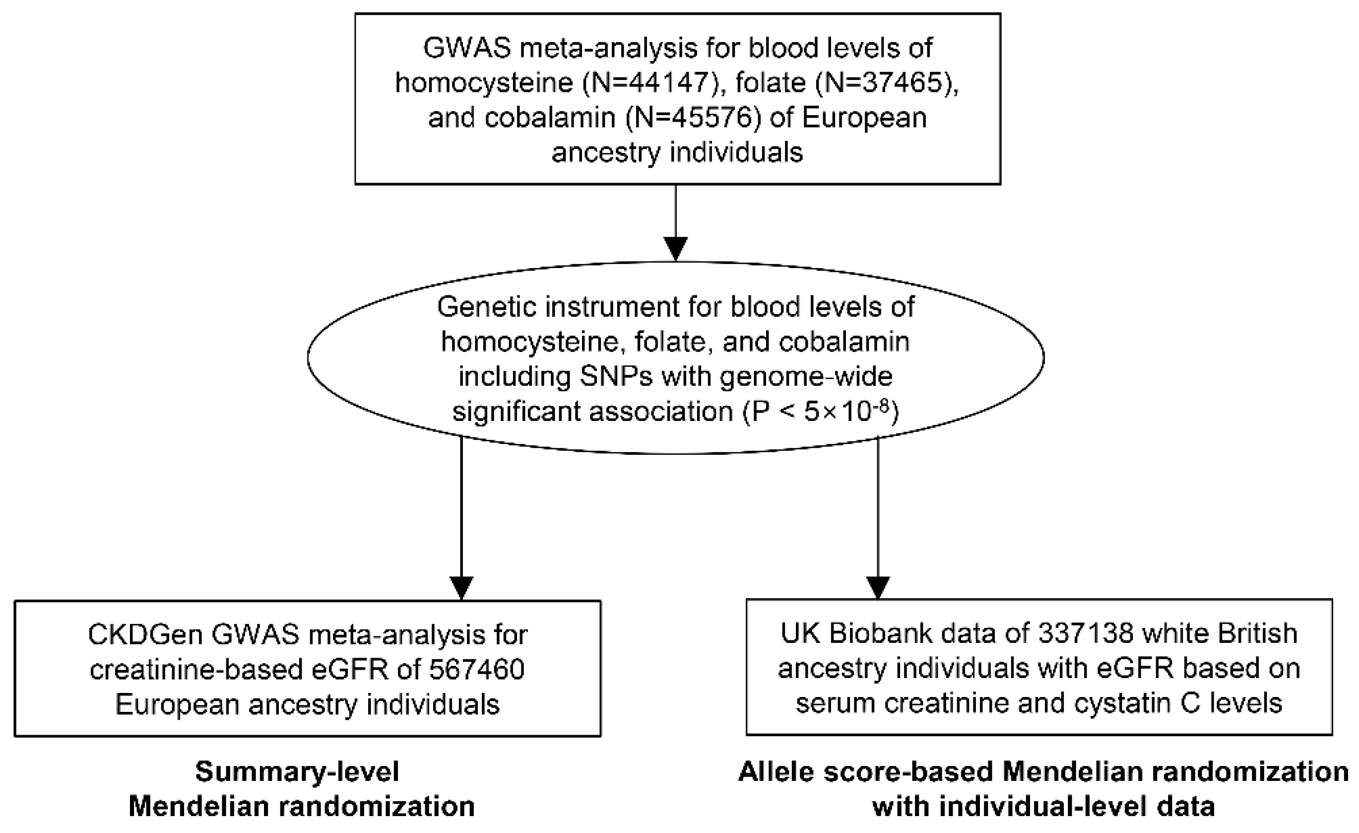
The study was performed in accordance with the Declaration of Helsinki. The study was approved by the Institutional Review Boards of Seoul National University Hospital (No. E-2070-048-1140) and the U.K. Biobank consortium (application No. 53799). As the study investigated anonymous databases or summary-level data, the requirement for informed consent was waived by the institutional review boards.

2.2. Study Setting

This study was an MR analysis including outcome assessment in two population-scale datasets (Figure 1). Genetic instruments for blood homocysteine, folate, and cobalamin levels were introduced from a previous genome-wide association study (GWAS) analyses [16,17]. First, the genetic instrument was applied to the summary-level MR, and the outcome summary statistics for a kidney function marker, eGFR, were provided by the CKDGen consortium, which is the largest in scale to date [18]. Second, the MR analysis was performed with individual-level data from the U.K. Biobank for replication.

2.3. Genetic Instrument

The study utilized the previous GWAS meta-analysis results for blood total homocysteine, folate, and cobalamin levels [16,17]. The single nucleotide polymorphisms (SNPs) reaching the genome-wide significance threshold (p < 5 × 10⁻⁸) for the exposure phenotypes were selected from the previous GWAS, which included 44,147, 37,465, and 45,576 individuals of European ancestry for total homocysteine, folate, and cobalamin levels, respectively [16,17]. The utilized SNPs were not in linkage disequilibrium (r2 < 0.1 in the 1000 G European population). The summary statistics of the genetic instrument for blood total homocysteine (N of SNPs = 18), folate (N of SNPs = 3), and cobalamin levels (N of SNPs = 14) are presented in Table 1. As the studied exposure biomarkers are in a metabolic pathway, genetic correlation between the instruments for each studied biomarker was identified; 1 SNP on the methylenetetrahydrofolate reductase (MTHFR) gene overlapped, and another SNP was in linkage disequilibrium between the genetic instrument for homocysteine and folate level. Similarly, 4 SNPs of the genetic instrument for homocysteine level were correlated with the 4 SNPs predicting cobalamin levels.

2.4. Considerations for Key Assumption of MR

The MR analysis requires that three assumptions be met to demonstrate causal effects [13]. First, the relevance assumption is that the genetic instrument should be strongly associated with the exposure of interest, and the assumption was met by utilizing the SNPs reaching genome-wide significant level association. Second, the independence association is that the genetic instrument should not be associated with confounders. We performed well-known MR sensitivity analysis available for summary-level MR, relaxing this assumption for some of the instruments [19,20]. Third, the exclusion-restriction assumption is that the causal effects should be through the exposure of interest and cannot be formally tested. However, median-based sensitivity MR methods relax this assumption in up to 50% of the genetic instruments and are thus considered a sensitivity analysis for this assumption [20]. Further, we tested the causal effects from a single variant that is biologically proven for its effect on the homocysteine metabolic pathway and the causal estimates from the single-variant MR, which would be minimally biased from a horizontal pleiotropic pathway.

2.5. Summary-Level MR with the CKDGen Data

As the genetic instruments were from GWAS results of individuals of European ancestry, the CKDGen data for log-transformed eGFR, determined by serum creatinine levels, of 567,460 individuals of European ancestry were downloaded from the public repository (URL: https://ckdgen.imbi.uni-freiburg.de/, last accessed date 7 March 2021) [18]. The study meta-analyzed 85 GWAS results; the population had a median age of 50.1 years with 48% males, and the median eGFR value was 91.4 mL/min/1.73 m2.
In the summary-level MR, the SNPs that were nonoverlapping were excluded during the harmonization of the summary statistics [21]. To ensure that the effects were from genetic predisposition for the studied biomarkers to eGFR, rather than the reverse direction, Steiger filtering was performed as previously described [22].
The main MR method was the conventional fixed-effects inverse variance weighted method. As the inverse variance weighted method can be biased from a pleiotropic effect, additional sensitivity MR analysis is necessary to test the attainment of the MR assumptions. First, MR-Egger regression with bootstrapped standard errors was performed, and MR-Egger regression provides pleiotropy-robust causal estimates [19]. Additionally, the MR-Egger intercept p values were calculated, which is the formal test for detecting the presence of directional pleiotropy. Second, the penalized weighted median method was implemented, which derives valid causal estimates even under conditions when invalid instruments are present [20]. The median-based method relaxes the independence and the exclusion-restriction assumption for up to 50% of the instrumented weights. Third, we performed the contamination mixture method, which detects groups of genetic variants with similar causal estimates and performs robust MR analysis in the presence of invalid instruments [23].
In a separate analysis, we instrumented a single variant (rs1801133), which is on the MTHFR gene, which codes the enzyme involved in homocysteine–folate metabolism. Implementing a single biologically proven variant has advantages, as the analysis would give pleiotropy-robust results, although the instrumental power generally decreases by limiting the number of SNPs included in the genetic instrument. The SNP has been well studied for its effect on homocysteine levels and health outcomes mediated by blood homocysteine or folate levels [24]. The SNP showed the strongest association strength with blood homocysteine level in the currently implemented GWAS meta-analysis (p = 4.34 × 10−104). As the effect from the single SNP would hardly be from a horizontal pleiotropic pathway, the causal estimates from the single variant would certainly attain the key MR assumptions. In this analysis, the Wald ratio method was the MR analysis tool used to yield causal estimates.
The effect sizes were transformed to a % change in eGFR units. The summary-level MR analysis was performed by the TwoSampleMR package in R (version 4.0.2, the R foundation) [25], and two-sided p values <0.05 were considered significant.

2.6. Allele-Score-Based MR with Individual-Level Data in the U.K. Biobank

The U.K. Biobank is a prospective population-based cohort of >500,000 individuals aged 40–69 years collected from 2006 to 2010 in the United Kingdom [26,27]. The data have strength for our MR analysis, as the U.K. Biobank cohort is independent from the CKDGen data. Thus, the analysis with the data would be an independent replication of the summary-level MR results. In addition, as the data are phenotyped for multiple clinical characteristics, adjustments for important comorbidities related to kidney function were possible. The limitation of the data is that healthy volunteer bias is present, and the U.K. Biobank participants had lower prevalence of CKD than the general population [28].
For the analysis, we included the U.K. Biobank data of unrelated individuals of white British ancestry. Those who were outliers in terms of heterozygosity or missing rate and those with sex chromosome aneuploidy were excluded, resulting in 337,138 individuals assessed for allele-score-based MR [15]. The median age was 58 years, and 46% were males; the median eGFR was 92.5 mL/min/1.73 m2 determined by the Chronic Kidney Disease Epidemiology Collaboration (CKD-EPI) equation based on serum creatinine levels.
We calculated allele scores for the exposures by multiplying the gene dosage matrix with the effect sizes of the genetic instrument by using PLINK 2.0 (version alpha 2.3) [29]. The associations between the allele scores and eGFR were investigated by linear regression analysis, adjusted for age, sex, genotype measurement batch, and the first 10 principal components. Additional sensitivity analysis was performed, including hypertension, diabetes mellitus, and obesity as the covariates in the regression model.

3. Results

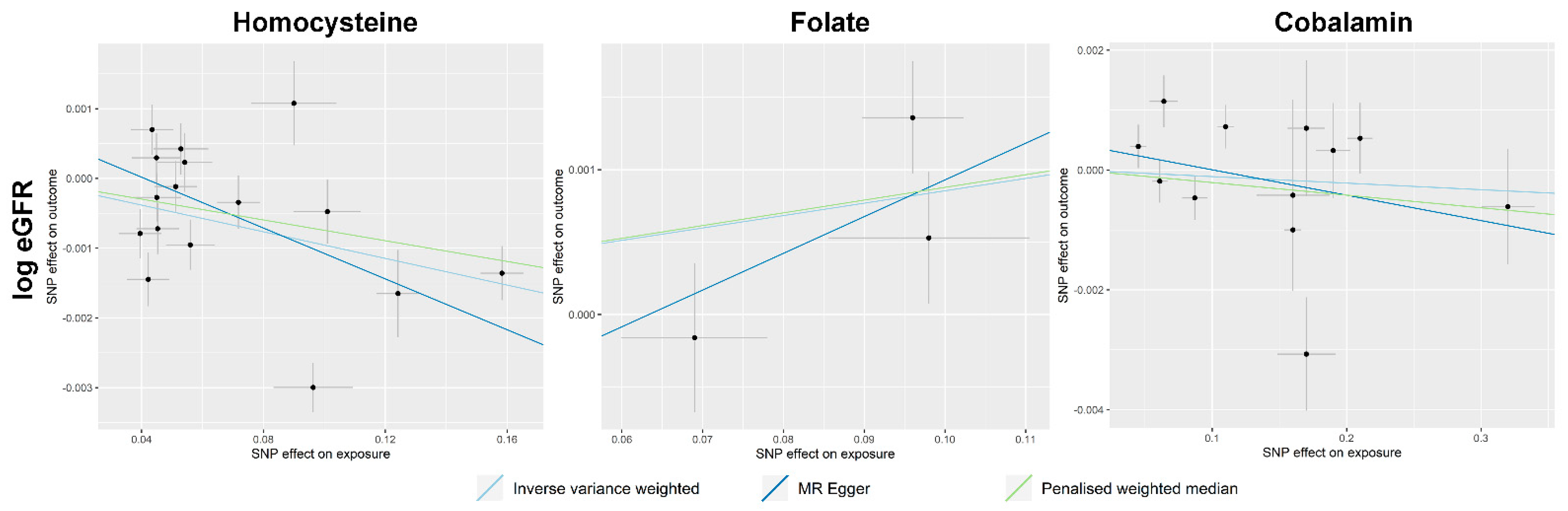
3.1. Summary-Level MR Results

In the summary-level MR, 16 genetic variants remained in the genetic instrument for homocysteine, as two SNPs were excluded due to nonoverlapping. The genetic instrument for folate included three SNPs, as none of them were disregarded during the harmonization process. There were 12 SNPs remaining as the genetic instrument for cobalamin as two SNPs were disregarded due to nonoverlapping. Steiger filtering showed that the causal effects from the genetic variants were from exposures to outcome, and none of the SNPs were disregarded by the filtering process.
The scatter plots demonstrating the summary-level MR results are presented in Figure 2. The causal estimates demonstrate that high total homocysteine levels were causally linked to low eGFR values (Table 2). The results were significant in both the MR-Egger and penalized weighted median methods, and the MR-Egger intercept p value indicates the absence of significant directional pleiotropy. On the other hand, high genetically predicted folate levels were significantly associated with high eGFR values, and the findings were consistent throughout the utilized MR sensitivity analyses. However, genetically predicted cobalamin levels were nonsignificantly associated with eGFR outcomes in the CKDGen data.
When the single SNP rs1801133 was implemented as the genetic instrument, the homocysteine-increasing genetic predisposition was significantly associated with lower eGFR (per 1 standard deviation increase in the genetic predisposition, eGFR change −0.85 (95% confidence interval −1.33; −0.38) %.

3.2. Allele-Score-Based MR Results

A high allele score for blood total homocysteine level was significantly associated with low eGFR (Table 3). Even after phenotypical hypertension, diabetes mellitus, and obesity status were additionally adjusted, the results remained significant. On the other hand, a high allele score for folate level was significantly associated with high eGFR values. The results were similar when the phenotypical covariates were adjusted. The allele score for cobalamin level was nonsignificantly associated with the outcomes, similar to the summary-level MR results.

4. Discussion

In this MR analysis, we identified that a high total blood homocysteine level was causally linked to low eGFR in the summary-level MR with the largest genetic dataset to date for eGFR provided by the CKDGen consortium. The findings were consistent for pleiotropy-robust MR sensitivity analysis, and the results were replicated by independent population-scale data from the U.K. Biobank. Therefore, this study supports that a high blood homocysteine level is a causative factor for reduced kidney function.
Observational associations between blood homocysteine levels and kidney function or cardiovascular disease have been repeatedly reported. However, the blood level of a biomarker can be increased due to impaired kidney clearance (e.g., reverse causation), and whether blood homocysteine levels can cause decreased kidney function cannot be answered by previous observational findings. A previous subanalysis of a randomized trial including Chinese individuals reported that folate supplementation, with a drop in blood homocysteine levels, can delay the progression of kidney function impairment [10]. However, the study was limited to an Asian ethnic population, and a replicative finding for kidney function outcome has rarely been reported. Additionally, conflicting results with composite vitamin B supplementation were reported in Canadian diabetic kidney disease patients [12]. Therefore, additional studies investigating the causal effects of blood homocysteine levels on kidney function are warranted, and in this study, this was performed through MR analysis, which has the strength to identify causal estimates from a genetically predictable biomarker. Through our efforts to attain the MR assumptions and as the direction of the genetic effect was inspected, the consistent findings of our MR analysis support that a high blood homocysteine level causally decreases kidney function. Our study has strength, as the study findings were replicated in two population-scale datasets and included individuals of European ancestry in whom the effects of homocysteine on kidney function had yet to be reported. Furthermore, the study population was not limited to those with advanced CKD, and most of the studied individuals had preserved eGFR; thus, the adverse effects of blood homocysteine on kidney function may be present in the general population.
Homocysteine is a nonprotein amino acid present in the methionine metabolism pathway. In this pathway, folate is activated by the MTHFR enzyme, which converts 5,10-methylenetetrahydrofolate to 5-methyletetrahydrofolate, which is necessary for converting homocysteine to another amino acid. High concentrations of blood homocysteine have been reported to cause pathologic vascular changes [30]. Homocysteine binds to proteins and modifies their function through the so-called homocysteinylation process, which occurs proportionally to the blood homocysteine level. When homocysteine binds with a thiol group, the redox status of a protein can be altered, further leading to oxidative stress injury [31]. Homocysteine can induce collagen synthesis, resulting in vascular endothelial dysfunction and stiffness [32]. In addition, homocysteine has been reported to be linked to inflammation, which may further contribute to the consequent facilitation of atherosclerotic injury [33]. Such effects of homocysteine on vascular injury may be the mechanism of the adverse causal effects of high blood homocysteine levels, supported by the MR analysis of small vessel stroke in a previous study and kidney function in the current analysis [14].
Based on our findings, a future clinical trial may target homocysteine-lowering therapy to prevent kidney function loss. As folic acid supplementation is the most well-recognized method of lowering blood homocysteine levels, such an intervention may initially be considered. The benefits of folate supplementation for the cardiovascular outcome of CKD patients have been suggested by a previous meta-analysis, particularly when homocysteine was effectively lowered [34]. However, some contradictory reports were present in the United States and Canada, and the efficacy of vitamin B supplementation to lower blood homocysteine levels was absent as the secondary prevention of cardiovascular diseases or diabetic kidney disease [11,12,35]. One of the previous studies addressed a nonvitamin homocysteine-lowering strategy that may be considered due to possible vitamin B toxicity in those with reduced kidney function who are already receiving a folate-fortified diet [12]. Therefore, although folate supplementation may be a worthwhile intervention for lowering blood homocysteine levels to prevent kidney function impairment, particularly in European or Asian countries without a folate fortification policy, other future strategies that effectively lower blood homocysteine levels may also be considered. Specifically, multiple vitamin B supplementation in populations receiving a folate-fortified diet may be discouraged, considering the possibility of a harmful effect of additive intake of vitamin B [12,35]. Furthermore, regarding the previous negative results in a population with mostly established kidney function impairment [12], if a folate supplement is being trialed, the intervention may target primary, rather than secondary, prevention of CKD in the general population without reduced kidney clearance. Those with a high blood homocysteine level or with folate deficiency may be the primary target group for such trials.
The current study has several limitations. First, the genetic effect sizes in MR analysis cannot directly reflect the potential clinical effect size of the related intervention [36]. The actual effects of the homocysteine-lowering strategy on kidney function should be answered in a future clinical trial, and the possibility that the efficacy may be different according to the trialed homocysteine-lowering intervention should be taken into account. Second, as the eGFR outcome was from those with relatively preserved kidney function, the effects of homocysteine levels in those with advanced CKD cannot be investigated herein. Thus, the study cannot advocate a trial for secondary prevention of CKD by homocysteine-lowering interventions, which was also discouraged by a previous clinical trial [12]. Third, the study was based on individuals of European ancestry; thus, the findings may not be generalizable to those with other ethnic backgrounds. Last, the study outcome estimated kidney function based on creatinine values, which may be affected by one’s body shape or diet. A future clinical study may consider including measured kidney function or more robust kidney function parameters.
In conclusion, a high blood homocysteine level is causally linked to a low eGFR. A future clinical trial investigating the efficacy of homocysteine-lowering interventions is warranted for the primary prevention of kidney function impairment.

Author Contributions

The corresponding author attests that all listed authors meet the authorship criteria and that no others meeting the criteria have been omitted. S.P., H.L., K.K., K.W.J., and D.K.K. contributed to the conception and design of the study. S.L., Y.K., S.C., Y.C.K., S.S.H., J.P.L., K.W.J., C.S.L., Y.S.K., and D.K.K. provided statistical advice and interpreted the data. S.P. and K.K. performed the main statistical analysis, assisted by S.L., and Y.K., H.L., J.P.L., K.W.J., C.S.L., Y.S.K., and D.K.K. provided advice regarding the data interpretation. Y.C.K., S.S.H., H.L., J.P.L., K.W.J., C.S.L., and Y.S.K. provided material support during the study. S.P. and D.K.K. had full access to all data in the study and take responsibility for the integrity of the data and the accuracy of the data analysis. All authors participated in drafting the manuscript. All authors reviewed the manuscript and approved the final version to be published.

Funding

This work was supported by the Industrial Strategic Technology Development Program Development of biocore technology (10077474, Development of early diagnosis technology for acute/chronic renal failure) funded by the Ministry of Trade, Industry & Energy (MOTIE, Korea) and a grant from SNU R&DB Foundation (800-20190571). The study was performed independently by the authors.

Institutional Review Board Statement

The study was approved by the Institutional Review Boards of Seoul National University Hospital (No. E-2070-048-1140).

Informed Consent Statement

The need for acquiring informed consent was waived because this study analyzed anonymous public database and summary statistics.

Data Availability Statement

The data described in the manuscript will be made available from the U.K. Biobank consortium after acquiring approval (URL: https://biobank.ctsu.ox.ac.uk/crystal/docs.cgi?id=1, last accessed 7 March 2021). The code book and analytic code will be made available by the corresponding author upon reasonable request.

Acknowledgments

The study was based on the data provided by the U.K. Biobank consortium (application No. 53799). We thank the investigators of the previous studies who provided valuable genetic summary statistics for this study.

Conflicts of Interest

The authors declare no conflict of interest.

References

  1. Bikbov, B.; Purcell, C.A.; Levey, A.S.; Smith, M.; Abdoli, A.; Abebe, M.; Adebayo, O.M.; Afarideh, M.; Agarwal, S.K.; Agudelo-Botero, M.; et al. Global, regional, and national burden of chronic kidney disease, 1990-2017: A systematic analysis for the Global Burden of Disease Study 2017. Lancet 2020, 395, 709–733. [Google Scholar] [CrossRef] [Green Version]
  2. Soinio, M.; Marniemi, J.; Laakso, M.; Lehto, S.; Rönnemaa, T. Elevated plasma homocysteine level is an independent predictor of coronary heart disease events in patients with type 2 diabetes mellitus. Ann. Intern. Med. 2004, 140, 94–100. [Google Scholar] [CrossRef] [Green Version]
  3. Nygård, O.; Nordrehaug, J.E.; Refsum, H.; Ueland, P.M.; Farstad, M.; Vollset, S.E. Plasma Homocysteine Levels and Mortality in Patients with Coronary Artery Disease. N. Engl. J. Med. 1997, 337, 230–237. [Google Scholar] [CrossRef] [PubMed]
  4. Heinz, J.; Kropf, S.; Luley, C.; Dierkes, J. Homocysteine as a Risk Factor for Cardiovascular Disease in Patients Treated by Dialysis: A Meta–analysis. Am. J. Kidney Dis. 2009, 54, 478–489. [Google Scholar] [CrossRef] [PubMed]
  5. Moustapha, A.; Naso, A.; Nahlawi, M.; Gupta, A.; Arheart, K.L.; Jacobsen, D.W.; Robinson, K.; Dennis, V.W. Prospective study of hy-perhomocysteinemia as an adverse cardiovascular risk factor in end-stage renal disease. Circulation 1998, 97, 138–141. [Google Scholar] [CrossRef] [PubMed]
  6. Bostom, A.G.; Lathrop, L. Hyperhomocysteinemia in end-stage renal disease: Prevalence, etiology, and potential relationship to arteriosclerotic outcomes. Kidney Int. 1997, 52, 10–20. [Google Scholar] [CrossRef] [PubMed] [Green Version]
  7. Friedman, A.N.; Hunsicker, L.G.; Selhub, J.; Bostom, A.G. Total Plasma Homocysteine and Arteriosclerotic Outcomes in Type 2 Diabetes with Nephropathy. J. Am. Soc. Nephrol. 2005, 16, 3397–3402. [Google Scholar] [CrossRef] [PubMed] [Green Version]
  8. Menon, V.; Sarnak, M.J.; Greene, T.; Wang, X.; Pereira, A.A.; Beck, G.J.; Kusek, J.W.; Selhub, J.; Collins, A.J.; Levey, A.S.; et al. Relationship Between Homocysteine and Mortality in Chronic Kidney Disease. Circulation 2006, 113, 1572–1577. [Google Scholar] [CrossRef]
  9. Huo, Y.; Li, J.; Qin, X.; Huang, Y.; Wang, X.; Gottesman, R.F.; Tang, G.; Wang, B.; Chen, D.; He, M.; et al. Efficacy of folic acid therapy in primary prevention of stroke among adults with hypertension in China: The CSPPT randomized clinical trial. JAMA 2015, 313, 1325–1335. [Google Scholar] [CrossRef] [PubMed]
  10. Xu, X.; Qin, X.; Li, Y.; Sun, D.; Wang, J.; Liang, M.; Wang, B.; Huo, Y.; Hou, F.F. Efficacy of Folic Acid Therapy on the Progression of Chronic Kidney Disease: The Renal Substudy of the China Stroke Primary Prevention Trial. JAMA Intern. Med. 2016, 176, 1443–1450. [Google Scholar] [CrossRef]
  11. Martí-Carvajal, A.J.; Solà, I.; Lathyris, D.; Dayer, M. Homocysteine-lowering interventions for preventing cardiovascular events. Cochrane Database Syst. Rev. 2017, 8, Cd006612. [Google Scholar] [CrossRef] [PubMed]
  12. House, A.A.; Eliasziw, M.; Cattran, D.C.; Churchill, D.N.; Oliver, M.J.; Fine, A.; Dresser, G.K.; Spence, J.D. Effect of B-vitamin therapy on progression of diabetic nephropathy: A randomized controlled trial. JAMA 2010, 303, 1603–1609. [Google Scholar] [CrossRef] [PubMed] [Green Version]
  13. Davies, N.M.; Holmes, M.V.; Smith, G.D. Reading Mendelian randomisation studies: A guide, glossary, and checklist for clinicians. BMJ 2018, 362, k601. [Google Scholar] [CrossRef] [PubMed] [Green Version]
  14. Larsson, S.C.; Traylor, M.; Markus, H.S. Homocysteine and small vessel stroke: A mendelian randomization analysis. Ann. Neurol. 2019, 85, 495–501. [Google Scholar] [CrossRef] [PubMed] [Green Version]
  15. Park, S.; Lee, S.; Kim, Y.; Lee, Y.; Kang, M.W.; Kim, K.; Kim, Y.C.; Han, S.S.; Lee, H.; Lee, J.P.; et al. Short or Long Sleep Duration and CKD: A Mendelian Randomization Study. J. Am. Soc. Nephrol. 2020, 31, 2937–2947. [Google Scholar] [CrossRef] [PubMed]
  16. van Meurs, J.B.; Pare, G.; Schwartz, S.M.; Hazra, A.; Tanaka, T.; Vermeulen, S.H.; Cotlarciuc, I.; Yuan, X.; Mälarstig, A.; Bandinelli, S.; et al. Common genetic loci influencing plasma homocysteine concentrations and their effect on risk of coronary artery disease. Am. J. Clin. Nutr. 2013, 98, 668–676. [Google Scholar] [CrossRef] [PubMed]
  17. Grarup, N.; Sulem, P.; Sandholt, C.H.; Thorleifsson, G.; Ahluwalia, T.S.; Steinthorsdottir, V.; Bjarnason, H.; Gudbjartsson, D.F.; Magnusson, O.T.; Sparsø, T.; et al. Genetic Architecture of Vitamin B12 and Folate Levels Uncovered Applying Deeply Sequenced Large Datasets. PLoS Genet. 2013, 9, e1003530. [Google Scholar] [CrossRef] [PubMed] [Green Version]
  18. Wuttke, M.; Li, Y.; Li, M.; Sieber, K.B.; Feitosa, M.F.; Gorski, M.; Tin, A.; Wang, L.; Chu, A.Y.; Hoppmann, A.; et al. A catalog of genetic loci associated with kidney function from analyses of a million individuals. Nat. Genet. 2019, 51, 957–972. [Google Scholar] [CrossRef] [Green Version]
  19. Bowden, J.K.; Smith, G.D.; Burgess, S. Mendelian randomization with invalid instruments: Effect estimation and bias detection through Egger regression. Int. J. Epidemiol. 2015, 44, 512–525. [Google Scholar] [CrossRef] [Green Version]
  20. Bowden, J.; Smith, G.D.; Haycock, P.C.; Burgess, S. Consistent Estimation in Mendelian Randomization with Some Invalid Instruments Using a Weighted Median Estimator. Genet. Epidemiol. 2016, 40, 304–314. [Google Scholar] [CrossRef] [PubMed] [Green Version]
  21. Hartwig, F.P.; Davies, N.M.; Hemani, G.; Smith, G.D. Two-sample Mendelian randomization: Avoiding the downsides of a powerful, widely applicable but potentially fallible technique. Int. J. Epidemiol. 2016, 45, 1717–1726. [Google Scholar] [CrossRef] [PubMed] [Green Version]
  22. Hemani, G.; Tilling, K.; Davey Smith, G. Orienting the causal relationship between imprecisely measured traits using GWAS summary data. PLoS Genet. 2017, 13, e1007081. [Google Scholar]
  23. Burgess, S.; Foley, C.N.; Allara, E.; Staley, J.R.; Howson, J.M.M. A robust and efficient method for Mendelian randomization with hundreds of genetic variants. Nat. Commun. 2020, 11, 1–11. [Google Scholar] [CrossRef] [PubMed] [Green Version]
  24. Jin, H.; Cheng, H.; Chen, W.; Sheng, X.; Levy, M.A.; Brown, M.J.; Tian, J. An evidence-based approach to globally assess the covari-ate-dependent effect of the MTHFR single nucleotide polymorphism rs1801133 on blood homocysteine: A systematic review and meta-analysis. Am. J. Clin. Nutr. 2018, 107, 817–825. [Google Scholar] [CrossRef] [Green Version]
  25. Hemani, G.; Zheng, J.; Elsworth, B.; Wade, K.H.; Haberland, V.; Baird, D.; Laurin, C.; Burgess, S.; Bowden, J.; Langdon, R.; et al. The MR-Base platform supports systematic causal inference across the human phenome. eLife 2018, 7. [Google Scholar] [CrossRef]
  26. Bycroft, C.; Freeman, C.; Petkova, D.; Band, G.; Elliott, L.T.; Sharp, K.; Motyer, A.; Vukcevic, D.; Delaneau, O.; O’Connell, J.; et al. The UK Biobank resource with deep phenotyping and genomic data. Nat. Cell Biol. 2018, 562, 203–209. [Google Scholar] [CrossRef] [PubMed] [Green Version]
  27. Sudlow, C.; Gallacher, J.; Allen, N.; Beral, V.; Burton, P.; Danesh, J.; Downey, P.; Elliott, P.; Green, J.; Landray, M.; et al. UK Biobank: An Open Access Resource for Identifying the Causes of a Wide Range of Complex Diseases of Middle and Old Age. PLoS Med. 2015, 12, e1001779. [Google Scholar] [CrossRef] [Green Version]
  28. Fry, A.; Littlejohns, T.J.; Sudlow, C.; Doherty, N.; Adamska, L.; Sprosen, T.; Collins, R.; E Allen, N. Comparison of Sociodemographic and Health-Related Characteristics of UK Biobank Participants with Those of the General Population. Am. J. Epidemiol. 2017, 186, 1026–1034. [Google Scholar] [CrossRef] [Green Version]
  29. Chang, C.C.; Chow, C.C.; Tellier, L.C.A.M.; Vattikuti, S.; Purcell, S.M.; Lee, J.J. Second-generation PLINK: Rising to the challenge of larger and richer datasets. GigaScience 2015, 4, 7. [Google Scholar] [CrossRef] [PubMed]
  30. Stamler, J.S.; Osborne, J.A.; Jaraki, O.; Rabbani, L.E.; Mullins, M.; Singel, D.; Loscalzo, J. Adverse vascular effects of homocysteine are modulated by endothelium-derived relaxing factor and related oxides of nitrogen. J. Clin. Investig. 1993, 91, 308–318. [Google Scholar] [CrossRef]
  31. Škovierová, H.; Vidomanová, E.; Mahmood, S.; Sopková, J.; Drgová, A.; Červeňová, T.; Halašová, E.; Lehotský, J. The Molecular and Cellular Effect of Homocysteine Metabolism Imbalance on Human Health. Int. J. Mol. Sci. 2016, 17, 1733. [Google Scholar] [CrossRef]
  32. Sen, U.; Mishra, P.K.; Tyagi, N.; Tyagi, S.C. Homocysteine to Hydrogen Sulfide or Hypertension. Cell Biophys. 2010, 57, 49–58. [Google Scholar] [CrossRef] [PubMed] [Green Version]
  33. Au-Yeung, K.K.; Woo, C.W.; Sung, F.L.; Yip, J.C.; Siow, Y.L.; O, K. Hyperhomocysteinemia activates nuclear factor-kappaB in endothelial cells via oxidative stress. Circ. Res. 2004, 94, 28–36. [Google Scholar] [CrossRef] [Green Version]
  34. Qin, X.; Huo, Y.; Langman, C.B.; Hou, F.; Chen, Y.; Matossian, D.; Xu, X.; Wang, X. Folic Acid Therapy and Cardiovascular Disease in ESRD or Advanced Chronic Kidney Disease: A Meta-Analysis. Clin. J. Am. Soc. Nephrol. 2010, 6, 482–488. [Google Scholar] [CrossRef] [PubMed]
  35. Bønaa, K.H.; Njølstad, I.; Ueland, P.M.; Schirmer, H.; Tverdal, A.; Steigen, T.; Wang, H.; Nordrehaug, J.E.; Arnesen, E.; Rasmussen, K. Homocysteine Lowering and Cardiovascular Events after Acute Myocardial Infarction. N. Engl. J. Med. 2006, 354, 1578–1588. [Google Scholar] [CrossRef] [PubMed] [Green Version]
  36. Burgess, S.; Butterworth, A.; Malarstig, A.; Thompson, S.G. Use of Mendelian randomisation to assess potential benefit of clinical intervention. BMJ 2012, 345, e7325. [Google Scholar] [CrossRef] [Green Version]
Figure 1. Study flow diagram.
Nutrients 13 00906 g001
Figure 2. Scatter plots showing the results for summary-level Mendelian randomization analysis results. The x-axes indicate the effect for exposure variables, and the y-axes indicate the effect for outcome (log-transformed eGFR). The analysis was performed for 567,460 individuals of European ancestry of the CKDGen consortium.
Nutrients 13 00906 g002
Table 1. The summary statistics of the genetic instrument for blood total homocysteine (N of SNPs = 18), folate (N of SNPs = 3), and cobalamin levels (N of SNPs = 14).
Genetically Predicted ExposureSNPEffect AlleleEffect Allele FrequencyBeta p Value
Homocysteiners1801133A0.340.15834.34E-104
rs2275565T0.21−0.05421.96E-10
rs7422339A0.330.08644.58E-27
rs9369898A0.620.04492.17E-10
rs7130284T0.07−0.12421.88E-20
rs154657A0.470.09631.74E-43
rs234709T0.45−0.07183.90E-24
rs4660306T0.330.04352.33E-09
rs548987C0.130.05971.12E-08
rs42648A0.4−0.03951.97E-08
rs1801222A0.340.04538.43E-10
rs2251468A0.65−0.05121.28E-12
rs838133A0.450.04227.48E-09
rs12134663A0.8−0.1012.54E-21
rs12780845A0.650.05297.80E-10
rs957140A0.45−0.0452.43E-10
rs12921383T0.87−0.098.22E-11
rs2851391T0.470.0561.70E-12
Folaters1801133G0.670.0969.50E-53
rs17421511G0.830.0981.80E-15
rs652197C0.180.0691.20E-14
Cobalaminrs602662A0.60.162.40E-139
rs34324219C0.880.211.10E-111
rs34528912T0.040.172.10E-15
rs117456053G0.980.161.90E-09
rs1801222G0.590.113.30E-75
rs56077122A0.330.0874.80E-21
rs2336573T0.030.328.40E-59
rs1131603C0.0550.194.90E-49
rs5753231C0.790.0647.50E-10
rs41281112C0.950.178.90E-35
rs1141321C0.630.0613.60E-26
rs3742801T0.290.0451.70E-13
rs2270655G0.940.0662.20E-13
rs12272669A0.00220.513.00E-09
The genetic effect sizes are to reflect one standard deviation increase in the phenotype.
Table 2. Summary-level Mendelian randomization results with the CKDGen data.
Genetically Predicted
Exposure
MR MethodMR-Egger Intercept
p Value
Beta of eGFR Change (%)
and 95% CI
p Value for
Causal Estimate
HomocysteineInverse variance weighted0.54−0.95 (−1.21, −0.69)<0.001
MR-Egger−1.81 (−2.59, −1.02)<0.001
Penalized weighted median−0.74 (−1.15, −0.33)<0.001
Contamination
mixture
−0.60 (−1.09, −0.20)<0.001
FolateInverse variance weighted0.510.86 (0.30, 1.42)0.002
MR-Egger2.57 (0.70, 4.47)0.001
Penalized weighted median0.88 (0.20, 1.56)0.011
Contamination
mixture
0.90 (0.10, 1.61)<0.001
CobalaminInverse variance weighted0.33−0.11 (−0.33, 0.11)0.333
MR-Egger−0.42 (−0.84, 0.00)0.025
Penalized weighted median−0.21 (−0.59, 0.17)0.276
Contamination
mixture
−0.10 (−0.40, 0.20)0.092
MR = Mendelian randomization, CI = confidence interval; The analysis was performed with the summary statistics for log-transformed estimated glomerular filtration rate (eGFR) from the genome-wide association study meta-analysis conducted by the CKDGen consortium, which included 567,460 individuals of European ancestry. The effect sizes of the causal estimates were from a one standard deviation increase in the genetic predisposition for the plasma biomarkers towards eGFR change (%).
Table 3. Allele-score-based Mendelian randomization results with the U.K. Biobank data.
OutcomeGenetically Predicted ExposureMain Analysis aClinical Covariates Adjusted b
Beta for eGFR (Continuous) and 95% CIpBeta for eGFR (Continuous) and 95% CIp
eGFR (mL/min/1.73 m2) Homocysteine−0.108 (−0.148, −0.068)1.6 × 10⁻⁷−0.106 (−0.147, −0.066)2.3 × 10⁻⁷
Folate0.067 (0.027, 0.108)0.0010.061 (0.021, 0.101)0.003
Cobalamin0.005 (−0.035, 0.046)0.8020.004 (−0.037, 0.044)0.855
eGFR = estimated glomerular filtration rate, CI = confidence interval; All allele scores were scaled to a one standard deviation increase; a The logistic regression model was adjusted for age, sex, genotype measurement batch, and the first 10 principal components of the genetic information. The analysis was performed in 337,138 individuals of white British ancestry in the U.K. Biobank cohort and a total of 321,260 individuals had creatinine-based eGFR values. b Phenotypical hypertension, diabetes mellitus, and obesity were added to the main model. The analysis was performed with 333,107 individuals of white British ancestry from the U.K. Biobank with complete information for the additional covariates, and among them, 317,737 individuals had available creatinine-based eGFR values.
Publisher’s Note: MDPI stays neutral with regard to jurisdictional claims in published maps and institutional affiliations.

