
Worldwide Overview of Neospora spp. Infection in Equids Diagnosed by Serological Tests: Systematic Review and Meta-Analysis
 and
and 
Abstract
:1. Introduction
2. Results
3. Discussion
4. Materials and Methods
4.1. Data Base
4.2. Selection Criteria
4.3. Meta-Analysis Approach
5. Conclusions
Author Contributions
Funding
Conflicts of Interest
References
- Mimoun, L.; Steinman, A.; Kliachko, Y.; Tirosh-Levy, S.; Schvartz, G.; Blinder, E.; Baneth, G.; Mazuz, M.L. Neospora spp. Seroprevalence and Risk Factors for Seropositivity in Apparently Healthy Horses and Pregnant Mares. Animals 2022, 12, 2699. [Google Scholar] [CrossRef] [PubMed]
- Bezerra, R.A.; Lima, B.A.; Alvares, F.B.V.; Rossi, G.A.M.; Braga, F.R.; Melo, R.P.B.; Mota, R.A.; Vilela, V.L.R.; Feitosa, T.F. Detection of Anti-Neospora caninum IgG in Blood Serum and Colostrum Samples in Naturally Infected Sheep and in Their Newborn Offspring. Pathogens 2022, 11, 1263. [Google Scholar] [CrossRef] [PubMed]
- Feitosa, T.F.; Costa, F.T.R.; Bezerra, R.A.; Alvares, F.B.V.; Ferreira, L.C.; Mota, R.A.; Gennari, S.M.; Pena, H.F.J.; Azevedo, S.S.; Vilela, V.L.R. Vertical transmission and kinetic of antibodies anti-Neospora caninum in naturally infected lambs in the semiarid region of Brazil. Rev. Bras. Parasitol. Vet. 2021, 30, e010621. [Google Scholar] [CrossRef] [PubMed]
- Tirosh-Levy, S.; Savitsky, I.; Blinder, E.; Mazuz, M.L. The involvement of protozoan parasites in sheep abortions—A ten-year review of diagnostic results. Vet. Parasitol. 2022, 303, 109664. [Google Scholar] [CrossRef]
- Sánchez-Sánchez, R.; Vázquez-Calvo, Á.; Fernández-Escobar, M.; Regidor-Cerrillo, J.; Benavides, J.; Gutiérrez, J.; Gutiérrez-Expósito, D.; Crespo-Ramos, F.J.; Ortega-Mora, L.M.; Álvarez-García, G. Dynamics of Neospora caninum-Associated Abortions in a Dairy Sheep Flock and Results of a Test-and-Cull Control Programme. Pathogens 2021, 10, 1518. [Google Scholar] [CrossRef]
- Lindsay, D.S.; Dubey, J.P. Neosporosis, Toxoplasmosis, and Sarcocystosis in Ruminants: An Update. Vet. Clin. Food Anim. Pract. 2020, 36, 205–222. [Google Scholar] [CrossRef]
- Dubey, J.P.; Schares, G. Neosporosis in animals—The last five years. Vet. Parasitol. 2011, 180, 90–91. [Google Scholar] [CrossRef]
- Bártová, E.; Sedlák, K.; Kobédová, K.; Budíková, M.; Joel Atuman, Y.; Kamani, J. Seroprevalence and risk factors of Neospora spp. and Toxoplasma gondii infections among horses and donkeys in Nigeria, West Africa. Acta Parasitol. 2017, 62, 606–609. [Google Scholar] [CrossRef]
- Marsh, A.E.; Howem, D.K.; Wang, G.; Barr, B.C.; Cannon, N.; Conrad, P.A. Differentiation of Neospora hughesi from Neospora caninum based on their immunodominant surface antigen, SAG1 and SRS2. Int. J. Parasitol. 1999, 29, 1575–1582. [Google Scholar] [CrossRef]
- Pusterla, N.; Conrad, P.A.; Packham, A.E.; Mapes, S.M.; Finno, C.J.; Gardner, I.A.; Barr, B.C.; Ferraro, G.L.; Wilson, W.D. Endogenous transplacental transmission of Neospora hughesi in naturally infected horses. J. Parasitol. 2011, 97, 281–285. [Google Scholar] [CrossRef]
- Tavares, T.C.; Pimentel, M.M.L.; Câmara, F.V.; Lopes, K.R.; Dias, R.V.C. Análise biométrica dos equinos utilizados para tração no Município de Mossoró—RN, Brasil. Rev. Bras. Hig. Sanidade Anim. 2015, 9, 425–438. [Google Scholar] [CrossRef]
- Molento, M.B.; Vilela, V.L.R. Health evaluation of donkeys: Parasite control methods and a model for challenge infections. Braz. J. Vet. Res. Anim. Sci. 2021, 58, e174275. [Google Scholar] [CrossRef]
- Costa, P.W.L.; Oliveira, C.S.M.; Bezerra, R.A.; Alvares, F.B.V.; Formiga, V.H.A.S.; Martins, M.R.D.D.; Feitosa, T.F.; Vilela, V.L.R. Anti-Toxoplasma gondii and Anti-Neospora caninum Antibodies in Urban Traction Equids in Northeast Brazil: Seroprevalence and Risk Factors. Trop. Med. Infect. Dis. 2023, 8, 234. [Google Scholar] [CrossRef]
- Wapenaar, W.; Barkema, H.W.; VanLeeuwen, J.A.; McClure, J.T.; O’Handley, R.M.; Kwok, O.C.H.; Thulliez, P.; Dubey, J.P.; Jenkins, M.C. Comparison of serological methods for the diagnosis of Neospora caninum infection in cattle. Vet. Parasitol. 2007, 143, 166–173. [Google Scholar] [CrossRef] [PubMed]
- Javanmardi, E.; Majidiani, H.; Shariatzadeh, S.A.; Anvari, D.; Shamsinia, S.; Ghasemi, E.; Kordi, B.; Shams, M.; Asghari, A. Global seroprevalence of Neospora spp. in horses and donkeys: A systematic review and meta-analysis. Vet. Parasitol. 2020, 288, 109299. [Google Scholar] [CrossRef]
- Abu-Halaweh, M.; Abo-Shehada, M.N.; Khalil, R. Age, gender and climate associations with the seroprevalence of Neospora species infection in horses in Jordan. Rev. Bras. Parasitol. Vet. 2020, 29, e016019. [Google Scholar] [CrossRef]
- Bártová, E.; Machaèové, T.; Sedlék, K.; Budíkové, M.; Mariani, U.; Veneziano, V. Seroprevalence of antibodies of Neospora spp. and Toxoplasma gondii in horses from southern Italy. Folia Parasitol. 2015, 62, 1. [Google Scholar] [CrossRef]
- Blanco, R.D.; Patarroyo, J.H.; Vargas, M.I.; Cardona, J.A.; Araújo, L.S.; Gomez, V.E. Ocorrência de anticorpos anti-Neospora spp. em jumentos (Equus asinus) no estado de Sucre—Colômbia. Arq. Bras. Med. Vet. Zootec. 2014, 66, 450–454. [Google Scholar] [CrossRef]
- Cazarotto, C.J.; Balzan, A.; Grosskopf, R.K.; Boito, J.P.; Portella, L.P.; Vogel, F.F.; Favero, J.F.; Cucco, D.D.C.; Biazus, A.H.; Machado, G.; et al. Horses seropositive for Toxoplasma gondii, Sarcocystis spp. and Neospora spp.: Possible risk factors for infection in Brazil. Microbiol. Pathog. 2016, 99, 30–35. [Google Scholar] [CrossRef]
- Cong, W.; Nie, L.-B.; Qin, S.-Y.; Wang, W.-L.; Qian, A.-D.; Meng, Q.-F. Prevalence of Neospora spp. in donkeys in China. Parasite 2018, 25, 16. [Google Scholar]
- Cruz, I.; Vinhas, A.R.; Dubcy, J.P.; Cardoso, L.; Cotovio, M.; Lopes, A.P. First report of antibodies to Neospora spp. in horses from Portugal. Rev. Bras. Parasitol. Veterinária 2019, 28, 161–163. [Google Scholar] [CrossRef] [PubMed]
- Galvão, C.M.M.Q.; Rezende-Gondim, M.M.; Chaves, A.C.R.; Schares, G.; Ribas, J.R.L.; Gondim, L.F.P. Brazilian donkeys (Equus asinus) have a low exposure to Neospora spp. Rev. Bras. Parasitol. Vet. 2015, 24, 340–344. [Google Scholar] [CrossRef] [PubMed]
- Gennari, S.M.; Pena, H.F.J.; Lindsay, D.S.; Lopes, M.G.; Soares, H.S.; Cabral, A.D.; Vitaliano, S.N.; Amaku, M. Prevalence of antibodies against Neospora spp. and Sarcocystis neurona in donkeys from northeastern Brazil. Braz. J. Vet. Parasitol. 2016, 25, 109–111. [Google Scholar] [CrossRef]
- Jiménez, D.; Romero-Zuñiga, J.J.; Dolz, D. Serosurveillance of infectious agents in equines of the Central Valley of Costa Rica. Open Vet. J. 2014, 4, 107–112. [Google Scholar] [PubMed]
- Luza, M.; Serrano-Martínez, E.; Tantaleán, M.; Quispe, M.; Casas, G. Primer reporte de Neospora caninum, en caballos de carrera de Lima, Perú. Salud Tecnol. Vet. 2013, 1, 40–45. [Google Scholar] [CrossRef]
- Llano, H.A.B.; Soares, R.M.; Acevedo-gutierrez, L.Y.; Rodas, J.D.; Polo, G.; Borges-Silva, W.; Jesus, R.F.; Gondim, L.F.P. Seroepidemiology of Sarcocystis neurona and Neospora spp. in horses, donkeys, and mules from Colombia. Acta Trop. 2021, 220, 105970. [Google Scholar] [CrossRef]
- Machacová, T.; Bártová, E.; Di Loria, A.; Sedlák, K.; Guccione, J.; Fulgione, D.; Veneziano, V. Seroprevalence and risk factors of Neospora spp. in donkeys from Southern Italy. Vet. Parasitol. 2013, 198, 201–204. [Google Scholar] [CrossRef]
- Moreira, T.R.; Sarturi, C.; Stelmachtchuk, F.N.; Andersson, E.; Norlander, E.; Oliveira, F.L.C.; Portela, J.M.; Marcili, A.; Emanuelson, U.; Gennari, S.M.; et al. Prevalence of antibodies against Toxoplasma gondii and Neospora spp. in equids of Western Para, Brazil. Acta Trop. 2019, 189, 39–45. [Google Scholar] [CrossRef]
- Moura, A.B.; Silva, M.O.; Farias, J.A.; Vieira-Neto, A.; Souza, A.P.; Sartor, A.A.; Fonteque, J.H.; Bunn, S. Neospora spp. Antibodies in horses from two geographical regions of the state of Santa Catarina, Brazil. Rev. Bras. Parasitol. Vet. 2013, 22, 597–601. [Google Scholar] [CrossRef]
- Nazir, M.M.; Ayaz, M.M.; Ahmed, A.N.; Rasheed, I.; Faraz, A.; Akram, Q.; Akhtar, S.; Maqbool, A.; Tabassum, S.; Zheng, Y.; et al. Prevalence and risk factors for IgG antibodies to Neospora spp. in three types of equids from Southern Punjab, Pakistan. Acta Trop. 2018, 188, 240–243. [Google Scholar] [CrossRef]
- Oliveira, S.; Silva, N.Q.B.; Silveira, I.; Labruna, M.B.; Gennari, S.M.; Pena, H.F.J. Occurrences of antibodies against Toxoplasma gondii, Neospora spp., and Sarcocystis neurona in horses and dogs in the municipality of Pauliceia, São Paulo, Brazil. Braz. J. Vet. Res. Anim. Sci. 2017, 54, 277–282. [Google Scholar] [CrossRef]
- Padilla-Díaz, K.J.; Medina-Esparza, L.; Cruz-Vázquez, C.; Vitela-Mendoza, I.; Gómez-Leyva, J.F.; Quezada-Tristán, T. Detection of anti-Neospora spp. antibodies associated with different risk factors in horses from Mexico. Rev. Mex. Cienc. Pecuárias 2021, 12, 194–204. [Google Scholar] [CrossRef]
- Ribeiro, M.J.; Rosa, M.H.; Bruhn, F.R.; Garcia, A.M.; Rocha, C.M.; Guimaraes, A.M. Seroepidemiology of Sarcocystis neurona, Toxoplasma gondii and Neospora spp. among horses in the south of the state of Minas Gerais, Brazil. Rev. Bras. Parasitol. Vet. 2016, 25, 142–150. [Google Scholar] [CrossRef] [PubMed]
- Spohr, K.A.H.; Borges, A.M.C.M.; Ribeiros, T.M.P.; Jaymes, V.S.; Godoy, I.; Nakazato, L.; Dutra, V.; Aguiar, D.M. Fatores de risco associados à prevalência de anticorpos anti-Sarcocystis neurona, Neospora spp. e Toxoplasma gondii em equinos de Roraima, Amazônia. Pesqui. Vet. Bras. 2018, 38, 1337–1343. [Google Scholar] [CrossRef]
- Talafha, A.Q.; Abutarbush, S.M.; Rutley, D.L. Seroprevalence and potential risk factors associated with Neospora spp. infection among asymptomatic horses in Jordan. Korean J. Parasitol. 2015, 53, 163–167. [Google Scholar] [CrossRef]
- Tavalla, M.; Sabaghan, M.; Abdizadeh, R.; Khademvatan, S.; Rafiei, A.; Piranshahi, A.R. Seroprevalence of Toxoplasma gondii and Neospora spp. infections in Arab Horses, southwest of Iran. Jundishapur J. Microbiol. 2015, 8, 1–5. [Google Scholar] [CrossRef]
- Tirosh-Levy, S.; Steinman, A.; Minderigiu, A.; Arieli, O.; Savitski, I.; Fleiderovitz, L.; Edery, N.; Schvartz, G.; Mazuz, L.M. High Exposure to Toxoplasma gondii and Neospora Spp. in Donkeys in Israel: Serological Survey and Case Reports. Animals 2020, 10, 1921. [Google Scholar] [CrossRef]
- Waap, H.; Oliveira, U.V.; Nunes, T.; Gomes, J.; Gomes, T.; Barwald, A.; Munhoz, A.D.; Schares, G. Serological survey of Neospora spp. and Besnoitia spp. in horses in Portugal. Vet. Parasitol. Reg. Stud. Rep. 2020, 20, 100391. [Google Scholar] [CrossRef]
- Villalobos, E.M.C.; Furman, K.E.; Lara, M.; Cunha, E.M.S.; Finger, M.A.; Busch, A.P.B.; Barros, I.R.; Deconto, I.; Dornbusch, P.T.; Biondo, A.W. Detection of Neospora sp. antibodies in cart horses from urban areas of Curitiba, Southern Brazil. Braz. J. Veterenary Parasitol. 2012, 21, 68–70. [Google Scholar] [CrossRef]
- Borenstein, M.; Hedges, L.V.; Higgins, J.P.; Rothstein, H.R. Introduction to Meta-Analysis; John Wiley & Sons: Hoboken, NJ, USA, 2011. [Google Scholar]
- Moher, D.; Liberati, A.; Tetzlaff, J.; Altman, D.G. Preferred reporting items for systematic reviews and meta-analyses: The PRISMA statement. Int. J. Surg. 2010, 8, 336–341. [Google Scholar] [CrossRef]
- R Core Team. R: A Language and Environment for Statistical Computing; R Foundation for Statistical Computing: Vienna, Austria, 2020; Available online: https://www.r-project.org/ (accessed on 15 May 2023).
- Balduzzi, S.; Rücker, G.; Schwarzer, G. How to perform a meta-analysis with R: A practical tutorial. Evid.-Based Ment. Health 2019, 22, 153–160. [Google Scholar] [CrossRef] [PubMed]


| Number of Studies | Sample | Positive | Combined Prevalency (95% CI) | Heterogeneity | ||
|---|---|---|---|---|---|---|
| p | I2 | |||||
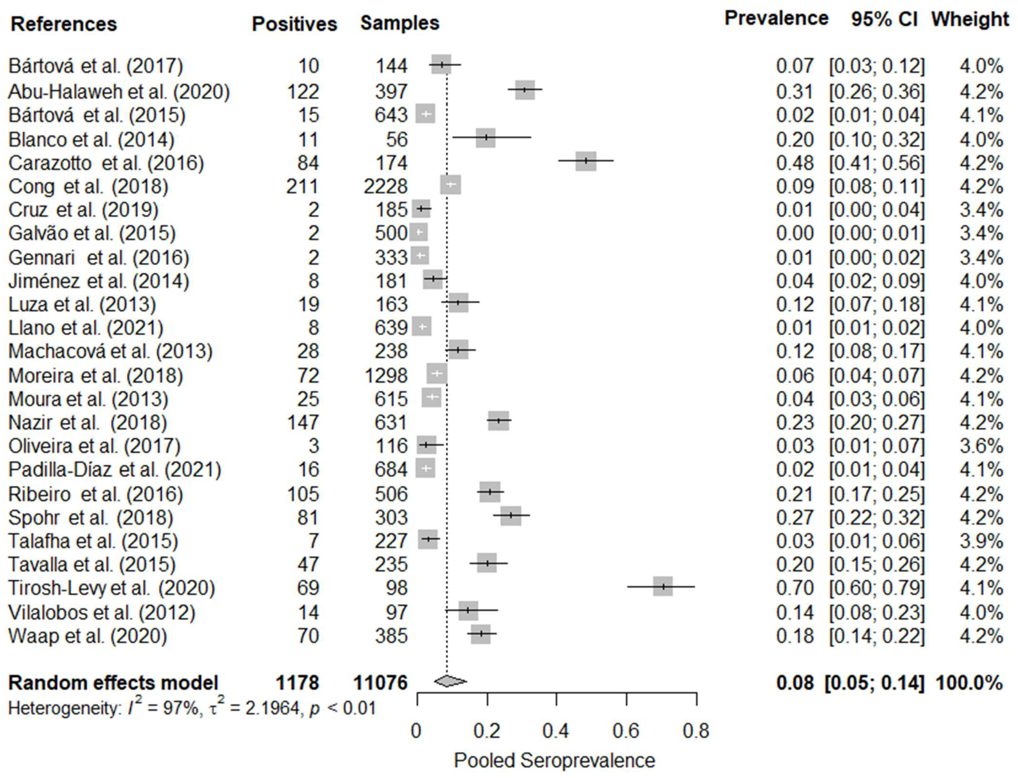
| Overall combined prevalence | 25 | 11,076 | 1178 | 8.34% (4.78–14.17%) | <0.01 | 97.30% |
| Continent | ||||||
| Africa | 1 | 144 | 10 | 6.94% (3.78–12.43%) | Not applicable | |
| Asia | 6 | 3816 | 603 | 20.48% (7.66–44.43%) | <0.01 | 98.30% |
| Central/North America | 2 | 865 | 24 | 3.07% (1.65–5.64%) | 0.14 | 55.00% |
| Europe | 4 | 1451 | 115 | 5.48% (1.49–18.19%) | <0.01 | 95.80% |
| South America | 12 | 4800 | 426 | 6.87% (2.81–15.82%) | <0.01 | 97.30% |
| Specie | ||||||
| Donkeys | 10 | 3954 | 397 | 7.36% (2.08–22.94%) | <0.01 | 97.10% |
| Horses | 19 | 6950 | 756 | 8.50% (5.03–13.99%) | <0.01 | 96.90% |
| Mules | 3 | 172 | 25 | 6.07% (0.71–36.83%) | <0.01 | 83.70% |
| Diagnosis | ||||||
| ELISA | 7 | 3746 | 414 | 7.91% (3.71–16.08%) | <0.01 | 95.10% |
| IFAT | 17 | 7095 | 717 | 8.08% (3.74–16.61%) | <0.01 | 97.90% |
| MAT | 1 | 235 | 47 | 20.00% (15.37–25.60%) | Not applicable | |
Disclaimer/Publisher’s Note: The statements, opinions and data contained in all publications are solely those of the individual author(s) and contributor(s) and not of MDPI and/or the editor(s). MDPI and/or the editor(s) disclaim responsibility for any injury to people or property resulting from any ideas, methods, instructions or products referred to in the content. |
© 2023 by the authors. Licensee MDPI, Basel, Switzerland. This article is an open access article distributed under the terms and conditions of the Creative Commons Attribution (CC BY) license (https://creativecommons.org/licenses/by/4.0/).
Share and Cite
Costa, P.W.L.; Alvares, F.B.V.; Araújo, H.G.; Limeira, C.H.; Braga, F.R.; Feitosa, T.F.; Vilela, V.L.R. Worldwide Overview of Neospora spp. Infection in Equids Diagnosed by Serological Tests: Systematic Review and Meta-Analysis. Parasitologia 2023, 3, 260-268. https://0-doi-org.brum.beds.ac.uk/10.3390/parasitologia3030027
Costa PWL, Alvares FBV, Araújo HG, Limeira CH, Braga FR, Feitosa TF, Vilela VLR. Worldwide Overview of Neospora spp. Infection in Equids Diagnosed by Serological Tests: Systematic Review and Meta-Analysis. Parasitologia. 2023; 3(3):260-268. https://0-doi-org.brum.beds.ac.uk/10.3390/parasitologia3030027
Chicago/Turabian StyleCosta, Paulo Wbiratan Lopes, Felipe Boniedj Ventura Alvares, Hosaneide Gomes Araújo, Clécio Henrique Limeira, Fabio Ribeiro Braga, Thais Ferreira Feitosa, and Vinícius Longo Ribeiro Vilela. 2023. "Worldwide Overview of Neospora spp. Infection in Equids Diagnosed by Serological Tests: Systematic Review and Meta-Analysis" Parasitologia 3, no. 3: 260-268. https://0-doi-org.brum.beds.ac.uk/10.3390/parasitologia3030027