Article

WO3 Photocatalyst Containing Copper Inactivates SARS-CoV-2 Pango Lineage A and Omicron BA.2 Variant in Visible Light and in Darkness

1
Laboratory of Global Infectious Diseases Control Science, Graduate School of Agricultural and Life Sciences, The University of Tokyo, 1-1-1 Yayoi, Bunkyo-ku, Tokyo 113-8657, Japan
2
Department of Veterinary Science, National Institute of Infectious Diseases, Toyama, Shinjuku-ku, Tokyo 162-8640, Japan
3
Advintage Co., Ltd., 1-1-1-705 Ebisuminami, Shibuya-ku, Tokyo 150-0022, Japan
4
Centre for Advanced Materials and Energy Sciences, Universiti Brunei Darussalam, Gadong BE1410, Brunei
5
Laboratory of Global Animal Resource Science, Graduate School of Agricultural and Life Sciences, The University of Tokyo, 1-1-1 Yayoi, Bunkyo-ku, Tokyo 113-8657, Japan
*
Author to whom correspondence should be addressed.
Submission received: 20 June 2022 / Revised: 8 August 2022 / Accepted: 13 August 2022 / Published: 16 August 2022
(This article belongs to the Special Issue Emerging and Re-emerging Viral Infectious Diseases)

Abstract

:
Severe acute respiratory syndrome coronavirus 2 (SARS-CoV-2) is the causative agent of coronavirus disease 2019, which has been a global pandemic. Since SARS-CoV-2 is transmitted through contaminated surfaces and aerosols, environmental disinfection is important to block the spread of the virus. Photocatalysts are attractive tools for virus inactivation and are widely used as air purifiers and coating materials. However, photocatalysts are inactive in the dark, and some of them need to be excited with light of a specific wavelength. Therefore, photocatalysts that can effectively inactivate SARS-CoV-2 in indoor environments are needed. Here, we show that a WO3 photocatalyst containing copper inactivated the SARS-CoV-2 WK-521 strain (Pango lineage A) upon irradiation with white light in a time- and concentration-dependent manner. Additionally, this photocatalyst also inactivated SARS-CoV-2 in dark conditions due to the antiviral effect of copper. Furthermore, this photocatalyst inactivated not only the WK-521 strain but also the Omicron variant BA.2. These results indicate that the WO3 photocatalyst containing copper can inactivate indoor SARS-CoV-2 regardless of the variant, in visible light or darkness, making it an effective tool for controlling the spread of SARS-CoV-2.

1. Introduction

Severe acute respiratory syndrome coronavirus 2 (SARS-CoV-2) is the causative agent of coronavirus disease 2019 (COVID-19), which has had an unprecedented impact on modern human civilization [1] and resulted in more than 6.3 million deaths globally as of early June 2022. Despite the development of drugs and vaccines, the number of infected people continues to increase. Although the transmission route of SARS-CoV-2 is still being debated, it is generally believed to be transmitted through the airborne route, surface contamination, and fecal-oral transmission [2]. Thus, the inactivation of the virus in the air and on surfaces is essential for controlling its transmission. In addition, the genome of SARS-CoV-2 has mutated rapidly, and several variants are reported. Mutations in the virus help it to evade the host immune system and to acquire drug resistance. Therefore, despite the presence of vaccines and drugs, it is important to find effective ways to inactivate the virus to prevent the spread of infection, regardless of the variant. It is reported that SARS-CoV-2 can be inactivated by photocatalysts [3], heat [4], ultraviolet (UV) light [5,6] and disinfectants such as ethanol [7]. In particular, since photocatalysts are harmless to the human body, unlike UV light, they have recently received great attention. It has been proposed that they can be applied for disinfection of living and working spaces without evacuating people.
Photocatalysts are excited by light and exhibit a strong oxidation-reduction reaction generating reactive oxygen species (ROS), such as hydroxyl (·OH) and superoxide radicals (O2), on their surface [8]. Using this oxidation-reduction reaction, photocatalysts kill microorganisms, such as bacteria and fungi, and inactivate viruses such as influenza virus, hepatitis C virus, vesicular stomatitis virus, enterovirus, herpes virus, Zika virus, human coronavirus, bovine coronavirus, human norovirus, murine norovirus, SARS coronavirus, and bacteriophages [8,9,10,11,12,13,14,15]. Many compounds such as titanium dioxide (TiO2), tungsten trioxide (WO3), zinc oxide (ZnO), cadmium sulfide (CdS), and iron (III) oxide (Fe2O3) are known to exhibit photocatalysis and are being actively researched. In particular, TiO2 and WO3 have been reported to inactivate SARS-CoV-2, and are very promising as antiviral materials [3,10,16]. In addition, photocatalysts damage the viral morphology, RNA and proteins, leading to the inactivation of SARS-CoV-2 [3,16]. Therefore, it is expected that photocatalysts can inactivate SARS-CoV-2, regardless of the rapidly evolving variants.
On the other hand, photocatalysts have three limitations: First, the photocatalytic reaction occurs only on the surface of the photocatalyst. Therefore, it is necessary to coat all the surfaces to avoid contamination or to use it together with a circulator such as an air purifier. Second, the wavelength of light that can be used to excite the photocatalysts is limited. The wide bandgap (larger than 3 eV) of TiO2, which is the most common photocatalyst, limits the wavelength of the excitation light to the UV region [17]. Thus, narrowing the band gap of TiO2 is very important for using the TiO2 photocatalyst under visible light [18]. For example, mixing TiO2 with silicane (SiH) narrows the band gap (2.082 eV), and it can be excited with visible light [19]. Third, since light is necessary for the excitation of photocatalysts, the photocatalytic reaction does not occur in dark conditions, such as while sleeping.
In this study, to overcome these limitations, we used a WO3 photocatalyst containing copper that can be applied to a surface by spraying. WO3 coating kept the surface clear of viral contamination. In addition, unlike TiO2, WO3 could be excited by room light even without mixing with any other compounds such as SiH. Therefore, a light source with a specific wavelength was not required. Furthermore, mixing copper with WO3 particles is expected to enable the photocatalyst to inactivate the virus even in the dark due to the effect of copper. However, there is only one study that reported the inactivation of the SARS-CoV-2 Pango lineage A by a WO3 photocatalyst [16]. In this study, we investigated the SARS-CoV-2 inactivation ability of a WO3 photocatalyst both in white light (irradiated by a light emitting diode (LED)) and in darkness, confirmed the time- and concentration-dependency of SARS-CoV-2 inactivation by the WO3 photocatalyst, and analyzed the effectiveness of this photocatalyst against different variants of SARS-CoV-2, according to Japanese Industrial Standards (JIS).

2. Results

2.1. Characterization of the WO3 Photocatalyst

First, to confirm the cytotoxicity of the WO3 photocatalyst, we added it to the culture media of Vero E6/TMPRSS2 cells and incubated the cells for 48 h. Cell viability was measured using the water-soluble tetrazolium salt (WST-8) assays. As shown in Figure 1A, the absorbance of WST-8 decreased in a concentration-dependent manner, which indicated that the WO3 photocatalyst affected cell viability modestly at the half-maximal cytotoxic concentration (CC50) of 3.0 mg/mL. In addition, the ability of the photocatalyst to decompose organic matter was confirmed in the methylene blue degradation assay. Methylene blue was degraded in a time-dependent manner at a degradation speed of 1.5 nmol/min. These results suggested that the WO3 photocatalyst was harmless and had a strong ability to decompose organic matter.

2.2. Inactivation of SARS-CoV-2 WK-521 Strain by the WO3 Photocatalyst

To confirm the inactivation ability of WO3 photocatalyst against SARS-CoV-2, the WK-521 strain was placed on the WO3 coated glass and irradiated with 1000 lx light (Figure 2A). As shown in Figure 2B, the titer of SARS-CoV-2 WK-521 strain placed on WO3 coated glass significantly decreased after irradiation with light for 240 min compared to before irradiation. In addition, the infectivity of SARS-CoV-2 WK-521 strain placed on WO3 coated glass in a dark place also decreased. However, this decrease was not to the extent observed in the illuminated sample. Indeed, the mean antiviral activity values were 3.04 and 1.50 with and without light conditions, respectively (Figure 2C). This decrement in the dark condition might be due to the antiviral effect of the copper contained in the WO3 photocatalyst. Indeed, the photocatalyst solution without WO3 exerted an antiviral effect on human coronavirus 229E (HCoV-229E) (Figure S1). On the other hand, no decrease in the titer was observed in the samples placed on the glass without WO3 coating for 240 min, with or without exposure to light. These results showed that the excitation light itself had no antiviral effect; the decrease in the titer was due to the effect of the LED-WO3 photocatalytic reaction in light and due to the antiviral effect of copper in darkness.

2.3. Time- and Dose-Dependency of the Antiviral Effects of WO3 Photocatalysts

Next, to confirm the time-dependence of the antiviral effect of the WO3 photocatalyst, the SARS-CoV-2 WK-521 strain was placed on WO3-coated glass and irradiated with light for 0, 60, 120 and 240 min. As shown in Figure 3A, the viral titer decreased in a time-dependent manner, and the mean antiviral activity values were 0.66, 1.08 and 2.25 for 60-, 120- and 240-min light exposures, respectively (Figure 3B). In addition, to confirm the antiviral effect of the WO3 photocatalyst, the SARS-CoV-2 WK-521 strain was placed on glasses coated with 10, 30 or 100 mg of WO3 and irradiated with LED light for 0 or 240 min. As shown in Figure 3C, in the group irradiated with light for 240 min, a decrease in the titer was observed in all concentrations of WO3 photocatalyst compared to the group not exposed to light (0 min), which was significant in the 30 and 100 mg coatings. The antiviral activity values were 2.00, 1.25 and 0.83 for 100, 30 and 10 mg of WO3, respectively, indicating that the titer of SARS-CoV-2 WK-521 strain was decreased by the WO3 photocatalyst in a dose-dependent manner (Figure 3D). In contrast, there was no difference in viral titers among various concentrations on WO3-coated glass in the group which was not irradiated by LED light (0 min, Figure 3C). Our results demonstrated that photocatalysis is the mechanism of inactivation of SARS-CoV-2 WK-521 strain by WO3, photocatalytic inactivation of SARS-CoV-2 WK-521 strain by WO3 was dose- and time-dependent.

2.4. WO3 Photocatalysts Inactivate SARS-CoV-2 Omicron Variant BA.2
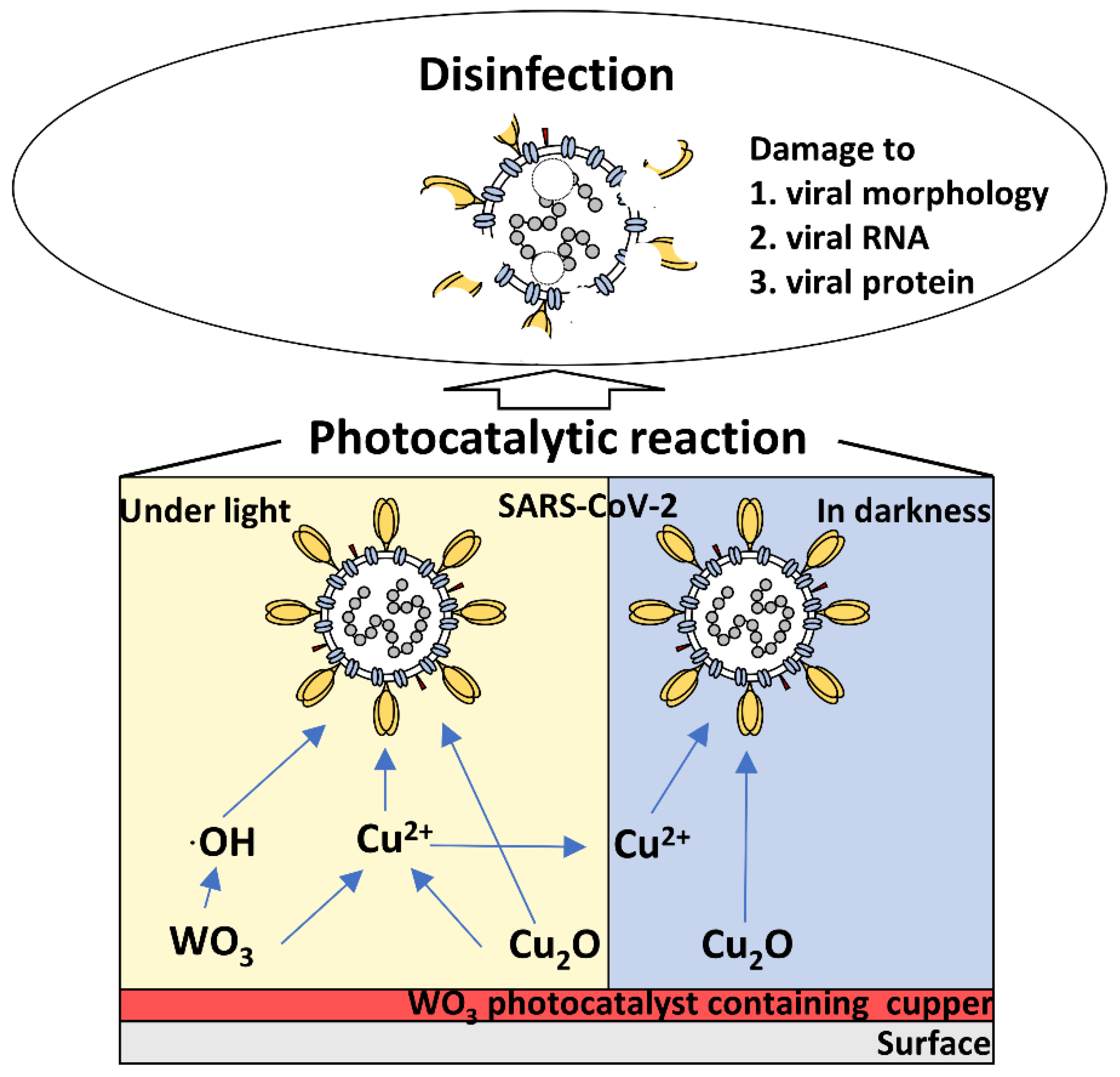
Finally, we clarified whether the WO3 photocatalyst exerted an antiviral effect against SARS-CoV-2 Omicron variant BA.2. The Omicron variant BA.2 was placed on a WO3-coated glass (150 μL with a titer of 1 × 10⁷ 50% tissue culture infective dose (TCID50)/mL and irradiated with LED light for 0 to 240 min. As shown in Figure 4A, exposure to light for 240 min reduced the titer of this variant, similar to what was observed in the WK-521 strain. In addition, the mean antiviral activity after 240 min of photocatalytic reaction on WO3 was 3.17, which was comparable to that observed in the WK-521 strain (Figure 4B). This result indicated that the WO3 photocatalyst inactivates SARS-CoV-2 regardless of the variant.

3. Discussion

In this study, we demonstrated that the WO3 photocatalyst containing copper effectively inactivated SARS-CoV-2. Indeed, our results provide evidence that WO3 photocatalytic reaction for 240 min significantly decreased the infectivity of the SARS-CoV-2 WK-521 strain. Additionally, the copper present in the photocatalyst enabled it to inactivate the virus even in darkness. Furthermore, the WO3 photocatalyst containing copper decreased the SARS-CoV-2 WK-521 strain titers in a time-and dose-dependent manner, confirming the photocatalysis induced the inactivation of the virus. Our results are supported by a previous report that showed the effective inactivation of SARS-CoV-2 by a WO3-based visible light-responsive photocatalyst under different temperatures and exposure durations [16]. Notably, we demonstrated the effectiveness of a 240 min photocatalytic reaction involving the WO3 photocatalyst containing copper, not only against the SARS-CoV-2 WK-521 strain but also against the Omicron variant BA.2, as indicated by the decreased viral titers comparable with those of the WK-521 strain. These results suggest that the WO3 photocatalyst exerts an antiviral effect regardless of the variant. The present study is the first to report that a WO3 photocatalytic reaction can inactivate SARS-CoV-2, regardless of the variant.
It was previously reported that the mechanisms involved in the inactivation of SARS-CoV-2 by photocatalysis are damage to viral morphology, RNA, and protein [3,16]. In this study, the inactivation of the SARS-CoV-2 WK-521 strain and the Omicron variant BA. 2 by a WO3 photocatalyst containing copper was demonstrated. This suggests that even if the virus is mutated, a photocatalytic reaction by WO3 can achieve viral inactivation by damaging the viral protein, RNA and lipid bilayer, irrespective of the variant. Hence, this WO3 photocatalyst containing copper could be effective against potential variants of SARS-CoV-2 that may develop in the future.
WO3 photocatalyst containing copper inactivated SARS-CoV-2 not only upon irradiation with light but also in dark conditions as well. It has been reported previously that copper nanoparticles can inactivate SARS-CoV-2 [20]. In addition, copper oxide nanoclusters grafted with titanium dioxide also inactivated SARS-CoV-2 alpha, beta, gamma and delta variants under illumination and in dark conditions as well [21]. These observations suggest that copper is responsible for the inactivation of the SARS-CoV-2 WK-521 strain under dark conditions observed in this study, indicating that the WO3 photocatalyst containing copper can be effective even at night time.
The WO3 photocatalyst containing copper can inactivate SARS-CoV-2 under light and in darkness, regardless of the variant. Findings from previous reports suggest the inactivation of the WO3 photocatalyst containing copper was owing to the damage caused to viral morphology, RNA, and proteins (Figure 5) [3,16,21]. Under light, this damage may be induced by hydroxy radicals and copper ions generated by the WO3 photocatalyst and solid-state copper. Conversely, in darkness, solid-state copper and copper ions retained after the photocatalytic reaction may inactivate the virus. The WO3 photocatalyst containing copper is considered to efficiently inactivate SARS-CoV-2 by these mechanisms, regardless of the variant.
Since a WO3 photocatalyst containing copper was excited by a white LED in this study, it is evident that this photocatalyst works effectively in an indoor environment without the necessity for any specific light source. In addition, unlike UV light, the WO3 photocatalyst is harmless to the human body. Although evidence from recent studies suggests that SARS-CoV-2 infection from contaminated surfaces is not as relevant [22,23,24], its possibility should not be ignored, especially when considering indoor spaces with a high probability of infection. Therefore, we may expect that the WO3 photocatalyst containing copper can be used for the disinfection of surfaces that are touched regularly by multiple individuals, such as the buttons on an elevator and straps of a train, to prevent viral spread. In addition, findings from our previous study showed that an air purifier with a TiO2 photocatalyst could inactivate SARS-CoV-2 in aerosols [3]. Therefore, the WO3 photocatalyst may also inactivate SARS-CoV-2 in aerosols if used in an air purifier. Thus, this study demonstrated that a WO3 photocatalyst containing copper could be effectively applied to control SARS-CoV-2 transmission and mitigate the ongoing COVID-19 pandemic.

4. Materials and Methods

4.1. Virus and Cell Culture

Vero E6 cells, which express the transmembrane serine protease TMPRSS2 (Vero E6/TMPRSS2 (JCRB1819), were maintained in Dulbecco’s modified Eagle’s medium (DMEM, Thermo Fisher Scientific, Waltham, MA, USA) supplemented with 10% heat-inactivated fetal bovine serum (FBS, Thermo Fisher Scientific) at 37 °C with 5% CO2. MRC-5 cells (CCL-171) were maintained in Eagle’s minimal essential medium (EMEM, Thermo Fisher Scientific) supplemented with 10% heat-inactivated FBS at 37 °C with 5% CO2. The WK-521 strain (Pango lineage A; 2019-nCoV/Japan/TY/WK-521/2020) [25] and Omicron variant BA.2 (hCoV-19/JPN/TY40-385/2022 strain) of SARS-CoV-2 were cultured and quantified using Vero E6/TMPRSS2 cells in DMEM containing 2% FBS. HCoV-229E was cultured and quantified in MRC-5 cells in DMEM containing 2% FBS. The infectivity of SARS-CoV-2 and HCoV-229E was calculated by their titration in Vero E6/TMPRSS2 cells and MRC-5 cells, respectively, using the TCID50 assay and the Reed–Muench method [26].

4.2. Cytotoxicity Assay

Vero cells (5 × 10⁴ cells/well) were seeded overnight in 24-well plates and treated with 0, 0.5, 1, 2, 4, or 8 mg/mL WO3 photocatalyst for 48 h. The cells were washed with PBS and cultured in 1 mL of fresh DMEM plus 30 µL of WST-8, and the WST-8 assays were performed at 37 °C for 90 min. The optical density at 450 nm (OD450) was measured using an Ensight Perkin Elmer multimode plate reader (Perkin Elmer, Milan, Italy). CC50 was calculated from the linear regression of OD450 vs. compound concentration.

4.3. Degradation of Methylene Blue

A glass coated with 100 mg of the WO3 photocatalyst containing copper (NFE2-W; Chemical Technology Co., Ltd., Takaishi, Japan) was put in 25 mL of 12.5 μM methylene blue solution. The WO3 photocatalyst was excited with a white LED (BBZ T13 Silver; Dongguan Oushi Electronic Technology Co., Ltd., Dongguan, China) every 5 min for 60 min. To confirm the effect of the WO3 photocatalyst on methylene blue, methylene blue was collected at each time point, and the absorbance at 660 nm was measured using an Ensight Perkin Elmer multimode plate reader. The degradation speed of methylene blue was calculated from the linear regression of OD450 vs. the photocatalytic reaction time.

4.4. Inactivation of SARS-CoV-2 and HCoV-229E by the WO3 Photocatalytic Reaction

The photocatalytic reaction was performed according to JIS R1752:2020 [27] with a minor modification (Figure 2A). Briefly, filter paper was placed at the bottom of the 10 cm dish and wetted with 4 mL sterilized water for moisture preservation. To avoid directly touching the filter paper, a plastic tube was placed on the filter paper, and glass coated with 100 mg WO3 photocatalyst containing copper was placed on top of the plastic tube. One hundred and fifty microliters of the WK-521 strain with a titer of 1 × 10⁶ TCID50/mL, Omicron variant BA.2 with a titer of 1 × 10⁷ TCID50/mL, or HCoV-229E with a titer of 1.47 × 10⁵ TCID50/mL was placed on the WO3-coated glass and spread by covering it with a film. Glass without the WO3 coating was used as a negative control. To confirm the antiviral effect of copper, glass coated with a photocatalyst without WO3 was used for HCoV-229E. The samples were then illuminated with 1000 lx light using a white LED for 240 min or not illuminated. After illumination, the samples were washed by immersing in 8 mL phosphate-buffered saline (PBS). As time control, the virus was immediately collected after placing the droplet on glass with or without WO3 coating (0 min). To confirm the time dependency, the SARS-CoV-2 WK-521 strain was placed on the 100 mg WO3-coated glass and illuminated for 0, 60, 120 and 240 min. To observe the concentration dependency, the SARS-CoV-2 WK-521 strain was placed on glass coated with 100, 30 or 10 mg WO3 and illuminated for 0 and 240 min. The SARS-CoV-2 and HCoV-229E titers in all experiments were measured using the TCID50 assay.
The photocatalytic inactivation efficiency was defined as follows:
Antiviral activity value = log10 (Nt) − log10 (N0)
where Nt represents the virus titer of the photocatalytically treated specimens after irradiation for t hours; N0 represents the virus titer of the photocatalytically treated specimens just after inoculation (0 min).

4.5. Statistical Analysis

Statistical comparisons were performed using Student’s t-test. p-values < 0.05 were considered statistically significant.

5. Conclusions

This is the first report showing that a WO3 photocatalyst inactivates Omicron variant BA.2 as well as the SARS-CoV-2 WK-521 strain, indicating the effectiveness of this photocatalyst against the virus, regardless of the variant. In addition, a WO3 photocatalyst containing copper can inactivate the virus using a simple white light or even in dark conditions, indicating its potential for wide application. In conclusion, a WO3 photocatalyst containing copper could be a very effective tool for controlling the spread of SARS-CoV-2.

Supplementary Materials

The following supporting information can be downloaded at: https://0-www-mdpi-com.brum.beds.ac.uk/article/10.3390/pathogens11080922/s1, Figure S1: Antiviral effect of WO3 photocatalyst with or without WO3 for HCoV-229E.

Author Contributions

Conceived and designed the experiments: Y.A. and R.M. Conducted and performed the experiments: K.M., R.M. and Y.A. Analyzed the data: R.M. and Y.A. Supervised this experiment: Y.A., K.M., K.H., Y.M. (Yosuke Mori) and Y.M. (Yasunobu Matsumoto). Contributed reagents/materials/analysis tools: Y.A., K.M., K.H., Y.M. (Yosuke Mori) and T.K. Wrote the paper: R.M. and Y.A. All authors have read and agreed to the published version of the manuscript.

Funding

This work was supported by a grant from the Office for Novel Coronavirus Disease Control, Cabinet Secretariat, Government of Japan (COVID-19 AI and Simulation Project) and by grant-in-aid from Advintage Co., Ltd.

Data Availability Statement

Not applicable.

Acknowledgments

We would like to thank Kaltech Co., Ltd. (https://kaltec.co.jp/en/) for helping the organization of our laboratory and Editage (www.editage.com) for English language editing.

Conflicts of Interest

The authors declare no conflict of interest.

References

  1. Wu, F.; Zhao, S.; Yu, B.; Chen, Y.M.; Wang, W.; Song, Z.G.; Hu, Y.; Tao, Z.W.; Tian, J.H.; Pei, Y.Y.; et al. A new coronavirus associated with human respiratory disease in China. Nature 2020, 579, 265–269. [Google Scholar] [CrossRef]
  2. Mukhra, R.; Krishan, K.; Kanchan, T. Possible modes of transmission of Novel coronavirus SARS-CoV-2: A review. Acta Bio Med. Atenei Parm. 2020, 91, e2020036. [Google Scholar] [CrossRef]
  3. Matsuura, R.; Lo, C.W.; Wada, S.; Somei, J.; Ochiai, H.; Murakami, T.; Saito, N.; Ogawa, T.; Shinjo, A.; Benno, Y.; et al. SARS-CoV-2 Disinfection of Air and Surface Contamination by TiO2 Photocatalyst-Mediated Damage to Viral Morphology, RNA, and Protein. Viruses 2021, 12, 650. [Google Scholar] [CrossRef]
  4. Saadatpour, F.; Mohammadipanah, F. Physicochemical susceptibility of SARS-CoV-2 to disinfection and physical approach of prophylaxis. Health Sci. Rep. 2020, 3, e213. [Google Scholar] [CrossRef]
  5. Heilingloh, C.S.; Aufderhorst, U.W.; Schipper, L.; Dittmer, U.; Witzke, O.; Yang, D.; Zheng, X.; Sutter, K.; Trilling, M.; Alt, M.; et al. Susceptibility of SARS-CoV-2 to UV irradiation. Am. J. Infect. Control 2020, 48, 1273–1275. [Google Scholar] [CrossRef]
  6. Lo, C.W.; Matsuura, R.; Iimura, K.; Wada, S.; Shinjo, A.; Benno, Y.; Nakagawa, M.; Takei, M.; Aida, Y. UVC disinfects SARS-CoV-2 by induction of viral genome damage without apparent effects on viral morphology and proteins. Sci. Rep. 2021, 11, 13804. [Google Scholar] [CrossRef]
  7. Kratzel, A.; Todt, D.; V’kovski, P.; Steiner, S.; Gultom, M.; Thao, T.T.N.; Ebert, N.; Holwerda, M.; Steinmann, J.; Niemeyer, D.; et al. Inactivation of Severe Acute Respiratory Syndrome Coronavirus 2 by WHO-Recommended Hand Rub Formulations and Alcohols. Emerg. Infect. Dis. 2020, 26, 1592–1595. [Google Scholar] [CrossRef]
  8. Foster, H.A.; Ditta, I.B.; Varghese, S.; Steele, A. Photocatalytic disinfection using titanium dioxide: Spectrum and mechanism of antimicrobial activity. Appl. Microbiol. Biotechnol. 2011, 90, 1847–1868. [Google Scholar] [CrossRef]
  9. Nakano, R.; Ishiguro, H.; Yao, Y.; Kajioka, J.; Fujishima, A.; Sunada, K.; Minoshima, M.; Hashimoto, K.; Kubota, Y. Photocatalytic inactivation of influenza virus by titanium dioxide thin film. Photochem. Photobiol. Sci. 2012, 11, 1293–1298. [Google Scholar] [CrossRef]
  10. Tong, Y.; Shi, G.; Hu, G.; Hu, X.; Han, L.; Xie, X.; Xu, Y.; Zhang, R.; Sun, J.; Zhong, J. Photo-catalyzed TiO(2) inactivates pathogenic viruses by attacking viral genome. Chem. Eng. J. 2021, 414, 128788. [Google Scholar] [CrossRef]
  11. Khaiboullina, S.; Uppal, T.; Dhabarde, N.; Subramanian, V.R.; Verma, S.C. Inactivation of Human Coronavirus by Titania Nanoparticle Coatings and UVC Radiation: Throwing Light on SARS-CoV-2. Viruses 2021, 13, 19. [Google Scholar] [CrossRef] [PubMed]
  12. Yoshizawa, N.; Ishihara, R.; Omiya, D.; Ishitsuka, M.; Hirano, S.; Suzuki, T. Application of a Photocatalyst as an Inactivator of Bovine Coronavirus. Viruses 2020, 12, 1372. [Google Scholar] [CrossRef] [PubMed]
  13. Park, D.; Shahbaz, H.M.; Kim, S.H.; Lee, M.; Lee, W.; Oh, J.W.; Lee, D.U.; Park, J. Inactivation efficiency and mechanism of UV-TiO2 photocatalysis against murine norovirus using a solidified agar matrix. Int. J. Food Microbiol. 2016, 238, 256–264. [Google Scholar] [CrossRef] [PubMed]
  14. Han, W.; Zhang, B.; Cao, W.; Yang, D.; Taira, I.; Okamoto, Y.; Arai, J.I.; Yan, X. The inactivation effect of photocatalytic titanium apatite filter on SARS virus. Prog. Biochem. Biophys. 2004, 31, 982–985. [Google Scholar]
  15. Syngouna, V.I.; Chrysikopoulos, C.V. Inactivation of MS2 bacteriophage by titanium dioxide nanoparticles in the presence of quartz sand with and without ambient light. J. Colloid Interface Sci. 2017, 497, 117–125. [Google Scholar] [CrossRef]
  16. Uema, M.; Yonemitsu, K.; Momose, Y.; Ishii, Y.; Tateda, K.; Inoue, T.; Asakura, H. Effect of the Photocatalyst under Visible Light Irradiation in SARS-CoV-2 Stability on an Abiotic Surface. Biocontrol. Sci. 2021, 26, 119–125. [Google Scholar] [CrossRef]
  17. Tao, J.; Luttrell, T.; Batzill, M. A two-dimensional phase of TiO2 with a reduced bandgap. Nat. Chem. 2011, 3, 296–300. [Google Scholar] [CrossRef]
  18. Bono, N.; Ponti, F.; Punta, C.; Candiani, G. Effect of UV Irradiation and TiO2-Photocatalysis on Airborne Bacteria and Viruses: An Overview. Materials 2021, 14, 1075. [Google Scholar] [CrossRef]
  19. Niu, M.; Cheng, D.; Cao, D. SiH/TiO2 and GeH/TiO2 heterojunctions: Promising TiO2-based photocatalysts under visible light. Sci. Rep. 2014, 4, 4810. [Google Scholar] [CrossRef]
  20. Foffa, I.; Losi, P.; Quaranta, P.; Cara, A.; Al Kayal, T.; D’Acunto, M.; Presciuttini, G.; Pistello, M.; Soldani, G. A Copper nanoparticles-based polymeric spray coating: Nanoshield against SARS-CoV-2. J. Appl. Biomater. Funct. Mater. 2022, 20, 22808000221076326. [Google Scholar] [CrossRef]
  21. Nakano, R.; Yamaguchi, A.; Sunada, K.; Nagai, T.; Nakano, A.; Suzuki, Y.; Yano, H.; Ishiguro, H.; Miyauchi, M. Inactivation of various variant types of SARS-CoV-2 by indoor-light-sensitive TiO2-based photocatalyst. Sci. Rep. 2022, 12, 5804. [Google Scholar] [CrossRef]
  22. Gonçalves, J.; da Silva, P.G.; Reis, L.; Nascimento, M.S.J.; Koritnik, T.; Paragi, M.; Mesquita, J.R. Surface contamination with SARS-CoV-2: A systematic review. Sci Total Environ. 2021, 798, 149231. [Google Scholar] [CrossRef] [PubMed]
  23. Mondelli, M.U.; Colaneri, M.; Seminari, E.M.; Baldanti, F.; Bruno, R. Low risk of SARS-CoV-2 transmission by fomites in real-life conditions. Lancet Infect. Dis. 2021, 21, e112. [Google Scholar] [CrossRef]
  24. Centers for Disease Control and Prevention, 2021. Science Brief: SARS-CoV-2 and Surface (Fomite) Transmission for Indoor Community Environments. Available online: https://www.cdc.gov/coronavirus/2019-ncov/more/science-and-research/surface-transmission.html#print (accessed on 4 July 2022).
  25. Matsuyama, S.; Nao, N.; Shirato, K.; Kawase, M.; Saito, S.; Takayama, I.; Nagata, N.; Sekizuka, T.; Katoh, H.; Kato, F.; et al. Enhanced isolation of SARS-CoV-2 by TMPRSS2-expressing cells. Proc. Natl. Acad. Sci. USA 2020, 117, 7001–7003. [Google Scholar] [CrossRef] [PubMed]
  26. Reed, L.J.; Muench, H. A simple method of estimating fifty per cent endpoints. Am. J. Epidemiol. 1938, 27, 493–497. [Google Scholar] [CrossRef]
  27. JIS R 1752:2020; Fine Ceramics (Advanced Ceramics, Advanced Technical Ceramics)—Test Method for Antibacterial Activity of Photocatalytic Materials and Efficacy under Indoor Lighting Environment. Japanese Standards Association: Tokyo, Japan, 2020.
Figure 1. Cytotoxicity and methylene blue degradation potential of the WO3 photocatalyst. (A) The half-maximal cytotoxic concentration (CC50) was calculated from the water-soluble tetrazolium salt (WST-1) assay. Each dot and error bar represent the mean ± standard deviation (SD) from three independent experiments. (B) To confirm the ability of the WO3 photocatalyst to decompose organic matter, WO3-coated glass was placed in 25 mL of 12.5 μM of methylene blue and irradiated with white light. Methylene blue was collected every 5 min, and the absorbance at 660 nm was measured. The methylene blue contents were calculated from the OD value. The degradation speed of methylene blue was calculated from linear regression analysis.
Pathogens 11 00922 g001
Figure 2. Inactivation of SARS-CoV-2 WK-521 strain by the WO3 photocatalyst. (A) A wet filter paper was placed in a 10 cm dish to avoid dryness. Glass with or without a coating of WO3 photocatalyst containing copper (100 mg) was placed on a plastic tube, which was in turn placed on the filter paper to avoid direct contact with the filter paper. SARS-CoV-2 WK-521 strain (150 μL) with a titer of 1 × 10⁶ 50% tissue culture infective dose (TCID50)/mL was placed on the coated or uncoated glass. The WO3 photocatalyst was excited by white LED light with 1000 lx for 0 or 240 min. To confirm the effect of WO3 photocatalyst containing copper in dark conditions, SARS-CoV-2 WK-521 strain was placed on the glass with or without a coating of the photocatalyst and kept in the dark for 240 min. (B) Titers of SARS-CoV-2 WK-521 strain were measured using the TCID50 assay with Vero E6/TMPRSS2 cells. Assays were performed in at least 6 wells, and the values represent the mean ± standard deviation (SD) of two independent experiments. Statistical comparisons were performed using Student’s t-test. Asterisk indicates a statistically significant difference (* p < 0.05). (C) Antiviral activity value was calculated using the formula: (the log10 titer of SARS-CoV-2 WK-521 strain of 240min sample) − (the log10 titer of SARS-CoV-2 WK-521 strain of 0 min sample of the same sample).
Pathogens 11 00922 g002
Figure 3. Time- and dose-dependency of the antiviral effects of WO3 photocatalysts. (A) To confirm time-dependency, SARS-CoV-2 WK-521 strain (150 μL) with a titer of 1 × 10⁶ 50% tissue culture infective dose (TCID50)/mL was placed on glass coated with WO3 photocatalyst (100 mg). WO3 photocatalyst was excited by white LED light with 1000 lx for 0, 60, 120 or 240 min. The titer of SARS-CoV-2 WK-521 strain was measured using the TCID50 assay with Vero E6/TMPRSS2 cells. Assays were performed in at least 6 wells, and the values represent the mean ± standard deviation (SD) of two independent experiments. (B) Antiviral activity value was calculated using the formula: (the log10 titer of SARS-CoV-2 of each time point sample) − (the log10 titer of SARS-CoV-2 of 0 min sample of same sample). (C) To confirm concentration-dependency, SARS-CoV-2 WK-521 strain (150 μL) with a titer of 1 × 10⁶ TCID50/mL was placed on glass coated with 10, 30 or 100 mg WO3 photocatalyst. WO3 photocatalyst was excited by white LED light with 1000 lx for 0 or 240 min. Titers of SARS-CoV-2 were measured using the TCID50 assay with Vero E6/TMPRSS2 cells. Assays were performed in at least 6 wells, and the values represent the mean ± SD of two independent experiments. (D) Antiviral activity value was calculated using the formula: (the log10 titer of SARS-CoV-2 WK-521 strain of 240 min sample) - (the log10 titer of SARS-CoV-2 WK-521 strain of 0 min sample of same concentration sample). Statistical comparisons were performed using Student’s t-test. Asterisk indicates a statistically significant difference (* p < 0.05; ** p < 0.01).
Pathogens 11 00922 g003
Figure 4. WO3 photocatalysts inactivate SARS-CoV-2 Omicron variant BA.2. (A) SARS-CoV-2 Omicron variant BA.2 (150 μL) with a titer of 1 × 10⁷ 50% tissue culture infective dose (TCID50)/mL was placed on glass coated with WO3 photocatalyst (100 mg). WO3 photocatalyst was excited using a white LED light with 1000 lx for 0 or 240 min. The titer of SARS-CoV-2 was measured using the TCID50 assay with Vero E6/TMPRSS2 cells. Assays were performed in at least 6 wells, and the values represent the mean ± standard deviation (SD) of two independent experiments. (B) Antiviral activity value was calculated using the formula: (the log10 titer of SARS-CoV-2 of 240 min sample) − (the log10 titer of SARS-CoV-2 of 0 min sample). Statistical comparisons were performed using Student’s t-test. Asterisk indicates a statistically significant difference (** p < 0.01).
Pathogens 11 00922 g004
Figure 5. Schematic representation and the hypothesis of the inactivation mechanism of the WO3 photocatalyst containing copper. The findings of this study revealed that the WO3 photocatalyst containing copper inactivates SARS-CoV-2 under light or in darkness, regardless of the variant. Based on the results from previous studies, hydroxy radicals and copper ions considered to be generated by the WO3 photocatalyst and solid-state copper inactivate the virus under light, whereas in the dark, the virus is considered to be inactivated by the antiviral activity of solid-state copper and copper ions retained after the photocatalytic reaction [3,16,21]. In addition, the inactivation of SARS-CoV-2 might be attributed to the damage caused to the viral morphology, RNA, and proteins [3,16,21].
Pathogens 11 00922 g005
Publisher’s Note: MDPI stays neutral with regard to jurisdictional claims in published maps and institutional affiliations.

