Article

Persistence of Legionella in Routinely Disinfected Heater-Cooler Units and Heater Units assessed by Propidium Monoazide qPCR

Department of Public Health and Pediatrics, University of Turin, 10126 Torino, Italy
*
Author to whom correspondence should be addressed.
Submission received: 28 October 2020 / Revised: 20 November 2020 / Accepted: 21 November 2020 / Published: 23 November 2020
(This article belongs to the Section Waterborne/Foodborne/Airborne Pathogens)

Abstract

:
Background: Evidence to date indicates that heater–cooler units (HCUs) and heater units (HUs) can generate potentially infectious aerosols containing a range of opportunistic pathogens such as Mycobacterium chimaera, other non-tuberculous mycobacterial (NTM) species, Pseudomonas aeruginosa and Legionella spp. Our purpose was to determine the extent of Legionella contamination and total viable count (TVC) in HCUs and HUs and to analyze the relationship by water system design of devices of two different brands (LivaNova vs. Maquet). Methods: Legionella spp. were detected and quantified by our optimized PMA-qPCR protocol; TVCs were assessed according to ISO protocol 6222. Analyses were performed in the first sampling round and after six months of surveillance. Results: Overall, Legionella spp. was detected in 65.7% of devices. In the second sampling round, Legionella positivity rates were significantly lower in water samples from the Maquet devices compared to the LivaNova ones (27.3% vs. 61.5%). LivaNova HCUs also yielded more Legionella, and aquatic bacteria counts than Maquet in both first and second-round samples. Conclusions: We recommend that all surgical patients and staff exposed to aerosols from thermoregulatory devices should be followed up for Legionella infection and that microbiological surveillance on such devices should be conducted regularly as precautionary principle.

1. Introduction

Legionellae are ubiquitously present in water environments, either natural or man-made. Even though these bacteria are usually found at low concentrations in natural aquatic environments, mounting evidence has shown how artificial water environments with particular physical and chemical conditions can favor the growth of these pathogens [1]. Documented sources include cooling towers [2,3,4], hospital water systems [5], swimming pools [6,7], domestic water systems and showers [8,9], ice-making machines [10,11], whirlpool spas [12,13], hospital pool water for water birthing [14,15], hot springs [16,17], fountains [18], dental units [19], soil [20], automobile windshield washer fluid [21] and wastewater treatment plants [22].
In many nosocomial Legionella outbreaks [5,23] water distribution systems are the most frequent source of infection. The key to legionellosis prevention are proper maintenance of water systems in which Legionella spp. grows (disinfection, water system supply maintenance), staff training and the implementation of a clinical surveillance [24].
Evidence to date indicates that heater–cooler units (HCUs) used during cardiopulmonary bypass surgeries can generate potentially infectious aerosols containing a range of opportunistic pathogens such as Mycobacterium chimaera [25], other non-tuberculous mycobacterial (NTM) species [26,27], Pseudomonas aeruginosa, Stenotrophomonas maltophilia and fungi [28,29]. In the US and Europe, non-tuberculous mycobacteria (NTM) infections have been identified in patients undergoing cardiothoracic surgeries [28,30].
Heater units (HUs) are used in extracorporeal membrane oxygenation (ECMO), to provide prolonged cardiopulmonary support in acute respiratory distress syndrome. To date, there have been three reports of HU contamination with Mycobacterium chimaera world-wide [31,32,33] yet any link between HU contamination and patient infection with Mycobacterium chimaera is still to be determined.
The isolation of Legionella spp. from several HCUs [28,34,35] has raised concern that also these pathogens may be transmitted to the surgical staff through aerosolization of the water contained in these devices. While it is unlikely that respiratory exposure to Legionella from HCUs occurs in patients undergoing cardiothoracic surgery due to the closed-circuit ventilation of these units, there is also a theoretical risk of exposure to Legionella-containing aerosols for same-unit patients that are not being kept on closed circuit ventilation.
To date, however, there is lack of consistent evidence supporting a correlation between airborne Legionella transmission in operating theatres and HCU or HU usage. According to Public Health England surveillance data (1 January 2007 to 1 November 2016), no cases of Legionnaires’ disease were identified in healthcare workers expected to have been exposed to Legionella in cardiac surgery setting, and no Legionella-derived endocarditis cases were reported in cardiothoracic surgery patients [36]. In the U.S., at the University of Washington Medical Center, Seattle, two out of four Legionella-infected patients had previously undergone surgery involving the use of CardioQuip HCUs positive for Legionella spp. [34]. Of note, the manufacturer of these devices, had recently been implicated in three NTM (Mycobacterium abscessus) patient infections at a single facility involving the use of the MCH-1000 devices CardioQuip (Bryan, TX, USA). The Food and Drug Administration (FDA) has alerted healthcare providers to the risk of infection when using CardioQuip’s heater–cooler device during cardiothoracic surgery [37].
As the aforementioned reports leave open the possibility that Legionella transmission may occur even in the absence of a direct contact of the patient/care provider with the HCU and HU water, it has been proposed that contaminated devices’ water may leak onto other parts of the device, thereby favoring bacterial spread in the operating theatre through the aerosol [38].
Here, we report the results of the first molecular surveillance of Legionella contamination of water from HCUs and HUs devices using a propidium monoazide quantitative polymerase chain reaction (PMA-qPCR) method. Our study purpose was to determine the extent of Legionella contamination in HCUs and HUs devices while assessing the long-term efficacy of the recommended decontamination protocols against this pathogen. We also measured the total viable counts (TVCs) of aerobic heterotrophic bacteria at 36 °C and 22 °C to evaluate the relationship between Legionella contamination and the microbial quality of the water samples.

2. Materials and Methods

2.1. Devices

Thermoregulatory devices, heater units (HUs) or heater–cooler units (HCUs) are engaged to adjust the blood temperature within the extracorporeal circuits in acute respiratory distress syndrome, in cardiopulmonary bypass and during cardiac surgery. The devices include tanks (capacity around 24 L) that provide temperature-controlled water to external heat exchangers or to warming/cooling blankets through closed water circuits. The devices heat the water up to 41 °C, an optimal condition for multiplication of bacteria such as Mycobacteria spp. and Legionella spp. Pumps that move water into the patient’s circuit can create aerosol that is pushed out of the devices by cooling fans, thereby favoring the aerodispersion of bacteria in the operating room. This occurs when the water tanks of the devices are not closed with a sealing lid.
The HCUs from different brands can produce and release aerosol with different modality (Figure 1) as shown in previous studies [28,39] in which relevant differences in HCU40 design compared to 3T were identified: air flow direction, location of cooling ventilators, continuous cooling of the water tank at 4 °C and an electronic alarm that activates when disinfection is required.
Regarding HU35, the design itself may ensure patient safety during ECMO (Figure 2) since ventilators used to cool the sealed water tank (capacity around 1.5 L) are located directly under the tank.
The disinfection of devices is performed according to the manufacturers’ updated procedures: (a) for the HCU Stockert 3T, disinfection with peracetic acid (3.3% of Puristeril 340; Fresenius Medical Care, Bad Homburg, Germany) or with sodium hypochlorite (1.3% of Clorox regular bleach, Clorox Co., Oakland, CA, USA) every 14 days, along with weekly changes of water in the presence of 100 ppm of hydrogen peroxide is recommended [40]; (b) for the HCU40, the disinfection protocol includes a weekly treatment with 2% chloramine-T [41]; (c) for the HU35, the disinfection protocol includes a treatment with 2% chloramine-T and to be performed weekly and after each single use.
The thermoregulatory devices tanks are filled, according to the manufacturer’s recommendations, with filtered water with a terminal filter of pore size of 0.2 µm.
We analyzed water samples taken from 35 thermoregulatory devices, of which 13 were HCU Stockert 3T manufactured by LivaNova PLC (Sorin Group Deutschland GmbH, Norderstedt, Germany), 8 were HCU40 and 14 were HU35 both manufactured by Maquet Getinge Group (Rastatt, Germany).

2.2. Water Sampling

Follow-up investigation was carried out in nine cardiac surgery facilities and one pediatric cardiac surgery suite, all located in the Piedmont region (northwestern Italy). Thirty-five devices (21 HCU and 14 HU) were analyzed in the context of periodic analyses in our laboratory from 2017 to 2020. For each HCU, one liter of water sample was collected from both circuits (i.e., the patient circuit and the cardioplegia circuit) before disinfection, whereas for HU devices a water sample of 0.5 liter was collected. Each sample was stored in a sterile plastic bottle containing sodium thiosulfate (10% w/v) to neutralize chlorinated water which may change the microbe numbers during storage from the time of sampling to the time of analysis.
In this study, we assessed bacterial contamination in water samples obtained from HCUs and HU devices at the first sampling round (before disinfection) and compared it with that in samples obtained after six months of surveillance, hereafter defined as second sampling round. During this surveillance period, the surgical personnel carried out monthly disinfection cycles of the devices according to the manufacturer’s instructions.

2.3. Sample Preparation, DNA Extraction and PMA Treatment

The samples were processed for Legionella detection using our optimized PMA-qPCR protocol, as previously described [42]. Briefly, water sample was filtered through a 0.45 μm polycarbonate filter (Millipore, Billerica, MA, USA), overlaid with 500 μL of propidium monoazide (PMA) (50 μM) in 90 mm Petri dishes and incubated in the dark for 10 min followed by a 10 min exposure to a 500 W light on ice at a distance of 20 cm from the light source. After irradiation, the filter was added to the lysis buffer for DNA extraction according to the manufacturer’s instructions (Aquadien™, Bio-Rad, Marnes-la-Coquette, France). Extracted genomic DNA was then analyzed by qPCR to detect the presence of amplifiable sequences.

2.4. Detection and Quantification of Legionella by qPCR

The analyses were performed by “iQ-Check™ Quanti Legionella spp.”, according to the manufacturer’s instructions (Bio-Rad, Marnes-la-Coquette, France). The iQ-Check™ Quanti Legionella spp. kit amplifies and quantifies a fragment of approximately 100 bp from the 5S rRNA gene of Legionella spp. using molecular beacon probes. The qPCR data were then analyzed by Bio-Rad CFX Manager IDE (Bio-Rad, Hercules, CA, USA).

2.5. Total Viable Count (TVC)

One milliliter of undiluted samples and 1 mL of 1:10 diluted samples, in Page’s saline solution, were tested on yeast extract agar using the pour plate method according to the UNI EN ISO protocol 6222 [43]. The number of colony-forming units (CFUs) per sample was calculated after 7 days of incubation at 22 °C or after 5 days of incubation at 36 °C, according to the US standard method [44]. Results were reported as CFU/mL.

2.6. Statistical Analysis
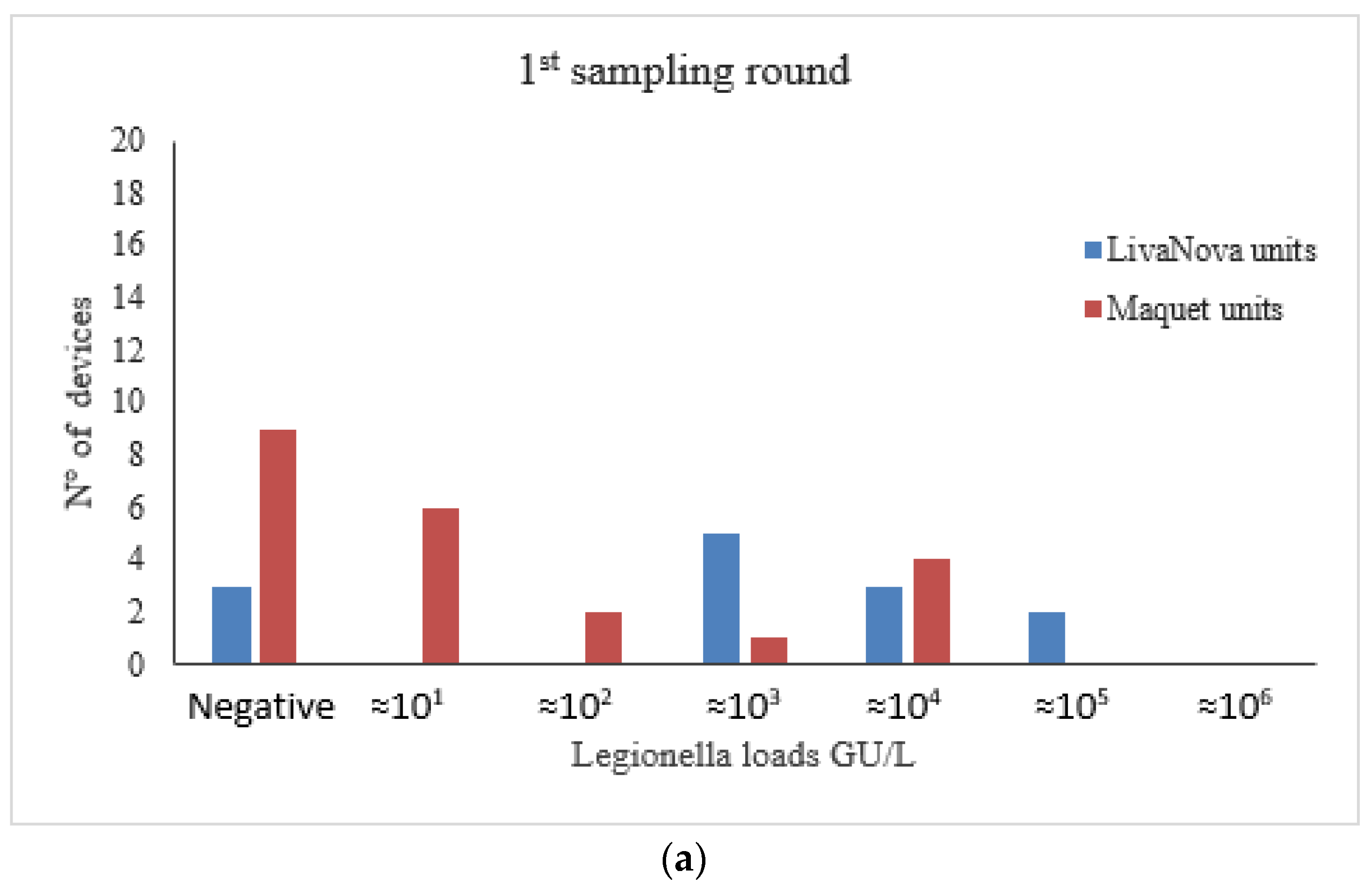
The statistical software R (version 4.0.2) [45] was used to perform all calculations and draw all plots. The proportions were compared using Fisher’s exact test. The Mann–Whitney U test was adopted to evaluate between-sample differences (LivaNova vs. Maquet) in Legionella quantification (genomic units per liter, GU/L) and TVC data. The correlation between respective Legionella counts and TVCs was evaluated using Kendall’s tau correlation coefficient. Finally, correlation differences between the two sampling rounds (first vs. second) were investigated by Dunn and Clark’s Z test for correlation coefficients in dependent samples [46], as implemented by the R package “cocor” [47], after converting each tau coefficient into the corresponding parametric coefficient r, as described by Walker [48]. For all tests, the level of significance was set at α = 0.05.

3. Results

Legionella spp. was detected in 65.7% of devices, with a slightly higher percentage of Legionella-positive samples observed in LivaNova vs. Maquet devices (76.9% vs. 59.1%).
When we analyzed a second water sample taken from the same devices after six months of monthly disinfection, we found on average a lower total number of contaminated devices (40.0%) compared to the first sampling round, albeit this difference only attained borderline significance (p = 0.0547).
We next analyzed the relationship between the different water system design of devices’ brand and the contamination rate. Interestingly, the percentage of contaminated machines in the second sampling round varied consistently (p = 0.0666) only among Maquet devices. Fully detailed results by device and brand are summarized in Table 1.
The comparison between specific thermoregulatory devices (Stockert 3T, HCU40 and HU35) did not yield statistically significant difference both in the first and in the second sampling round (data not shown).
Legionella load ranged from 1.2 × 101 to 5.9 × 10⁶ GU/L. Data frequency distributions are shown in Figure 3a,b.
Samples collected from LivaNova maintain higher microbial loads despite the disinfection cycles, compared to Maquet device. The statistical analysis showed a significant difference compared to the concentrations of Legionella found in the Maquet ones (Figure 3b, Table 2).
Counts relative to the first and second sampling round revealed that the HCUs manufactured by LivaNova had a significantly higher counts of both aquatic bacteria and Legionella compared to Maquet devices (Table 2).
As the TVC analysis showed a clear correlation between the results obtained at 22 °C and 36 °C in both the first (τ = 0.9592; p < 0.0001) and second round (τ = 0.8809; p < 0.0001), we opted to report and discuss hereafter only the TVC results relative to the 22 °C condition.
Overall, aquatic bacteria were detected in 14 of the 35 water samples (40.0%) from both the first and second sampling round (Table 3). Importantly, the devices manufactured by LivaNova yielded a significantly higher number of positive samples (≥1 CFU/mL) than those manufactured by Maquet (p = 0.012), again with identical results for each sampling round (Table 3, Figure 4a,b).
Legionella counts and TVC in water samples from the 1st and 2nd sampling round at 22 °C appeared to be positively correlated, with a moderate association reaching statistical significance (τ = 0.3984, p = 0.0038) (Figure 5a). This correlation was more evident among Maquet devices (τ = 0.3609, p = 0.0494), whereas it was weaker and not significant in LivaNova devices (τ = 0.2809, p = 0.2194). In contrast, the same analysis failed to find any correlation between TVC at 22 °C and Legionella counts in samples drawn during the second round, either considering all samples (τ = 0.1663, p = 0.2489) or one of the two brands (LivaNova: τ = −0.0902, p = 0.6946; Maquet: τ = 0.1461, p = 0.4591) (Figure 5b).
Finally, Dunn and Clark’s Z test for dependent samples detected a difference between the respective correlation coefficients of the two sampling rounds, although not statistically significant (p = 0.0831) probably due to the limited sample size (n = 35).

4. Discussion

High microbial load in HCU or HU water, possibly containing pathogenic bacteria such as NTM and Legionella spp., poses a potential risk of infection for patients and personnel in operating theatres. This contamination can be mainly ascribed to the presence of waterborne bacteria: the microorganisms in HCU and HU biofilms primarily originate from tap water when tap water is added to the water tanks of thermoregulatory devices without filtration [49,50], a procedure strongly discouraged by the manufacturers. However, the risk of aerosol emission is related to the design of water system devices and to the internal ventilation systems [25,39,51]. In fact, the risk of infection arising from the use of HU35 is foresee-ably low, due to the air-tight and closed system design device (without the external cooling fan) and the minimally invasive nature of ECMO [52].
FDA began receiving reports of non-tuberculous mycobacteria (NTM) infections linked to the devices in 2010, leading to a 2015 warning letter to LivaNova [53] and subsequent design changes intended to cut the risk. Regulatory communications on the topic have centered on LivaNova, but FDA has also received reports of contamination linked to devices from Cincinnati Sub-Zero, Maquet and Terumo [54].
In order to reduce the risk of aerosol transmission, LivaNova has recently upgraded its HCU 3T manufactured before 2016 with a new internal sealing. In addition, it has equipped them with a vacuum pump aspirating liquids into the central suction system of the hospital, which reduces—albeit not fully eliminating—the exhaust emissions from the rear of the machine [53]. On 25 February 2020 LivaNova received FDA clearance for 3T heater–cooler device modification [55]. Although regular and effective decontamination and microbiological surveillance is crucial to mitigate the risk of infection due to M. chimaera [32,56,57] and other opportunistic pathogens, such as Legionella, the theoretical risk of aerosolization remains high when these devices are decontaminated [58,59] or emptied, or when their circuits are damaged.
This study is the first to quantify Legionella in HCUs and HU35 devices using the PMA-qPCR method. Previous reports assessed the extent of Legionella contamination in thermoregulatory devices using either bacterial culture [28] or PCR [35], with the latter method being used to examine three devices only. In our study, besides analyzing a much larger number of devices from numerous regional hospitals, we were able to inhibit PCR amplification of DNA from dead cells by including PMA treatment in our protocol, which allowed us to obtain a Legionella recovery rate of 65.2%. Thus, based on our data, we strongly recommend using the PMA-qPCR method to achieve optimal detection and quantification of Legionella in artificial aquatic environments.
Between-brand comparison (LivaNova vs. Maquet) of Legionella positivity rates showed that the proportion varied consistently in the second sampling round (Table 1), with a significantly lower percentage of Legionella-positive water samples from the Maquet devices compared to those by LivaNova (27.3% vs. 61.5%). The differences in Legionella positivity rates between thermoregulatory water systems (Stockert 3T, HCU 40 and HU 35) will be investigated on further water samples collected during the regional surveillance and analyzed by our laboratory (Regional Reference Laboratory).
The HCUs manufactured by LivaNova also yielded higher Legionella and aquatic bacteria counts than those manufactured by Maquet in both first- and second-round samples, a difference that was statistically significant (Figure 3 and Figure 4; Table 2). Moreover, we found a positive correlation between Legionella and TVCs in first-round samples, in particular among Maquet devices (Figure 5a). These results confirm the findings of a previous study on dental units showing that the concentration of heterotrophic bacteria is associated with Legionella when the detection of this pathogen is carried out by PCR [19]. The lack of correlation between Legionella and TVCs in the second-round samples (Figure 5b) is probably due to a difference in the long-term effectiveness of the disinfection program against aquatic flora [49] compared to that used for Legionella.
The results obtained in this study showed that the decontamination protocol recommended by LivaNova (3.3% Puristeril 340), and systematically implemented by cardiac surgery teams, failed to significantly decrease the microbial load (including Legionella) during the six months of this observation. In our previous analysis [36], we demonstrated that the disinfection procedures were effective in reducing TVCs of bacteria in both LivaNova and Maquet devices, with the exception of NTM species. However, in that instance, we had analyzed water samples obtained shortly after disinfection and compared them with the pre-disinfection condition. Instead, samples analyzed in the present study were collected before disinfection and after six months of monthly disinfection, with matched comparisons showing that these devices can be recolonized during this time period.
These results are in line with what has been previously observed in dental unit waterlines and flexible endoscopes [19,60,61,62,63,64], where waterborne bacteria persisting in waterline effluent water, despite previous disinfection, can efficiently recolonize the water circuits of these devices. Given the well-established tolerance of biofilm to disinfectants, it is tempting to speculate that Legionella recolonization may be due to biofilm formation and persistence in these devices. Indeed, following continuous exposure to antimicrobial agents, biofilms have been shown to favor the selection of resistant bacteria that can repopulate the water environment thought to have been decontaminated.
Probably the difference in microbial load observed between LivaNova and Maquet devices is to be ascribed to the use of peracetic acid (Puristeril) for disinfecting LivaNova devices. Some published studies on fixative properties of disinfectants have, in fact, confirmed that organic polymers forming the biofilm matrix might be coagulated by the acidic pH of peracetic acid, thereby creating a greater barrier for the disinfectant to reach bacterial cells [65,66,67]. This phenomenon could be due to a lower efficacy of the disinfectant solution in removing the components of mature biofilms and/or to a modification of the biofilm structure after repeated treatments: the latter would make it less sensitive to the action of the peracetic acid formulation. In our experience, 3.3%-diluted Puristeril maintains a pH of ≈3.5, which may have contributed to biofilm consolidation.

5. Conclusions

In conclusion, here we report significant Legionella spp. contamination in water samples from previously disinfected thermoregulatory devices. This study confirms that the manufacturer’s disinfection procedures fail to be effective in the long term against Legionella, as well as other pathogens, particularly for LivaNova Stockert 3T HCUs, which seem to provide a greater chance for microbial recolonization and biofilm consolidation during the intervals between disinfections.
The lack of Legionnaires’ disease case in the surgical setting may lead one to believe that the risk of Legionella exposure for surgical staff and patients is actually very low, though it must be remembered that Legionnaires’ disease is likely to be underdiagnosed, and the true incidence may be underestimated. Therefore, the results of the present study seem to suggest that microbiological surveillance on HCUs or HU devices should be conducted regularly as a precautionary principle. Further analyses are needed to confirm the impact of the device’s design on the possibility of Legionella transmission so as to determine the extent to which surgical patients and staff exposed to aerosols from these devices should be followed up for legionella infection.

Author Contributions

Substantial contribution to conception and design of the study: S.D. and M.G.; formal analysis: M.G. and G.M.; data curation: J.G.; drafting and critically revising the manuscript: S.D., M.G., J.G. and C.M.Z.; approval of the final submitted version: S.D., M.G., C.M.Z., J.G. and G.M. All authors have read and agreed to the published version of the manuscript.

Funding

This work was supported by a grant from the Department fund DITS_RILO_18_01, Department of Public Health and Pediatrics, University of Turin.

Conflicts of Interest

None of the authors declares to have a conflict of interest that could influence or bias the content of the paper.

References and Notes

  1. Fields, B.S.; Benson, R.F.; Besser, R.E. Legionella and Legionnaires’ disease: 25 years of investigation. Clin. Microbiol. Rev. 2002, 15, 506–526. [Google Scholar] [CrossRef] [PubMed] [Green Version]
  2. Hammami, N.; Laisnez, V.; Wybo, I.; Uvijn, D.; Broucke, C.; Van Damme, A.; Van Zandweghe, L.; Bultynck, W.; Temmerman, W.; Van De Ginste, L.; et al. A cluster of Legionnaires’ disease in Belgium linked to a cooling tower, August-September 2016: Practical approach and challenges. Epidemiol. Infect. 2019, 147, e326. [Google Scholar] [CrossRef] [PubMed] [Green Version]
  3. Paranjape, K.; Bédard, É.; Whyte, L.G.; Ronholm, J.; Prévost, M.; Faucher, S.P. Presence of Legionella spp. in cooling towers: The role of microbial diversity, Pseudomonas, and continuous chlorine application. Water Res. 2020, 169, 115252. [Google Scholar] [CrossRef] [PubMed]
  4. Wüthrich, D.; Gautsch, S.; Spieler-Denz, R.; Dubuis, O.; Gaia, V.; Moran-Gilad, J.; Hinic, V.; Seth-Smith, H.M.; Nickel, C.H.; Tschudin-Sutter, S.; et al. Air-conditioner cooling towers as complex reservoirs and continuous source of Legionella pneumophila infection evidenced by a genomic analysis study in 2017, Switzerland. Euro Surveill. 2019, 24. [Google Scholar] [CrossRef] [PubMed] [Green Version]
  5. Cunha, B.A.; Burillo, A.; Bouza, E. Legionnaires’ disease. Lancet 2016, 387, 376–385. [Google Scholar] [CrossRef]
  6. Coetzee, N.; Duggal, H.; Hawker, J.; Ibbotson, S.; Harrison, T.G.; Phin, N.; Laza-Stanca, V.; Johnston, R.; Iqbal, Z.; Rehman, Y.; et al. An outbreak of Legionnaires’ disease associated with a display spa pool in retail premises, Stoke-on-Trent, United Kingdom, July 2012. Euro Surveill. 2012, 17, 20271. [Google Scholar]
  7. Dabrera, G.; Naik, F.; Phin, N. Legionellosis incidents associated with spa pools, England, 2002–2018. Public Health 2020, 185, 232–234. [Google Scholar] [CrossRef]
  8. Hayes-Phillips, D.; Bentham, R.; Ross, K.; Whiley, H. Factors Influencing Legionella Contamination of Domestic Household Showers. Pathogens 2019, 8, 27. [Google Scholar] [CrossRef] [Green Version]
  9. Erdoğan, H.; Arslan, H. Domestically Acquired Legionnaires’ Disease: Two Case Reports and a Review of the Pertinent Literature. Balkan Med. J. 2016, 33, 350–353. [Google Scholar] [CrossRef]
  10. Graman, P.S.; Quinlan, G.A.; Rank, J.A. Nosocomial Legionellosis Traced to a Contaminated Ice Machine. Infect. Control Hosp. Epidemiol. 1997, 18, 637–640. [Google Scholar] [CrossRef]
  11. Bencini, M.A.; Yzerman, E.P.F.; Koornstra, R.H.T.; Nolte, C.C.M.; den Boer, J.W.; Bruin, J.P. A case of Legionnaires’ disease caused by aspiration of ice water. Arch. Environ. Occup. Health 2005, 60, 302–306. [Google Scholar] [CrossRef] [PubMed]
  12. Brousseau, N.; Lévesque, B.; Guillemet, T.A.; Cantin, P.; Gauvin, D.; Giroux, J.-P.; Gingras, S.; Proulx, F.; Côté, P.-A.; Dewailly, E. Contamination of public whirlpool spas: Factors associated with the presence of Legionella spp., Pseudomonas aeruginosa and Escherichia coli. Int J. Environ. Health Res. 2013, 23, 1–15. [Google Scholar] [CrossRef] [PubMed]
  13. Centers for Disease Control and Prevention (CDC). Surveillance data from public spa inspections—United States, May-September 2002. MMWR Morb. Mortal. Wkly. Rep. 2004, 53, 553–555. [Google Scholar]
  14. Fritschel, E.; Sanyal, K.; Threadgill, H.; Cervantes, D. Fatal Legionellosis after Water Birth, Texas, USA, 2014. Emerg Infect. Dis 2015, 21, 130–132. [Google Scholar] [CrossRef] [PubMed]
  15. Franzin, L.; Cabodi, D.; Scolfaro, C.; Gioannini, P. Microbiological investigations on a nosocomial case of Legionella pneumophila pneumonia associated with water birth and review of neonatal cases. Infez. Med. 2004, 12, 69–75. [Google Scholar] [PubMed]
  16. Ishizaki, N.; Sogawa, K.; Inoue, H.; Agata, K.; Edagawa, A.; Miyamoto, H.; Fukuyama, M.; Furuhata, K. Legionella thermalis sp. nov., isolated from hot spring water in Tokyo, Japan. Microbiol. Immunol. 2016, 60, 203–208. [Google Scholar] [CrossRef] [Green Version]
  17. Ghilamicael, A.M.; Boga, H.I.; Anami, S.E.; Mehari, T.; Budambula, N.L.M. Potential human pathogenic bacteria in five hot springs in Eritrea revealed by next generation sequencing. PLoS ONE 2018, 13, e0194554. [Google Scholar] [CrossRef] [Green Version]
  18. Smith, S.S.; Ritger, K.; Samala, U.; Black, S.R.; Okodua, M.; Miller, L.; Kozak-Muiznieks, N.A.; Hicks, L.A.; Steinheimer, C.; Ewaidah, S.; et al. Legionellosis Outbreak Associated With a Hotel Fountain. Open Forum Infect. Dis. 2015, 2. [Google Scholar] [CrossRef] [Green Version]
  19. Ditommaso, S.; Giacomuzzi, M.; Ricciardi, E.; Zotti, C.M. Cultural and Molecular Evidence of Legionella spp. Colonization in Dental Unit Waterlines: Which Is the Best Method for Risk Assessment? Int. J. Environ. Res. Public Health 2016, 13, 211. [Google Scholar] [CrossRef]
  20. Loh, C.H.; Soni, R. Exposure to potting soils and compost material as potential sources of Legionella pneumophilia in Australia. Respir. Med. Case Rep. 2020, 31, 101156. [Google Scholar] [CrossRef]
  21. Schwake, D.O.; Alum, A.; Abbaszadegan, M. Automobile windshield washer fluid: A potential source of transmission for Legionella. Sci. Total Environ. 2015, 526, 271–277. [Google Scholar] [CrossRef] [PubMed]
  22. Lund, V.; Fonahn, W.; Pettersen, J.E.; Caugant, D.A.; Ask, E.; Nysaeter, A. Detection of Legionella by cultivation and quantitative real-time polymerase chain reaction in biological waste water treatment plants in Norway. J. Water Health 2014, 12, 543–554. [Google Scholar] [CrossRef] [PubMed]
  23. Sabria, M.; Yu, V.L. Hospital-acquired legionellosis: Solutions for a preventable infection. Lancet Infect. Dis. 2002, 2, 368–373. [Google Scholar] [CrossRef]
  24. McDade, J.E. Legionella and the Prevention of Legionellosis. Emerg. Infect. Dis. 2008, 14, 1006. [Google Scholar] [CrossRef]
  25. Sommerstein, R.; Rüegg, C.; Kohler, P.; Bloemberg, G.; Kuster, S.P.; Sax, H. Transmission of Mycobacterium chimaera from Heater-Cooler Units during Cardiac Surgery despite an Ultraclean Air Ventilation System. Emerg. Infect. Dis. 2016, 22, 1008–1013. [Google Scholar] [CrossRef] [PubMed] [Green Version]
  26. Baker, A.W.; Lewis, S.S.; Alexander, B.D.; Chen, L.F.; Wallace, R.J.; Brown-Elliott, B.A.; Isaacs, P.J.; Pickett, L.C.; Patel, C.B.; Smith, P.K.; et al. Two-Phase Hospital-Associated Outbreak of Mycobacterium abscessus: Investigation and Mitigation. Clin. Infect. Dis. 2017, 64, 902–911. [Google Scholar] [CrossRef] [Green Version]
  27. Allen, K.B.; Yuh, D.D.; Schwartz, S.B.; Lange, R.A.; Hopkins, R.; Bauer, K.; Marders, J.A.; Delgado Donayre, J.; Milligan, N.; Wentz, C. Nontuberculous Mycobacterium Infections Associated With Heater-Cooler Devices. Ann. Thorac. Surg. 2017, 104, 1237–1242. [Google Scholar] [CrossRef] [Green Version]
  28. Chand, M.; Lamagni, T.; Kranzer, K.; Hedge, J.; Moore, G.; Parks, S.; Collins, S.; Del Ojo Elias, C.; Ahmed, N.; Brown, T.; et al. Insidious Risk of Severe Mycobacterium chimaera Infection in Cardiac Surgery Patients. Clin. Infect. Dis. 2017, 64, 335–342. [Google Scholar] [CrossRef] [Green Version]
  29. Haller, S.; Höller, C.; Jacobshagen, A.; Hamouda, O.; Abu Sin, M.; Monnet, D.L.; Plachouras, D.; Eckmanns, T. Contamination during production of heater-cooler units by Mycobacterium chimaera potential cause for invasive cardiovascular infections: Results of an outbreak investigation in Germany, April 2015 to February 2016. Euro Surveill. 2016, 21. [Google Scholar] [CrossRef] [Green Version]
  30. Stammers, A.H.; Riley, J.B. The Heater Cooler as a Source of Infection from Nontuberculous Mycobacteria. J. Extra Corpor. Technol. 2016, 48, 55–59. [Google Scholar]
  31. Achermann, Y.; Rössle, M.; Hoffmann, M.; Deggim, V.; Kuster, S.; Zimmermann, D.R.; Bloemberg, G.; Hombach, M.; Hasse, B. Prosthetic valve endocarditis and bloodstream infection due to Mycobacterium chimaera. J. Clin. Microbiol. 2013, 51, 1769–1773. [Google Scholar] [CrossRef] [Green Version]
  32. Garvey, M.I.; Ashford, R.; Bradley, C.W.; Bradley, C.R.; Martin, T.A.; Walker, J.; Jumaa, P. Decontamination of heater-cooler units associated with contamination by atypical mycobacteria. J. Hosp. Infect. 2016, 93, 229–234. [Google Scholar] [CrossRef] [PubMed]
  33. Ditommaso, S.; Giacomuzzi, M.; Memoli, G.; Zotti, C.M. Real-time PCR, the best approaches for rapid testing for Mycobacterium chimaera detection in heater cooler units and extracorporeal membrane oxygenation. Perfusion 2020, 267659120963878. [Google Scholar] [CrossRef] [PubMed]
  34. Aleccia, J. Operating-Room Machines Test Positive for Legionella at UW Medicine. Available online: https://www.seattletimes.com/seattle-news/health/operating-room-machines-test-positive-for-legionella-at-uwmc/ (accessed on 25 September 2020).
  35. Thomas, S.; Stevenson, D.; Otu, A.A.; Vergidis, P.; Barker, J.; Ashworth, A.; Exton, P.; Richardson, M.; George, R.; Moore, G. Microbial contamination of heater cooler units used in extracorporeal membrane oxygenation is not aerosolized into the environment: A single-center experience. Infect. Control Hosp. Epidemiol. 2020, 41, 242–244. [Google Scholar] [CrossRef] [PubMed]
  36. Public Health England Infections Associated with Heater Cooler Units Used in Cardiopulmonary Bypass and ECMO 2017.
  37. Food and Drug Administration. FDA’s Ongoing Evaluation and Continued Monitoring of Reports of Nontuberculous Mycobacteria Infections Associated with Heater-Cooler Devices. FDA 2020.
  38. Food and Drug Administration Nontuberculous Mycobacterium (NTM) Infections Associated with Heater-Cooler Devices (HCD) during Cardiothoracic Surgery 2016.
  39. Kuehl, R.; Banderet, F.; Egli, A.; Keller, P.M.; Frei, R.; Döbele, T.; Eckstein, F.; Widmer, A.F. Different Types of Heater-Cooler Units and Their Risk of Transmission of Mycobacterium chimaera During Open-Heart Surgery: Clues From Device Design. Infect. Control Hosp. Epidemiol. 2018, 39, 834–840. [Google Scholar] [CrossRef] [PubMed]
  40. LivaNova Deutschland GmbH Heater-Cooler System 3T Operating Instructions. Version 02/2020—CP_IFU_16-XX-XX_USA_021. Available online: https://livanovamediaprod.azureedge.net/livanova-media/livanova-public/media/resources01/cp_ifu_16-xx-xx_usa_021.pdf?ext=.pdf (accessed on 20 November 2020).
  41. Maquet Getinge GroupbH. Instructions for Use Heater Cooler unit HCU 40. Revision 1.0, Issue Date 2016-11. Available online: https://www.tga.gov.au/sites/default/files/instructions_for_use_hcu_40_mcv-ga-10000733-en_nonus.pdf (accessed on 20 November 2020).
  42. Ditommaso, S.; Ricciardi, E.; Giacomuzzi, M.; Arauco Rivera, S.R.; Zotti, C.M. Legionella in water samples: How can you interpret the results obtained by quantitative PCR? Mol. Cell. Probes 2015, 29, 7–12. [Google Scholar] [CrossRef]
  43. International Standards Organization ISO 6222: Water quality—Enumeration of culturable micro-organisms—Colony count by inoculation in a nutrient agar culture medium 1999.
  44. Eaton, A.D.; Clesceri, L.S.; Greenberg, A.E.; Franson, M.A.H.; American Public Health Association; American Water Works Association; Water Environment Federation. Standard Methods for the Examination of Water and Wastewater; American Public Health Association: Washington, DC, USA, 1995. [Google Scholar]
  45. R Development Core Team A Language and Environment for Statistical Computing 2019.
  46. Dunn, O.J.; Clark, V. Correlation Coefficients Measured on the Same Individuals. J. Am. Stat. Assoc. 1969, 64, 366–377. [Google Scholar] [CrossRef]
  47. Diedenhofen, B.; Musch, J. cocor: A comprehensive solution for the statistical comparison of correlations. PLoS ONE 2015, 10, e0121945. [Google Scholar] [CrossRef] [Green Version]
  48. Walker, D. JMASM9: Converting Kendall’s Tau For Correlational Or Meta-Analytic Analyses. J. Mod. Appl. Stat. Methods 2003, 2. [Google Scholar] [CrossRef] [Green Version]
  49. Falkinham, J.O.; Hilborn, E.D.; Arduino, M.J.; Pruden, A.; Edwards, M.A. Epidemiology and Ecology of Opportunistic Premise Plumbing Pathogens: Legionella pneumophila, Mycobacterium avium, and Pseudomonas aeruginosa. Environ. Health Perspect. 2015, 123, 749–758. [Google Scholar] [CrossRef] [Green Version]
  50. Tsintzou, A.; Vantarakis, A.; Pagonopoulou, O.; Athanassiadou, A.; Papapetropoulou, M. Environmental Mycobacteria in Drinking Water Before and After Replacement of the Water Distribution Network. Water Air Soil Pollut. 2000, 120, 273–282. [Google Scholar] [CrossRef]
  51. Götting, T.; Klassen, S.; Jonas, D.; Benk, C.; Serr, A.; Wagner, D.; Ebner, W. Heater-cooler units: Contamination of crucial devices in cardiothoracic surgery. J. Hosp. Infect. 2016, 93, 223–228. [Google Scholar] [CrossRef] [PubMed]
  52. Trudzinski, F.C.; Schlotthauer, U.; Kamp, A.; Hennemann, K.; Muellenbach, R.M.; Reischl, U.; Gärtner, B.; Wilkens, H.; Bals, R.; Herrmann, M.; et al. Clinical implications of Mycobacterium chimaera detection in thermoregulatory devices used for extracorporeal membrane oxygenation (ECMO), Germany, 2015 to 2016. Euro Surveill. 2016, 21. [Google Scholar] [CrossRef] [PubMed]
  53. LivaNova Receives U.S. FDA 510(k) Clearance for LifeSPARC Advanced Circulatory Support System | LivaNova PLC. Available online: https://investor.livanova.com/news-releases/news-release-details/livanova-receives-us-fda-510k-clearance-lifesparc-advanced (accessed on 23 October 2020).
  54. Food and Drug Administration Sorin Group Deutschland GmbH—12/29/2015. Available online: https://www.fda.gov/inspections-compliance-enforcement-and-criminal-investigations/warning-letters/sorin-group-deutschland-gmbh-12292015 (accessed on 13 November 2020).
  55. Food and Drug Administration. UPDATE: Reduce the Risk of Cardiac Surgery Infection While Using the LivaNova Heater-Cooler System 3T: FDA Safety Communication. FDA 2020.
  56. Falkinham, J.O. Disinfection and cleaning of heater-cooler units: Suspension- and biofilm-killing. J. Hosp. Infect. 2020. [Google Scholar] [CrossRef]
  57. Chan, T.; Ling, M.L.; Teng, S.Y.; Chiu, K.Y.; James, E.M. Microbiological monitoring of heater-cooler unit to keep free of Mycobacterium chimaera infection. Perfusion 2019, 34, 9–14. [Google Scholar] [CrossRef]
  58. Ditommaso, S.; Giacomuzzi, M.; Memoli, G.; Zotti, C.M. Failure to eradicate non-tuberculous mycobacteria upon disinfection of heater-cooler units: Results of a microbiological investigation in northwestern Italy. J. Hosp. Infect. 2020. [Google Scholar] [CrossRef]
  59. Walker, J.; Moore, G.; Collins, S.; Parks, S.; Garvey, M.I.; Lamagni, T.; Smith, G.; Dawkin, L.; Goldenberg, S.; Chand, M. Microbiological problems and biofilms associated with Mycobacterium chimaera in heater-cooler units used for cardiopulmonary bypass. J. Hosp. Infect. 2017, 96, 209–220. [Google Scholar] [CrossRef] [Green Version]
  60. Santos, L.C.S.; Parvin, F.; Huizer-Pajkos, A.; Wang, J.; Inglis, D.W.; Andrade, D.; Hu, H.; Vickery, K. Contribution of usage to endoscope working channel damage and bacterial contamination. J. Hosp. Infect. 2020, 105, 176–182. [Google Scholar] [CrossRef] [PubMed]
  61. Cowen, A.E. The clinical risks of infection associated with endoscopy. Can. J. Gastroenterol. 2001, 15, 321–331. [Google Scholar] [CrossRef] [PubMed]
  62. Walker, J.T.; Bradshaw, D.J.; Finney, M.; Fulford, M.R.; Frandsen, E.; ØStergaard, E.; Ten Cate, J.M.; Moorer, W.R.; Schel, A.J.; Mavridou, A.; et al. Microbiological evaluation of dental unit water systems in general dental practice in Europe. Eur. J. Oral Sci. 2004, 112, 412–418. [Google Scholar] [CrossRef] [PubMed]
  63. Cholley, A.C.; Traoré, O.; Hennequin, C.; Aumeran, C. Klebsiella pneumoniae survival and regrowth in endoscope channel biofilm exposed to glutaraldehyde and desiccation. Eur. J. Clin. Microbiol. Infect. Dis. 2020, 39, 1129–1136. [Google Scholar] [CrossRef] [PubMed]
  64. Arvand, M.; Hack, A. Microbial contamination of dental unit waterlines in dental practices in Hesse, Germany: A cross-sectional study. Eur. J. Microbiol. Immunol. 2013, 3, 49–52. [Google Scholar] [CrossRef] [PubMed] [Green Version]
  65. Pineau, L.; Desbuquois, C.; Marchetti, B.; Luu Duc, D. Comparison of the fixative properties of five disinfectant solutions. J. Hosp. Infect. 2008, 68, 171–177. [Google Scholar] [CrossRef]
  66. Akinbobola, A.B.; Sherry, L.; Mckay, W.G.; Ramage, G.; Williams, C. Tolerance of Pseudomonas aeruginosa in in-vitro biofilms to high-level peracetic acid disinfection. J. Hosp. Infect. 2017, 97, 162–168. [Google Scholar] [CrossRef] [PubMed] [Green Version]
  67. Meyer, B.; Eschborn, S.; Schmidt, M.; Gabriel, H.; Brill, F.H.H. Advantage of pH-neutral peracetic acid over peracetic acid in reduction of viable count of biofilms. J. Hosp. Infect. 2020, 104, 603–604. [Google Scholar] [CrossRef]
Figure 1. Detailed description of heater–cooler units (HCU) devices: common features and differences (ventilators in red, water tank in blue, red dotted arrows representing airflow direction, blue dotted arrows representing potential direction of aerosol leakage) (source: Kuhel et al. 2018) [39].
Pathogens 09 00978 g001
Figure 2. Detailed description of HU35 devices. (Provided by Getinge).
Pathogens 09 00978 g002
Figure 3. Legionella spp. concentration in thermoregulatory devices water samples collected at the first sampling round. (a) and after six months of monthly disinfection at the second sampling round (b).
Pathogens 09 00978 g003aPathogens 09 00978 g003b
Figure 4. TVC concentration in thermoregulatory devices water samples collected at the first sampling round. (a) and after six months of monthly disinfection at the second sampling round (b).
Pathogens 09 00978 g004
Figure 5. (a) Correlation between Legionella counts and TVC at 22 °C in the first sampling round. (b) Correlation between Legionella counts and TVC at 22 °C in the second sampling round.
Pathogens 09 00978 g005
Table 1. Frequency of Legionella-positive devices according to brand. Fisher’s exact test was used for between-round and between-brand (LivaNova vs. Maquet) positivity rate comparisons.
TotalLivaNova UnitsMaquet UnitsComparison between Brand p-Value
Number of units351322
1st round Legionella-positive (%)23 (65.7)10 (76.9)13 (59.1) *0.4630
2nd round Legionella-positive (%)14 (40.0)8 (61.5)6 (27.3) **0.0751
comparison between round
(p-value)
0.05470.67280.0666
* (5 HCU 40 and 8 HU35) ** (3 HCU 40 and 3 HU35).
Table 2. Legionella counts and TVC in water samples from the 1st and 2nd sampling round. Mann–Whitney–Wilcoxon U test was used for between-brand (LivaNova vs. Maquet) count comparisons.
Count TypeAll Units
Median (Q1–Q3)
LivaNova Units
Median (Q1–Q3)
Maquet Units
Median (Q1–Q3)
Comparison between Brand p-Value
1st roundLegionella72 (0–10,615)1418 (1113–57,824)28 (0–497)p = 0.037
TVC (22 °C)0 (0–149)10 (0–3000)0 (0–0)p = 0.010
2nd roundLegionella0 (0–156)279 (0–138,912)0 (0–9)p = 0.020
TVC (22 °C)0 (0–170)67 (0–3000)0 (0–0)p = 0.011
Table 3. Frequency of total viable count (TVC)-positive devices according to brand. Fisher’s exact test was used for between-brand (LivaNova vs. Maquet) positivity rate comparisons.
TotalLivaNova UnitsMaquet UnitsComparison between Brand p-Value
Number of units351322
1st round TVC-positive (%)14 (40.0)9 (69.2%)5 (22.7%) *0.012
2nd round TVC-positive (%)14 (40.0)9 (69.2%)5 (22.7%) **0.012
* (4 HCU40 and 1 HU35) ** (4 HCU40 and 1 HU35).
Publisher’s Note: MDPI stays neutral with regard to jurisdictional claims in published maps and institutional affiliations.

