Review

Confocal Microscopy and Anterior Segment Optical Coherence Tomography Imaging of the Ocular Surface and Bleb Morphology in Medically and Surgically Treated Glaucoma Patients: A Review

1
IRCCS-Fondazione Bietti, Rome, Via Livenza, 3, 00198 Rome, Italy
2
Department of Surgical & Clinical, Diagnostic and Pediatric Sciences, Section of Ophthalmology, University of Pavia—IRCCS Fondazione Policlinico San Matteo, 27100 Pavia, Italy
3
Department of Ophthalmology, Icahn School of Medicine at Mount Sinai Hospital, New York, NY 10029, USA
4
Ophthalmology Clinic, Department of Medicine and Aging Science, University G. d’Annunzio of Chieti—Pescara, 66100 Chieti, Italy
5
Department of Clinical Sciences and Translational Medicine, University of Rome Tor Vergata, Viale Oxford 81, 00133 Rome, Italy
*
Author to whom correspondence should be addressed.
Pharmaceuticals 2021, 14(6), 581; https://0-doi-org.brum.beds.ac.uk/10.3390/ph14060581
Submission received: 1 May 2021 / Revised: 14 June 2021 / Accepted: 14 June 2021 / Published: 18 June 2021
(This article belongs to the Special Issue Ocular Surface Disease and Glaucoma Treatments)

Abstract

:
Glaucoma patients often suffer from ocular surface disease (OSD) caused by the chronic administration of topical anti-glaucoma medications, especially in cases of long-term therapy with preserved or multiple drugs. Additionally, glaucoma surgery may determine ocular surface changes related to the formation and location of the filtering bleb, the application of anti-mitotic agents, and the post-operative wound-healing processes within the conjunctiva. Recently, several studies have evaluated the role of advanced diagnostic imaging technologies such as in vivo confocal microscopy (IVCM) and anterior segment-optical coherence tomography (AS-OCT) in detecting microscopic and macroscopic features of glaucoma therapy-related OSD. Their clinical applications are still being explored, with recent particular attention paid to analyzing the effects of new drug formulations and of minimally invasive surgical procedures on the ocular surface status. In this review, we summarize the current knowledge about the main changes of the ocular surface identified at IVCM and AS-OCT in glaucoma patients under medical therapy, or after surgical treatment.

1. Introduction

Glaucoma is a chronic optic neuropathy characterized by death of retinal ganglion cells and their axons, leading to corresponding visual field loss [1]. It is estimated that approximately 76 million people have glaucoma in 2020 and it is expected that the number will increase to 112 million by 2040 [2].
Glaucoma is a multifactorial disease in which an elevated intraocular pressure (IOP) has been identified as the major risk factor for disease onset and progression to blindness [3,4]. Therefore, the main purpose of treatment is to preserve the patient’s visual function and quality of life (QoL) by lowering IOP with medical, laser, and/or surgical therapies [5,6].
According to the European Glaucoma Society Guidelines, the first approach to control IOP is topical monotherapy [7]. However, up to 50–75% of patients require a combined therapy with two or more drugs in order to reach their target IOP with consequent effects on the health status of the ocular surface [5,8,9].
Long-term use of antiglaucoma drugs, history of therapy changes due to ocular surface intolerance, repeated daily instillations, and the action of active compounds and preservatives may result in the onset of the ocular surface disease (OSD), which can compromise the patient’s adherence to therapy, satisfaction, QoL, and treatment outcome [10,11,12]. Moreover, the drugs induced alterations of the ocular surface and may increase the rate of failure of glaucoma filtration surgery, which, unfortunately, may further contribute to the OSD worsening [13,14].
OSD is a multifactorial symptomatic disorder characterized by an imbalance in the homeostasis between the tear film and the ocular surface morpho-functional unit with the involvement of the conjunctiva, cornea, Meibomian glands (MGs), and lacrimal glands [15,16]. It has been reported to affect approximately 15% of the general elderly population [17] and 48% to 59% of medically treated glaucoma patients [18,19].
Reported symptoms of OSD are dryness, grittiness, burning, irritation, itching, tearing, foreign body sensation, transient visual disturbance, and blurred vision [11].
Signs are conjunctival hyperemia, modifications in tear film stability and osmolarity, eyelid inflammation, and toxic or immune-allergic blepharitis related to MGs’ dysfunction [20,21]. In addition, OSD is characterized by many subclinical changes relevant to ocular surface physiology such as a decrease in the density of goblet cells (GCs), inflammatory cells’ infiltration, and a reduction in corneal sensitivity and in the number and density of the corneal sub-basal nerve fibers [21].
In clinical settings, the diagnosis of OSD is based on slit lamp examination, tear film break-up time [22], Schirmer test score, and corneal and conjunctival staining [23].
Additionally, ex vivo histologic techniques and impression cytology are useful, even in asymptomatic patients, to identify an abnormal expression of interleukins and inflammatory markers [24].
However, ex vivo techniques are significantly invasive, and detailed morphological and quantitative analysis of the ocular surface microstructures are better performed using dedicated ophthalmological imaging platforms.
In vivo confocal microscopy (IVCM) is a diagnostic tool able to perform in vivo high-resolution ocular tissue images in real time. IVCM provides accurate microstructural information of the ocular surface and adnexa at the cellular level in a quick and non-invasive manner with a resolution comparable to that of histologic methods [15,25].
In the last years, the advent of new generation anterior segment optical coherence tomography (AS-OCT) in clinical practice allowed to obtain additional valuable information at the tissue level, including the ocular surface epithelia and the tear film, in a noninvasive way [26].
In this review, we describe the main detrimental effects induced by medical and surgical glaucoma therapy on the ocular surface, with particular attention to their appearance at IVCM and AS-OCT.

2. Methods

A literature review was performed using the PubMed Database, which was searched using the following phrases: ocular surface, glaucoma, ocular hypertension, medical therapy, preserved and preservative-free glaucoma medications, glaucoma filtration surgery, IVCM, and AS-OCT. In total, this research yielded more than 130 publications. All available abstracts were reviewed for relevancy to the specific topic under discussion and 115 articles were deemed appropriate to be included. Detailed information was extracted into a summary outline and then expanded to produce this review. Non-English language studies and papers irrelevant to the ocular surface and medical and surgical glaucoma management were excluded.

3. Medical Treatment

3.1. Ocular Surface and Medical Therapy: Subclinical Inflammation

Five different classes of medications are available for the topical management of glaucoma, including β-adrenergic antagonists, prostaglandin analogs (PGA), cholinergic agents, adrenergic agonists, and carbonic anhydrase inhibitors, each demonstrating varying acceptable levels of safety and efficacy [11].
The mechanisms by which antiglaucoma drugs have an effect on the ocular surface may be either allergic or toxic. The former is usually detected at the initiation of therapy, whereas the latter is more frequent and related to the chronic action of both active compounds and preservatives [27,28].
Benzalkonium chloride (BAK), a quaternary ammonium compound with bacteriostatic, bactericidal, and surfactant properties, is the most common preservative used in ophthalmic preparations including IOP-lowering medications [29].
Its effects on the ocular surface have been extensively studied in both preclinical and clinical studies [30]. BAK induces time- and concentration-dependent cytotoxic effects on the ocular surface cells characterized by inflammatory cell infiltration, overexpression of class II antigens, adhesion molecules, chemokines, chemokine receptors, interleukins, reduction in GCs’ density, expression of cell death markers, and induction of apoptosis [30,31].
In more detail, the administration of preserved latanoprost or timolol eye drops was associated with an increased expression of cytokines such as interleukin-6, interleukin-8, and interleukin-10 in the conjunctival epithelial cells [32]. Additionally, in patients receiving topical beta-blockers, corneal staining, conjunctival GCs’ loss, and squamous metaplasia have been shown [33], with the frequency of these subclinical effects reported to be higher with preserved than with preservative-free (PF) eye drops [32,34].
However, the direct effects of the active compounds cannot be ruled out. Baudouin et al. [35] reported that the expression of human leucocyte antigen-DR, a marker of inflammation, was higher in patients treated with preserved drugs or multitreatment than in untreated eyes, and it was slightly more elevated in the PF group than in the control group, suggesting a low level of subclinical inflammation induced by the active compound.
Moreover, topical application of 0.005% PF latanoprost on murine eye induced ocular surface inflammation, including activation of P38-NF-кB pathway, production of inflammatory cytokines, and CD4+ T cells’ infiltration [36], and promoted cell apoptosis; in a more macroscopic way, it decreased tear production, induced conjunctival GCs’ loss, and disrupted the corneal epithelial barrier [36].

3.2. Morphologic Changes: In Vivo Confocal Microscopy
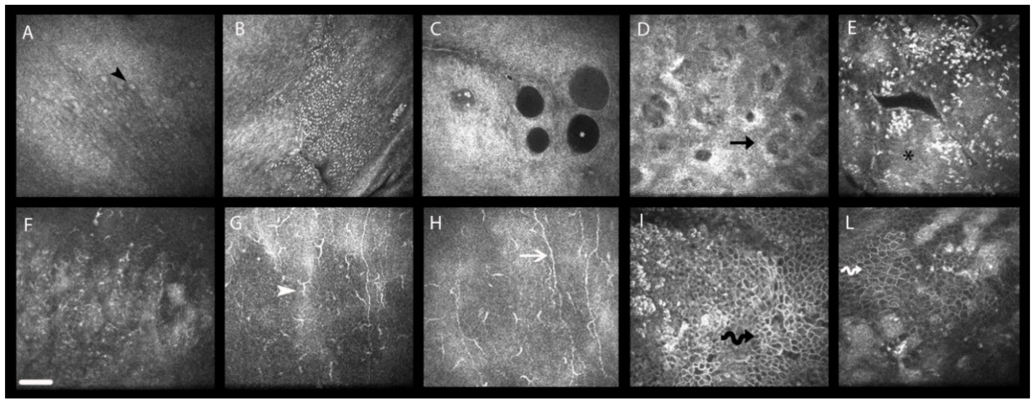
In the last two decades, IVCM has been progressively used to study the sub-clinical and clinically relevant changes of the ocular surface in patients with medically controlled glaucoma [24] (Figure 1) (Table 1 and Table 2).
Several structures can be analyzed including bulbar and palpebral conjunctiva, cornea, limbus, and MGs; in detail, it has been used to the examine the GCs’ and MGs’ density, the Vogt’s palisades and transition epithelium of the corneoscleral limbus, corneal epithelial and endothelial cell density, stromal keratocytes, and several parameters related to the sub-basal corneal nerve plexus [37].

3.2.1. Conjunctiva

Antiglaucoma drugs may induce modifications of the conjunctiva from the epithelium to stroma.
Ciancaglini et al. described at IVCM the presence of epithelial microcysts in the bulbar conjunctiva in untreated ocular hypertension (OH) and in topically treated primary open-angle glaucoma (POAG) patients [38]. A higher microcyst density and area were found in patients on combined β-blocker/prostaglandin therapy when compared with PGA monotherapy, suggesting that combined therapy may enhance the aqueous humor outflow more than monotherapy [38]. Moreover, the authors did not exclude that a higher BAK concentration in the combined therapy could be a contributing factor for the increased epithelial microcysts density and area [38].
The presence of epithelial microcysts may have different explanations: they may be considered an adaptive mechanism in eyes with reduced aqueous humor trabecular outflow, but also a sign of degenerated GCs, or a hallmark of epithelial disruption [25,39].
Other epithelial changes found with IVCM are squamous metaplasia, desquamation, keratinization, thickening, subepithelial fibrosis, conjunctiva-associated lymphoid tissue (CALT) activation, dendritic cells’ (DCs) activation, and GCs’ loss [40,41].
GCs are essential for the mucin production, which guarantee the adhesion between the tear film and epithelia. Therefore, GCs’ loss decreases mucin production, and thus the tear film stability; this leads to a reduced tear clearance, with the development of dry eye and conjunctival inflammation [25].
To date, few studies were specifically conducted to determine the effects of distinct classes and regimen of topical drugs on the density of GCs in patients with glaucoma with different results. Ciancaglini et al. [42] found a marked decrease of GCs and a worse score of epithelial regularity in patients receiving preserved levobunolol with respect to those treated with the unpreserved formulation, suggesting the toxicity of preservatives on the ocular surface. Similarly, Frezzotti et al. [43] reported a significant increase in the cumulative grading score of epithelial irregularity and a reduction in the intra-epithelial GCs’ density at IVCM after 12 months of treatment with preserved timolol; conversely, these changes did not occur in controls and patients treated with PF timolol 0.1% gel formulation. However, even if the duration of both studies was limited to a short period of time (6 and 12 months, respectively) [42,43], it is possible to suppose that preservatives could exert their toxic effect on the conjunctiva after a few months of therapy.
Differently, PGAs seem to have distinct effect on GCs [44]. PF tafluprost increased GCs density until the sixth month in naïve glaucomatous patients, suggesting the aptitude of prostaglandin derivatives to stimulate mucin secretion [45]. An increase in GCs’ density at IVCM was also reported in patients receiving bimatoprost 0.01% with respect to those treated with the bimatoprost 0.03% formulation [46]. This result seems to suggest that the concentration of the active drug is more influential than the concentration of BAK in the tolerability of chronic therapy. However, a weakness of this study is represented by its open-label design and by the strict inclusion criteria that allow to analyse the safety of bimatoprost and BAK only in the glaucomatous eyes with relatively healthy ocular surface compared with the average glaucoma patient usually being treated [46].
Additionally, less pronounced conjunctival modifications were found in patients treated with preservative PGA, in comparison with patients controlled with other classes of IOP lowering medications. Specifically, PGA induced a lower reduction in GCs’ density when compared with other drug classes and no changes in the subepithelial collagen fiber diameter and expression of subepithelial fibrosis when compared with controls [47]. Moreover, GCs’ loss is even more frequent in patients treated with two drugs compared with patients in monotherapy [41].
Conjunctival stromal changes, generally less frequently observed than epithelial modifications, are represented by infiltration of inflammatory cells, fibroblasts proliferation, and activation with connective tissue deposition [40].
Finally, IVCM allows to study the effects of medications on CALT; this structure is mainly located in the lamina propria of the tarsal conjunctiva and characterized by a layer of specialized secretory lympho-epithelium, lymphoid follicles, crypt-associated lymphoid structures, and high endothelial venules [48]. At IVCM, the follicular CALT appears as defined round structures hosting hyper-reflective cellular elements in a collagen scaffold [48,49].
Confocal studies, conducted on animals, documented an intrafollicular infiltration of inflammatory cells after instillation of BAK alone or BAK-preserved PGA, whereas the PF-PGA did not activate the conjunctival immune response [50,51]. Overall, the immune-inflammatory reaction positively correlated with the presence of preservative and its concentration, with the adjunctive evidence that the change of BAK with less toxic preservatives, such as polyquaternium-1, in part preserved follicles from the infiltration of inflammatory cells [52]. The effects of antiglaucoma drugs on human CALT in vivo are very limited, but the available reported information agrees with those described in animal models [25]. These reported findings may support the evidence that antiglaucoma medications induce an immune-related ocular surface disorder in the lymphoid structures related for the most part to the use of preservatives.

3.2.2. Cornea

Topical IOP-lowering medications may induce significant modifications of corneal epithelial layers, sub-basal nerve plexus, stroma, and endothelium; all corneal changes can be detected with IVCM, with different confocal features positively correlated with clinical OSD symptoms [53].
Martone et al. [54] compared the long-term effects of PF and preserved antiglaucoma medications on the cornea in POAG and OH patients. They reported a reduced density of superficial epithelial cells in all groups of patients treated with preservative formulations, suggesting a toxic effect of BAK on these cells. The preservative groups also showed an increased density of basal epithelial cells, probably related to a proliferative stimulus induced from the superficial layer [54].
Medical therapy may also induce changes in corneal DCs, normally located within the limbal and central epithelia of the cornea [55]. In patients with glaucoma, the DCs’ density was reported to be two to three times higher at the limbus, and more than ten times higher in the central cornea, than that in healthy eyes. This density was also higher in multi-treated patients and in patients taking preserved drugs or with a high cumulative daily dose of BAK [55]. This was the first study that specifically investigated the DCs in the entire cornea in glaucomatous patients, suggesting that they may have a role in the development of the OSD. However, given the retrospective nature of the study, it was not clear whether the DCs’ density increase was primary related to the drugs’ toxicity or was a consequence of the iatrogenic dry eye [55].
As reported in these previous reports, the epithelium was the corneal layer more frequently altered by the antiglaucoma therapy. However, the inflammatory processes occurring within the epithelium have been also hypothesized to determine stromal changes through the promotion of apoptotic phenomena and increased stromal proteolytic activity [25,54].
Several studies have evaluated stromal reflectivity, keratocyte density, and sub-basal plexus nerves parameters such as density, tortuosity, and reflectivity; nerve fiber changes are crucial, as nerve fibers regulate the corneal trophism, which is essential to maintain a healthy ocular surface [56].
The increase in keratocyte density in the entire stroma was reported in patients under PGA therapy for at least three years, suggesting that PGA induces a disruption of the architecture of the extracellular matrix through the activation of metalloproteinases and inhibition of tissue inhibitors of metalloproteinases [57].
Different results were reported in a previous study of Baratz et al. [58]. They reported no significant differences in keratocyte density or corneal endothelial cells morphology between treated and untreated eyes. However, they showed a significant decrease in the density of sub-basal plexus nerves in the treated group of the Ocular Hypertension Treatment Study after six years of therapy. These contrasting results were probably related by the fact that the author [58] studied the actions of the glaucoma therapy in general without differentiating between the selective effects that each class of hypotensive drugs may have on the stroma.
The reduction in the number and density of sub-basal plexus nerves and an increased number of nerve beading and tortuosity at confocal microscopy were recognized in all therapy regimens [56], but were more evident in the case of treatment with preserved medications [54,59].
A recent study of Saini et al. reported a significant reduction in nerves’ number, length, and density in patients controlled with two or more topical antiglaucoma medications with preservatives (latanoprost 0.005%, brimonidine and timolol 0.5%) [60].
Inversely, PF formulations seem to be associated with less toxicity on the ocular surface. In 2013, Rossi et al. [61] conducted a prospective study to evaluate the corneal changes at IVCM after twelve months of therapy with PF tafluprost in naïve glaucoma patients and in patients switched to tafluprost from a previous different monotherapy. They showed that patients that switched from preservative monotherapy to tafluprost presented an improvement of several confocal parameters, such as epithelial cells’ density, keratocyte activation, number of sub-basal nerves, and nerve tortuosity, suggesting a possible reversible effect of the ocular surface toxicity [61].
In another prospective study, the same author performed IVCM of the cornea in three groups of patients: naïve patients treated with PF tafluprost 0.0015%; naïve patients treated with preserved bimatoprost 0.003% or preserved travoprost 0.004%; and patients on topical preserved prostaglandin or beta-blocker monotherapy that need a switch [62]. They found that naïve-to-therapy patients did not show statistically significant changes from baseline regarding nerves’ parameters such as number, tortuosity, and beading like formations, whereas the switch to PF tafluprost in patients on topical preserved monotherapy improved the nerve status [62].
However, different results, probably related to the different IVCM employed and treatment duration, were reported by Rolle et al. [63]. In a cross-sectional study, they found corneal alterations on patients treated with PF-tafluprost 0.0015% or PF-timolol 0.1% for three years, confirming that the active compound itself may have a toxic effect on the ocular surface.
Antiglaucoma drugs may also determine polymegathism, pleomorphism, and reduction in density of the corneal endothelial cells [56], and may affect the corneoscleral limbus in terms of worsening of transitional epithelium regularity, inflammation of Vogt’s palisades, and increase in DCs [55,64].
Mastropasqua et al. [64] studied the immunoinflammatory effects of antiglaucoma medications on the corneoscleral limbus and found that the irregularity of the transition epithelium, DCs’ activation, and fibrosis of the Vogt’s palisades around the entire limbal circumference were more common in patients receiving BAK-preserved drugs or controlled with multitherapy. These alterations were correlated with the increased expression of both human leucocyte antigen-DR and interleukin-6 at impression cytology, suggesting that the inflammation may be the main mechanism involved in the limbal alterations [64].

3.2.3. Meibomian Glands

MGs are holocrine glands located in the tarsal plate of the eyelids and synthesize the meibum, a lipoid complex essential to stabilize the tear film and decrease its evaporation [65]. IVCM allows to identify MGs and to obtain several MG-related parameters of the orifice and acinus [65].
The main MGs’ modifications induced by antiglaucoma therapy are represented by the reduction of glandular density and area, signs of glandular loss and decreased meibum production, an increased reflectivity of the acinar secretion, dilation of the ductal orifice, and inhomogeneity of MGs’ interstice and wall, which indicate glandular inflammation [66].
These findings are more evident in the case of therapy with preserved drugs. Specifically, Agnifili et al. [67] reported that preserved PGAs were more toxic than the PF formulation, whereas no differences were found between preserved and unpreserved β-blockers; this suggested that, besides the detrimental effect of BAK, active compounds, and especially PGA, also play a role in inducing MGs’ alterations.
Additionally, MGs’ changes are even more frequent in patients treated with two or more drugs compared with patients in monotherapy. It has been shown that the prostaglandin/timolol fixed combinations induce less modifications MGs and GCs compared with the unfixed combination of latanoprost + timolol, and these changes are even more contained in the PF formulation of bimatoprost/timolol [68]. These results sustain the higher tolerability for the ocular surface of fixed combinations over the unfixed regimens. The modification of the therapy regimen should be strongly considered in the case of glaucoma-related OSD, as it may lead to a potential reversibility of damage of GCs and MGs. These aspects should be taken into account especially when patients require a multiple therapy to control the disease.

3.3. Morphologic Changes: Anterior Segment Optical Coherence Tomography

In the last years, AS-OCT has gained growing importance in studying all the anterior segment structures of the eye, providing information from a more macroscopic point of view compared with IVCM [69].
In the field of glaucoma, AS-OCT allows to visualize the aqueous outflow pathways, the anterior chamber angle anatomy, and function, and to evaluate the alterations induced by IOP-lowering agents on the ocular surface including corneal epithelia, tear film, and tear meniscus (Table 1 and Table 2) [37,70,71].

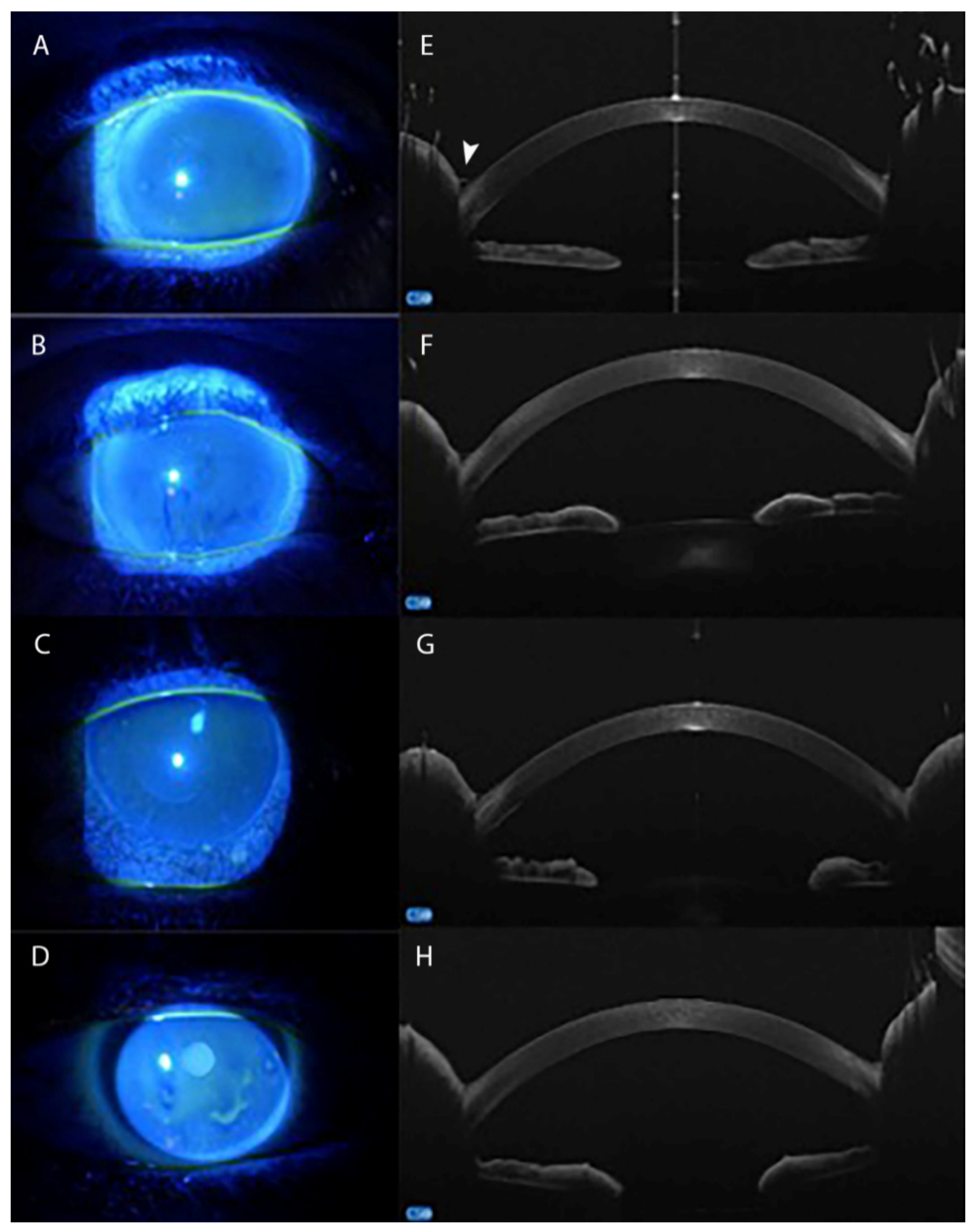
3.3.1. Tear Meniscus

More recently, studies have also used AS-OCT in clinical practice to evaluate the tear film and tear meniscus in patients affected by dry eye disease [72,73], with tear meniscus reduction being proposed as a biomarker of dry eye and, generally, OSD. However, to date, few data are available about the tear meniscus alterations, as detected at AS-OCT, in glaucomatous patients on medical therapy (Figure 2).
In a recent case-control observational study, Agnifili et al. [26] evaluated the tear meniscus morphometric modifications using AS-OCT in glaucomatous patients controlled with different therapy regimens. The authors observed that the tear meniscus height and area were significantly reduced in glaucoma patients compared with normal eyes. Moreover, no differences were detected between β-blockers and PGA monotherapy, where the presence of one or two preservative-containing eye drops did not modify the tear meniscus.
However, the tear meniscus height and area were significantly reduced in patients on multitherapy with two or more medications, with values similar to those observed in the case of dry eye [26]. These parameters were also positively correlated with ocular surface clinical tests such as break-up time, corneal fluorescein staining, and Schirmer Test I, and negatively with the ocular surface disease index questionnaire score, suggesting that the evaluation of tear meniscus at AS-OCT may allow to characterize some aspects of the glaucoma therapy-related OSD [26]. This was the first study conducted on patients with glaucoma; it was retrospective and included a small sample of patients. Therefore, it would be interesting to conduct further studies in order to confirm these results [26] and to differentiate the impact of the cumulative daily dose of preservative and the role of each active compound on the tear meniscus morphometry.

3.3.2. Corneal Epithelial Thickness

The chronic use of antiglaucoma drugs induces corneal epithelial changes, which may also lead to macroscopic alterations, such as corneal epithelial thickness modification, which can be further considered as an additional feature of glaucoma therapy-related OSD [74]. The measure of corneal epithelial thickness using AS-OCT allows to obtain information about the ocular surface status in the early stages of the glaucoma therapy-related OSD [75].
In a retrospective study, Cennamo et al. [75] measured corneal epithelial thickness in glaucoma patients and evaluated changes in thickness based on the number of corneal microvilli using scanning electron microscopy. The authors showed that the early epithelial damage in patients on therapy with preservative glaucoma medications is characterized by corneal epithelial thickness reduction and expression of a cellular damage, which initially involves corneal epithelial microvilli. However, the prolonged use of drugs may induce an advanced damage of epithelial microvilli with epithelial sub-oedema and inflammatory cells’ infiltration, which finally determined an increase in corneal epithelial thickness [75]. However, this study did not take into consideration factors that could contribute to corneal epithelial damage such as treatment duration and types of antiglaucoma drugs and preservatives.
Batawi et al. [76] compared the epithelial, stromal, and central corneal thickness between POAG and healthy eyes assessed by AS-OCT that, differently from confocal microscopy and ultrasound pachimetry, can measure the corneal epithelium and stromal layer separately. The authors found that POAG patients had lower absolute stromal and total thickness values compared with healthy eyes. Moreover, thickness negatively correlated with the number of antiglaucoma medications, suggesting that the preservative amount may probably affect the corneal epithelium [76].
Differently, a recent study did not find significant differences in corneal epithelial thickness among patients under different types and durations of treatment, while the only factor influencing corneal epithelial thickness was represented by age [77]. In a similar study, Dogan et al. [78] reported that there was no significant correlation between corneal thickness and glaucoma type, duration of therapy, number of drugs, and daily drops.
Table 1. Summary of clinical studies evaluating the main morphological changes induced by beta-blocker and prostaglandin analogue monotherapy on the ocular surface using IVCM and AS-OCT.
Ocular TissueAuthorsYearTechniqueStudy PopulationSpecific Therapy
(Preserved/Unpreserved)
Main Results
ConjunctivaCiancaglini M. et al. [38]2008IVCM aUntreated OH b
POAG c
Healthy controls
Preserved timolol 0.5%In glaucoma patients with respect to healthy controls:
Evidence of conjunctival EM d in OH b and POAG c groups [38]
Increase in DCs’e density and SFD f [47]
Comparison between BB i and PGA j and between preserved and PF h medications:
Decrease of GCs’ l density [42,43,47] and high score of epithelial regularity in the preserved BB i group compared with the PF h–BB i group
Increase of GCs’ l density in patients treated with PF h tafluprost [45], Bimatoprost 0.01% [46], and travoprost 0.004% [47]
Preserved levobunolol 0.5%
Latanoprost 0.005%
Travoprost 0.004%
Bimatoprost 0.03%
Ciancaglini M. et al. [42]2008IVCM a, IC gOH bPreserved levobunolol
POAG cPF h levobunolol
Mastropasqua L. et al. [45]2013LSCM k, IC gPOAG cPreserved latanoprost
Healthy controlsPF h tafluprost
Figus M. et al. [46]2014LSCM kPOAG cBimatoprost 0.01%
Bimatoprost 0.03%
Frezzotti P. et al. [43]2014IVCM aOH b
POAG c
Healthy controls
Preserved timolol
PF h timolol
Zhu W. et al. [47]2015IVCM aPOAG cCarteolol hydrochloride 2%
Healthy controlsTravoprost 0.004%
CorneaBaratz K. et al. [58]2006IVCM aOH b (medication group) OH b (observation group)timolol 0.5%In glaucoma patients with respect to healthy controls:
Decrease in sub-basal plexus nerve density [56,58,63]
Decrease in endothelial cell density [56]
Increase in nerve tortuosity [56,63]
Increase in keratocyte stromal density [54,57]
Increase in stromal reflectivity [63]
Increase in DCs’ n density [55]
Decrease of corneal thickness measurements at AS-OCT r [75,76,77,78]
Comparison between preserved and PF h medications:
Less confocal changes of the cornea parameters (epithelial, endothelial, and dendritic cells’ density; sub-basal nerve reflectivity; tortuosity; and beading) in glaucoma patients treated with PF h medications [55,59] and in patients on preserved monotherapy that switched to PF h therapy [54,55,61,62]
At limbus: higher limbal inflammation, irregularity of the transition epithelium, DCs’ n activation, and fibrosis of the Vogt’s palisades in patients receiving BAK q preserved drugs [64]
betaxolol 0.25%
Latanoprost 0.005%
Unoprostone 0.15%
Martone G. et al. [54]2009IVCM aOH b
POAG c
Healthy controls
Preserved timolol 0.5%
PF h timolol 0.5%
Bergonzi C. et al. [57]2010IVCM aPOAG cBB i (not specified)
PGA j (not specified)
Ranno S. et al. [56]2011IVCM aPOAG cBB i (not specified)
Healthy controlsPGA j (not specified)
Rossi G.C.M. et al. [61]2013IVCM aOH b
OAG m
Healthy controls
PF h Tafluprost
Fogagnolo P. et al. [59]2015IVCM aOH b
POAG c
PXG o
NTG p
Unpreserved tafluprost 0.0015%
Preserved latanoprost 0.005% +
BAK q 0.02%
Mastropasqua R. et al. [64]2015LSCM k, IC gPOAG c
Sjogren syndrome-dry eye
Healthy controls
Preserved timolol 0.5%
PF h timolol 0.5%
Preserved bimatoprost 0.001%
PF h tafluprost 0.015%
Mastropasqua R. et al. [55]2016IVCM aOAG m
Sjogren syndrome-dry eye
Healthy controls
Preserved timolol 0.5%
PF h timolol 0.5%
Preserved bimatoprost 0.001%
PF h tafluprost 0.015%
Rolle T. et al. [63]2017IVCM aOH b
POAG c
Healthy controls
PF h Timolol 0.1%
Tafluprost 0.0015%
Rossi G.C.M. et al. [62]2019IVCM aOH b
OAG m
Healthy controls
Preserved BB i (not specified)
PF h Tafluprost
Preserved bimatoprost 0.003%
Preserved travoprost 0.004%
Cennamo G. et al. [75]2018AS-OCT r, SEM sOAG m
Healthy controls
Preserved monotherapy
(not specified)
Batawi H. et al. [76]2018AS-OCT rPOAG cTimolol 0.005%
Healthy controlsLatanoprost
Montorio D. et al. [77]2020AS-OCT rPOAG cBB i (not specified)
Healthy controlsPGA j (not specified)
Dogan E. et al. [78]2020AS-OCT rPOAG c
PACG t
PXG o
Healthy controls
BB i (not specified)
PGA j (not specified)
Tear MeniscusAgnifili L. et al. [26]2020AS-OCT rPOAG c
EDE u
Healthy controls
BB i (not specified)Decrease in TMH v and TMA w in glaucoma patients with respect to healthy subjects
PGA j (not specified)TMH v and TMA w negatively correlated with OSDI x score
a IVCM = in vivo confocal microscopy; b OH = ocular hypertension; c POAG = primary open angle glaucoma; d EM = epithelial microcysts; e DCs = dendritic cells; f SFD = subepithelial collagen fiber diameter; g IC = impression citology; h PF = preservative free; i BB = beta-blocker; j PGA = prostaglandin analogue; k LSCM = laser scanning confocal microscopy; l GCs = goblet cells; m OAG = open angle glaucoma; n DCs = dendritic cells; o PXG = pseudoesfoliative glaucoma; p NTG = normal tension glaucoma; q BAK = benzalkonium chloride; r AS-OCT = anterior segment optical coherence tomography; s SEM = scanning electron microscopy; t PACG = primary angle closure glaucoma; u EDE = evaporative dry eye; v TMH = tear meniscus height; w TMA = tear meniscus area; x OSDI = ocular surface disease index.
Table 2. Summary of clinical studies evaluating the main morphological changes induced by combined therapy on the ocular surface using IVCM and AS-OCT.
Ocular TissueAuthorsYearTechniqueStudy PopulationSpecific TherapyMain Results
ConjunctivaCiancaglini M. et al. [38]2008IVCM aUntreated OH b
POAG c
Healthy controls
Unfixed CT d:In the case of multitherapy with respect to monotherapy:
Higher EM e density and area [38]
Higher DCs’ f density [47]
Greater GCs’ g loss [41]
Less MGs’ k and GCs’ g modifications in the case of fixed CT d and PF l formulation [68]
latanoprost/timolol
travoprost/timolol
bimatoprost/timolol
Zhu W. et al. [47]2015IVCM aPOAG cCT d:
Healthy controlsTwo or three drugs (not specified)
Di Staso S. et al. [41]2018IVCM aPOAG c
PXG h
PG i
DED j
Healthy controls
Fixed CT d:
latanoprost/timolol
travoprost/timolol
bimatoprost/timolol
Unfixed CT d:
bimatoprost/timolol
Agnifili L. et al. [68]2018IVCM aPOAG cFixed CT d:
PXG hprostaglandin/timolol
PG iUnfixed CT d:
Healthy controlslatanoprost + timolol
CorneaBaratz K. et al. [58]2006IVCM aOH b (medication group)Fixed CT d:In the case of multitherapy with respect to healthy controls:
Decrease in central corneal sub-basal nerve fiber number, length, and density and increase in basal epithelial cells’ density [60]
Decrease of corneal thickness measurements at AS-OCT o [77,78]
In the case of multitherapy with respect to monotherapy:
Higher decrease in sub-basal plexus nerve density [58]
Higher DCs’ f density [55]
At limbus: higher limbal inflammation, irregularity of the transition epithelium, DCs’ f activation, and fibrosis of the Vogt’s palisades [64]
Discording results in the corneal thickness parameters at AS-OCT o with regard to the number of drugs used [76,77,78]
OH b (observation group)Dorzolamide/timolol
Mastropasqua R. et al. [64]2015LSCM m
IC n
POAG c
Sjogren syndrome-dry eye
Healthy controls
Fixed CT d:
preserved latanoprost/timolol
preserved dorzolamide/timolol
preserved bimatroprost/brimonidine/timolol
preserved latanoprost/dorzolamide/timolol
Unfixed CT d:
preserved bimatoprost/timolol
preserved brimonidine/timolol
Mastropasqua R. et al. [55]2016IVCM aOAG p
Sjogren syndrome-dry eye
Healthy controls
Fixed CT d:
preserved latanoprost/timolol
preserved dorzolamide/timolol
preserved brimonidine/timolol
preserved bimatroprost/brimonidine/timolol
Unfixed CT d:
preserved bimatoprost/timolol
Saini M. et al. [60]2017IVCM aOH b
POAG c
PACG q
JOAG r
NTG s
Healthy controls
Preserved CT d:
timolol/brimonidine
timolol/latanoprost
latanoprost/brimonidine
Batawi H. et al. [76]2018AS-OCT oPOAG c
Healthy controls
CT d (not specified)
Montorio D. et al. [77]2020AS-OCT oPOAG c
Healthy controls
CT d (not specified)
Dogan E. et al. [78]2020AS-OCT oPOAG c
PACG q
PXG h
Healthy controls
CT d (not specified)
Tear MeniscusAgnifili L. et al. [26]2020AS-OCT oPOAG c
EDE t
Healthy controls
Two or more drugs (not specified)Smaller TMH u and TMA v in the case of multitherapy with respect to monotherapy
a IVCM = in vivo confocal microscopy; b OH = ocular hypertension; c POAG = primary open angle glaucoma; d CT = combined therapy; e EM = epithelial microcysts; f DCs = dendritic cells; g GCs = coblet cells; h PXG = pseudoesfoliative glaucoma; i PG = pigmentary glaucoma; j DED = dry eye disease; k MGs = Meibomian glands; l PF = preservative free; m LSCM = laser scanning confocal microscopy; n IC = impression citology; o AS-OCT = anterior segment optical coherence tomography; p OAG = open angle glaucoma; q PACG = primary angle closure glaucoma; r JOAG = juvenile open angle glaucoma; s NTG = normal tension glaucoma; t EDE = evaporative dry eye; u TMH = tear meniscus height; v TMA = tear meniscus area.

4. Surgical Treatment

4.1. Ocular Surface and Surgical Therapy

Surgical treatment is considered when medical therapy is not able to control the IOP and to stabilize the progression of the disease or in the case of hypersensitive reactions to antiglaucoma drugs [79].
Trabeculectomy is the gold standard and the most effective filtering procedure. It is characterised by the formation of an intrascleral fistula draining the aqueous humor from the anterior chamber to the subconjunctival space, which is commonly referred to as a filtering bleb [80]. However, trabeculectomy may widely alter the ocular surface, causing a persistent clinical or subclinical inflammatory process [30,81].
Studies have demonstrated that patients with glaucoma filtering bleb may experience ocular discomfort in terms of burning, tearing, dry eye like signs, pain, and dysesthesia [14,82,83]. These symptoms may be related to the formation and location of the bleb, the application of anti-mitotic agents, and the post-operative wound-healing processes within the conjunctiva that may contribute to the ocular surface changes [40,84].
On the other hand, the cessation of chronic use of IOP-lowering agents and the topical instillation of steroids after surgery may have a positive effect on the ocular surface status, which is probably related to the end of the chronic inflammation and the toxic action of the medications [84].
Recently, new surgical procedures called minimally-invasive glaucoma surgeries (MIGS) have been introduced in order to reduce the invasiveness of conventional filtering surgery. They may be considered an appropriate choice for patients with concomitant OSD and glaucoma [16]. These procedures spare or induce minimal trauma on the conjunctiva, resulting in a reduction of the post-operative inflammatory reaction and in less irritation to the ocular surface [85].

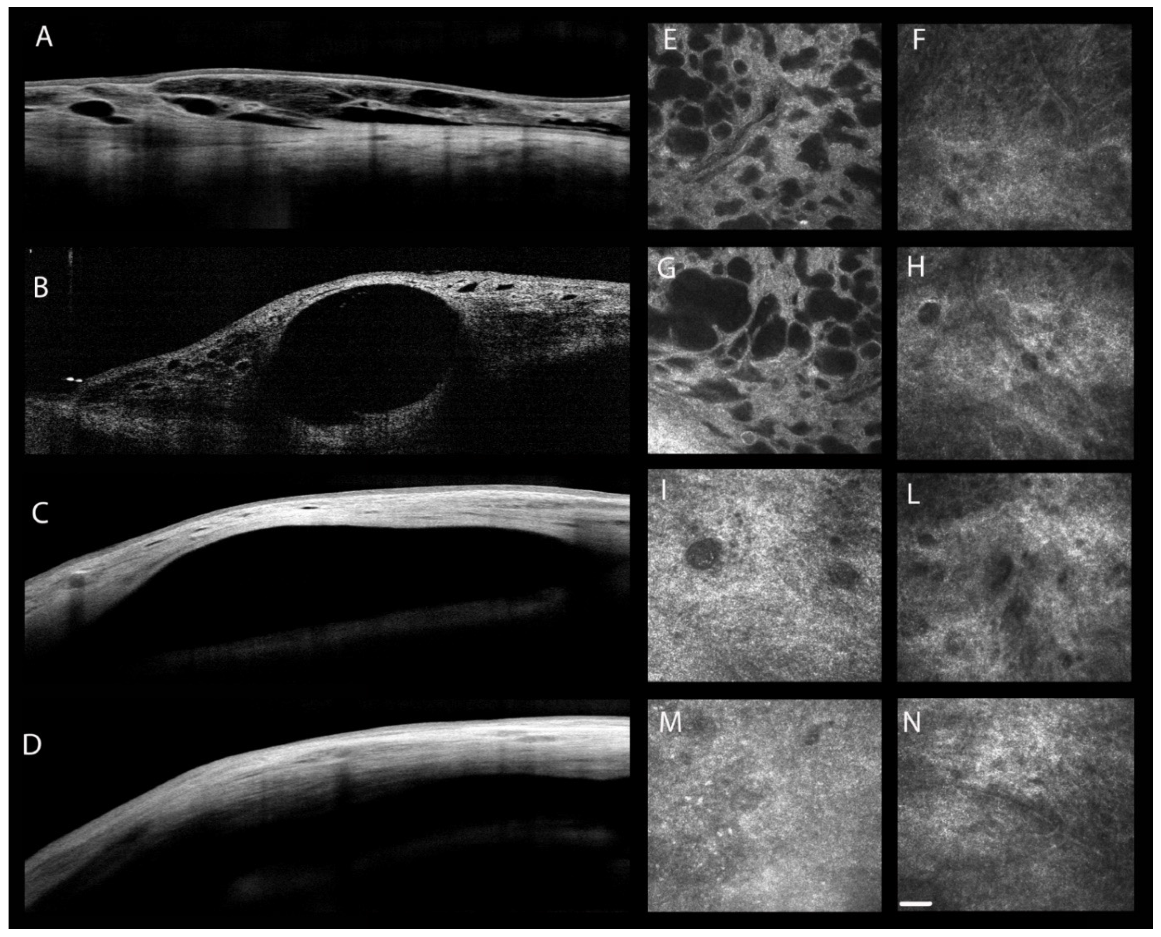
4.2. Morphologic Changes: In Vivo Confocal Microscopy

In the glaucoma surgical field, IVCM is applied to study the trans-scleral aqueous outflow, to differentiate functioning from non-functioning conjunctival filtering blebs, and to follow the conjunctival wound-healing process [86] (Table 3 and Figure 3). The IVCM use after filtering surgery would be appropriate in order to help clinicians in identifying the early signs of failure and to adopt appropriate procedures, such as laser suture lysis or needling with or without anti-metabolite injection, before the IOP rises and the bleb fails.
Non-functioning bleb presents a hyper-reflective subepithelial tissue with dense collagenous connective tissue and blood vessels and few or no epithelial microcysts [25,87,88,89].
Inversely, a functioning filtering bleb is characterized by a normal conjunctival epithelium with a hypo-reflective subepithelial tissue and numerous epithelial microcysts, which are considered as in vivo indicators of the trans-bleb aqueous humor passage [87,90,91].
At IVCM, epithelial microcysts appear as round or oval-shaped optically clear structures that may contain inflammatory cells and amorphous material and are surrounded by a hyper-reflective wall [40].
Ciancaglini et al. [90] evaluated the IVCM characteristics of the bulbar conjunctiva in POAG patients before and one month after trabeculectomy. They reported a fivefold increase in conjunctival epithelial microcysts’ density and area in correspondence to the bleb after surgery suggesting that is probably related to the enhancement of the aqueous filtration through the sclera and conjunctiva [90].
On the conjunctival surface of functioning bleb, the presence of a great number of atypical GCs may be also observed, which appear as hypo-reflective structures that show weak or no mucin marker 5AC immunostaining at impression cytology [92]. The epithelial microcysts seem to correspond to these GCs containing aqueous humor, suggesting that the transconjunctival aqueous outflow through the bleb wall epithelium occurs at the level of GCs [93].
In a prospective, cross-sectional study, Agnifili et al. [93] correlated the preoperative conjunctival GCs’ density and mucin positivity, using IVCM and impression cytology, with the 12-month success in patients undergoing trabeculectomy. The authors reported higher postoperative values of microcysts’ density and area in patients with a successful surgery compared with failed cases. A strong, positive correlation was found between these parameters and the baseline GCs’ density and the mucin marker 5AC, thus confirming a direct active role of GCs in vehiculating the aqueous humor [93].
Based on these reported findings, it is interesting to underline that the surgical success could also depend on the GCs’ density available before surgery. Therefore, it is mandatory to suggest a careful selection of the glaucoma medications used in the medical management of glaucoma in order to preserve the integrity of this critical cell population.
Other preoperative conjunctival parameters (as expression of the inflammatory status of the conjunctiva) were considered as predictive tools for the surgery outcome. Mastropasqua et al. [94] reported that high preoperative levels of DCs, low levels of GCs, and a hyper-reflective stroma in correspondence to the site of surgery significantly increased the risk of bleb dysfunction and failure.
In a recent six-month, prospective, case-control study, Agnifili et al. [84] compared the in vivo and ex vivo characteristics of the ocular surface before and after successful filtration surgery. They showed an increase in GCs’ density and a reduction in MGs’ density, MGs’ inhomogeneity, limbal DCs’ density, sub-basal corneal nerve inhomogeneity, and conjunctival human leucocyte antigen-DR positivity in the surgical group compared with the medical control group [84]. Based on these findings, the authors concluded that the ocular surface improved after glaucoma filtration surgery [84].
The filtering procedure may also induce different stromal features according to the bleb filtering ability. Functioning filtering bleb shows a high density of stromal cystic spaces, absence of encapsulated cysts in the stroma, small diameters, and less tortuosity of stromal vessels [95,96]. As for epithelial microcysts, stromal cystic spaces were considered as a sign of aqueous humor passage through the bleb wall [25]. Conversely, early unfavorable signs are few cystic stromal spaces and large vessel diameters [95]. Moreover, Guthoff et al. [95] showed that functioning bleb presented a trabecular or reticular stromal pattern, whereas non-functioning bleb is characterized by a compacted or corrugated stromal pattern.
Additionally, conjunctival and stromal modifications at the bleb site may be determined by the use of anti-fibrotic agents during surgery.
Ciancaglini et al. [88] did not report any microscopic differences between patients who received intraoperative mitomycin C (MMC) and patients who did not. However, different studies showed the presence of hyper-reflective microdots in the superficial epithelium layer, which probably represented necrotic or inflammatory cells, and the presence of a rarified stroma with large cystic non-encapsulated spaces in the case of MMC administration [87,96].
Finally, only a few studies were performed regarding the effects of MIGS on the ocular surface [85,97,98].
A one-year prospective study of Fea et al. [97] evaluated the efficacy, safety, and bleb morphology following implantation of XEN 45 gel stent combined or not with cataract surgery. The authors reported an increase in mean microcysts’ area and density associated with a reduction of density of subepithelial connective tissue in the postoperative period [97]. Additionally, the stromal density was significantly lower in the case of successful surgery. These findings could suggest that the stent implantation may induce a new or increase an alternative aqueous humor outflow [97].
Moreover, Sacchi et al. [98] retrospectively analyzed the conjunctival filtering bleb characteristics at IVCM after XEN gel implantation and trabeculectomy. IVCM showed a greater mean microcysts area in the case of trabeculectomy, suggesting that the larger scleral ostium obtained with this procedure led to a greater aqueous outflow towards the subconjunctival space [98].
Recently, Baiocchi et al. [85] compared the IVCM characteristics in glaucoma patients treated with XEN 45 gel stent implant, trabeculectomy, or medical therapy. In the case of XEN 45 Gel Stent in the subconjunctival space, the IVCM documented the presence of regular corneal epithelial cells with microcysts and normo-reflective subepithelial nerve plexus. These findings are an expression of a lower rate of ocular surface inflammation. Differently, the combined procedure determined a higher inflammation rate that was also more pronounced in the case of trabeculectomy or medical therapy [85]. Therefore, from the results of this study, we may suggest that the XEN implant may be a good and alternative option in the hypertensive glaucoma management. This surgery, if well timed, could represent a solution in patients where a successful surgical outcome after trabeculectomy could be compromised by severe ocular surface inflammation due to long-term antiglaucoma topical therapy.

4.3. Morphologic Changes: Anterior Segment Optical Coherence Tomography

AS-OCT provides a high resolution, objective, macroscopic detailed evaluation of the filtering bleb morphology and function after glaucoma surgery by analyzing, in a non-invasive manner, the conjunctival and subconjunctival characteristics (Table 3) [30]. It may be also used to determine which blebs are suitable for needling [99], to analyze bleb changes after laser suture lysis [100,101], and to predict the functioning of bleb at a later stage or the need to plan a bleb revision surgery [102,103]. Despite this consolidated knowledge about the AS-OCT conjunctival and subconjunctival changes following glaucoma surgery, at this moment, there is a lack of information regarding the ability of AS-OCT to detect the effects of surgery on the tear meniscus and corneal epithelia.
Generally, AS-OCT macroscopic bleb features after surgery are in agreement with microscopic characteristics as seen with confocal microscopy; however, these techniques have a different target in the filtering bleb analysis [25,88].
Different post-trabeculectomy bleb parameters are analyzed using AS-OCT including length and height of the inner bleb space, maximum and minimum bleb wall thickness, bleb wall density, subepithelial connective tissue density, and number of microcysts [88,104].
Features of bleb morphology associated with successful IOP-lowering are reported in many studies (Figure 3) [88,102,104,105,106]. Functioning filtering blebs are characterized by an increased bleb height, high density of epithelial microcysts, few conjunctival fibrosis, large internal fluid-filled cavity, thick bleb wall, and low reflectivity appearance of the bleb wall that may be an expression of loose connective tissue [88,103,106,107]. Conversely, failed blebs show opposite features. Moreover, the reflective appearance of the bleb wall and the width of filtration opening analyzed by AS-OCT are considered important predictors of post-trabeculectomy outcomes [108].
AS-OCT has been also performed to study the bleb morphology after MIGS [97,98,109,110,111].
Two prospective 12-month studies analyzed the bleb morphology and reflectivity after XEN gel stent implant and reported that patients with a low bleb-wall reflectivity were more likely to have a good surgical outcome [97,109]. Furthermore, blebs with subconjunctival separation morphology; diffuse distribution of fluid; and small, diffuse, and hypo-reflective cysts were associated to a low risk of failure [110].
Finally, two recent studies compared the macroscopic conjunctival filtering bleb features after XEN gel stent implant and trabeculectomy [98,111]. The filtering blebs formed when using the XEN gel stent are smaller and with fewer intra-bleb cystic cavities, and are associated with less fibrosis and a thicker epithelium than the trabeculectomy blebs [111]. Moreover, the XEN bleb showed a small area of bleb-wall sub-epithelium cyst-like structure; expression of the smaller scleral ostium obtained with the 45 µm lumen of the XEN gel implant compared with the trabeculectomy; and a low bleb wall reflectivity, which is a surrogate sign of fibrosis strongly correlated with the surgical outcome [98].
These results should be taken into consideration, indicating that each type of surgery is associated with specific morphological features of the filtration bleb. Therefore, the knowledge of these aspects detected at AS-OCT may help a clinician to predict the risk of filtration failure in the very early post-operative period.

4.4. Vascular Changes: Optical Coherence Tomography Angiography

Recently, optical coherence tomography angiography (OCT-A) has been gaining a role in post-operative filtration bleb assessment. OCT-A provides, in a quick and non-invasive way, information about the bleb-wall vasculature that cannot be adequately explored with the available ophthalmologic imaging platforms [112,113].
The bleb vascularity is related to the conjunctival wound-healing process; a higher conjunctival vascularization after surgery is, in fact, correlated with higher levels of inflammation and increased risk of filtration failure [112,114].
Few studies have evaluated the relationship between filtering bleb vascularization at OCT-A and surgical success after glaucoma surgery (Table 3) [112,113,114,115,116].
Yin et al. [112] explored the association between the vessel area in the operative region and the surgical outcomes post-trabeculectomy. They reported a positive correlation between the vessel area at one month and the IOP at six months after trabeculectomy [112].
Similar results were presented by Hayek et al. [115]. They found a significant correlation between preoperative conjunctival vascular density and mean IOP at six months postoperatively. Specifically, a low preoperative conjunctival vessel density on the surgical site was associated with a lower post-surgical IOP, fewer needlings, and less use of IOP-lowering drugs [115].
Moreover, other OCT-A features such as the colour and brightness densities of the bleb-wall exhibited a good correlation with the vessel grading using conventional clinical bleb grading systems, suggesting that these OCT-A parameters may be considered as biomarkers of the bleb vascularity and outcome after surgery [116]. Seo et al. [114] compared bleb vascularity parameters at OCT-A between MMC-augmented trabeculectomy and phacotrabeculectomy. They found differences in the conjunctival vascularity density according to the type of surgery. In the phacotrabeculectomy group, the bleb color and brightness densities were significantly higher than in the trabeculectomy group, suggesting that the combined procedure may probably lead to increased inflammation, which may lead to changes in vessels on the bleb [114].
Finally, Mastropasqua et al. [113] evaluated the filtration bleb features after XEN gel stent implantation using OCT-A. Successful filtration blebs presented a lower bleb wall vessel density and numerous and large areas of vessel displacement within the bleb wall. The authors suggested that these OCT-A signs may be considered angiographic biomarkers of a good aqueous humour percolation through the bleb wall layers, which can help to distinguish functioning from non-functioning filtering blebs [113].
Table 3. Summary of clinical studies evaluating the main features of functioning glaucoma filtering bleb using IVCM, AS-OCT, and OCT-A.
AuthorsYearTechniqueStudy PopulationSurgical ProcedureMain Results
Labbè A. et al. [87]2005IVCM aOAG bTrabeculectomyFunctioning filtering bleb at IVCM a:
increase in conjunctival EM d density and area [87,88,89,90,93,97]
low density of connective tissue [88,91,97]
trabecular or reticular stromal pattern with high density of stromal cystic spaces [95]
small diameters and less tortuosity of stromal vessels [95,96]
evidence of numerous atypical GCs i with weak or no mucin marker 5AC immunostaining [92,93]
less signs of inflammation in the case of XEN 45 gel stent implant with respect to combined procedure, trabeculectomy, or medical therapy [85]
Functioning filtering bleb at AS-OCT j:
low reflectivity appearance of the bleb wall [88,97,109,110]
cystic pattern [109,110]
few conjunctival fibrosis [88]
diffuse distribution of fluid and small, diffuse, and hyporeflective cysts [110]
Thinner and flatter hypo-reflective bleb wall and fewer intra-bleb cystic cavities in the case of XEN 45 gel stent implant with respect to trabeculectomy bleb [98,111]
At OCT-A l:
rarefied vascular network with low vessel density and numerous and large vessel displacement areas within the bleb-wall [113]
positive correlation between conjunctival vessel density and area and IOP m after surgery [112,115]
positive correlation between the colour and brightness densities of the bleb wall after surgery and the conventional vascular score bleb grading systems [116]. Colour and brightness densities of the bleb wall were significantly higher in the case of the combined procedure [114]
Opposite features in the case of non-functioning filtering bleb.
Guthoff R. et al. [95]2006IVCM aOAG b
CACG c
Trabeculectomy
Messmer E. et al. [96]2006IVCM aPOAG e
PXG f
NTG g
Secondary glaucoma
Trabeculectomy
Amar N. et al. [92]2008IVCM a
IC h
OAG bTrabeculectomy
Ciancaglini M. et al. [88]2008IVCM a
AS-OCT j
POAG e
PXG f
Trabeculectomy
Sbeity Z. et al. [89]2009IVCM aPOAG e
Secondary glaucoma
Trabeculectomy
Ciancaglini M. et al. [90]2009IVCM aPOAG eTrabeculectomy
Morita K. et al. [91]2012IVCM a
UBM k
POAG e
PXG f
Trabeculectomy
Agnifili L. et al. [93] 2016IVCM a
IC h
OAG b
Healthy controls
Trabeculectomy
Fea A. et al. [97]2017IVCM a
AS-OCT j
POAG eXEN 45 gel stent implant
Olate-Perez A. et al. [109]2017AS-OCT jPOAG eXEN 45 gel stent implant + cataract surgery
Yin X. et al. [112]2018OCT-A lPrimary GlaucomaTrabeculectomy
Hayek S. et al. [115]2019OCT-A lOAG b
CACG c
Trabeculectomy
Lenzhofer M. et al. [110]2019AS-OCT jOAG bXEN 45 gel stent implant
Teus M.A. et al. [111]2019AS-OCT jPOAG e
Healthy controls
XEN 45 gel stent implant
Trabeculectomy
Seo J.H. et al. [116]2019OCT-A lPOAG e
Secondary glaucoma
Trabeculectomy
Seo J.H. et al. [114]2019OCT-A lPOAG e
PACG n
Secondary glaucoma
Trabeculectomy
Phacotrabeculectomy
Baiocchi S. et al. [85]2020IVCM aPOAG eXEN 45 gel stent implant
Trabeculectomy
Sacchi M. et al. [98]2020AS-OCT j
IVCM a
POAG e
PXG f
NTG g
XEN 45 gel stent implant
Trabeculectomy
Mastropasqua R. et al. [113]2020OCT-A lPOAG e
PXG f
PG o
XEN gel stent implant
a IVCM = in vivo confocal microscopy; b OAG = open angle glaucoma; c CACG = chronic angle closure glaucoma; d EM = epithelial microcysts; e POAG = primary open angle glaucoma; f PXG = pseudoesfoliative glaucoma; g NTG = normal tension glaucoma; h IC = impression citology; i GCs = goblet cells; j AS- OCT = anterior segment optical coherence tomography; k UBM = ultrasound biomicroscopy; l OCT-A = optical coherence tomography angiography; m IOP = intraocular pressure; n PACG = primary angle closure glaucoma; o PG = pigmentary glaucoma.

5. Conclusions

Several studies have demonstrated the direct effects of glaucoma medical and surgical therapy on the tissues composing the ocular surface system.
New diagnostic tools such as IVCM and AS-OCT have progressively gained importance in the diagnosis and monitoring of glaucoma-related OSD. They allow to define, in a non-invasive way, the micro- and macrostructural changes of the ocular surface induced by medical therapy and surgical procedures, in order to detect the early signs of ocular surface modifications and the relationship between the ocular surface health status and the failure of filtering surgery.
The introduction of new drug formulations, slow-release devices, and MIGS procedures may induce less ocular surface toxicity, resulting in a positive effect on the patient’s QoL and satisfaction. Therefore, the coexistence of glaucoma and OSD should have been taken into account by physicians in order to choose the right therapy regimen and the suitable surgical timing and procedure.

Author Contributions

Conceptualization: C.C., L.A. and F.O.; methodology: C.C. and F.O.; data collection: C.C., I.R., G.R., M.M., L.T. and A.C.V.V.; interpretation of data: C.C., I.R., G.R., M.M., L.T., A.C.V.V., G.M., A.H., L.Q. and F.O.; writing—original draft preparation: C.C., L.A. and F.O.; critical revision for important intellectual content: L.A., L.Q., A.H., G.M. and F.O.; final approval of the version to be published: C.C., I.R., G.R., M.M., L.T., A.C.V.V., L.A., G.M., A.H., L.Q. and F.O. All authors have read and agreed to the published version of the manuscript.

Funding

This work received no external funding. A.H. is supported by NIH grant (R01EY030851), NSF DMS (1853222/2021192), and in part by a Challenge Grant award from Research to Prevent Blindness, NY.

Institutional Review Board Statement

Not applicable.

Informed Consent Statement

Not applicable.

Data Availability Statement

No new data were created or analyzed in this study. Data sharing is not applicable to this article.

Acknowledgments

The contribution of IRCCS Fondazione Bietti to this work was supported by the Italian Ministry of Health and by Fondazione Roma. A.H. would like to disclose that he received remuneration from Adom, Qlaris, Luseed, and Cipla for serving as a consultant, and he serves on the board of Adom, Qlaris, and Phileas Pharma. A.H. holds an ownership interest in AdOM, Luseed, Oxymap, Qlaris, Phileas Pharma, and QuLent. All relationships listed above are pursuant to Icahn School of Medicine’s policy on outside activities.

Conflicts of Interest

The authors declare no conflict of interest.

Abbreviations

AS-OCTanterior segment optical coherence tomography
BAKbenzalkonium chloride
CALTconjunctiva-associated lymphoid tissue
DCsdendritic cells
GCsgoblet cells
IOPintraocular pressure
IVCMin vivo confocal microscopy
MGsMeibomian glands
MIGSminimally-invasive glaucoma surgeries
MMCmitomycin C
OCToptical coherence tomography angiography
OHocular hypertension
OSDocular surface disease
PFpreservative-free
PGAprostaglandin analogs
POAGprimary open angle glaucoma
QoLquality of life

References

  1. Quigley, H.A. Glaucoma. Lancet 2011, 377, 1367–1377. [Google Scholar] [CrossRef]
  2. Tham, Y.C.; Li, X.; Wong, T.Y.; Quigley, H.A.; Aung, T.; Cheng, C.Y. Global prevalence of glaucoma and projections of glaucoma burden through 2040: A systematic review and meta-analysis. Ophthalmology 2014, 121, 2081–2090. [Google Scholar] [CrossRef] [PubMed]
  3. Agis Investigators. The Advanced Glaucoma Intervention Study (AGIS): 7. The relationship between control of intraocular pressure and visual field deterioration. The AGIS Investigators. Am. J. Ophthalmol. 2000, 130, 429–440. [Google Scholar] [CrossRef]
  4. Leske, M.C.; Heijl, A.; Hussein, M.; Bengtsson, B.; Hyman, L.; Komaroff, E.; Early Manifest Glaucoma Trial Group. Factors for glaucoma progression and the effect of treatment: The early manifest glaucoma trial. Arch. Ophthalmol. 2003, 121, 48–56. [Google Scholar] [CrossRef] [PubMed]
  5. Lichter, P.R.; Musch, D.C.; Gillespie, B.W.; Guire, K.E.; Janz, N.K.; Wren, P.A.; Mills, R.P.; CIGTS Study Group. Interim clinical outcomes in the Collaborative Initial Glaucoma Treatment Study comparing initial treatment randomized to medications or surgery. Ophthalmology 2001, 108, 1943–1953. [Google Scholar] [CrossRef]
  6. Maier, P.C.; Funk, J.; Schwarzer, G.; Antes, G.; Falck-Ytter, Y.T. Treatment of ocular hypertension and open angle glaucoma: Meta-analysis of randomised controlled trials. BMJ 2005, 331, 134. [Google Scholar] [CrossRef] [Green Version]
  7. European Glaucoma Society. Terminology and Guidelines for Glaucoma. 5th Edition. Available online: https://www.eugs.org/eng/egs_guidelines_reg.asp?l=1 (accessed on 18 June 2021).
  8. Roberti, G.; Tanga, L.; Manni, G.; Riva, I.; Verticchio, A.C.; Berardo, F.; Carnevale, C.; Oddone, F. Tear Film, Conjunctival and Corneal Modifications Induced by Glaucoma Treatment. Curr. Med. Chem. 2019, 26, 4253–4261. [Google Scholar] [CrossRef]
  9. Boso, A.L.M.; Gasperi, E.; Fernandes, L.; Costa, V.P.; Alves, M. Impact of Ocular Surface Disease Treatment in Patients with Glaucoma. Clin. Ophthalmol. 2020, 14, 103–111. [Google Scholar] [CrossRef]
  10. Skalicky, S.E.; Goldberg, I.; McCluskey, I. Ocular surface disease and quality of life in patients with glaucoma. Am. J. Ophthalmol. 2012, 153, 1–9.e2. [Google Scholar] [CrossRef]
  11. Kaštelan, S.; Tomic, M.; Soldo, K.; Salopek-Rabatic, J. How ocular surface disease impacts the glaucoma treatment outcome. BioMed Res. Int. 2013, 2013, 696328. [Google Scholar] [CrossRef] [Green Version]
  12. Stalmans, I.; Lemij, H.; Clarke, J.; Baudouin, C.; GOAL study group. Signs and Symptoms of Ocular Surface Disease: The Reasons for Patient Dissatisfaction with Glaucoma Treatments. Clin. Ophthalmol. 2020, 14, 3675–3680. [Google Scholar] [CrossRef]
  13. Baudouin, C. Ocular surface and external filtration surgery: Mutual relationships. Dev. Ophthalmol. 2012, 50, 64–78. [Google Scholar]
  14. Ji, H.; Zhu, Y.; Zhang, Y.; Li, Z.; Ge, J.; Zhuo, Y. Dry Eye Disease in Patients with Functioning Filtering Blebs after Trabeculectomy. PLoS ONE 2016, 11, e0152696. [Google Scholar] [CrossRef]
  15. Villani, E.; Sacchi, M.; Magnani, F.; Nicodemo, A.; Williams, S.E.I.; Rossi, A.; Ratiglia, R.; De Cillà, S.; Nucci, P. The Ocular Surface in Medically Controlled Glaucoma: An In Vivo Confocal Study. Investig. Ophthalmol. Vis. Sci. 2016, 57, 1003–1010. [Google Scholar] [CrossRef]
  16. Zhang, X.; Vadoothker, S.; Munir, W.M.; Saeedi, O. Ocular Surface Disease and Glaucoma Medications: A Clinical Approach. Eye Contact Lens 2019, 45, 11–18. [Google Scholar] [CrossRef] [PubMed]
  17. Schein, O.D.; Muñoz, B.; Tielsch, J.M.; Bandeen-Roche, K.; West, S. Prevalence of dry eye among the elderly. Am. J. Ophthalmol. 1997, 124, 723–728. [Google Scholar] [CrossRef]
  18. Leung, E.W.; Medeiros, F.A.; Weinreb, R.N. Prevalence of ocular surface disease in glaucoma patients. J. Glaucoma 2008, 17, 350–355. [Google Scholar] [CrossRef] [PubMed] [Green Version]
  19. Fechtner, R.D.; Godfrey, D.G.; Budenz, D.; Stewart, J.A.; Stewart, W.C.; Jasek, M.C. Prevalence of ocular surface complaints in patients with glaucoma using topical intraocular pressure-lowering medications. Cornea 2010, 29, 618–621. [Google Scholar] [CrossRef] [PubMed]
  20. Wong, A.B.C.; Wang, M.T.M.; Liu, K.; Prime, Z.K.; Danesh-Meyer, H.V.; Craig, J.P. Exploring topical anti-glaucoma medication effects on the ocular surface in the context of the current understanding of dry eye. Ocul. Surf. 2018, 16, 289–293. [Google Scholar] [CrossRef]
  21. Baudouin, C.; Kolko, M.; Melik-Parsadaniantz, S.; Messmer, E.M. Inflammation in Glaucoma: From the back to the front of the eye, and beyond. Prog. Retin. Eye Res. 2020, 100916. [Google Scholar] [CrossRef]
  22. Yazdani, M.; Fiskådal, J.; Chen, X.; Utheim, O.A.; Ræder, S.; Vitelli, V.; Utheim, T.P. Tear Film Break-Up Time and Dry Eye Disease Severity in a Large Norwegian Cohort. J. Clin. Med. 2021, 10, 884. [Google Scholar] [CrossRef]
  23. Begley, C.; Caffery, B.; Chalmers, R.; Situ, P.; Simpson, T.; Nelson, J.D. Review and analysis of grading scales for ocular surface staining. Ocul. Surf. 2019, 17, 208–220. [Google Scholar] [CrossRef]
  24. Villani, E.; Baudouin, C.; Efron, N.; Hamrah, P.; Kojima, T.; Patel, S.V.; Pflugfelder, S.C.; Zhivov, A.; Dogru, M. In vivo confocal microscopy of the ocular surface: From bench to bedside. Curr. Eye Res. 2014, 39, 213–231. [Google Scholar] [CrossRef] [PubMed] [Green Version]
  25. Mastropasqua, L.; Agnifili, L.; Mastropasqua, R.; Fasanella, V.; Nubile, M.; Toto, L.; Carpineto, P.; Ciancaglini, M. In vivo laser scanning confocal microscopy of the ocular surface in glaucoma. Microsc. Microanal. 2014, 20, 879–894. [Google Scholar] [CrossRef]
  26. Agnifili, L.; Brescia, L.; Scatena, B.; Oddone, F.; Figus, M.; Perna, F.; Doronzo, E.; D’Ugo, E.; Mastropasqua, L. Tear Meniscus Imaging by Anterior Segment-Optical Coherence Tomography in Medically Controlled Glaucoma. J. Glaucoma 2020, 29, 374–380. [Google Scholar] [CrossRef]
  27. Steven, D.W.; Alaghband, P.; Lim, K.S. Preservatives in glaucoma medication. Br. J. Ophthalmol. 2018, 102, 1497–1503. [Google Scholar] [CrossRef] [PubMed] [Green Version]
  28. Baudouin, C. Detrimental effect of preservatives in eyedrops: Implications for the treatment of glaucoma. Acta Ophthalmol. 2008, 86, 716–726. [Google Scholar] [CrossRef] [PubMed]
  29. Tressler, C.S.; Beatty, R.; Lemp, M.A. Preservative use in topical glaucoma medications. Ocul. Surf. 2011, 9, 140–158. [Google Scholar] [CrossRef]
  30. Baudouin, C. Ocular Surface and External Filtration Surgery: Mutual Relationships. Dev. Ophthalmol. 2017, 59, 67–79. [Google Scholar] [PubMed]
  31. Baudouin, C.; Liang, H.; Hamard, P.; Riancho, L.; Creuzot-Garcher, C.; Warnet, J.M.; Brignole-Baudouin, F. The ocular surface of glaucoma patients treated over the long term expresses inflammatory markers related to both T-helper 1 and T-helper 2 pathways. Ophthalmology 2008, 115, 109–115. [Google Scholar] [CrossRef] [PubMed]
  32. Pisella, P.J.; Debbasch, C.; Hamard, P.; Creuzot-Garcher, C.; Rat, P.; Brignole, F.; Baudouin, C. Conjunctival proinflammatory and proapoptotic effects of latanoprost and preserved and unpreserved timolol: An ex vivo and in vitro study. Investig. Ophthalmol. Vis. Sci. 2004, 45, 1360–1368. [Google Scholar] [CrossRef] [PubMed] [Green Version]
  33. Aydin Kurna, S.; Acikgoz, S.; Altun, A.; Ozbay, N.; Sengor, T.; Okan Olcaysu, O. The effects of topical antiglaucoma drugs as monotherapy on the ocular surface: A prospective study. J. Ophthalmol. 2014, 2014, 460483. [Google Scholar] [CrossRef] [PubMed]
  34. Mohammed, I.; Kulkarni, B.; Faraj, L.A.; Abbas, A.; Dua, H.S.; King, A.J. Profiling ocular surface responses to preserved and non-preserved topical glaucoma medications: A 2-year randomized evaluation study. Clin. Exp. Ophthalmol. 2020, 48, 973–982. [Google Scholar] [CrossRef]
  35. Baudouin, C.; Hamard, P.; Liang, H.; Creuzot-Garcher, C.; Bensoussan, L.; Brignole, F. Conjunctival epithelial cell expression of interleukins and inflammatory markers in glaucoma patients treated over the long term. Ophthalmology 2004, 111, 2186–2192. [Google Scholar] [CrossRef]
  36. Yang, Y.; Huang, C.; Lin, X.; Wu, Y.; Ouyang, W.; Tang, L.; Ye, S.; Wang, Y.; Li, W.; Zhang, X.; et al. 0.005% Preservative-Free Latanoprost Induces Dry Eye-Like Ocular Surface Damage via Promotion of Inflammation in Mice. Investig. Ophthalmol. Vis. Sci. 2018, 59, 3375–3384. [Google Scholar] [CrossRef] [Green Version]
  37. Schmidl, D.; Schlatter, A.; Chua, J.; Tan, B.; Garhöfer, G.; Schmetterer, L. Novel Approaches for Imaging-Based Diagnosis of Ocular Surface Disease. Diagnostics 2020, 10, 589. [Google Scholar] [CrossRef]
  38. Ciancaglini, M.; Carpineto, P.; Agnifili, L.; Nubile, M.; Fasanella, V.; Mastropasqua, L. Conjunctival modifications in ocular hypertension and primary open angle glaucoma: An in vivo confocal microscopy study. Investig. Ophthalmol. Vis. Sci. 2008, 49, 3042–3048. [Google Scholar] [CrossRef] [PubMed] [Green Version]
  39. Baudouin, C.; Labbé, A.; Liang, H.; Pauly, A.; Brignole-Baudouin, F. Preservatives in eyedrops: The good, the bad and the ugly. Prog. Retin. Eye Res. 2010, 29, 312–334. [Google Scholar] [CrossRef]
  40. Mastropasqua, L.; Agnifili, L.; Mastropasqua, R.; Fasanella, V. Conjunctival modifications induced by medical and surgical therapies in patients with glaucoma. Curr. Opin. Pharmacol. 2013, 13, 56–64. [Google Scholar] [CrossRef] [PubMed]
  41. Di Staso, S.; Agnifili, L.; Ciancaglini, M.; Murano, G.; Borrelli, E.; Mastropasqua, L. In Vivo Scanning Laser Confocal Microscopy of Conjunctival Goblet Cells in Medically-controlled Glaucoma. In Vivo 2018, 32, 437–443. [Google Scholar]
  42. Ciancaglini, M.; Carpineto, P.; Agnifili, L.; Nubile, M.; Fasanella, V.; Lanzini, M.; Calienno, R.; Mastropasqua, L. An in vivo confocal microscopy and impression cytology analysis of preserved and unpreserved levobunolol-induced conjunctival changes. Eur. J. Ophthalmol. 2008, 18, 400–407. [Google Scholar] [CrossRef] [PubMed]
  43. Frezzotti, P.; Fogagnolo, P.; Haka, G.; Motolese, I.; Iester, M.; Bagaglia, S.A.; Mittica, P.; Menicacci, C.; Rossetti, L.; Motolese, E. In vivo confocal microscopy of conjunctiva in preservative-free timolol 0.1% gel formulation therapy for glaucoma. Acta Ophthalmol. 2014, 92, e133–e140. [Google Scholar] [CrossRef] [PubMed]
  44. Di Staso, S.; Agnifili, L.; Cecannecchia, S.; Di Gregorio, A.; Ciancaglini, M. In Vivo Analysis of Prostaglandins-induced Ocular Surface and Periocular Adnexa Modifications in Patients with Glaucoma. In Vivo 2018, 32, 211–220. [Google Scholar] [PubMed] [Green Version]
  45. Mastropasqua, L.; Agnifili, L.; Fasanella, V.; Curcio, C.; Ciabattoni, C.; Mastropasqua, R.; Toto, L.; Ciancaglini, M. Conjunctival goblet cells density and preservative-free tafluprost therapy for glaucoma: An in vivo confocal microscopy and impression cytology study. Acta Ophthalmol. 2013, 91, e397–e405. [Google Scholar] [CrossRef]
  46. Figus, M.; Nardi, M.; Piaggi, P.; Sartini, M.; Guidi, G.; Martini, L.; Lazzeri, S. Bimatoprost 0.01% vs bimatoprost 0.03%: A 12-month prospective trial of clinical and in vivo confocal microscopy in glaucoma patients. Eye 2014, 28, 422–429. [Google Scholar] [CrossRef]
  47. Zhu, W.; Kong, X.; Xu, J.; Sun, X. Effects of Long-Term Antiglaucoma Eye Drops on Conjunctival Structures: An In Vivo Confocal Microscopy Study. J. Ophthalmol. 2015, 2015, 165475. [Google Scholar] [CrossRef]
  48. Mastropasqua, R.; Agnifili, L.; Fasanella, V.; Nubile, M.; Gnama, A.A.; Falconio, G.; Perri, P.; Di Staso, S.; Mariotti, C. The Conjunctiva-Associated Lymphoid Tissue in Chronic Ocular Surface Diseases. Microsc. Microanal. 2017, 23, 697–707. [Google Scholar] [CrossRef]
  49. Agnifili, L.; Mastropasqua, R.; Fasanella, V.; Di Staso, S.; Mastropasqua, A.; Brescia, L.; Mastropasqua, L. In vivo confocal microscopy of conjunctiva-associated lymphoid tissue in healthy humans. Investig. Ophthalmol. Vis. Sci. 2014, 55, 5254–5262. [Google Scholar] [CrossRef] [Green Version]
  50. Liang, H.; Baudouin, C.; Pauly, A.; Brignole-Baudouin, F. Conjunctival and corneal reactions in rabbits following short- and repeated exposure to preservative-free tafluprost, commercially available latanoprost and 0.02% benzalkonium chloride. Br. J. Ophthalmol. 2008, 92, 1275–1282. [Google Scholar] [CrossRef] [PubMed]
  51. Liang, H.; Baudouin, C.; Labbe, A.; Riancho, L.; Brignole-Baudouin, F. Conjunctiva-associated lymphoid tissue (CALT) reactions to antiglaucoma prostaglandins with or without BAK-preservative in rabbit acute toxicity study. PLoS ONE 2012, 7, e33913. [Google Scholar] [CrossRef]
  52. Liang, H.; Brignole-Baudouin, F.; Riancho, L.; Baudouin, C. Reduced in vivo ocular surface toxicity with polyquad-preserved travoprost versus benzalkonium-preserved travoprost or latanoprost ophthalmic solutions. Ophthalmic Res. 2012, 48, 89–101. [Google Scholar] [CrossRef]
  53. Baghdasaryan, E.; Tepelus, T.C.; Vickers, L.A.; Huang, P.; Chopra, V.; Sadda, S.R.; Lee, O.L. Assessment of Corneal Changes Associated with Topical Antiglaucoma Therapy Using In Vivo Confocal Microscopy. Ophthalmic Res. 2019, 61, 51–59. [Google Scholar] [CrossRef]
  54. Martone, G.; Frezzotti, P.; Tosi, G.M.; Traversi, C.; Mittica, V.; Malandrini, A.; Pichierri, P.; Balestrazzi, A.; Motolese, P.A.; Motolese, I.; et al. An in vivo confocal microscopy analysis of effects of topical antiglaucoma therapy with preservative on corneal innervation and morphology. Am. J. Ophthalmol. 2009, 147, 725–735.e1. [Google Scholar] [CrossRef]
  55. Mastropasqua, R.; Agnifili, L.; Fasanella, V.; Lappa, A.; Brescia, L.; Lanzini, M.; Oddone, F.; Perri, P.; Mastropasqua, L. In Vivo Distribution of Corneal Epithelial Dendritic Cells in Patients with Glaucoma. Investig. Ophthalmol. Vis. Sci. 2016, 57, 5996–6002. [Google Scholar] [CrossRef] [PubMed]
  56. Ranno, S.; Fogagnolo, P.; Rossetti, L.; Orzalesi, N.; Nucci, P. Changes in corneal parameters at confocal microscopy in treated glaucoma patients. Clin. Ophthalmol. 2011, 5, 1037–1042. [Google Scholar] [CrossRef] [Green Version]
  57. Bergonzi, C.; Giani, A.; Blini, M.; Marchi, S.; Luccarelli, S.; Staurenghi, G. Evaluation of prostaglandin analogue effects on corneal keratocyte density using scanning laser confocal microscopy. J. Glaucoma 2010, 19, 617–621. [Google Scholar] [CrossRef] [PubMed]
  58. Baratz, K.H.; Nau, C.H.; Winter, E.J.; McLaren, J.W.; Hodge, D.O.; Herman, D.C.; Bourne, W.M. Effects of glaucoma medications on corneal endothelium, keratocytes, and subbasal nerves among participants in the ocular hypertension treatment study. Cornea 2006, 25, 1046–1052. [Google Scholar] [CrossRef] [PubMed]
  59. Fogagnolo, P.; Dipinto, A.; Vanzulli, E.; Maggiolo, E.; De Cilla’, S.; Autelitano, A.; Rossetti, L. A 1-year randomized study of the clinical and confocal effects of tafluprost and latanoprost in newly diagnosed glaucoma patients. Adv. Ther. 2015, 32, 356–369. [Google Scholar] [CrossRef] [PubMed] [Green Version]
  60. Saini, M.; Vanathi, M.; Dada, T.; Agarwal, T.; Dhiman, R.; Khokhar, S. Ocular surface evaluation in eyes with chronic glaucoma on long term topical antiglaucoma therapy. Int. J. Ophthalmol. 2017, 10, 931–938. [Google Scholar]
  61. Rossi, G.C.M.; Blini, M.; Scudeller, L.; Ricciardelli, G.; Depolo, L.; Amisano, A.; Bossolesi, L.; Pasinetti, G.M.; Bianchi, P.E. Effect of preservative-free tafluprost on keratocytes, sub-basal nerves, and endothelium: A single-blind one-year confocal study on naïve or treated glaucoma and hypertensive patients versus a control group. J. Ocul. Pharmacol. Ther. 2013, 29, 821–825. [Google Scholar] [CrossRef]
  62. Rossi, G.C.M.; Scudeller, L.; Lumini, C.; Mirabile, A.V.; Picasso, E.; Bettio, F.; Pasinetti, G.M.; Bianchi, P.E. An in vivo confocal, prospective, masked, 36 months study on glaucoma patients medically treated with preservative-free or preserved monotherapy. Sci. Rep. 2019, 9, 4282. [Google Scholar] [CrossRef] [Green Version]
  63. Rolle, T.; Spinetta, R.; Nuzzi, R. Long term safety and tolerability of Tafluprost 0.0015% vs Timolol 0.1% preservative-free in ocular hypertensive and in primary open-angle glaucoma patients: A cross sectional study. BMC Ophthalmol. 2017, 17, 136. [Google Scholar] [CrossRef] [Green Version]
  64. Mastropasqua, R.; Agnifili, L.; Fasanella, V.; Curcio, C.; Brescia, L.; Lanzini, M.; Fresina, M.; Mastropasqua, L.; Marchini, G. Corneoscleral limbus in glaucoma patients: In vivo confocal microscopy and immunocytological study. Investig. Ophthalmol. Vis. Sci. 2015, 56, 2050–2058. [Google Scholar] [CrossRef]
  65. Binotti, W.W.; Bayraktutar, B.; Cuneyt Ozmen, M.; Cox, S.M.; Hamrah, P. A Review of Imaging Biomarkers of the Ocular Surface. Eye Contact Lens 2020, 46 (Suppl. 2), S84–S105. [Google Scholar] [CrossRef]
  66. Fasanella, V.; Agnifili, L.; Mastropasqua, R.; Brescia, L.; Di Staso, F.; Ciancaglini, M.; Mastropasqua, L. In Vivo Laser Scanning Confocal Microscopy of Human Meibomian Glands in Aging and Ocular Surface Diseases. BioMed Res. Int. 2016, 2016, 7432131. [Google Scholar] [CrossRef] [PubMed] [Green Version]
  67. Agnifili, L.; Fasanella, V.; Costagliola, C.; Ciabattoni, C.; Mastropasqua, R.; Frezzotti, P.; Mastropasqua, L. In vivo confocal microscopy of meibomian glands in glaucoma. Br. J. Ophthalmol. 2013, 97, 343–349. [Google Scholar] [CrossRef]
  68. Agnifili, L.; Mastropasqua, R.; Fasanella, V.; Brescia, L.; Scatena, B.; Oddone, F.; Mastropasqua, L. Meibomian Gland Features and Conjunctival Goblet Cell Density in Glaucomatous Patients Controlled with Prostaglandin/Timolol Fixed Combinations: A Case Control, Cross-sectional Study. J. Glaucoma 2018, 27, 364–370. [Google Scholar] [CrossRef] [PubMed]
  69. Jiao, H.; Hill, L.J.; Downie, L.E.; Chinnery, H.R. Anterior segment optical coherence tomography: Its application in clinical practice and experimental models of disease. Clin. Exp. Optom. 2019, 102, 208–217. [Google Scholar] [CrossRef] [PubMed] [Green Version]
  70. Hommer, A.; Schmidl, D.; Kromus, M.; Bata, A.M.; Fondi, K.; Werkmeister, R.M.; Baar, C.; Schmetterer, L.; Garhöfer, G. Effect of changing from preserved prostaglandins to preservative-free tafluprost in patients with glaucoma on tear film thickness. Eur. J. Ophthalmol. 2018, 28, 385–392. [Google Scholar] [CrossRef]
  71. Ang, M.; Baskaran, M.; Werkmeister, R.M.; Chua, J.; Schmidl, D.; Dos Santos, V.A.; Garhöfer, G.; Mehta, J.S.; Schmetterer, L. Anterior segment optical coherence tomography. Prog. Retin. Eye Res. 2018, 66, 132–156. [Google Scholar] [CrossRef] [PubMed]
  72. Raj, A.; Dhasmana, R.; Nagpal, R.C. Anterior Segment Optical Coherence Tomography for Tear Meniscus Evaluation and its Correlation with other Tear Variables in Healthy Individuals. J. Clin. Diagn. Res. 2016, 10, NC01–NC04. [Google Scholar] [CrossRef] [PubMed]
  73. Akiyama, R.; Usui, T.; Yamagami, S. Diagnosis of Dry Eye by Tear Meniscus Measurements Using Anterior Segment Swept Source Optical Coherence Tomography. Cornea 2015, 34 (Suppl. 11), S115–S120. [Google Scholar] [CrossRef] [PubMed]
  74. Mathews, P.M.; Ramulu, P.Y.; Friedman, D.S.; Utine, C.A.; Akpek, E.K. Evaluation of ocular surface disease in patients with glaucoma. Ophthalmology 2013, 120, 2241–2248. [Google Scholar] [CrossRef] [Green Version]
  75. Cennamo, G.; Montorio, D.; Del Prete, S.; Del Prete, A.; Cennamo, G. Anterior-Segment Optical Coherence Tomography and Scanning Electron Microscopy to Evaluate Corneal Epithelial Changes in Patients Undergoing Glaucoma Therapy. Cornea 2018, 37, 1522–1526. [Google Scholar] [CrossRef]
  76. Batawi, H.; Lollett, I.V.; Maliakal, C.; Wellik, S.R.; Anderson, M.G.; Feuer, W.; Karp, C.L.; Galor, A. A Comparative Study of Central Corneal Epithelial, Stromal, and Total Thickness in Males with and Without Primary Open-Angle Glaucoma. Cornea 2018, 37, 712–719. [Google Scholar] [CrossRef]
  77. Montorio, D.; Cennamo, G.; Breve, M.A.; Fiore, U.; Reibaldi, M.; Brescia Morra, V.; Cennamo, G. Evaluation of corneal epithelial thickness in glaucomatous patients using anterior-segment optical coherence tomography. J. Biophotonics 2020, 13, e201900095. [Google Scholar] [CrossRef]
  78. Dogan, E.; Çakir, B.K.; Aksoy, N.O.; Celik, E.; Erkorkmaz, U. Effects of topical antiglaucomatous medications on central corneal epithelial thickness by anterior segment optical coherence tomography. Eur. J. Ophthalmol. 2020, 30, 1519–1524. [Google Scholar] [CrossRef]
  79. Weinreb, R.N.; Aung, T.; Medeiros, F.A. The pathophysiology and treatment of glaucoma: A review. JAMA 2014, 311, 1901–1911. [Google Scholar] [CrossRef] [PubMed] [Green Version]
  80. Razeghinejad, M.R.; Fudemberg, S.J.; Spaeth, G.L. The changing conceptual basis of trabeculectomy: A review of past and current surgical techniques. Surv. Ophthalmol. 2012, 57, 1–25. [Google Scholar] [CrossRef]
  81. Cvenkel, B.; Kopitar, A.N.; Ihan, A. Correlation between filtering bleb morphology, expression of inflammatory marker HLA-DR by ocular surface, and outcome of trabeculectomy. J. Glaucoma 2013, 22, 15–20. [Google Scholar] [CrossRef] [PubMed]
  82. Budenz, D.L.; Hoffman, K.; Zacchei, A. Glaucoma filtering bleb dysesthesia. Am. J. Ophthalmol. 2001, 131, 626–630. [Google Scholar] [CrossRef]
  83. Neves Mendes, C.R.; Yudi Hida, R.; Kasahara, N. Ocular surface changes in eyes with glaucoma filtering blebs. Curr. Eye Res. 2012, 37, 309–311. [Google Scholar] [CrossRef] [PubMed]
  84. Agnifili, L.; Brescia, L.; Oddone, F.; Sacchi, M.; D’Ugo, E.; Di Marzio, G.; Perna, F.; Costagliola, C.; Mastropasqua, R. The ocular surface after successful glaucoma filtration surgery: A clinical, in vivo confocal microscopy, and immune-cytology study. Sci. Rep. 2019, 9, 11299. [Google Scholar] [CrossRef] [PubMed] [Green Version]
  85. Baiocchi, S.; Mazzotta, C.; Sgheri, A.; Di Maggio, A.; Bagaglia, S.A.; Posarelli, M.; Ciompi, L.; Meduri, A.; Tosi, G.M. In vivo confocal microscopy: Qualitative investigation of the conjunctival and corneal surface in open angle glaucomatous patients undergoing the XEN-Gel implant, trabeculectomy or medical therapy. Eye Vis. 2020, 7, 15. [Google Scholar] [CrossRef] [Green Version]
  86. Kymionis, G.D.; Diakonis, V.F.; Shehadeh, M.M.; Pallikaris, A.I.; Pallikaris, I.G. Anterior segment applications of in vivo confocal microscopy. Semin. Ophthalmol. 2015, 30, 243–251. [Google Scholar] [CrossRef]
  87. Labbé, A.; Dupas, B.; Hamard, P.; Baudouin, C. In vivo confocal microscopy study of blebs after filtering surgery. Ophthalmology 2005, 112, 1979. [Google Scholar] [CrossRef]
  88. Ciancaglini, M.; Carpineto, P.; Agnifili, L.; Nubile, M.; Lanzini, M.; Fasanella, V.; Mastropasqua, L. Filtering bleb functionality: A clinical, anterior segment optical coherence tomography and in vivo confocal microscopy study. J. Glaucoma 2008, 17, 308–317. [Google Scholar] [CrossRef]
  89. Sbeity, Z.; Palmiero, P.M.; Tello, C.; Liebmann, J.M.; Ritch, R. Noncontact in vivo scanning laser microscopy of filtering blebs. J. Glaucoma 2009, 18, 479–483. [Google Scholar] [CrossRef]
  90. Ciancaglini, M.; Carpineto, P.; Agnifili, L.; Nubile, M.; Fasanella, V.; Mattei, P.A.; Mastropasqua, L. Conjunctival characteristics in primary open-angle glaucoma and modifications induced by trabeculectomy with mitomycin C: An in vivo confocal microscopy study. Br. J. Ophthalmol. 2009, 93, 1204–1209. [Google Scholar] [CrossRef] [Green Version]
  91. Morita, K.; Gao, Y.; Saito, Y.; Higashide, T.; Kobayashi, A.; Ohkubo, S.; Sugiyama, K. In vivo confocal microscopy and ultrasound biomicroscopy study of filtering blebs after trabeculectomy: Limbus-based versus fornix-based conjunctival flaps. J. Glaucoma 2012, 21, 383–391. [Google Scholar] [CrossRef]
  92. Amar, N.; Labbé, A.; Hamard, P.; Dupas, B.; Baudouin, C. Filtering blebs and aqueous pathway an immunocytological and in vivo confocal microscopy study. Ophthalmology 2008, 115, 1154–1161.e4. [Google Scholar] [CrossRef]
  93. Agnifili, L.; Fasanella, V.; Mastropasqua, R.; Frezzotti, P.; Curcio, C.; Brescia, L.; Marchini, G. In Vivo Goblet Cell Density as a Potential Indicator of Glaucoma Filtration Surgery Outcome. Investig. Ophthalmol. Vis. Sci. 2016, 57, 2928–2935. [Google Scholar] [CrossRef] [Green Version]
  94. Mastropasqua, R.; Fasanella, V.; Brescia, L.; Oddone, F.; Mariotti, C.; Di Staso, S.; Agnifili, L. In Vivo Confocal Imaging of the Conjunctiva as a Predictive Tool for the Glaucoma Filtration Surgery Outcome. Investig. Ophthalmol. Vis. Sci. 2017, 58, BIO114–BIO120. [Google Scholar] [CrossRef] [PubMed]
  95. Guthoff, R.; Klink, T.; Schlunck, G.; Grehn, F. In vivo confocal microscopy of failing and functioning filtering blebs: Results and clinical correlations. J. Glaucoma 2006, 15, 552–558. [Google Scholar] [CrossRef] [PubMed]
  96. Messmer, E.M.; Zapp, D.M.; Mackert, M.J.; Thiel, M.; Kampik, A. In vivo confocal microscopy of filtering blebs after trabeculectomy. Arch. Ophthalmol. 2006, 124, 1095–1103. [Google Scholar] [CrossRef] [PubMed] [Green Version]
  97. Fea, A.M.; Spinetta, R.; Cannizzo, P.M.L.; Consolandi, G.; Lavia, C.; Aragno, V.; Germinetti, F.; Rolle, T. Evaluation of Bleb Morphology and Reduction in IOP and Glaucoma Medication following Implantation of a Novel Gel Stent. J. Ophthalmol. 2017, 2017, 9364910. [Google Scholar] [CrossRef] [PubMed]
  98. Sacchi, M.; Agnifili, L.; Brescia, L.; Oddone, F.; Villani, E.; Nucci, P.; Mastropasqua, L. Structural imaging of conjunctival filtering blebs in XEN gel implantation and trabeculectomy: A confocal and anterior segment optical coherence tomography study. Graefes Arch. Clin. Exp. Ophthalmol. 2020, 258, 1763–1770. [Google Scholar] [CrossRef]
  99. Guthoff, R.; Guthoff, T.; Hensler, D.; Grehn, F.; Klink, T. Bleb needling in encapsulated filtering blebs: Evaluation by optical coherence tomography. Ophthalmologica 2010, 224, 204–208. [Google Scholar] [CrossRef]
  100. Singh, M.; Aung, T.; Friedman, D.S.; Zheng, C.; Foster, P.J.; Nolan, W.P.; See, J.L.; Smith, S.D.; Chew, P.T.K. Anterior segment optical coherence tomography imaging of trabeculectomy blebs before and after laser suture lysis. Am. J. Ophthalmol. 2007, 143, 873–875. [Google Scholar] [CrossRef]
  101. Sng, C.C.A.; Singh, M.; Chew, P.T.K.; Ngo, C.S.; Zheng, C.; Tun, T.A.; See, J.L.S.; Ang, M.; Loon, S.C.; Aung, T. Quantitative assessment of changes in trabeculectomy blebs after laser suture lysis using anterior segment coherence tomography. J. Glaucoma 2012, 21, 313–317. [Google Scholar] [CrossRef]
  102. Shan, J.; DeBoer, C.; Xu, B.Y. Anterior Segment Optical Coherence Tomography: Applications for Clinical Care and Scientific Research. Asia Pac. J. Ophthalmol. 2019, 8, 146–157. [Google Scholar] [CrossRef]
  103. Raj, A.; Bahadur, H. Morphological analysis of functional filtering blebs with anterior segment optical coherence tomography: A short-term prediction for success of trabeculectomy. Eur. J. Ophthalmol. 2020, 1120672120924340. [Google Scholar] [CrossRef]
  104. Kawana, K.; Kiuchi, T.; Yasuno, Y.; Oshika, T. Evaluation of trabeculectomy blebs using 3-dimensional cornea and anterior segment optical coherence tomography. Ophthalmology 2009, 116, 848–855. [Google Scholar] [CrossRef]
  105. Mastropasqua, R.; Fasanella, V.; Agnifili, L.; Curcio, C.; Ciancaglini, M.; Mastropasqua, L. Anterior segment optical coherence tomography imaging of conjunctival filtering blebs after glaucoma surgery. BioMed Res. Int. 2014, 2014, 610623. [Google Scholar] [CrossRef]
  106. Kokubun, T.; Tsuda, S.; Kunikata, H.; Himori, N.; Yokoyama, Y.; Kunimatsu-Sanuki, S.; Nakazawa, T. Anterior-Segment Optical Coherence Tomography for Predicting Postoperative Outcomes after Trabeculectomy. Curr. Eye Res. 2018, 43, 762–770. [Google Scholar] [CrossRef]
  107. Meziani, L.; Hassani, R.T.J.; El Sanharawi, M.; Brasnu, E.; Liang, H.; Hamard, P.; Baudouin, C.; Labbe, A. Evaluation of Blebs After Filtering Surgery with En-Face Anterior-Segment Optical Coherence Tomography: A Pilot Study. J. Glaucoma 2016, 25, e550–e558. [Google Scholar] [CrossRef] [PubMed]
  108. Tsutsumi-Kuroda, U.; Kojima, S.; Fukushima, A.; Nakashima, K.I.; Iwao, K.; Tanihara, H.; Inoue, T. Early bleb parameters as long-term prognostic factors for surgical success: A retrospective observational study using three-dimensional anterior-segment optical coherence tomography. BMC Ophthalmol. 2019, 19, 155. [Google Scholar] [CrossRef] [Green Version]
  109. Olate-Pérez, A.; Pérez-Torregrosa, V.T.; Gargallo-Benedicto, A.; Neira-Ibáñez, P.; Cerdà-Ibáñez, M.; Osorio-Alayo, V.; Barreiro-Rego, A.; Duch-Samper, A. Prospective study of filtering blebs after XEN45 surgery. Arch. Soc. Esp. Oftalmol. 2017, 92, 366–371. [Google Scholar] [CrossRef] [PubMed]
  110. Lenzhofer, M.; Strohmaier, C.; Hohensinn, M.; Hitzl, W.; Sperl, P.; Gerner, M.; Steiner, V.; Moussa, S.; Krall, E.; Reitsamer, H.A. Longitudinal bleb morphology in anterior segment OCT after minimally invasive transscleral ab interno Glaucoma Gel Microstent implantation. Acta Ophthalmol. 2019, 97, e231–e237. [Google Scholar] [CrossRef]
  111. Teus, M.A.; Moreno-Arrones, J.P.; Castaño, B.; Castejon, M.A.; Bolivar, G. Optical coherence tomography analysis of filtering blebs after long-term, functioning trabeculectomy and XEN® stent implant. Graefes Arch. Clin. Exp. Ophthalmol. 2019, 257, 1005–1011. [Google Scholar] [CrossRef] [PubMed] [Green Version]
  112. Yin, X.; Cai, Q.; Song, R.; He, X.; Lu, P. Relationship between filtering bleb vascularization and surgical outcomes after trabeculectomy: An optical coherence tomography angiography study. Graefes Arch. Clin. Exp. Ophthalmol. 2018, 256, 2399–2405. [Google Scholar] [CrossRef] [PubMed]
  113. Mastropasqua, R.; Brescia, L.; Di Antonio, L.; Guarini, D.; Giattini, D.; Zuppardi, E.; Agnifili, L. Angiographic biomarkers of filtering bleb function after XEN gel implantation for glaucoma: An optical coherence tomography-angiography study. Acta Ophthalmol. 2020, 98, e761–e767. [Google Scholar] [CrossRef]
  114. Seo, J.H.; Lee, Y.; Shin, J.H.; Kim, Y.A.; Park, K.H. Comparison of conjunctival vascularity changes using optical coherence tomography angiography after trabeculectomy and phacotrabeculectomy. Graefes Arch. Clin. Exp. Ophthalmol. 2019, 257, 2239–2255. [Google Scholar] [CrossRef]
  115. Hayek, S.; Labbé, A.; Brasnu, E.; Hamard, P.; Baudouin, C. Optical Coherence Tomography Angiography Evaluation of Conjunctival Vessels During Filtering Surgery. Transl. Vis. Sci. Technol. 2019, 8, 4. [Google Scholar] [CrossRef] [PubMed] [Green Version]
  116. Seo, J.H.; Kim, Y.A.; Park, K.H.; Lee, Y. Evaluation of Functional Filtering Bleb Using Optical Coherence Tomography Angiography. Transl. Vis. Sci. Technol. 2019, 8, 14. [Google Scholar] [CrossRef] [PubMed] [Green Version]
Figure 1. In vivo confocal microscopy (IVCM) of the ocular surface tissues in multi-treated medically controlled glaucoma. (AE) Confocal frames taken from a patient controlled with a preserved fixed combination of timolol and dorzolamide and a preservative-free prostaglandin analog (PGA) (three eyedrops per day). (A) Goblet cells (GCs). GCs appear as hyper-reflective elements (black arrowhead) dispersed within the epithelium, and often appear scattered with an evident reduction of their density. (B) Inflammatory infiltrates. Inflammatory infiltrates appear as clusters of small hyper-reflective and mono-nucleate elements (presumably lymphocytes) infiltrating the epithelium of the tarsal or bulbar conjunctiva. (C) Epithelial microcysts. These structures (white asterisk) represent hallmarks of aqueous humor outflow stimulation through the uveo-scleral route, rather than detrimental effects induced by medications. (D) Meibomian glands. These glands appear markedly reduced in their dimension, with hyper-reflectivity of acinar wall and interstice; the black arrow indicates a glandular acinus. (E) CALT. Roundish immune follicles (black asterisk) appear infiltrated by numerous small hyper-reflective mono-nucleate cells (presumably lymphocytes). (FL) Confocal frames taken from a patient controlled with a preserved fixed combination of timolol and dorzolamide, PGA, and brimonidine (five preserved eyedrops per day). (F,L) Limbal transition epithelium. The transition epithelium of the limbus appears irregular with scattered and highly hyper-reflective inflammatory elements (L), and with evident features of cellular polymegathism (undulated white arrow). (GI) Cornea. The sub-epithelial layer and the Bowman’s membrane (G,H) present infiltration and activation of numerous dendritic cells (white arrowhead), with alterations of sub-basal nerve plexus morphology (white arrow). The corneal epithelium (I) appears markedly irregular with a higher degree of cellular polymorphism and polymegatysm. Bar represents 50 µm.
Pharmaceuticals 14 00581 g001
Figure 2. Anterior segment-optical coherence tomography (AS-OCT) of tear meniscus in medically controlled glaucoma. Fluorescein appearance of the tear meniscus and tear film in patients controlled with a preservative-free or preserved PGA mono-therapy (A,B), or with two or more drugs per day (C,D). AS-OCT shows the progressive reduction of the tear meniscus height (arrowhead) with increasing the number of medications, and the cumulative daily dose of preservative, required to control the disease (EH).
Pharmaceuticals 14 00581 g002
Figure 3. AS-OCT and IVCM conjunctival bleb features after glaucoma filtration surgery. (AD) Filtration bleb imaged by AS-OCT. Diffuse (A) or cystic (B) filtration bleb after completely successful trabeculectomy, showing numerous, differently sized hypo-reflective spaces filled with aqueous humor; incapsulated (C) and flat (D) filtration bleb after a failed trabeculectomy without evidence of hypo-reflective intra-bleb wall spaces. (EN) Bleb-wall imaged by IVCM. In diffuse (E,F) or cystic (G,H) functioning filtration blebs, the bleb-wall epithelium (E,G) shows several microcysts with a loosely arranged stroma (F,H), indicating a good aqueous humor percolation and a minimal hydraulic resistivity through the bleb-wall layers. Opposite features are present in incapsulated (I,L) or flat (M,N) non-functioning filtration blebs: the bleb-wall epithelium (I,M) shows rare microcysts, whereas the stroma (L,N) appears densely arranged. These features indicate an inefficient aqueous humor percolation through the bleb-wall layers. Bar represents 50 µm.
Pharmaceuticals 14 00581 g003
Publisher’s Note: MDPI stays neutral with regard to jurisdictional claims in published maps and institutional affiliations.

