
Chemical Characterization and Biological Evaluation of New Cobalt(II) Complexes with Bioactive Ligands, 2-Picolinehydroxamic Acid and Reduced Schiff Base N-(2-Hydroxybenzyl)alanine, in Terms of DNA Binding and Antimicrobial Activity

 ,
,  , and
, and
Abstract
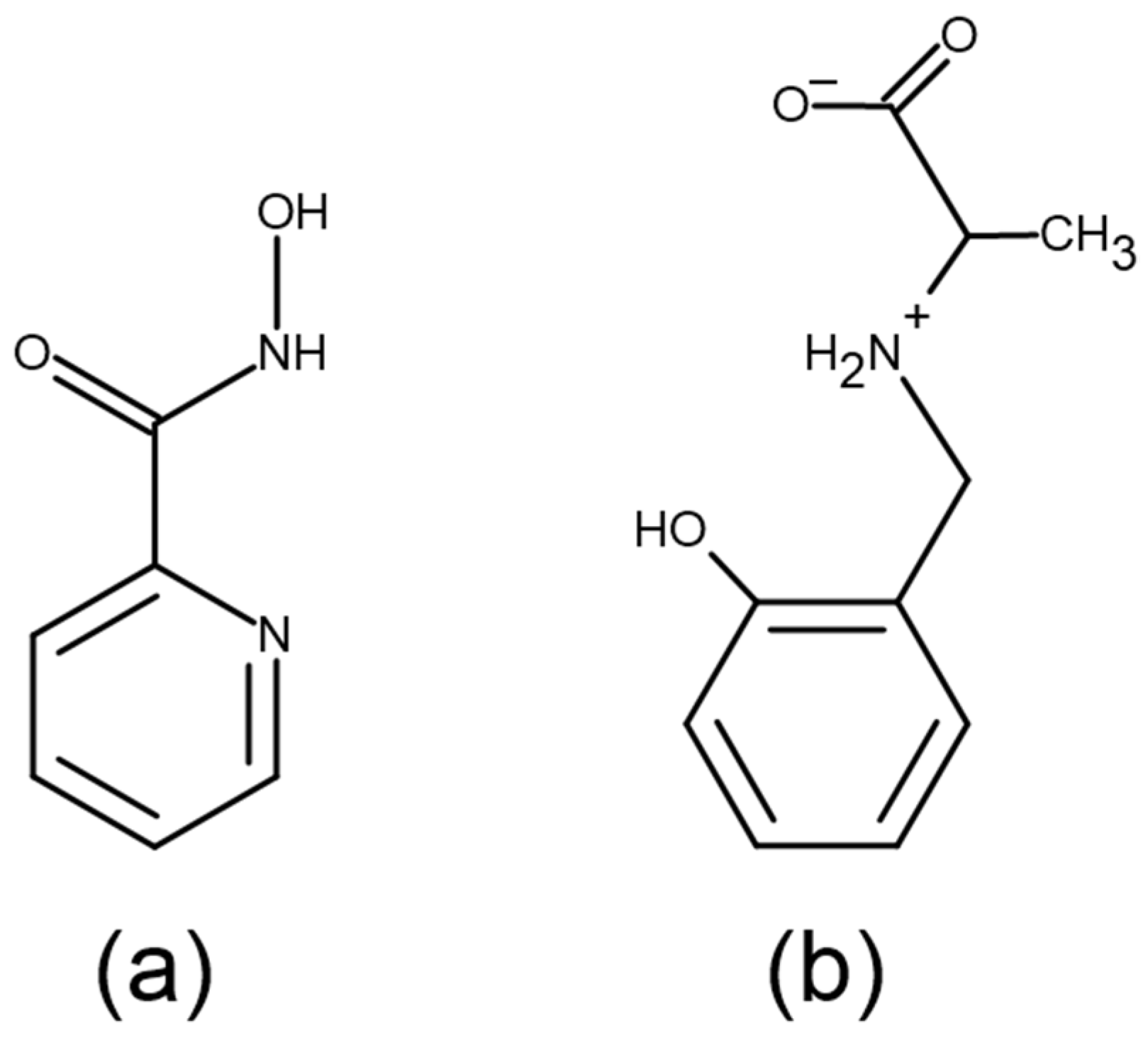
:1. Introduction
2. Results and Discussion
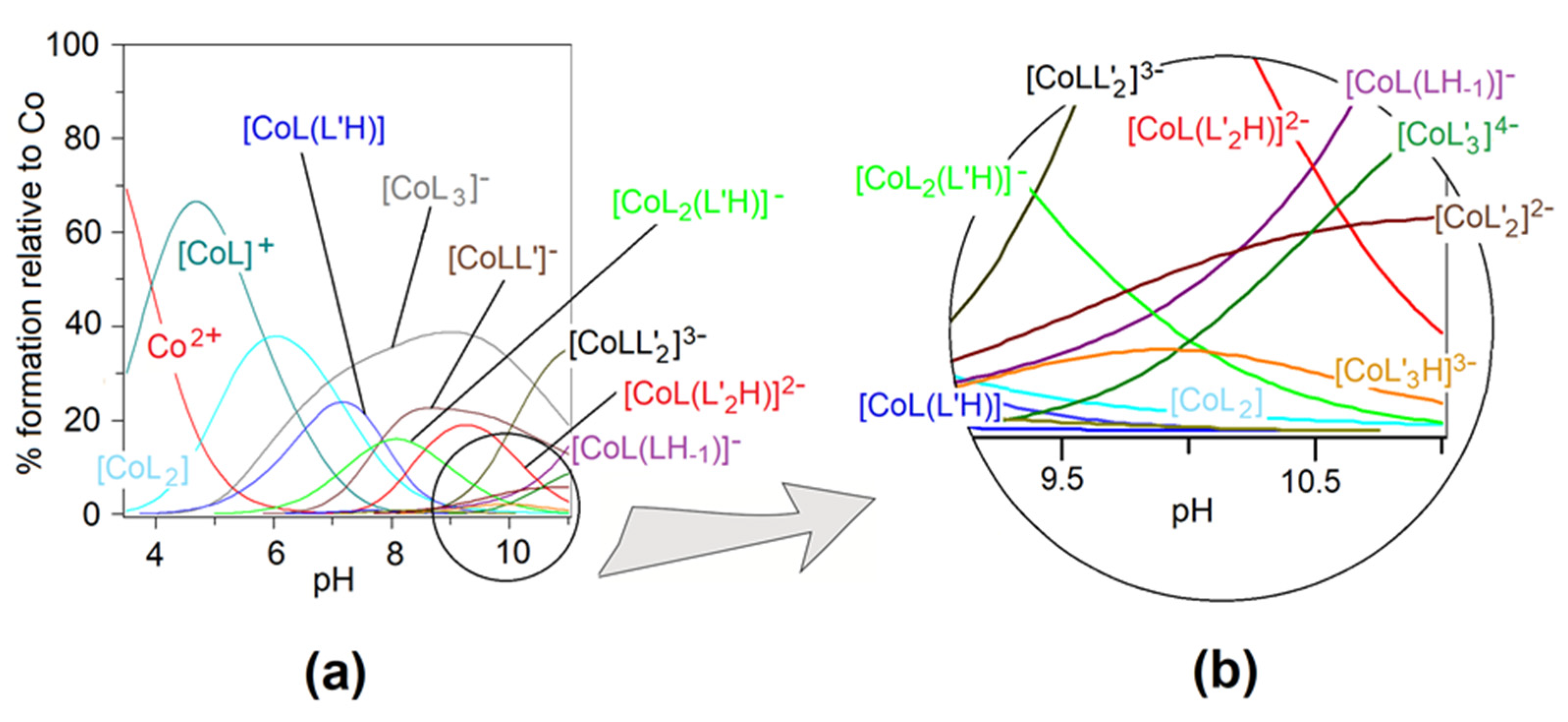
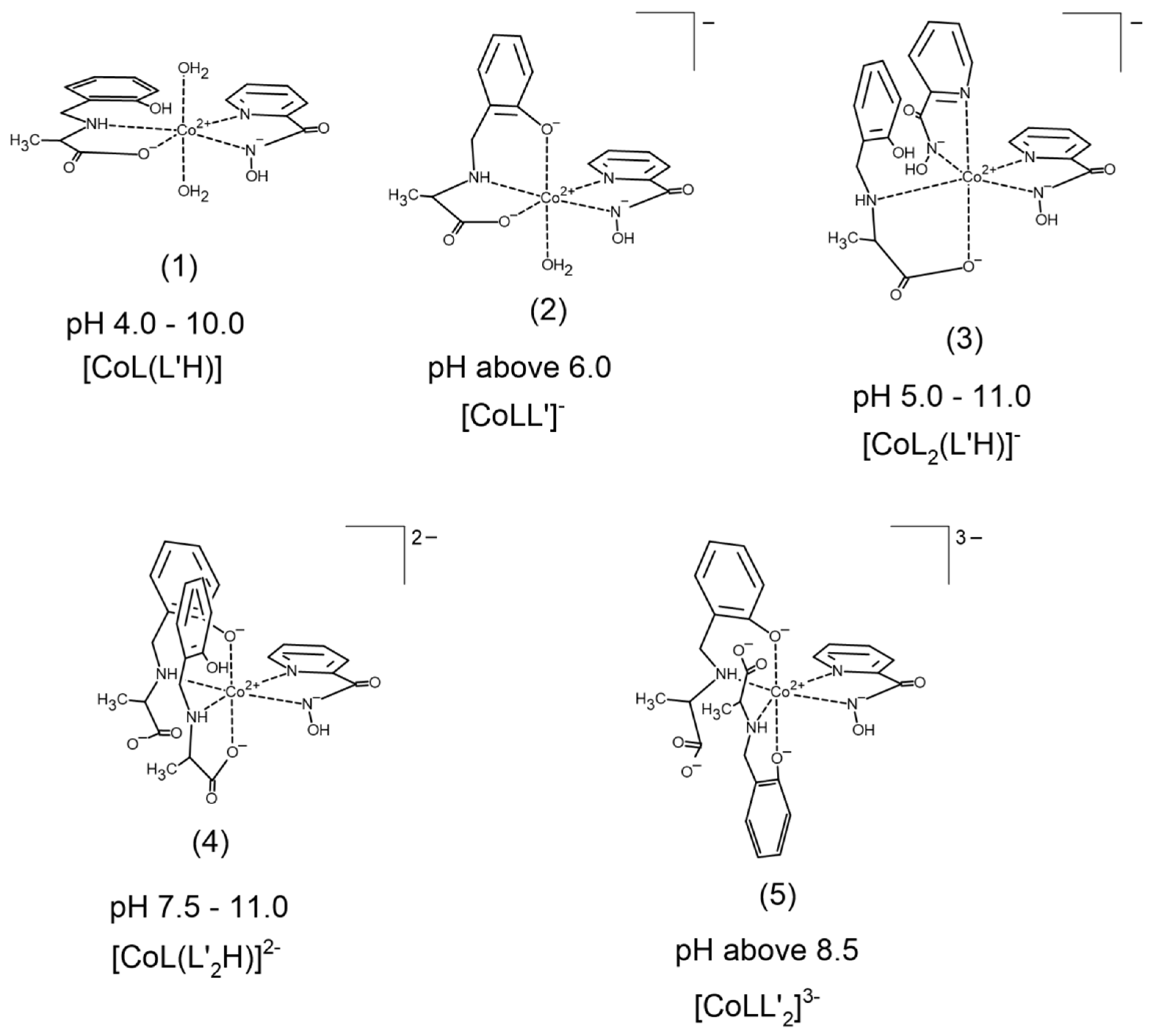
2.1. Heteroligand Complex Formation Equilibria
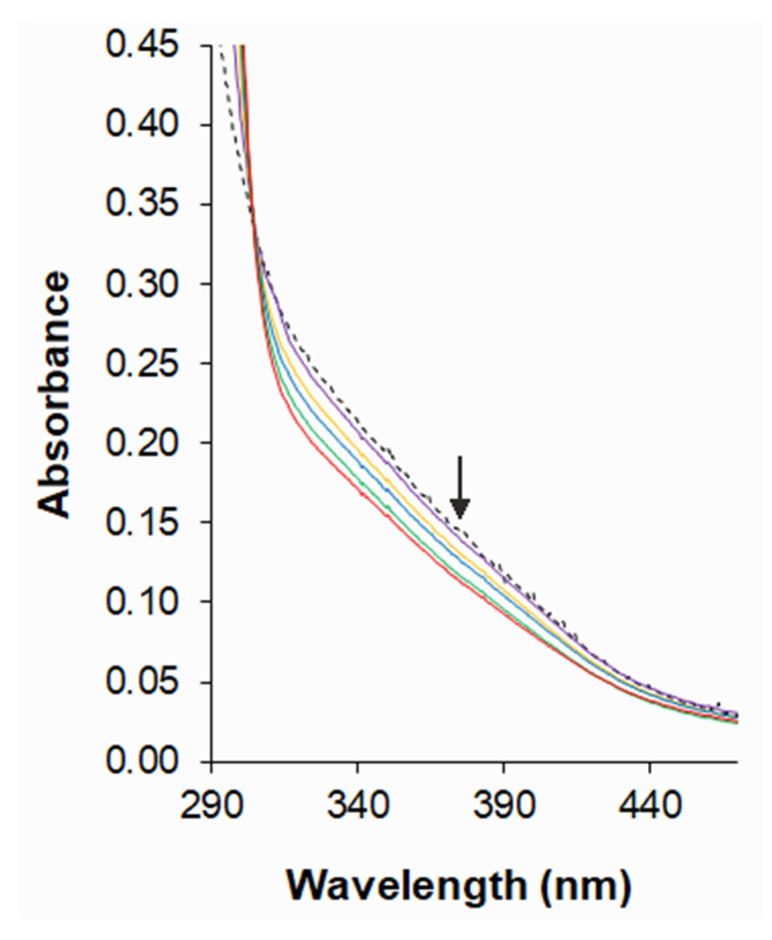
2.2. UV–Vis Spectra
2.3. DNA-Binding Studies of Co(II) Complexes
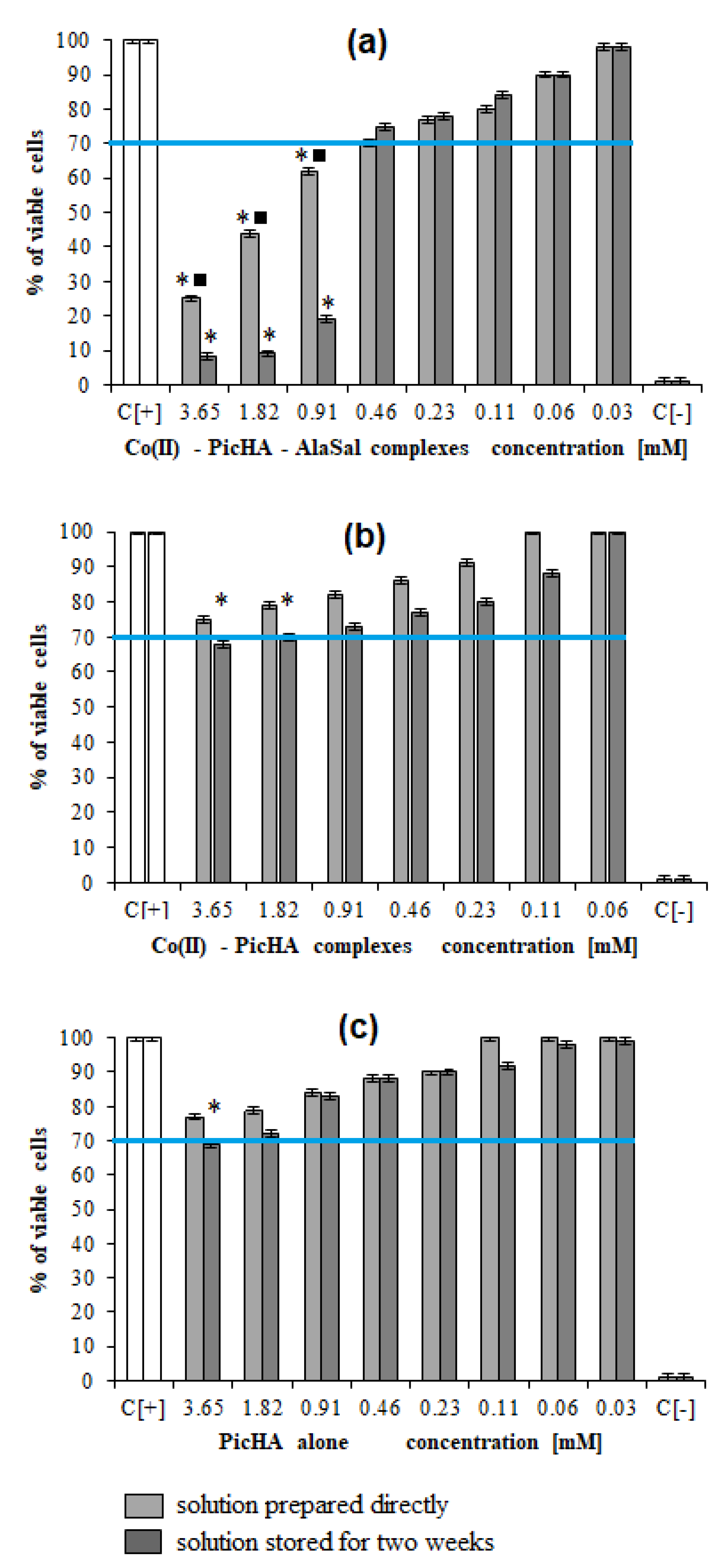
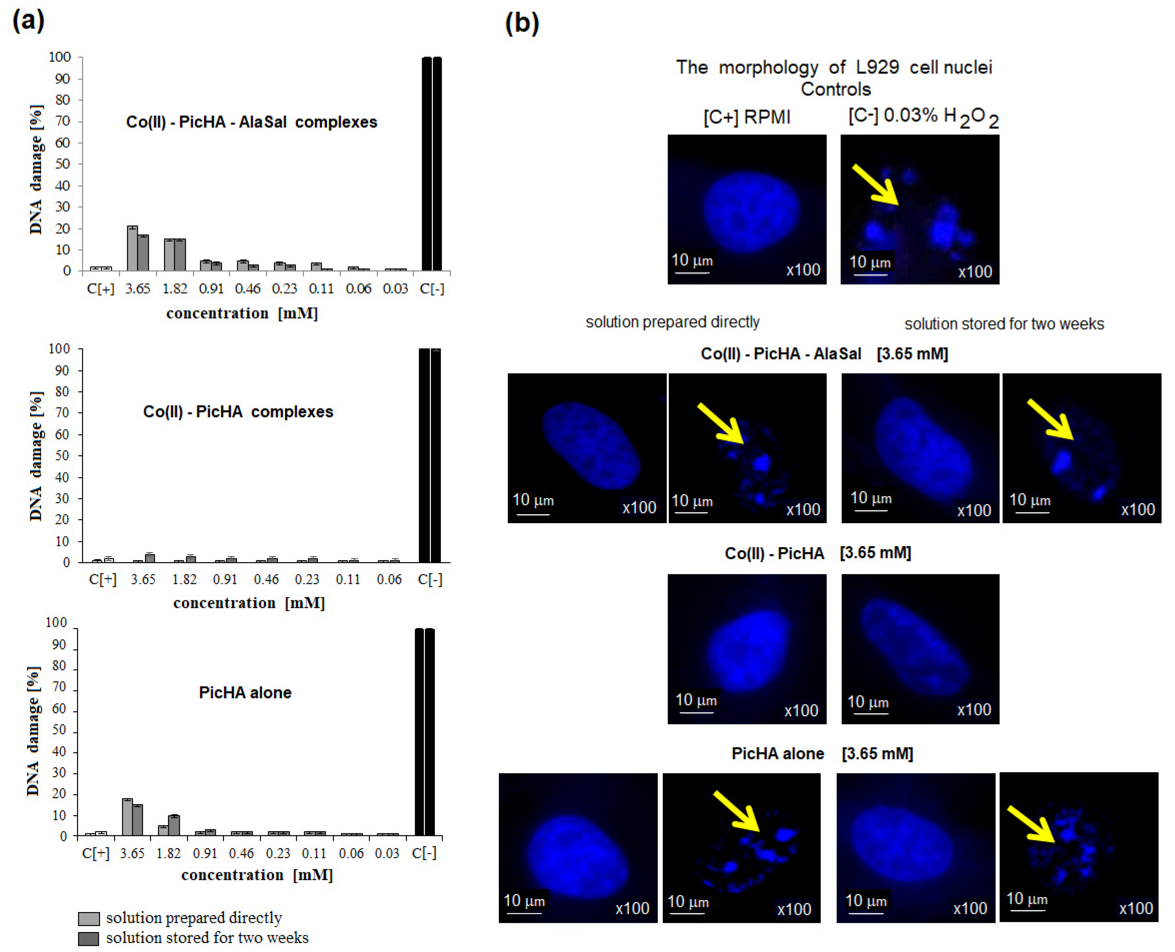
2.4. Biological Activity
3. Materials and Methods
3.1. Materials
3.2. pH-Metric Titrations
3.3. Spectrophotometric Measurements
3.4. DNA-Binding Experiment
3.5. Biological Assays
3.5.1. Antimicrobial Activity
3.5.2. Cell Cultures
Measurements of Cellular Metabolic Activity and Global Growth Inhibition
DAPI Staining of Cell Nuclei
3.5.3. Statistical Analysis
4. Conclusions
Supplementary Materials
Author Contributions
Funding
Institutional Review Board Statement
Informed Consent Statement
Data Availability Statement
Acknowledgments
Conflicts of Interest
References
- Tao, Z.-H.; Han, J.-X.; Fang, J.-Y. Helicobacter pylori infection and eradication: Exploring their impacts on the gastrointestinal microbiota. Helicobacter 2020, 25, 12754. [Google Scholar] [CrossRef]
- Alaghaz, A.-N.M.A.; Zayed, M.E.; Alharbi, S.A.; Ammar, R.A.A.; Chinnathambi, A. Synthesis, spectroscopic identification, thermal, potentiometric and antibacterial activity studies of 4-amino-5-mercapto-S-triazole Schiff’s base complexes. J. Mol. Struct. 2015, 1087, 60–67. [Google Scholar] [CrossRef]
- Zhou, J.; Kang, Y.; Chen, L.; Wang, H.; Liu, J.; Zeng, S.; Yu, L. The Drug-Resistance Mechanisms of Five Platinum-Based Antitumor Agents. Front. Pharmacol. 2020, 11, 343. [Google Scholar] [CrossRef] [Green Version]
- Xavier, C.P.R.; Pesic, M.; Vasconcelos, M.H. Understanding Cancer Drug Resistance by Developing and Studying Resistant Cell Line Models. Curr. Cancer Drug Targets 2016, 16, 226–237. [Google Scholar] [CrossRef]
- Richman, D.D. Antiviral drug resistance. Antivir. Res. 2006, 71, 117–121. [Google Scholar] [CrossRef]
- Tscherner, M.; Schwarzmüller, T.; Kuchler, K. Pathogenesis and Antifungal Drug Resistance of the Human Fungal Pathogen Candida glabrata. Pharmaceuticals 2011, 4, 169–186. [Google Scholar] [CrossRef] [Green Version]
- Karczewska, E.; Wojtas, I.; Sito, E.; Trojanowska, D.; Budak, A.; Zwolinska-Wcislo, M.; Wilk, A. Assessment of co-existence of Helicobacter pylori and Candida fungi in diseases of the upper gastrointestinal tract. J. Physiol. Pharmacol. Off. J. Pol. Physiol. Soc. 2009, 60, 33–39. [Google Scholar]
- Zabrodina, G.S.; Kalakutskaya, L.V.; Lukoyanova, O.V.; Lopatina, T.I.; Katkova, M.A. Water-soluble heteroligand complexes of 2-methyl-4-oxo-4H-pyran-3-olatoneodymium(III) with amino acids. Russ. J. Gen. Chem. 2014, 84, 923–926. [Google Scholar] [CrossRef]
- Dharmaraja, J.; Subbaraj, P.; Esakkidurai, T.; Shobana, S. Studies on Ni(II), Cu(II) and Zn(II) complexes with 2-aminobenzamide and some bioactive imidazole enzyme constituents. J. Coord. Chem. 2015, 68, 4314–4344. [Google Scholar] [CrossRef]
- Sutradhar, M.; Alegria, E.C.B.A.; Ferretti, F.; Raposo, L.R.; da Silva, M.F.C.G.; Baptista, P.V.; Fernandes, A.R.; Pombeiro, A.J.L. Antiproliferative activity of heterometallic sodium and potassium-dioxidovanadium(V) polymers. J. Inorg. Biochem. 2019, 200, 110811. [Google Scholar] [CrossRef]
- Sutradhar, M.; Rajeshwari; Barman, T.R.; Fernandes, A.R.; Paradinha, F.; Roma-Rodrigues, C.; da Silva, M.F.C.G.; Pombeiro, A.J.L. Mixed ligand aroylhydrazone and N-donor heterocyclic Lewis base Cu(II) complexes as potential antiproliferative agents. J. Inorg. Biochem. 2017, 175, 267–275. [Google Scholar] [CrossRef]
- Aljahdali, M.; El-Sherif, A.A. Equilibrium Studies of Binary and Mixed-Ligand Complexes of Zinc(II) Involving 2-(Aminomethyl)-Benzimidazole and Some Bio-Relevant Ligands. J. Solut. Chem. 2012, 41, 1759–1776. [Google Scholar] [CrossRef]
- Singh, Y.P.; Tej, G.N.V.C.; Pandey, A.; Priya, K.; Pandey, P.; Shankar, G.; Nayak, P.K.; Rai, G.; Chittiboyina, A.G.; Doerksen, R.J.; et al. Design, synthesis and biological evaluation of novel naturally-inspired multifunctional molecules for the management of Alzheimer’s disease. Eur. J. Med. Chem. 2020, 198, 112257. [Google Scholar] [CrossRef]
- Patole, J.; Shingnapurkar, D.; Padhye, S.; Ratledge, C. Schiff base conjugates of p-aminosalicylic acid as antimycobacterial agents. Bioorg. Med. Chem. Lett. 2006, 16, 1514–1517. [Google Scholar] [CrossRef]
- Salerno, D.; Beretta, G.L.; Zanchetta, G.; Brioschi, S.; Cristofalo, M.; Missana, N.; Nardo, L.; Cassina, V.; Tempestini, A.; Giovannoni, R.; et al. Platinum-Based Drugs and DNA Interactions Studied by Single-Molecule and Bulk Measurements. Biophys. J. 2016, 110, 2151–2161. [Google Scholar] [CrossRef] [Green Version]
- Khan, S.; Alothman, Z.A.; Mohammad, M.; Islam, M.S.; Slawin, A.; Wabaidur, S.M.; Islam, M.M.; Mir, M.H. Synthesis and characterization of a hydrogen bonded metal-organic cocrystal: Exploration of its DNA binding study. Polyhedron 2020, 180, 114454. [Google Scholar] [CrossRef]
- Joseph, J.; Rani, G.A. Antioxidant and Biochemical Activities of Mixed Ligand Complexes. Appl. Biochem. Biotechnol. 2014, 172, 867–890. [Google Scholar] [CrossRef]
- Weninger, A.; Baecker, D.; Obermoser, V.; Egger, D.; Wurst, K.; Gust, R. Synthesis and Biological Evaluation of Zeise’s Salt Derivatives with Acetylsalicylic Acid Substructure. Int. J. Mol. Sci. 2018, 19, 1612. [Google Scholar] [CrossRef] [Green Version]
- Ndagi, U.; Mhlongo, N.; Soliman, M.E. Metal complexes in cancer therapy—An update from drug design perspective. Drug Des. Dev. Ther. 2017, 11, 599–616. [Google Scholar] [CrossRef] [Green Version]
- Alzamil, N.O. Synthesis, DFT calculation, DNA-binding, antimicrobial, cytotoxic and molecular docking studies on new complexes VO(II), Fe(III), Co(II), Ni(II) and Cu(II) of pyridine Schiff base ligand. Mater. Res. Express 2020, 7, 065401. [Google Scholar] [CrossRef]
- Kirthan, B.; Prabhakara, M.; Naik, H.B.; Nayak, P.A.; Naik, E.I. Synthesis, characterization, DNA interaction and anti-bacterial studies of Cu(ii), Co(ii) and Ni(ii) metal complexes containing azo-dye ligand. Chem. Data Collect. 2020, 29, 100506. [Google Scholar] [CrossRef]
- Chang, E.L.; Simmers, C.; Knight, D.A. Cobalt Complexes as Antiviral and Antibacterial Agents. Pharmaceuticals 2010, 3, 1711–1728. [Google Scholar] [CrossRef] [Green Version]
- Singh, V.K.; Kadu, R.; Roy, H.; Raghavaiah, P.; Mobin, S.M. Phenolate based metallomacrocyclic xanthate complexes of CoII/CuII and their exclusive deployment in [2:2] binuclear N,O-Schiff base macrocycle formation and in vitro anticancer studies. Dalton Trans. 2016, 45, 1443–1454. [Google Scholar] [CrossRef] [PubMed]
- Baecker, D.; Obermoser, V.; Kirchner, E.A.; Hupfauf, A.; Kircher, B.; Gust, R. Fluorination as tool to improve bioanalytical sensitivity and COX-2-selective antitumor activity of cobalt alkyne complexes. Dalton Trans. 2019, 48, 15856–15868. [Google Scholar] [CrossRef] [PubMed]
- Woźniczka, M.; Świątek, M.; Pająk, M.; Gądek-Sobczyńska, J.; Chmiela, M.; Gonciarz, W.; Lisiecki, P.; Pasternak, B.; Kufelnicki, A. Complexes in aqueous cobalt(II)–2-picolinehydroxamic acid system: Formation equilibria, DNA-binding ability, antimicrobial and cytotoxic properties. J. Inorg. Biochem. 2018, 187, 62–72. [Google Scholar] [CrossRef] [PubMed]
- Woźniczka, M.; Sutradhar, M.; Pombeiro, A.J.L.; Świątek, M.; Pająk, M.; Gądek-Sobczyńska, J.; Chmiela, M.; Gonciarz, W.; Pasternak, B.; Kufelnicki, A. Equilibria in Aqueous Cobalt(II)—Reduced Schiff Base N-(2-hydroxybenzyl)alanine System: Chemical Characterization, Kinetic Analysis, Antimicrobial and Cytotoxic Properties. Molecules 2020, 25, 3462. [Google Scholar] [CrossRef]
- Sutradhar, M.; Andrade, M.A.; Carabineiro, S.A.C.; Martins, L.M.D.R.S.; da Silva, M.F.C.G.; Pombeiro, A.J.L. Oxido- and Dioxido-Vanadium(V) Complexes Supported on Carbon Materials: Reusable Catalysts for the Oxidation of Cyclohexane. Nanomaterials 2021, 11, 1456. [Google Scholar] [CrossRef]
- Sutradhar, M.; Alegria, E.C.B.A.; da Silva, M.F.C.G.; Martins, L.M.D.R.S.; Pombeiro, A.J.L. Aroylhydrazone Cu(II) Complexes in keto Form: Structural Characterization and Catalytic Activity towards Cyclohexane Oxidation. Molecules 2016, 21, 425. [Google Scholar] [CrossRef] [Green Version]
- Cozzi, P.G. Metal–Salen Schiff base complexes in catalysis: Practical aspects. Chem. Soc. Rev. 2004, 33, 410–421. [Google Scholar] [CrossRef]
- Al Zoubi, W.; Ko, Y.G. Organometallic complexes of Schiff bases: Recent progress in oxidation catalysis. J. Organomet. Chem. 2016, 822, 173–188. [Google Scholar] [CrossRef]
- Al Zoubi, W.; Al-Hamdani, A.A.S.; Kaseem, M. Synthesis and antioxidant activities of Schiff bases and their complexes: A review. Appl. Organomet. Chem. 2016, 30, 810–817. [Google Scholar] [CrossRef]
- Chohan, Z.H.; Arif, M.; Akhtar, M.A.; Supuran, C.T. Metal-Based Antibacterial and Antifungal Agents: Synthesis, Characterization, and In Vitro Biological Evaluation of Co(II), Cu(II), Ni(II), and Zn(II) Complexes with Amino Acid-Derived Compounds. Bioinorg. Chem. Appl. 2006, 2006, 83131. [Google Scholar] [CrossRef] [Green Version]
- Luo, Y.; Wang, J.; Ding, X.; Ni, R.; Li, M.; Yang, T.; Wang, J.; Jing, C.; You, Z. Syntheses, crystal structures and antimicrobial activities of polynuclear CoII, NiII and ZnII complexes derived from the N,N’-bis(4-fluorosalicylidene)-1,3-propanediamine Schiff base. Inorg. Chim. Acta 2021, 516, 120146. [Google Scholar] [CrossRef]
- Revathi, N.; Sankarganesh, M.; Raja, J.D.; Kumar, G.G.V.; Sakthivel, A.; Rajasekaran, R. Bio-active mixed ligand Cu(II) and Zn(II) complexes of pyrimidine derivative Schiff base: DFT calculation, antimicrobial, antioxidant, DNA binding, anticancer and molecular docking studies. J. Biomol. Struct. Dyn. 2021, 39, 3012–3024. [Google Scholar] [CrossRef]
- Ismail, B.A.; Nassar, D.A.; El–Wahab, Z.H.A.; Ali, O.A.M. Synthesis, characterization, thermal, DFT computational studies and anticancer activity of furfural-type schiff base complexes. J. Mol. Struct. 2021, 1227, 129393. [Google Scholar] [CrossRef]
- Shchur, I.; Shchegolkov, E.; Burgart, Y.; Kozitsina, A.; Ivanova, A.; Alyamovskaya, I.; Evstigneeva, N.; Gerasimova, N.; Ganebnykh, I.; Zilberberg, N.; et al. Metal complexes based on polyfluorosalicylic acids and their antimycotic and antimicrobial activity. Polyhedron 2019, 177, 114279. [Google Scholar] [CrossRef]
- Bandyopadhyay, S.; Das, A.; Mukherjee, G.N.; Cantoni, A.; Bocelli, G.; Chaudhuri, S.; Ribas, J. Equilibrium and structural studies on Co(II), Ni(II), Cu(II) and Zn(II) complexes with N-(2-benzimidazolyl)methyliminodiacetic acid: Crystal structures of Ni(II) and Cu(II) complexes. Inorg. Chim. Acta 2004, 357, 3563–3573. [Google Scholar] [CrossRef]
- Lou, B.-Y.; Xu, Y.; Yuan, D.-Q.; Han, L.; Hong, M.-C. Bis[N-(4-hydroxybenzyl)-D,L-alaninato]copper(II) tetrahydrate. Acta Crystallogr. Sect. E Struct. Rep. Online 2004, 60, m522–m523. [Google Scholar] [CrossRef] [Green Version]
- Abdelkarim, A.T.; El-Sherif, A.A. Potentiometric, Thermodynamics and Coordination Properties for Binary and Mixed Ligand Complexes of Copper(II) with Imidazole-4-acetic Acid and Tryptophan or Phenylalanine Aromatic Amino Acids. J. Solut. Chem. 2016, 45, 712–731. [Google Scholar] [CrossRef]
- Shobana, S.; Subramaniam, P.; Dharmaraja, J.; Arvindnarayan, S. Stability and Structural Studies on Ni(II)–5-Fluorouracil Mixed Ligand Complex Systems. J. Solut. Chem. 2016, 45, 334–358. [Google Scholar] [CrossRef]
- Woźniczka, M.; Vogt, A.; Kufelnicki, A. Equilibria in cobalt(II)–amino acid–imidazole system under oxygen-free conditions: Effect of side groups on mixed-ligand systems with selected L-α-amino acids. Chem. Cent. J. 2016, 10, 14. [Google Scholar] [CrossRef] [Green Version]
- Padmavathi, M.; Satyanarayana, S. Potentiometric and proton NMR studies on ternary metal (II) complexes containing thiaminepyrophosphate and a series of secondary ligands. Indian J. Chem. 1999, 38, 295–298. [Google Scholar]
- Patel, P.; Bhattacharya, P.K. Stereochemical factors affecting intramolecular interligand interaction in mixed ligand complexes. Indian J. Chem. 1993, 32, 506–510. [Google Scholar]
- Aljahdali, M.; El-Sherif, A.A.; Shoukry, M.M.; Mohamed, S.E. Potentiometric and Thermodynamic Studies of Binary and Ternary Transition Metal(II) Complexes of Imidazole-4-acetic Acid and Some Bio-relevant Ligands. J. Solut. Chem. 2013, 42, 1028–1050. [Google Scholar] [CrossRef]
- Lever, A.B.P. Inorganic Electronic Spectroscopy, 2nd ed.; Elsevier: Amsterdam, The Netherlands, 1984. [Google Scholar]
- Yamauchi, O.; Odani, A. Structure-stability relationship in ternary copper(II) complexes involving aromatic amines and tyrosine or related amino acids. Intramolecular aromatic ring stacking and its regulation through tyrosine phosphorylation. J. Am. Chem. Soc. 1985, 107, 5938–5945. [Google Scholar] [CrossRef]
- Sau, D.K.; Butcher, R.J.; Chaudhuri, S.; Saha, N. Synthesis and spectroscopic characterization of new cobalt(III) complexes with 5-methyl-3-formyl pyrazole 3-hexamethyleneiminyl thiosemicarbazone (HMPz3Hex): X-ray crystallographic identification of HMPz3Hex and [Co(MPz3Hex)2]Br·H2O with evidence for unusual rotation about the azomethine double bond of the ligand on complexation with cobalt(III). Polyhedron 2004, 23, 5–14. [Google Scholar] [CrossRef]
- Munteanu, C.R.; Suntharalingam, K. Advances in cobalt complexes as anticancer agents. Dalton Trans. 2015, 44, 13796–13808. [Google Scholar] [CrossRef]
- Ma, X.-F.; Li, D.-D.; Tian, J.-L.; Kou, Y.-Y.; Yan, S.-P. DNA binding and cleavage activity of reduced amino-acid Schiff base complexes of cobalt(II), copper(II), and cadmium(II). Transit. Met. Chem. 2009, 34, 475–481. [Google Scholar] [CrossRef]
- Zhang, Z.; Yang, Z.; Wu, Y.; Yuan, Z.; Du, J.; Li, L. Reduced amino acid Schiff base containing ruthenium(III) complexes: Synthesis, characterization, DNA interaction, and in vitro cytotoxicity. J. Biochem. Mol. Toxicol. 2020, 34, e22510. [Google Scholar] [CrossRef]
- Liu, X.-W.; Li, J.; Li, H.; Zheng, K.-C.; Chao, H.; Ji, L.-N. Synthesis, characterization, DNA-binding and photocleavage of complexes [Ru(phen)2(6-OH-dppz)]2+ and [Ru(phen)2(6-NO2-dppz)]2+. J. Inorg. Biochem. 2005, 99, 2372–2380. [Google Scholar] [CrossRef] [PubMed]
- Safyanova, I.S.; Ohui, K.A.; Omelchenko, I.V. Crystal structure of N-hydroxypicolinamide monohydrate. Acta Crystallogr. 2016, 72, 117–119. [Google Scholar] [CrossRef]
- Kelly, J.M.; Tossi, A.B.; McConnell, D.J.; OhUigin, C. A study of the interactions of some polypyridylruthenium(II) complexes with DNA using fluorescence spectroscopy, topoisomerisation and thermal denaturation. Nucleic Acids Res. 1985, 13, 6017–6034. [Google Scholar] [CrossRef] [PubMed]
- Shchegolkov, E.V.; Shchur, I.V.; Burgart, Y.V.; Slepukhin, P.A.; Evstigneeva, N.P.; Gerasimova, N.A.; Zilberberg, N.V.; Kungurov, N.V.; Saloutin, V.I.; Chupakhin, O.N. Copper(II) and cobalt(II) complexes based on methyl trifluorosalicylate and bipyridine-type ligands: Synthesis and their antimicrobial activity. Polyhedron 2021, 194, 114900. [Google Scholar] [CrossRef]
- Rao, R.N.; Panchangam, R.L.; Manickam, V.; Balamurali, M.M.; Chanda, K. Synthesis and Antitumor Activity Evaluation of Cyclometalated 2H- Indazole Ruthenium(II) and Iridium(III) Complexes. ChemPlusChem 2020, 85, 1800–1812. [Google Scholar] [CrossRef]
- Hase, J.; Kobashi, K. Inhibition of Proteus vulgaris Urease by Hydroxamic Acids. J. Biochem. 1967, 62, 293–299. [Google Scholar] [CrossRef]
- Stec, J.; Huang, Q.; Pieroni, M.; Kaiser, M.; Fomovska, A.; Mui, E.; Witola, W.H.; Bettis, S.; McLeod, R.; Brun, R.; et al. Synthesis, Biological Evaluation, and Structure–Activity Relationships of N-Benzoyl-2-hydroxybenzamides as Agents Active against P. falciparum (K1 strain), Trypanosomes, and Leishmania. J. Med. Chem. 2012, 55, 3088–3100. [Google Scholar] [CrossRef] [Green Version]
- Ohi, N.; Aoki, B.; Shinozaki, T.; Moro, K.; Noto, T.; Nehashi, T.; Okazaki, H.; Matsunaga, I. Semisynthetic β-lactam antibiotics. I. Synthesis and antibacterial activity of new ureidopenicillin derivatives having catechol moieties. J. Antibiot. 1986, 39, 230–241. [Google Scholar] [CrossRef] [Green Version]
- Law, B.Y.K.; Qu, Y.Q.; Mok, S.W.F.; Liu, H.; Zeng, W.; Han, Y.; Gordillo-Martinez, F.; Chan, W.-K.; Wong, K.M.-C.; Wong, V.K.W. New perspectives of cobalt tris(bipyridine) system: Anti-cancer effect and its collateral sensitivity towards multidrug-resistant (MDR) cancers. Oncotarget 2017, 8, 55003–55021. [Google Scholar] [CrossRef]
- Hynes, J.B. Hydroksylamine derivatives as potential antimalarial agents. 1. Hydroxamic acids. J. Med. Chem. 1970, 13, 1235–1237. [Google Scholar] [CrossRef]
- Koh, L.L.; Ranford, J.O.; Robinson, W.T.; Svensson, J.O.; Tan, A.L.C.; Wu, D. Model for the Reduced Schiff Base Intermediate between Amino Acids and Pyridoxal: Copper(II) Complexes of N-(2-Hydroxybenzyl)amino Acids with Nonpolar Side Chains and the Crystal Structures of [Cu(N-(2-hydroxybenzyl)-d,l-alanine)(phen)]·H2O and [Cu(N-(2-hydroxybenzyl)-d,l-alanine)(imidazole)]. Inorg. Chem. 1996, 35, 6466–6472. [Google Scholar] [CrossRef]
- Gans, P.; O’Sullivan, B. GLEE, a new computer program for glass electrode calibration. Talanta 2000, 51, 33–37. [Google Scholar] [CrossRef]
- Irving, H.; Miles, M.G.; Pettit, L.D. A study of some problems in determining the stoicheiometric proton dissociation constants of complexes by potentiometric titrations using a glass electrode. Anal. Chim. Acta 1967, 38, 475–488. [Google Scholar] [CrossRef]
- Gans, P.; Sabatini, A.; Vacca, A. Investigation of equilibria in solution. Determination of equilibrium constants with the HYPERQUAD suite of programs. Talanta 1996, 43, 1739–1753. [Google Scholar] [CrossRef]
- Pettit, L.D.; Powell, K.J. Stability Constants Database, IUPAC and Academic Software; Royal Society of Chemistry: London, UK, 1993. [Google Scholar]
- Alderighi, L.; Gans, P.; Ienco, A.; Peters, D.; Sabatini, A.; Vacca, A. Hyperquad simulation and speciation (HySS): A utility program for the investigation of equilibria involving soluble and partially soluble species. Coord. Chem. Rev. 1999, 184, 311–318. [Google Scholar] [CrossRef]
- Świątek, M.; Kufelnicki, A. Methal-ligand interaction of lanthanides with coumarin derivatives. Part I. Complexation of 3-(1-aminoethylidene-2H-chromene-2,4(3H)-dione with La(III), Ce(III), Nd(III) and Ho(III). Acta Pol. Pharm. 2012, 69, 1001–1007. [Google Scholar]
- Gaber, M.; El-Ghamry, H.A.; Fathalla, S.K. Synthesis, structural identification, DNA interaction and biological studies of divalent Mn, Co and Ni chelates of 3-amino-5-mercapto-1,2,4-triazole azo ligand. Appl. Organomet. Chem. 2020, 34, e5678. [Google Scholar] [CrossRef]
- Reichmann, M.E.; Rice, S.A.; Thomas, C.A.; Doty, P. A Further Examination of the Molecular Weight and Size of Desoxypentose Nucleic Acid. J. Am. Chem. Soc. 1954, 76, 3047–3053. [Google Scholar] [CrossRef]
- Mnich, E.; Kowalewicz-Kulbat, M.; Sicinska, P.; Hinc, K.; Obuchowski, M.; Gajewski, A.; Moran, A.P.; Chmiela, M. Impact of Helicobacter pylorion the healing process of the gastric barrier. World J. Gastroenterol. 2016, 22, 7536–7558. [Google Scholar] [CrossRef]
- Suzuki, N.; Murata-Kamiya, N.; Yanagiya, K.; Suda, W.; Hattori, M.; Kanda, H.; Bingo, A.; Fujii, Y.; Maeda, S.; Koike, K.; et al. Mutual reinforcement of inflammation and carcinogenesis by the Helicobacter pylori CagA oncoprotein. Sci. Rep. 2015, 5, 10024. [Google Scholar] [CrossRef] [Green Version]
- Huang, H.; Wang, J.; Zhang, J.; Cai, J.; Pi, J.; Xu, J.-F. Inspirations of Cobalt Oxide Nanoparticle Based Anticancer Therapeutics. Pharmaceutics 2021, 13, 1599. [Google Scholar] [CrossRef]


| Species | log10 βmll’h | Related Constants b |
|---|---|---|
| [CoL(L’H)] (1) | 21.40 (4) | 10.67 c |
| [CoLL’]− (2) | 13.56 (2) | 13.56 |
| [CoL2 (L’H)]− (3) | 25.61 (7) | 14.88 d |
| [CoL(L’2H)]2− (4) | 26.75 (5) | 16.02 e |
| [CoLL’2]3− (5) | 16.87 (5) | 16.87 |
| σ; na | 12.25; 753 |
| Microorganism | MIC/MBC/MFC (mM) | MIC = MBC/MFC (mM) | |||
|---|---|---|---|---|---|
| Co(II)–PicHA–AlaSal Complexes | Gentamicin | Amphotericin B | Amoxicillin | ||
| MIC | MBC/MFC | ||||
| Gram-negative bacteria | |||||
| Pseudomonas aeruginosa ATCC 27853 | 7.30 | >7.30 | <0.008 | - | - |
| Escherichia coli ATCC 25922 | 7.30 | >7.30 | <0.004 | - | - |
| Helicobacter pylori CCUC 17874 | 3.65 | 3.65 | - | - | <0.001 |
| Helicobacter pylori ATCC 700392 | 3.65 | 3.65 | - | - | <0.001 |
| Gram-positive bacteria | |||||
| Enterococcus faecalis ATCC 29212 | 7.30 | >7.30 | <0.26 | - | - |
| Staphylococcus aureus ATCC 29213 | 3.65 | >7.30 | <0.002 | - | - |
| Staphylococcus aureus ATCC 6538 | 3.65 | >7.30 | <0.002 | - | - |
| Staphylococcus epidermidis ATCC 12228 | 3.65 | >7.30 | <0.002 | - | - |
| Fungi | |||||
| Candida albicans ATTC 10231 | 3.65 | 3.65 | - | <0.001 | - |
| Candida glabrata ATCC 2001 | 3.65 | 3.65 | - | <0.001 | - |
| Candida parapsilosis ATCC 22019 | 3.65 | 7.30 | - | <0.001 | - |
Publisher’s Note: MDPI stays neutral with regard to jurisdictional claims in published maps and institutional affiliations. |
© 2021 by the authors. Licensee MDPI, Basel, Switzerland. This article is an open access article distributed under the terms and conditions of the Creative Commons Attribution (CC BY) license (https://creativecommons.org/licenses/by/4.0/).
Share and Cite
Woźniczka, M.; Lichawska, M.; Sutradhar, M.; Chmiela, M.; Gonciarz, W.; Pająk, M. Chemical Characterization and Biological Evaluation of New Cobalt(II) Complexes with Bioactive Ligands, 2-Picolinehydroxamic Acid and Reduced Schiff Base N-(2-Hydroxybenzyl)alanine, in Terms of DNA Binding and Antimicrobial Activity. Pharmaceuticals 2021, 14, 1254. https://0-doi-org.brum.beds.ac.uk/10.3390/ph14121254
Woźniczka M, Lichawska M, Sutradhar M, Chmiela M, Gonciarz W, Pająk M. Chemical Characterization and Biological Evaluation of New Cobalt(II) Complexes with Bioactive Ligands, 2-Picolinehydroxamic Acid and Reduced Schiff Base N-(2-Hydroxybenzyl)alanine, in Terms of DNA Binding and Antimicrobial Activity. Pharmaceuticals. 2021; 14(12):1254. https://0-doi-org.brum.beds.ac.uk/10.3390/ph14121254
Chicago/Turabian StyleWoźniczka, Magdalena, Marta Lichawska, Manas Sutradhar, Magdalena Chmiela, Weronika Gonciarz, and Marek Pająk. 2021. "Chemical Characterization and Biological Evaluation of New Cobalt(II) Complexes with Bioactive Ligands, 2-Picolinehydroxamic Acid and Reduced Schiff Base N-(2-Hydroxybenzyl)alanine, in Terms of DNA Binding and Antimicrobial Activity" Pharmaceuticals 14, no. 12: 1254. https://0-doi-org.brum.beds.ac.uk/10.3390/ph14121254