Review

Extracellular Vesicle-Based Therapeutics: Preclinical and Clinical Investigations

by
Natalia L. Klyachko
1,2,
Camryn J. Arzt
2,
Samuel M. Li
2,
Olesia A. Gololobova
1,2 and
Elena V. Batrakova
1,2,*
1
Center for Nanotechnology in Drug Delivery, University of North Carolina at Chapel Hill, Chapel Hill, NC 27599, USA
2
Eshelman School of Pharmacy, University of North Carolina at Chapel Hill, Chapel Hill, NC 27599, USA
*
Author to whom correspondence should be addressed.
Submission received: 5 November 2020 / Revised: 24 November 2020 / Accepted: 29 November 2020 / Published: 1 December 2020
(This article belongs to the Section Biologics and Biosimilars)

Abstract

:
Drug nanoformulations hold remarkable promise for the efficient delivery of therapeutics to a disease site. Unfortunately, artificial nanocarriers, mostly liposomes and polymeric nanoparticles, show limited applications due to the unfavorable pharmacokinetics and rapid clearance from the blood circulation by the reticuloendothelial system (RES). Besides, many of them have high cytotoxicity, low biodegradability, and the inability to cross biological barriers, including the blood brain barrier. Extracellular vesicles (EVs) are novel candidates for drug delivery systems with high bioavailability, exceptional biocompatibility, and low immunogenicity. They provide a means for intercellular communication and the transmission of bioactive compounds to targeted tissues, cells, and organs. These features have made them increasingly attractive as a therapeutic platform in recent years. However, there are many obstacles to designing EV-based therapeutics. In this review, we will outline the main hurdles and limitations for therapeutic and clinical applications of drug loaded EV formulations and describe various attempts to solve these problems.

Graphical Abstract

1. Introduction

Extracellular vesicles (EVs) have emerged as a new class of nanocarriers, triggering significant interest and enthusiasm. Extraordinary efforts have been made to develop new techniques that would make it possible to manufacture EV-based drug formulations for the treatment of various diseases, including cardiovascular diseases [1,2,3,4,5,6], regenerative disorders [7,8], infectious diseases [9], cancer [10,11,12,13,14,15,16,17,18,19], as well as autoimmune [20] and neurological disorders [17,21]. EVs are short- and long-distance mediators of intercellular communication that offer distinct advantages, uniquely positioning them as highly effective drug nanocarriers. They comprise various types of nanovesicles, including exosomes (30–120 nm), microvesicles (MVs) (50 nm–1μm), and apoptotic bodies (500–1000 nm) [22,23,24]. Notably, EVs consist of cellular membranes with multiple adhesive proteins on their surface [25,26] that enable efficient cell entry and delivery of therapeutic cargo.
The unique properties of EVs can be attributed to their biogenesis. Exosomes are initially produced by invagination of the endosomal membrane to create multivesicular bodies (MVB) [27]. In contrast, exosomes’ close relatives, MVs, are greater in size and bud directly from the plasma membrane. Therefore, exosomes and MVs originate from endosomal and plasma membranes, respectively. Apoptotic bodies form during the apoptotic process, when the cellular cytoskeleton breaks up, causing the membrane to bulge outward [28]. Different techniques have been developed for the characterization of EVs. Among them are nanoparticle tracking analysis (NTA) and dynamic light scattering (DLS), that provide information about: count (NTA) and size distribution (NTA and DLS); flow cytometry, western blotting, and mass spectrometry (MS) that can be used to characterize biochemical content of EVs; and several microscopy techniques Atomic Force Microscopy (AFM) and Cryogenic Transmission Electron Microscopy (CryTEM) that make it possible to assess EV morphology [29]. The structure, biogenesis and composition of EVs have been extensively described in several excellent reviews [7,18,21,30,31,32,33,34,35,36].
Similar to artificial nanocarriers, EVs can improve the fundamental characteristics of a free drug, such as its stability and solubility, and protect the drug against degradation in the bloodstream [31]. Relatively tight lipid bilayers in EV membranes can provide a sustained and prolonged release of the incorporated drug. Furthermore, contrary to most synthetic nanocarriers, EVs can cross biological barriers, including the blood brain barrier (BBB), making them especially valuable for the treatment of neurodegenerative disorders. It has been shown that EVs can cross the BBB from the brain to the bloodstream [37,38], as well as from blood to the CNS in vitro [39,40] and in vivo [41,42,43] under pathological conditions. However, whether EVs cross the BBB in the absence of pathology is still debated. Furthermore, these natural nanocarriers have low immunogenicity (especially, autologous EVs) and low cytotoxicity, which are usually substantial impediments for conventional synthetic nanoparticles. Finally, some types of EVs exert tissue tropism that makes it possible to target their formulations to specific cell types or migration towards inflamed tissues [44,45]. It is worth mentioning that bioinspired nanocarriers may have unique biological activity which is reflective of their origin, i.e., parent cells, that provides additional therapeutic efficacy to the incorporated drug [43]. These attractive features have contributed to the growing interest in EVs and inspired numerous studies aimed at their introduction to the field of drug delivery.
Despite these advantages, the clinical translation of EVs has been greatly slowed down due to a number of drawbacks, including upscaling processes of isolation and purification, as well as the lack of a means of efficiently loading these natural nanovesicles with therapeutics. Reliability, reproducibility, and donor-donor variations of EV formulations are still of significant concern. Furthermore, EV functional heterogenicity and limited yields represent serious obstacles for their future applications. Thus, depending on the mechanism of EV release, they may contain different proteins, active proteasomes, and even organelles (e.g., mitochondria) [46]. Inadequate targeting is another challenge for the clinical translation of different EV-based drug formulations. Herein, we will discuss how these hurdles can be overcome to introduce this unique biomimetic drug delivery system to the clinic.

2. Implications Related to Biological Activity Inherited from EV Origin

The biological activity of EVs released by various types of cells is vast and promising. Their ability to impact cells depends largely upon their protein markers and their cargo, which mimic the properties of their origin. Isolated EVs taken directly from specific types of cells, such as fibroblasts, neuronal cells, macrophages, and even cancer cells have a wide array of both pathogenic and therapeutic activities, largely depending their host cells. Therefore, one should pay special attention to the source of EVs and possible unwanted biological activity inherited from their parent cells. For example, EVs derived from diseased cells may contribute to the ability of a pathogen to spread throughout the body and evade the immune system [47]. Tumor-derived EVs are well-documented to express specific immune system markers such as MHC Class I and II molecules, death receptor ligands (FasL) and many others. The expression of these markers enables EVs to interact directly with prominent immune system cells such as T cells, B cells, and NK cells to encourage oncogenic activity and inhibit the immune system processes. Melanoma-derived EVs express FasL, which activates the Fas/FasL pathway to induce lymphocyte apoptosis, allowing tumors to evade cell-mediated cell death [47]. Next, EVs may contain prominent mediators that encourage angiogenic activity, metastasis, and mRNA transfer, leading to growth within the tumor microenvironment. Thus, gliomas, i.e., human brain and spinal cord tumors, express an oncogenic form of the epidermal growth factor receptor, EGFRvIII [48]. In mice, EVs containing EGFRvIII were shown to be released into the blood and fuse with tumor cells lacking EGFRvIII, conferring oncogenic activity upon previously benign cells. Moreover, cancer cell-derived EVs were shown to transport oncoproteins, including antigen MelanA/Mart-1 (melanoma), carcinoembryogenic antigen (CEA) (colon carcinoma), and HER2 (breast cancer) [49]. Finally, EVs can carry cancer-related miRNAs. Specifically, large amounts of small RNAs such as let-7, miR-1, miR-15, miR-16 and miR-375, which play an important role in cancer, were found in EVs [50]. Furthermore, Li et al. [51] studied the mechanism underlying the association between EVs and hypoxia during cancer progression. It was suggested that cancer cell-derived EVs mediate miRNA transfer and promote prometastatic behavior. Thus, oral squamous cell carcinoma (OSCC) cells secreted miR-21-rich EVs that ultimately contributed to the migration and invasion of OSCC cells [52]. In addition, miR-29a-3p carried by EVs from OSCC cells promoted M2-type macrophages polarization, and such macrophages enhanced the proliferation and migration of OSCC cells [53]. Hence, in many cases, it is preferable to use “clean” EVs without interior content that would not induce unwanted effects in patients. One approach to achieve this is to develop methods for the removal of the cargo of naive EVs without significant changes of the structure and content of their membranes. For example, Jang et al. [54] suggested using exosome-mimetic nanovesicles produced by the breakdown of monocytes via a serial extrusion through filters. These cell-derived nanovesicles should be depleted of their internal content inherited from parent cells.
Interestingly, EVs released by mesenchymal stem cells (MSCs) may deliver a bioactive cargo that inhibits or promotes tumor growth [55,56,57]. Thus, some studies indicated that MSC-derived EVs can play several roles in tumorigenesis, angiogenesis, and metastasis [42,58], although other studies showed tumor-suppressing effects [59,60,61,62]. These inconsistencies may be attributed to the source of parent MSCs, specifically, whether MSCs were obtained from cancer patients or healthy individuals [63]. Accordingly, nonmodified EVs may possess specific properties that would be beneficial to their therapeutic outcomes. For example, MSC-derived EVs have received much attention as potential therapeutic agents with regenerative properties [64,65,66,67,68,69,70], including protective effects in models of myocardial ischemia/reperfusion injury [65,66,71], pulmonary vascular disease [72], chronic myocardial infarction [73], and stroke [68,74,75,76]. Furthermore, EVs released by neural stem cells (NSCs) are known to promote neural tissue regeneration and functional recovery by releasing paracrine factors. In a recent report, Zhang et al. [77] demonstrated that the treatment of parent NSCs with interferon-gamma (IFN-γ) induced a generation of altered EVs that exerted improved therapeutic effects in an ischemic stroke rat model. Likewise, EVs derived from NSCs were shown to preserve and restore photoreceptors, decreasing apoptosis during retinal degeneration in rats [78]. Finally, EVs, particularly those produced by immune cells, are known to have immune-modulating, protective, and regenerative effects in conditions such as cardiovascular disease, atherosclerosis, and stroke [79]. Obviously, this additional biological activity may improve the therapeutic outcomes of drug-loaded formulations and should be considered when bio-inspired formulations are developed. For example, our earlier investigations demonstrated that naive EVs released by regenerative anti-inflammatory subtype of M2 macrophages produced synergistic neuroprotective effects in mouse models of Parkinson’s disease [43]. These effects were subtle but could be beneficial when added to the effects of incorporated therapeutics. Overall, these developments indicate that EVs can implement more than only inert carrier functions by being biological response modifiers. Further tailoring EVs may provide biologically active carriers that may be modified in accordance with the disease and produce, for example, the cytotoxic effects of EVs released by M1 macrophages for cancer treatment, or the neuroprotective effects of EVs released by M2 macrophages for the treatment of neurodegenerative disorders, and enhance the outcomes of their therapeutic cargo.

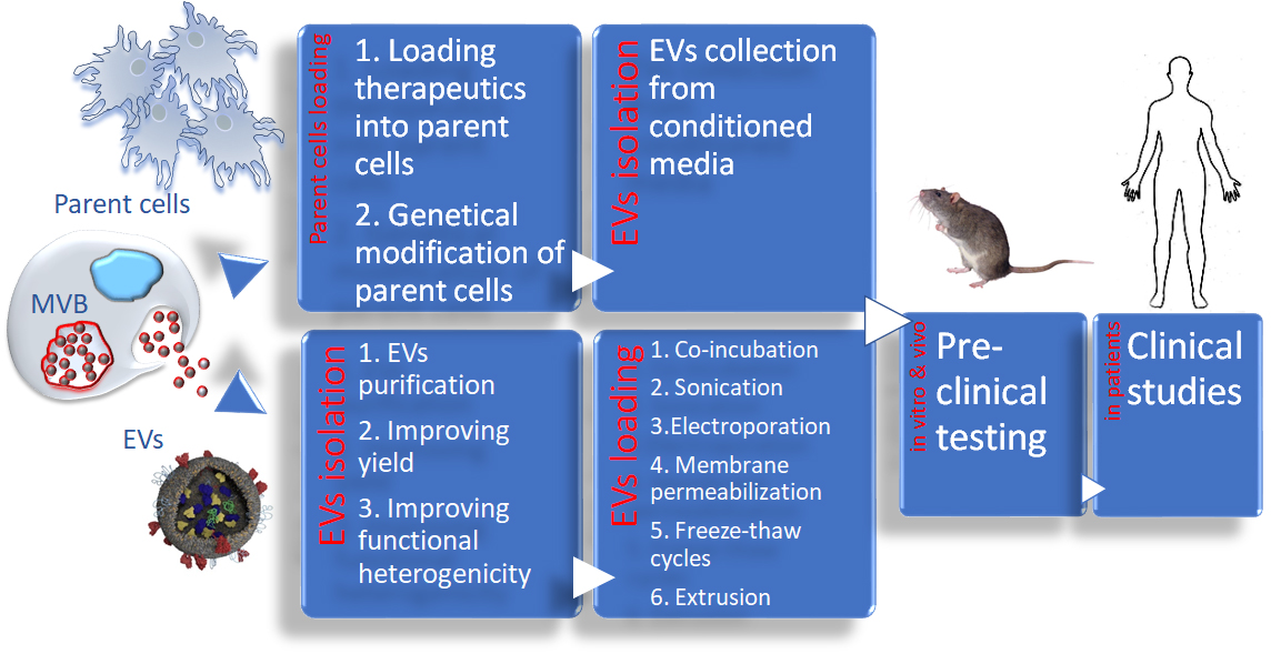
3. Improving Functional Heterogenicity and Yields of EVs Nanocarriers

EVs consist of various types of nanovesicles, namely exosomes, MVs, and apoptotic bodies [22,23,24]. It has shown to be difficult to separate EVs and MVs, mainly due to overlapping vesicle sizes and proteins expressed on their surface. Therefore, in most cases, a mixture of EVs and MVs is used to produce drug formulations [24]. It should also be noted that the absolute separation and definition of various EVs based on their size or biogenesis has yet to be established beyond doubt, and there is currently no consensus on markers that distinguish the origin of these vesicles once they have left the cell [80].
EVs can be isolated from conditioned cell culture media or bodily fluids by different methods, including differential centrifugation, filtration paired with centrifugation, concentration paired with ultracentrifugation, immunoaffinity chromatography, size exclusion chromatography, and polymer-based precipitation. Each isolation technique has advantages and disadvantages that should be considered in terms of being reproducible, specific, and feasible [81]. Differential ultracentrifugation combined with density gradient centrifugation are considered the “gold standard” for isolating EVs. This process involves applying a centrifugal force to a solution containing EVs, e.g., a conditioned cell culture media or biological fluids. It is worth noting that the type, quantity, and quality of EVs isolated by this method is sensitive to the g force, rotor type, angle of rotor sedimentation, radius of centrifugal force, pelleting efficiency, and solution viscosity. Gradient centrifugation requires extensive (62–90 h) centrifugation time [82], but provides a more uncontaminated EV isolate than ultracentrifugation alone. Of note, ultracentrifugation is associated with morphological alterations and partial aggregation of vesicles. Immunoaffinity chromatography is a more efficient method for isolating EVs as compared to differential ultracentrifugation and density gradient ultracentrifugation [81]. It requires a single easy step without using harsh chemicals. However, this method provides a relatively low yield and can be used for small volumes only. In addition, because this method of EV isolation depends on antibody recognition of EV proteins, only a subset of all EVs (those expressing the antibody-recognized protein) can be captured. Size exclusion chromatography (SEC) preserves the integrity and biological activity of EVs using gravity flow when vesicle structure and integrity remain intact. This is a fast and easy procedure that requires a small sample volume. However, a low concentrated sample needs an additional process for enrichment. Finally, polymer precipitation is relatively easy to use and does not require specialized equipment or a lengthy run time. However, it has been shown that this method coprecipitates nonvesicular contaminants such as lipoproteins, as well as polymer material [83]. Thus, Patel et al. [84] compared four EV isolation techniques for yield and purity. The polymer-based precipitation method had the maximum yield, followed by size-exclusion chromatography and differential ultracentrifugation. The immunoaffinity-based isolation method yielded the fewest EVs. Importantly, a high yield of EVs was accompanied by contaminations with serum proteins and chemical impurities, including high salt concentration, Sodium Dodecyl Sulfate (SDS), or Polyethylene glycol (PEG) contaminations after polymer-based precipitation. These issues may be addressed by pre- and post- isolation steps. Pre-isolation involves the removal of subcellular particles such as lipoproteins. Post-isolation involves removal of the polymer, typically by using a Sephadex G-25 column [82]. Therefore, considering large-scale clinical manufacturing [85], a level of segregation EVs from copurifying components may influence the functionality and therapeutic activity of the final product.
The translation of EV-based formulations into clinical practice requires compliance with existing regulatory frameworks [86]. EVs are a fairly heterogeneous population in terms of their biochemical composition, size, and the source [87]. Thus, the standardization and effective purification of large amounts of these nanovesicles is a critical, but still considerable, challenge. Specifically, the manufacturing of homogeneous drug nanoformulations, and production and quality control, are crucial requirements. Moleirinho et al. [88] developed a purification method using semicontinuous multicolumn chromatography, a robust, scalable and efficient tool for EV purification. Besides the higher recoveries obtained with the continuous system when comparing with batch chromatography, the EV properties were maintained during the purification process regarding their size and morphology. A fast and reliable method of isolating serum EVs was reported by Navajas et al. [89]. Using size-exclusion chromatography with qEV columns (Izon, Christchurch, New Zealand), a homogeneous population of EVs in terms of size, morphology, and protein composition was obtained.
Another challenge that has critical implications for the use of EV-based formulations is whether the sufficient number of these carriers can be generated [90]. Indeed, the EV yield per cell will impact the final production cost, as well as having clinical applications. In this respect, the choice of parent cells is very important. For example, MSCs are known to produce large numbers of EVs, suggesting that these cells may be efficient for EV production in a clinically applicable scale [65,91]. Several reports have indicated that specific treatments of EV producing cells could considerably increase the yield of these natural nanocarriers. For example, culturing dendritic cells (DCs) for a prolonged time [92] or at low pH [93] increased EV production up to ten-fold. Furthermore, the addition of neutral and cationic-bare liposomes enhanced EV secretion in a dose-dependent manner [94]. However, the possible contamination of EVs with liposomes is a serious concern associated with this method. Gao et al. [95] reported high yield of EVs using nitrogen cavitation that instantly disrupted neutrophils to form nanosized membrane vesicles. The authors indicated that this approach made it possible to increase the manufacture of EVs by 16-fold. Another option is to break parent cells, for example, monocytes/macrophages with simultaneous loading with anticancer agents, followed by the isolation of EV-like nanoparticles [54]. In attempting to mimic the function of EVs with nanovesicles, Jang et al. [54] utilized human U937 monocytic cells to produce nanovesicles with the ability to carry large amounts of therapeutics. While maintaining the plasma membrane proteins of the targeted cells, the drug-loaded nanovesicles were able to efficiently induce tumor cell death and increase the production yield of chemotherapeutics in relation to naturally occurring EVs by 100-fold. Of note, one should consider that the alteration of cell culture conditions can certainly increase yield, but the impact on the biological effect of EVs has to be crucially assessed for biosafety reasons.
Next, the mass production of EVs by membrane fusion with lipid-based materials was suggested in several reports [96,97,98]. The manufacture of large quantities of drug nanocarriers was achieved via a membrane extrusion technique [96] that allowed up to a 43-fold increase in the numbers of vesicles postisolation. The production of hybrid EVs was also proposed by Rayamajhi et al. [97]. EVs from mouse macrophages were hybridized with synthetic liposomes that increased the yield and retention of the EV functional properties. The manufacture of hybrid EVs was also suggested by De La Peña et al. [98]. This group utilized coated liposomes as artificial EVs, and discovered that the obtained nanocarriers functioned as naturally occurring EVs and efficiently activated immune responses [98]. Chemically-induced membrane blebbing was suggested for the fast production of large numbers of EV-like vesicles [99]. Different chemical agents, for example, sulfhydryl, paraformaldehyde, or dithiothreitol, were shown to lock the cell in a fixed physiological state and promote the release of vesicles from a plasma membrane to the conditioned media.
A different approach for the upscaling production of EVs was reported in a study conducted by Li et al. [100]. Instead of manufacturing EVs from animal cells, the authors utilized biocompatible bovine milk EVs (mEVs) that can be obtained inexpensively in large quantities [100]. mEVs were loaded with doxorubicin (Dox) and decorated with hyaluronan (HA), in order to direct them to CD44-overexpressing tumor cells. HA is a CD44-specific ligand which ensures that the EVs are directed to the cell membrane of the specified tumor cells. mEVs were able to deliver chemotherapeutics to tumor-specific cells in vitro and trigger apoptosis [100]. Crashed grapes were also suggested as an abounded source for EV-like nanoparticles [101]. Thus, the oral administration of EV-like nanovesicles from grapes facilitated intestinal regeneration in a mouse model of colitis that was induced by exposing mice to dextran sodium sulfate in drinking water. The EV-like nanovesicles prevented the colitis-associated reduction of both intestinal length and villus height. As a result, mice treated with grape-derived EV-like nanoparticles lived twice as long as untreated mice.

4. Loading EVs with Therapeutic Cargo

The closest relatives of natural nanocarriers EVs are synthetic vehicles, liposomes, and polymer-based nanoparticles. One of the major differences between these two drug delivery systems is that the loading of a drug into artificial vehicles can be achieved concurrently with their formation, when the drug is present at the time of nanoparticle formation. Contrary to synthetic-based formulations, therapeutics should be incorporated into EVs when they are already formed. It is worth noting that well-organized and tight EV membranes greatly restrict drug free penetration. In addition, substantial precautions should be made during drug loading to preserve the EV membrane structure, with all crucial proteins on their surface. This sets substantial restrictions on drug incorporation methods, and as a result, impedes the high loading efficacy of EV-based formulations.

4.1. Exogenous Loading of EVs

Regarding exogenous loading, naive EVs isolated from parent cells media can be loaded with therapeutic agents by a variety of methods. These procedures include: co-incubation with a drug at different temperatures [41,43,54,102,103,104,105,106,107,108,109,110], sonication of EVs/drug mixture, including water bath sonication [44,97], and probe sonication [43,109,111,112,113]; electroporation of EVs with siRNA and miRNA [114,115,116], or EVs with small molecule anticancer drugs, paclitaxel (Ptx) and Dox [111]; precomplexation of siRNA with cationic liposomes followed by the fusion with isolated EVs [117]; transient permeabilization of EVs membranes with saponin for enzymes and other proteins [43,44]; freeze-thaw cycles [43]; or extrusion procedure [43]. Notably, aside from therapeutics, these methods can be applied to imaging agents. Specifically, magnetic resonance imaging (MRI) of EVs loaded with superparamagnetic iron oxide nanoparticles was suggested for tracking EVs to lymph nodes [118]. The main cases of different loading procedures are outlined in Table 1.

4.1.1. Co-Incubation

Loading EVs with small molecule drugs by simple co-incubation can ultimately be utilized as a therapeutic treatment for a variety of diseases. Qu et al. [108] utilized this method of loading naive EVs isolated from mouse blood with dopamine by incubation with saturated solution of dopamine with 0.02% ascorbic acid for 24 h at room temperature (RT). The drug diffusion to the EVs luminal side was achieved via equalizing the concentrations. This novel drug delivery system was designed to allow the direct use of dopamine that would cross the BBB effectively with maximum biocompatibility and minimum toxicity in a mouse model of PD. The dopamine-loaded blood EVs were systemically injected into PD mice with acute brain inflammation caused by intracranial injection of 6-hydroxydopamine (6-OHDA). The encapsulation of dopamine in blood derived EVs significantly reduced its toxicity, and increased accumulation in the brain [108]. The authors suggested that blood EVs have a good natural targeting ability to the brain without any additional modification. Lipophilic small molecules, such as anticancer agents and anti-inflammatory agents, for example curcumin (Cur), can also be loaded by co-incubation with EVs. Sun et al. [41] reported that EVs from an EL-4 cell line were incubated with Cur at 22 °C, purified from nonincorporated drug, and injected intraperitoneally into mice with lipopolysaccharide (LPS)-induced septic shock. The results concluded that a significant reduction of inflammation was observed in LPS-intoxicated mice followed by treatment with Cur-incorporated EVs. Next, an anti-inflammatory drug, piceatannol, was loaded into neutrophil derived EVs by co-incubation via the pH gradient [95] using the same approach as was applied to Dox when loaded into liposomes [119]. Piceatannol is a weak acid molecule which increases the solubility in a basic environment. The pH gradient between the inner compartments of EVs (pH 9) and outside media (pH 4.5) made it possible to increase loading of piceatannol from 0.6% (drug(w)/vesicle(w)) with no pH gradient to 1.7% w/w. The authors reported that piceatannol-loaded EVs significantly alleviated acute lung inflammation/injury and sepsis induced by LPS. Co-incubation an acidophilic dye with a strong tumoricidal action, Acridine Orange (AO) with macrophage derived EVs was reported in [110]. AO charged EVs (EV-AO) showed an extended drug delivery time to melanoma cells as compared to free AO, improving drug cytotoxicity. Moreover, tumor reduction by EVs loaded with an anticancer agent, gemcitabine (Gem), was reported by Li et al. [109]. Thus, this chemotherapeutic drug has a history of significant side effects in the systemic treatment of pancreatic cancer. This off-site toxicity could be diminished by using EVs from the pancreatic cancer cell line Panc-1 as drug carriers. Gem-loaded EVs were able to suppress tumor growth or fully eliminate tumors in mouse model of pancreatic cancer.
The incorporation of high molecular weight therapeutics, such as protein and nucleic acids, into EVs has additional challenges related to the difficulty of penetration across the EV membranes for these large molecules, as well as their possible degradation during the loading procedure. A simple co-incubation with EVs at RT was also used for manufacture of EV-based formulation of a macromolecular therapeutic agent, brain-derived neurotrophic factor (BDNF) [45]. Macrophage-derived EVs were loaded with BDNF and intravenously injected into mice with brain inflammation [45]. It was reported that EVs were able to cross the BBB and deliver the protein cargo to the inflamed brain tissues. The increased brain penetration of EVs under neuroinflammation were attributed to interactions between the integrin lymphocyte function-associated antigen 1 (LFA1) on the EV surface, and intercellular adhesion molecule 1 (ICAM-1) overexpressed on the inflamed brain endothelium [45]. Finally, the use of the evolutionary conserved L-domain pathway was suggested for loading therapeutic proteins into EVs [122]. A target protein, Cre recombinase, was labeled with a specific WW tag that resulted in the specific interaction with L-domain containing protein Ndfip1 and packaging in EVs. The functional delivery of Cre recombinase in EVs to recipient cells was documented in transgenic mice upon intranasal administration [122]. Overall, the co-incubation method is relatively simple to scale up. Furthermore, this approach makes it possible to work with highly fragile therapeutic molecules, although it usually does not provide high loading efficiency of EVs nanocarriers.

4.1.2. Sonication

Another promising method for loading small molecule drugs, as well as macromolecules, is sonication. Treatment with ultrasound produces transient pores in EV membranes that allow drug diffusion inside the vesicles. This method may provide higher loading efficiency compared to simple co-incubation. Two different approaches could be utilized: water bath sonication and probe sonication in the presence of the drug. Water bath sonication is a mild procedure that can be used when a therapeutic with relatively low stability needs to be formulated. For example, lysosomal enzyme, tripeptidyl peptidase-1 (TPP1) was loaded into EVs under sonication in a water bath [44]. Up to 70 µg TPP1 was loaded into 1011 EVs particles with enzymatic activity 44,000 mU/min by this method. Accordingly, EVs significantly increased the stability of TPP1 against protease degradation and provided efficient TPP1 delivery to target cells in in vitro and in vivo models of late-infantile neuronal ceroid lipofuscinosis (LINCL). A robust brain accumulation of EV carriers and increased lifespan was recorded in LINCL mice following the intraperitoneal administration of TPP1-loaded EVs [44]. Next, water bath sonication followed by extrusion was used to load Dox into EV-mimetic hybrid vesicles [97]. Lamichhane et al. [123] reported loading functional small interfering RNA (siRNA) and microRNA (miRNA) into EVs via sonication in a water bath. EVs loaded with siRNA retained their integrity and mediated knockdown of target genes, leading to cognate protein downregulation. Of note, the overall delivery of labeled siRNA to cells via sonicated EVs was low (2.96%), especially given that the cell uptake of labeled EVs was >80% [123]. These results corroborate those in a recent publication by Albanese et al. [124]. The authors were able to achieve a substantial binding of EVs to a range of different target cells in vitro, but failed to detect a fusion between EVs and recipient cells, and the subsequent release of the EV cargo (miRNAs) into the cytoplasm. This raises a very important question about the mechanism of delivery of the therapeutic cargo into targeted cells. Specifically, whether the direct membrane fusion of EVs with plasma membrane or membranes of endocytic vesicle followed by release of therapeutic cargo into the cytoplasm occurred upon EV internalization by recipient cells.
A probe sonication can provide higher loading efficiency, but should be used with relatively stable therapeutics, such as small molecule anticancer agents, Ptx and Dox, as reported in [111,112]. These drugs were chosen as model drugs for their hydrophobic and hydrophilic properties, respectively [112]. Optimization sonication procedures, along with pH, and temperature of EVs/drug mixture, resulted in high loading efficiency of macrophage derived EV formulations. Thus, using a pH that is close to pI for Dox (i.e., decreasing the charge of the drug molecule and increasing its hydrophobicity) significantly increased Dox loading into EVs. Another important finding was that the temperature of the mixture drastically affected the drug loading. Specifically, the sonication of EVs with Ptx at room temperature (RT) with short periods of cooling in between the cycles was more efficient that sonication on ice that was maintained during sonication and intermediate periods [112]. Finally, the systemic administration of EVs loaded with Ptx and Dox resulted in the potent tumor growth suppression in mice with triple negative breast cancer (TNBC) solids, as compared to Taxol or Dox treated animals. Next, the probe sonication method was also used to load Dox into pH-sensitive EVs with hyaluronic acid grafted with deoxycholic acid (HDEA) [113]. The incorporation of HDEA allowed the researchers to accelerate release of encapsulated Dox in cancer cells due to the acidic pH-induced protonation of 3-(diethylamino)propylamine (DEAP) moieties [113]. In another work, Li et al. [43] used the sonication method to load a small molecule drug, Gem. Notably, the sonication method provided higher loading efficiency compared to co-incubation; however, ultrasound treatment may result in the destruction and inactivation of some therapeutic molecules.
Beside the incorporation of small molecule therapeutics, probe sonication was utilized to load macromolecules, such as proteins, enzymes, and growth factors. For example, a potent antioxidant, catalase was loaded into macrophage-derived EVs via a sonication procedure [43]. A reformation of EVs upon sonication resulted in high loading efficiency (around 26.1 ± 1.2%), sustained release, and catalase preservation against proteases degradation. About 940 ± 15 catalase molecules per nanocarrier were incorporated by sonication. Of note, the intensity of sonication upon loading may affect the morphology of the aggregates. Thus, “strong sonication” resulted in the appearance nonspherical associates with a variety of shapes, indicating the destruction of the initial EV structure [43]. In contrast, “mild sonication” with intermediate brakes for mixture cooling ensured the preservation of the EV structure and morphology, along with the high loading efficiency. Each mg of EV-based formulation of catalase (exoCAT) obtained by sonication contained approximately 1,376 ± 64.1 U of catalase activity in 4 × 10¹¹ EVs/mL. The systemic administration of exoCAT provided significant neuroprotective effects in mouse models of PD [43].

4.1.3. Electroporation

Electroporation is widely used to load small molecule drugs and nucleic acids. Takenaka et al. [116] utilized this method of exogenous loading to improve the delivery of Dox to lung cancer cells. The efficiency of Dox encapsulation into EVs was calculated to be 0.5%, based on the quantification of Dox using a spectrofluorometer. The cytotoxicity of Dox incorporated into EVs was examined in lung cancer cells in vitro and in vivo and compared to Dox incorporated into artificial liposomes. Interestingly, cotreatment with gefitinib (Gef), a tyrosine kinase inhibitor of epidermal growth factor receptor (EGFR), resulted in significant increases in Dox cytotoxicity in EV-based formulations, but not in liposome-based formulation. The results indicated that Gef can enhance cell-to-cell communication via EVs within the tumor microenvironment that was not applicable for synthetic liposomal nanocarriers. Thus, in some cases, using EVs instead of synthetic nanocarriers may completely alter the mechanism of drug therapeutic activity.
The electroporation method is widely used for loading small molecule drugs and nucleic acids into EVs [72,96,114,125,126]. Alvarez-Erviti et al. [114] pioneered this method, electroporating siRNA into DC-derived EVs. To reduce immunogenicity, autologous dendritic cells were used for EV production. To target the formulation to the brain, parent DCs were engineered to express Lamp2b, an EVs membrane protein, fused to the neuron specific RVG peptide. Systemically administered RVG-EVs loaded with exogenous siRNA produced strong mRNA (60%) and protein (62%) knockdown of BACE1, a therapeutic target in Alzheimer’s disease. The electroporation technique was also used to incorporate Alexa Fluor 488-tagged siRNA against MAPK1 into plasma EVs [117]. Fluorescently labeled siRNA was detected in recipient monocytes and lymphocytes isolated from the peripheral blood of healthy donors following incubation with electroporated EVs. EV-delivered siRNA caused selective downregulation of MAPK1 in recipient cells. Later, the same method was used to load EVs with miRNA to EGFR-expressing breast cancer cells [115]. In a recent study, the electroporation method was utilized to load tumor suppressor miRNAs into fibroblast-derived EVs to treat ovarian cancer [120]. Repeated intraperitoneal injections of miRNA-loaded EVs drastically inhibited peritoneal dissemination and reduced the tumor burden in a xenograft ovarian cancer mouse model. The electroporation of engineered extracellular vesicles allowed 15–20% encapsulation efficiency of siRNA and resulted in effective gene silencing, i.e., comparable to that of commercial Lipofectamine RNAiMax [96]. This is especially important considering that large quantities of these engendered EVs were manufactured by membrane fusion with synthetic lipids. Of note, electroporation may result in the aggregation of miRNA and co-isolation with EVs preparation [127]. Therefore, efficient purification of drug loaded EVs from nonincorporated miRNA should be developed to obtain reliable and reproducible therapeutic effects.

4.1.4. Transient Permeabilization with Saponin

To load therapeutic proteins such as catalase and TPP1, EV membranes were permeabilized with saponin [43,44]. For this purpose, EVs were first, pre-incubated with saponin to allow the selective removal membrane-bound cholesterol in the membranes, creating transient holes/pores in the EV bilayers, and thus, promoting drug loading (up to 6.3 µg/1011 particles for catalase, or 50 µg/1011 particles for TPP1 were incorporated). The successful delivery of these therapeutic enzymes to CNS manifested in the prevention of neurodegeneration and neuroinflammation in mouse models of Parkinson’s disease and a Lysosomal storage disorder, CLN2 [43,44]. Furthermore, modified calcium-mediated transfection led to the increased permeabilization and loading small RNAs into serum EVs [121].

4.1.5. Freeze-Thaw and Extrusion Cycles

Finally, freeze-thaw cycles, and extrusion of EVs in the presence of a therapeutic protein were suggested for drug incorporation into macrophage-derived EVs [43]. A formation of large aggregates was reported during loading via freeze-thaw cycles, probably due to interaction via adhesive proteins on the surface of EVs. On the contrary, extrusion procedure led to the formation of relatively unisize aggregates with the efficient drug incorporation (22%). Nevertheless, almost 50% of EVs were lost via extrusion procedure when 200 nm pore size polycarbonate extruder membranes were used. Next, the effect of loading procedures (extrusion, freeze-drying, and probe sonication) on the structure, lipid and protein content, as well as interaction with target cells was extensively studied by Saux et al. [128]. The authors advocated for extrusion, as the most promising loading procedure compared to sonication and freeze-drying. Of note, most of these studies on the beneficial effects of EVs are preliminary studies in animal models and their translational potential has not yet been assessed in patients.
Overall, a mixture of exosomes and MVs is generally used for manufacturing EV-based drug formulations, as was mentioned above. However, some studies indicate that the accumulation and therefore, therapeutic effects of these two types of vehicles might differ. Thus, in observing the differences in unloading methods of EVs, Saari et al. [106] analyzed the pathways of Ptx uptake in both exosomes and MVs from the human prostate cancer cell line PC-3 cells utilizing fluorescence lifetime imaging microscopy (FLIM). Results concluded that the drug delivery pathways of exosomes and MVs differ, demonstrating that exosomes mainly deliver by endocytosis, while MVs discharges its therapeutic cargo by fusion with the cell membrane, as well as endocytosis. Thus, one might consider advantages of using homogeneous population of exosomes or MVs for drug incorporation vs. manufacture cost. Indeed, efficient purification of loaded EVs from nonincorporated drug is of great importance for the assessment of their role in therapeutic nanomedicine applications. The contamination of free cargo may mask or even mislead the overall conclusions about efficiency of EV-based drug formulations.
Furthermore, the location of the therapeutic cargo, specifically, where the drug is situated, i.e., on the luminal or abluminal site of EVs, or incorporated into membrane of the vesicle will determine the level of drug protection and in the blood circulation, kinetics of drug release, and overall immunogenicity of EV-based formulation. The unique structure of EVs made of a hydrophobic lipid bilayer and a hydrophilic cavity allows for a loading of different therapeutic agents that have hydrophobic or hydrophilic properties [129]. Hence, therapeutic molecules may diffuse inside the EVs or simply adhered to their surface. In many cases, the location of the cargo is controlled by its solubility in water. For example, Haney at al. [112] utilized two chemotherapeutic agents, Dox and Ptx, as representatives of hydrophilic and hydrophobic small molecule antineoplastic agents, respectively. Dox, a relatively hydrophilic compound would be incorporated into EVs inner compartment, while Ptx, a highly hydrophobic compound, probably resided in the membranes of the EVs. The thorough characterization is crucial for the manufacture of reliable and reproducible EV-based formulations.

4.2. Exogenous Loading of Parent Cells

In exploring exogenous loading of parent cells, two methods are considered: the loading of therapeutic cargo into isolated parent cells, and the transfection of parent cells with therapeutics encoding plasmid DNA (pDNA). The main cases of different loading procedures are outlined in Table 2.
Regarding the loading of parent cells_with small molecule therapeutic agents, the cells can be incubated with the desired therapeutic agent, and then EVs with the incorporated drug are isolated from cell conditioned media [24]. Diaz Osterman et al. [130] utilized this method for loading of pancreatic adenocarcinoma cells (PANC-1) in vitro with Cur, which has potential anticancer and anti-inflammatory benefits. It was reported that Cur was detected in the lumen of the EVs. In another study, MSCs-derived EVs were loaded with Ptx by incubating the parent cells with the drug, and then isolating EVs [131]. MSCs were able to package and deliver active Ptx through their EVs, suggesting the possibility of using these cells as a factory to develop drugs with a higher cell-target specificity. The same loading procedure through parent cells (HepG2) was used to produce EVs carrying different anticancer agents: Ptx, Etoposide, Carboplatin, Irinotecan, Epirubicin, and Mitoxantrone [132]. The obtained EV-based drug formulations showed potent antiproliferative activity on human pancreatic cell line CFPAC-1 [132]. Loading parent cells with anticancer agents, Dox, Gentamicin, 5-Fluoracil, or Carboplatin was also proposed for manufacture monocyte-derived EVs [54]. This approach allows manufacturing relatively large batches of EV-based formulations. However, a high amount of the drug required for the loading into the cells that is also partially metabolized by parent cells during incubation diminish clinical potential of this method.
Regarding using macromolecular therapeutics for loading through parent cells, one should pay a special attention to preserve these drugs against degradation in host cells. Thus, our lab utilized this approach for loading macrophage-derived EVs with catalase [43,134]. For this purpose, first, catalase was incorporated into a polymer-based nanocontainer, and then loaded into parent cells. Importantly, the formulation design of this nanoparticle was different from the common approach for cell-free drug nanoformulation. Nanocarriers for cell-free delivery are typically size-restricted to avoid entrapment in monocytes, focusing on small size nanoparticles with a polyethylene-glycol (PEG) corona to perpetuate a stealth effect. On the contrary, the best nanoformulation for loading into parent cells had a relatively large size (c.a. 200 nm) that resulted in improved accumulation in macrophages, and drug reshuffling into EVs. The cross-linking of polymer-based nanoparticles with an excess of a nonbiodegradable linker ensured low cytotoxicity of nanoformulation and efficient catalase protection in the parent cells [43,133]. Released EVs loaded with catalase were efficiently accumulated in target cells and produced potent anti-inflammatory and neuroprotective effects in Parkinson’s disease mouse models. Maugeri at al. [140] reported loading mRNA encoding human erythropoietin (hEPO protein) into EVs through parent cells. The mRNA was incorporated into lipid-based nanoparticles (LNPs) to protect against degradation in the cells. hEPO-mRNA and the LNP components i.e., cationic ionizable lipids were accumulated in endosomes, and then incorporated into intraluminal vesicles of the late endosomes that were subsequently secreted into the extracellular environment as EVs. Systemically administered drug-loaded EVs delivered hEPO-mRNA to cells and produce human protein in mouse blood and major organs [140].
The second method of exogenous loading is the transfection of parent cells with pDNA encoding the desired therapeutic, and then isolation EVs from cell conditioned media. Mizrak et al. [135] utilized this technique for transduction of donor cells to isolate EVs that contained the therapeutic cytosine deaminase-uracil phosphoribosyltransferase (CD-UPRT) protein. The direct injection of drug-loaded EVs, as well as systemic therapy with 5-fluorouracil (5-FU), proved effective in the regression of glioblastomas. Of note, this approach allowed to deliver both the therapeutic mRNA and protein via EVs for cancer treatment. Transfection of macrophages cell line, RAW264.7 with miR-16 inhibitor using Lipofectamine 2000 was reported by Jang et al. [136]. The authors indicated that upregulated miR-16 can be transferred to tumor-associated macrophages via EVs and inhibit tumor associated macrophages (TAM) infiltration and M2 polarization, suppressing tumor growth in murine breast cancer model. Furthermore, EVs with incorporated chicken egg ovalbumin (OVA) were obtained by transfecting parent cells with plasmid that encodes fusion protein consisting of OVA and specific protein that is known to localize to EVs [137]. We also reported the development of two EV-based drug delivery system for catalase [138], and glial cell-line derived neurotropic factor (GDNF) [134]. For this purpose, parent cells, macrophages were genetically modified with pDNA encoding these therapeutic proteins and drug-loaded EVs were collected from cell concomitant media. We demonstrated that pre-transfected macrophages released EVs with encapsulated pDNA, mRNA and the encoded therapeutic protein [138]. Furthermore, EVs contained a considerable amount of the transcription factor, NF-κB, which is particularly involved in the utilized protein expression under CMV promoter. Accumulation of these EVs in the brain led to potent and sustained therapeutic effects in PD mouse models. EVs efficiently transferred their contents to contiguous neurons resulting in de novo protein synthesis in target cells. Thus, EVs represent a highly efficient packaging system that can be used for the delivery of proteins and genetic material to target cells. An interesting approach for incorporation of adeno-associated virus (AAV) capsids into EVs to diminish their immunogenicity and improve gene delivery was suggested by Maguire et al. [139]. It was reported that during production, a fraction of released AAV vectors were associated with EVs, termed vEVs (vector-EVs), which outperformed conventionally purified AAV vectors in transduction efficiency in vitro. Indeed, EVs isolation method from parent cells conditioned media is crucial for apparent therapeutic activity and its interpretation. For example, coprecipitation of nucleic acids is common in many isolation procedures, especially PEG-based precipitation and ultracentrifugation [127,141]. Nevertheless, manufacturing pure drug loaded EVs formulations through parental cells may be more feasible than through loading of naive EVs with considerable amount of nonincorporated drug in the media.

5. Challenges of Insufficient Targeting Efficiency of EVs Formulations to Disease Tissues

One of the bottlenecks known for EV-based formulations is insufficient targeting capability. Recent biodistribution studies of unmodified EVs after intravenous injection revealed a rapid accumulation of EVs in organs of the reticuloendothelial system (RES), such as the liver, kidney and spleen after systemic administration [142,143]. In this context, the need for EVs with enhanced targeting efficiencies is crucial. The addition of targeting moieties to EVs nanocarriers can be accomplished through their incorporation into naive EVs, or through genetic modification of parent cells. Regarding incorporation of vector moiety into naive EVs or parent cells, the molecule should consist of lipid-like part for efficient integration into EVs membranes and a targeting fragment. For example, anisamide (AA) vector moiety was used to target EVs to the sigma receptor overexpressed on many cancer cells lung cancer cells [111]. The efficient encoring of AA into EVs lipid bilayers was accomplished by using AA conjugated with lipid moiety, 1,2-distearoryl-sn-glycero-3-phospho ethanolamine-N-[methoxy(polyethyleneglycol-2000) (DSPE-PEG) [111]. The EVs vectorization was accomplished simultaneously upon Ptx loading via probe sonication of the EVs, Ptx, and DSPE-PEG-AA mixture. A profound ability to accumulate in cancer cells upon systemic administration and eradicate pulmonary metastases were demonstrated in mouse model of pulmonary metastases with Lewis Lung Carcinoma cells [111]. Wan et al. [144] reported vectorization EVs to cancer cells via incorporation of lipidated ligand, a nucleolin-targeting aptamer AS1411, that was covalently conjugated to cholesterol-poly(ethylene glycol) (cholesterol-PEG). The incorporation was achieved via mechanical extrusion of parent cells grafted with these target moieties. The vectorized EVs were then loaded with Ptx by probe sonication. The aptamer grafted EVs successfully delivered their therapeutic cargo Ptx in vitro and in vivo. Cell-type tropism is also an important factor in the achievement of tumor cell targeting [145]. Conjugation of BBB-targeting cyclo(Arg-Gly-Asp-D-Tyr-Lys) peptide, c(RGDyK), to the EVs surface by the copper-free click chemistry was reported by Tian et al. [146]. Specifically, this peptide is known to exhibit high affinity to integrin αvβ3 in reactive cerebral vascular endothelial cells after ischemia [147]. Systemic administration of BBB-targeted c(RGDyK)-EVs loaded with antioxidant curcumin (Cur) resulted in a strong suppression of the inflammatory response and cellular apoptosis in the lesion region in the transient middle cerebral artery occlusion (MCAO) mice model.
The second approach is widely used for targeting EVs through genetic modification of parent cells. In recent work, Alvarez-Erviti et al. vectorized EVs to the brain by transfecting parent dendritic cells (DCs) with plasmid encoding fusion protein consisting of lysosomal-associated membrane protein 2 (Lamp2b) and neuron-specific peptide derived from rabies virus glycoprotein (RVG) [114]. It was reported that RVG-vectorized EVs loaded with glyceraldehyde 3-phosphate dehydrogenase (GAPDH) siRNA successfully delivered the therapeutic siRNA in wild-type mice to the cells of neurovascular unit and caused efficient knock down of BACE1, which represents a therapeutic target in Alzheimer’s disease [114]. Ishikawa et al. [148] reported vectorization of EVs to cancer tissues with programmed cell death ligand 1 (PD-L1). To modify EVs surface, the insect cell line Spodoptera frugiperda 9 was infected with recombinant baculoviruses incorporating the PD-1 mutant gene to express the target membrane protein. Engineered EVs expressing the high-affinity PD-1 mutants effectively bound PD-L1 and PD-L1-expressing cancer cells, showing potential as a candidate in new therapy approaches.

6. EV-Based Drug Formulations in Clinic

An increasing amount of research effort has focused on moving EV-based formulations into the clinic. Currently, www.clinicaltrials.gov reports about 182 studies involving EVs as interventions or as a study object. Most clinical studies are concentrated on the use of EV as markers for diseases prognosis and diagnosis. Twenty-three clinical trials are focused on extracellular vesicles as a therapeutics or drug delivery agents (Table 3).
A large-scale production EV-based formulation is critical for success of clinical translation. As of today, the good manufacturing procedures (GMP) have been developed in Industry and Academia [149,150,151]. For example, a group in Wake Forest Institute for Regenerative Medicine labs has developed formulation of EVs loaded with therapeutic microRNAs. Furthermore, more than half a dozen clinical trials utilized EVs that proved feasibility and short-term safety of EVs formulations [152]. Thus, MolecuVax, Inc. developed EVs-based technology for delivery therapeutic peptides. Jazz Pharmaceutics Inc. is working on chemotherapeutics loaded into EVs to treat cancer. Finally, several companied manufactured drug-loaded EVs for treatment of neurodegenerative disorders (Stem cell Medicine Ltd. and Evox Therapeutics). In this case, it is very important that EVs formulations can be concentrated, lyophilized, and reconstituted in aqueous solutions without significant altering their properties [43].
The cellular origin for EVs varies from dendritic cells [153,154,155,156], reticulocytes [157], erythrocytes [158], monocytes [159], macrophages [160] to mesenchymal [161,162,163] and human iPSCs [164,165]. MSCs are the main source for EVs production for clinical trials. It has been reported that MSC-derived EVs can be easily scalable and safe for patients [166,167]. Phase I/II trial of intravenous administered of allogeneic mesenchymal stem cells derived EVs loaded with miR-124 in patients with chronic stroke (NCT03384433) found that dose up to 1.5 million cells/kg body weight was safe for patients (N = 15) and showed behavior end points improvement over 12 month [168]. EVs purified from monocyte cultures from 15 patients were used to treat advanced metastatic melanoma. The GMP process allowed harvesting about 5 × 10¹⁴ EVs MHC class II carriers [169]. In another trial, a total of 40 patients with advanced colorectal cancer received up to 2 × 10¹² EVs weekly for 4 weeks [170]. The therapies were safe, feasible, and efficient in induction of antigen-specific T lymphocyte response. Phase II clinical trial of vaccination with tumor antigen-loaded dendritic cell-derived EVs has been completed in 2018 by Gustave Roussy Center (France) (NCT01159288). The result showed an enhance antitumor immunity of NK cells in patients with advanced Non-Small Cell Lung Cancer [156].
Early Phase I trial NCT04270006 will use adipose stem cells derived EVs isolated from the patient and injections will be carried out to the patient’s periodontal pockets to identify regenerative efficiency. Preclinical in vivo results revealed significant regenerative effect compared to adipose stem cells and conventional treatment [171]. Effect of MSC-derived EVs on dry eye symptom in patients with chronic graft versus host diseases (NCT04213248) will be identified in Phase I/II clinical trial (N = 27). Patients will receive 10 µg/drop vesicles as an eye drop four times/day for 14 days and effect will be monitored during the 12 weeks of follow-up. Earlier study reported that EVs treatment can inhibit activation and infiltration of CD4 T cells [172]. Phase I clinical trial led by Dr. Gauri Varadhachary (NCT03608631) will identify dose and potential side effects of mesenchymal stromal cell-0derived EVs loaded with KrasG12D siRNA in treatment of pancreatic cancer with KrasG12D mutation. Thus, 28 participants will receive EVs systemically over 15-20 minutes on days 1, 4, and 10. Treatment repeats every 14 days for up to 3 courses and patients will be monitored up to 1 year. Next, safety and efficiency of EVs administered by nasal drop in patients with Alzheimer disease will be evaluated in the Phase I clinical trial in Ruijin Hospital (China). Nine participants will get 5, 10 or 20 µg of product two times a week for 12 weeks. Liver and kidney function and treatment-related adverse events will be monitored for 12 weeks, and cognitive function of participants as well as quality of life evaluation will be performed at baseline and 48 weeks after the treatment. Topical application of EVs will be appraised in the patients with dystrophic epidermolysis bullosa (NCT04173650) and ulcer (NCT04134676). In both studies participants will receive treatments directly on wounds and response will be measured after 14 days.
Several clinical trials were recently added to examine efficiency and safety of EVs products against novel coronavirus (SARS-COVID-2). Aerosol inhalation of MSC-derived EVs 2 × 10⁸ vesicles/3 mL at Day 1, Day 2, Day 3, Day 4, Day 5 will be assessed in the clinical trial in Ruijin Hospital (China) (NCT04276987). Inhalation of two types of MSC EVs twice a day during 10 days with 0.5–2 × 10¹⁰ particles in 3 mL on 90 participants will be evaluated in Medical Centre Dynasty Samara (Russia) (NCT04491240). Bone-marrow derived EVs will be administered via infusion to 60 participants with severe COVID-19 symptoms (NCT04493242). All-cause mortality and median days to recovery will be monitored. Finally, Landmark Hospital (Naples, Florida) will recruit 20 patients with COVID-19 to deliver acellular, minimally manipulated vesicles derived from human amniotic fluid. Product will be administered intravenously with 2–5 × 10¹¹ particles/mL in addition to the standard care to evaluate safety, side effects and treatment competence (NCT04384445). Safety and efficacy assessment will be observed in Phase I clinical trial led by Mustafa Cetin (NCT04389385). Donor originated COVID-19 specific T-cells derived EVs will be delivered to the patients with COVID-19 via inhalation. Treatment will be applied daily at the dose 2 × 10⁸ particles / 3 mL for 5 days.

7. Conclusions

EV-based therapeutics represent a next generation drug delivery system that could be a valuable tool for the therapy of various diseases. The advantages of these systems include ability to cross biological barriers, improve drug pharmacokinetics, increase therapeutic efficacy, and reduce toxic side effects commonly associated with the conventional synthetic nanocarriers. Furthermore, EVs nanocarriers can be chemically modified to incorporate various ligands for targeted drug delivery. Moreover, EVs carry multiple biological molecules which reflect their origin, making them a promising tool for assisting clinical diagnoses and evaluations of disease prognosis. Indeed, some technological, functional and safety features are still to be addressed. Nevertheless, the unprecedented level of attention and amount of work focused the development of EV-based formulations are grounds for optimism for their fast and extensive application in the clinic.

Author Contributions

Writing—original draft preparation, N.L.K., C.J.A., S.M.L., O.A.G., writing—review and editing E.V.B. All authors have read and agreed to the published version of the manuscript.

Funding

This research was funded by the M.J. Fox Foundation for Parkinson’s Research under grant 19-5960; Elsa U Pardee Foundation under grant 17-4676; Batten Disease Support and Research Association under grant A20-0944; and Eshelman Institute for Innovation grant UNC EII38-124.

Acknowledgments

We are very grateful to Sandra Lehrman, Stephen Lehrman, and Thomas Greenwood for financial support as well as various invaluable comments and suggestions. We are also thankful to the Assistant Dean for Development and Lead Development Officer for the Eshelman Institute for Innovation at UNC Kelly Collins for her assistance with communication the strategy and facilitating the support of this project.

Conflicts of Interest

The authors declare no conflict of interest.

References

  1. Chong, S.Y.; Lee, C.K.; Huang, C.; Ou, Y.H.; Charles, C.J.; Richards, A.M.; Neupane, Y.R.; Pavon, M.V.; Zharkova, O.; Pastorin, G.; et al. Extracellular Vesicles in Cardiovascular Diseases: Alternative Biomarker Sources, Therapeutic Agents, and Drug Delivery Carriers. Int. J. Mol. Sci. 2019, 20, 3272. [Google Scholar] [CrossRef] [PubMed] [Green Version]
  2. Boulanger, C.M.; Loyer, X.; Rautou, P.E.; Amabile, N. Extracellular vesicles in coronary artery disease. Nat. Rev. Cardiol. 2017, 14, 259–272. [Google Scholar] [CrossRef] [PubMed]
  3. Zamani, P.; Fereydouni, N.; Butler, A.E.; Navashenaq, J.G.; Sahebkar, A. The therapeutic and diagnostic role of exosomes in cardiovascular diseases. Trends Cardiovasc. Med. 2019, 29, 313–323. [Google Scholar] [CrossRef] [PubMed]
  4. Jia, G.; Sowers, J.R. Targeting endothelial exosomes for the prevention of cardiovascular disease. Biochim. Biophys. Acta Mol. Basis Dis. 2020, 1866, 165833. [Google Scholar] [CrossRef] [PubMed]
  5. Tikhomirov, R.; Donnell, B.R.; Catapano, F.; Faggian, G.; Gorelik, J.; Martelli, F.; Emanueli, C. Exosomes: From Potential Culprits to New Therapeutic Promise in the Setting of Cardiac Fibrosis. Cells 2020, 9, 592. [Google Scholar] [CrossRef] [PubMed] [Green Version]
  6. Wei, J.; Hollabaugh, C.; Miller, J.; Geiger, P.C.; Flynn, B.C. Molecular Cardioprotection and the Role of Exosomes: The Future Is Not Far Away. J. Cardiothorac. Vasc. Anesth. 2020. [Google Scholar] [CrossRef] [PubMed]
  7. Ramasubramanian, L.; Kumar, P.; Wang, A. Engineering Extracellular Vesicles as Nanotherapeutics for Regenerative Medicine. Biomolecules 2019, 10, 48. [Google Scholar] [CrossRef] [Green Version]
  8. Akbari, A.; Jabbari, N.; Sharifi, R.; Ahmadi, M.; Vahhabi, A.; Seyedzadeh, S.J.; Nawaz, M.; Szafert, S.; Mahmoodi, M.; Jabbari, E.; et al. Free and hydrogel encapsulated exosome-based therapies in regenerative medicine. Life Sci. 2020, 249, 117447. [Google Scholar] [CrossRef]
  9. Kumar, S.; Zhi, K.; Mukherji, A.; Gerth, K. Repurposing Antiviral Protease Inhibitors Using Extracellular Vesicles for Potential Therapy of COVID-19. Viruses 2020, 12, 486. [Google Scholar] [CrossRef]
  10. Bell, B.M.; Kirk, I.D.; Hiltbrunner, S.; Gabrielsson, S.; Bultema, J.J. Designer exosomes as next-generation cancer immunotherapy. Nanomedicine 2016, 12, 163–169. [Google Scholar] [CrossRef]
  11. Moore, C.; Kosgodage, U.; Lange, S.; Inal, J.M. The emerging role of exosome and microvesicle- (EMV-) based cancer therapeutics and immunotherapy. Int. J. Cancer 2017, 141, 428–436. [Google Scholar] [CrossRef] [PubMed]
  12. Masaoutis, C.; Mihailidou, C.; Tsourouflis, G.; Theocharis, S. Exosomes in lung cancer diagnosis and treatment. From the translating research into future clinical practice. Biochimie 2018, 151, 27–36. [Google Scholar] [CrossRef] [PubMed]
  13. Liu, C.; Su, C. Design strategies and application progress of therapeutic exosomes. Theranostics 2019, 9, 1015–1028. [Google Scholar] [CrossRef] [PubMed]
  14. Xie, X.; Wu, H.; Li, M.; Chen, X.; Xu, X.; Ni, W.; Lu, C.; Ni, R.; Bao, B.; Xiao, M. Progress in the application of exosomes as therapeutic vectors in tumor-targeted therapy. Cytotherapy 2019, 21, 509–524. [Google Scholar] [CrossRef] [PubMed]
  15. D’Agnano, I.; Berardi, A.C. Extracellular Vesicles, A Possible Theranostic Platform Strategy for Hepatocellular Carcinoma-An Overview. Cancers 2020, 12, 261. [Google Scholar] [CrossRef] [Green Version]
  16. Scavo, M.P.; Depalo, N.; Tutino, V.; de Nunzio, V.; Ingrosso, C.; Rizzi, F.; Notarnicola, M.; Curri, M.L.; Giannelli, G. Exosomes for Diagnosis and Therapy in Gastrointestinal Cancers. Int. J. Mol. Sci. 2020, 21, 367. [Google Scholar] [CrossRef] [Green Version]
  17. Chung, I.M.; Rajakumar, G.; Venkidasamy, B.; Subramanian, U.; Thiruvengadam, M. Exosomes: Current use and future applications. Clin. Chim. Acta 2020, 500, 226–232. [Google Scholar] [CrossRef]
  18. Jurj, A.; Zanoaga, O.; Braicu, C.; Lazar, V.; Tomuleasa, C.; Irimie, A.; Berindan-Neagoe, I. A Comprehensive Picture of Extracellular Vesicles and Their Contents. Molecular Transfer to Cancer Cells. Cancers 2020, 12, 298. [Google Scholar] [CrossRef] [Green Version]
  19. Tran, P.H.; Xiang, D.; Nguyen, T.N.; Tran, T.T.; Chen, Q.; Yin, W.; Zhang, Y.; Kong, L.; Duan, A.; Chen, K.; et al. Aptamer-guided extracellular vesicle theranostics in oncology. Theranostics 2020, 10, 3849–3866. [Google Scholar] [CrossRef]
  20. Sousa, C.; Pereira, I.; Santos, A.C.; Carbone, C.; Kovacevic, A.B.; Silva, A.M.; Souto, E.B. Targeting dendritic cells for the treatment of autoimmune disorders. Colloids Surf. B Biointerfaces 2017, 158, 237–248. [Google Scholar] [CrossRef]
  21. Ha, D.; Yang, N.; Nadithe, V. Exosomes as therapeutic drug carriers and delivery vehicles across biological membranes: Current perspectives and future challenges. Acta Pharm. Sin. B 2016, 6, 287–296. [Google Scholar] [CrossRef] [PubMed] [Green Version]
  22. Vader, P.; Mol, E.A.; Pasterkamp, G.; Schiffelers, R.M. Extracellular vesicles for drug delivery. Adv. Drug Deliv. Rev. 2016, 106, 148–156. [Google Scholar] [CrossRef] [PubMed]
  23. Villa, F.; Quarto, R.; Tasso, R. Extracellular Vesicles as Natural, Safe and Efficient Drug Delivery Systems. Pharmaceutics 2019, 11, 557. [Google Scholar] [CrossRef] [PubMed] [Green Version]
  24. Batrakova, E.V.; Kim, M.S. Using exosomes, naturally-equipped nanocarriers, for drug delivery. J. Control. Release 2015, 219, 396–405. [Google Scholar] [CrossRef] [PubMed] [Green Version]
  25. Thery, C.; Amigorena, S.; Raposo, G.; Clayton, A. Isolation and characterization of exosomes from cell culture supernatants and biological fluids. Curr. Protoc. Cell Biol. 2006, 30, 3–22. [Google Scholar] [CrossRef] [PubMed]
  26. Thery, C.; Ostrowski, M.; Segura, E. Membrane vesicles as conveyors of immune responses. Nat. Rev. Immunol. 2009, 9, 581–593. [Google Scholar] [CrossRef] [PubMed]
  27. Witwer, K.W.; Thery, C. Extracellular vesicles or exosomes? On primacy, precision, and popularity influencing a choice of nomenclature. J. Extracell. Vesicles 2019, 8, 1648167. [Google Scholar]
  28. Liu, D.; Kou, X.; Chen, C.; Liu, S.; Liu, Y.; Yu, W.; Yu, T.; Yang, R.; Wang, R.; Zhou, Y.; et al. Circulating apoptotic bodies maintain mesenchymal stem cell homeostasis and ameliorate osteopenia via transferring multiple cellular factors. Cell Res. 2018, 28, 918–933. [Google Scholar] [CrossRef] [Green Version]
  29. Van der Pol, E.; Coumans, F.A.; Grootemaat, A.E.; Gardiner, C.; Sargent, I.L.; Harrison, P.; Sturk, A.; van Leeuwen, T.G.; Nieuwland, R. Particle size distribution of exosomes and microvesicles determined by transmission electron microscopy, flow cytometry, nanoparticle tracking analysis, and resistive pulse sensing. J. Thromb. Haemost. 2014, 12, 1182–1192. [Google Scholar] [CrossRef]
  30. Vlassov, A.V.; Magdaleno, S.; Setterquist, R.; Conrad, R. Exosomes: Current knowledge of their composition, biological functions, and diagnostic and therapeutic potentials. Biochim. Biophys. Acta 2012, 1820, 940–948. [Google Scholar] [CrossRef]
  31. Turturici, G.; Tinnirello, R.; Sconzo, G.; Geraci, F. Extracellular membrane vesicles as a mechanism of cell-to-cell communication: Advantages and disadvantages. Am. J. Physiol. Cell Physiol. 2014, 306, C621–C633. [Google Scholar] [CrossRef] [PubMed] [Green Version]
  32. Aryani, A.; Denecke, B. Exosomes as a Nanodelivery System: A Key to the Future of Neuromedicine? Mol. Neurobiol. 2016, 53, 818–834. [Google Scholar] [CrossRef] [PubMed] [Green Version]
  33. Anand, S.; Samuel, M.; Kumar, S.; Mathivanan, S. Ticket to a bubble ride: Cargo sorting into exosomes and extracellular vesicles. Biochim. Biophys. Acta Proteins Proteom. 2019, 1867, 140203. [Google Scholar] [CrossRef] [PubMed]
  34. Elsharkasy, O.M.; Nordin, J.Z.; Hagey, D.W.; de Jong, O.G.; Schiffelers, R.M.; Andaloussi, S.E.; Vader, P. Extracellular vesicles as drug delivery systems: Why and how? Adv. Drug Deliv. Rev. 2020. [Google Scholar] [CrossRef] [PubMed]
  35. Tschuschke, M.; Kocherova, I.; Bryja, A.; Mozdziak, P.; Volponi, A.A.; Janowicz, K.; Sibiak, R.; Piotrowska-Kempisty, H.; Izycki, D.; Bukowska, D.; et al. Inclusion Biogenesis, Methods of Isolation and Clinical Application of Human Cellular Exosomes. J. Clin. Med. 2020, 9, 436. [Google Scholar] [CrossRef] [PubMed] [Green Version]
  36. Dai, H.I.; Vugmeyster, Y.; Mangal, N. Characterizing Exposure-Response Relationship for Therapeutic Monoclonal Antibodies in Immuno-Oncology and Beyond: Challenges, Perspectives, and Prospects. Clin. Pharmacol. Ther. 2020, 108, 1156–1170. [Google Scholar] [CrossRef] [PubMed]
  37. Matsumoto, J.; Stewart, T.; Sheng, L.; Li, N.; Bullock, K.; Song, N.; Shi, M.; Banks, W.A.; Zhang, J. Transmission of alpha-synuclein-containing erythrocyte-derived extracellular vesicles across the blood-brain barrier via adsorptive mediated transcytosis: Another mechanism for initiation and progression of Parkinson’s disease? Acta Neuropathol. Commun. 2017, 5, 71. [Google Scholar] [CrossRef] [Green Version]
  38. Winston, C.N.; Goetzl, E.J.; Akers, J.C.; Carter, B.S.; Rockenstein, E.M.; Galasko, D.; Masliah, E.; Rissman, R.A. Prediction of conversion from mild cognitive impairment to dementia with neuronally derived blood exosome protein profile. Alzheimers Dement. Diagn. Assess. Dis. Monit. 2016, 3, 63–72. [Google Scholar] [CrossRef] [Green Version]
  39. Chen, C.C.; Liu, L.; Ma, F.; Wong, C.W.; Guo, X.E.; Chacko, J.V.; Farhoodi, H.P.; Zhang, S.X.; Zimak, J.; Segaliny, A.; et al. Elucidation of Exosome Migration across the Blood-Brain Barrier Model In Vitro. Cell. Mol. Bioeng. 2016, 9, 509–529. [Google Scholar] [CrossRef]
  40. Kuroda, H.; Tachikawa, M.; Yagi, Y.; Umetsu, M.; Nurdin, A.; Miyauchi, E.; Watanabe, M.; Uchida, Y.; Terasaki, T. Cluster of Differentiation 46 Is the Major Receptor in Human Blood-Brain Barrier Endothelial Cells for Uptake of Exosomes Derived from Brain-Metastatic Melanoma Cells (SK-Mel-28). Mol. Pharm. 2019, 16, 292–304. [Google Scholar] [CrossRef]
  41. Sun, D.; Zhuang, X.; Xiang, X.; Liu, Y.; Zhang, S.; Liu, C.; Barnes, S.; Grizzle, W.; Miller, D.; Zhang, H.G. A novel nanoparticle drug delivery system: The anti-inflammatory activity of curcumin is enhanced when encapsulated in exosomes. Mol. Ther. 2010, 18, 1606–1614. [Google Scholar] [CrossRef] [PubMed]
  42. Yang, T.; Martin, P.; Fogarty, B.; Brown, A.; Schurman, K.; Phipps, R.; Yin, V.P.; Lockman, P.; Bai, S. Exosome delivered anticancer drugs across the blood-brain barrier for brain cancer therapy in Danio rerio. Pharm. Res. 2015, 32, 2003–2014. [Google Scholar] [CrossRef] [PubMed]
  43. Haney, M.J.; Klyachko, N.L.; Zhao, Y.; Gupta, R.; Plotnikova, E.G.; He, Z.; Patel, T.; Piroyan, A.; Sokolsky, M.; Kabanov, A.V.; et al. Exosomes as drug delivery vehicles for Parkinson’s disease therapy. J. Control. Release 2015, 207, 18–30. [Google Scholar] [CrossRef] [PubMed] [Green Version]
  44. Haney, M.J.; Klyachko, N.L.; Harrison, E.B.; Zhao, Y.; Kabanov, A.V.; Batrakova, E.V. TPP1 Delivery to Lysosomes with Extracellular Vesicles and their Enhanced Brain Distribution in the Animal Model of Batten Disease. Adv. Healthc. Mater. 2019, 8, e1801271. [Google Scholar] [CrossRef] [PubMed]
  45. Yuan, D.; Zhao, Y.; Banks, W.A.; Bullock, K.M.; Haney, M.; Batrakova, E.; Kabanov, A.V. Macrophage exosomes as natural nanocarriers for protein delivery to inflamed brain. Biomaterials 2017, 142, 1–12. [Google Scholar] [CrossRef]
  46. Marcoux, G.; Duchez, A.C.; Cloutier, N.; Provost, P.; Nigrovic, P.A.; Boilard, E. Revealing the diversity of extracellular vesicles using high-dimensional flow cytometry analyses. Sci. Rep. 2016, 6, 35928. [Google Scholar] [CrossRef]
  47. Van der Pol, E.; Boing, A.N.; Harrison, P.; Sturk, A.; Nieuwland, R. Classification, functions, and clinical relevance of extracellular vesicles. Pharmacol. Rev. 2012, 64, 676–705. [Google Scholar] [CrossRef] [Green Version]
  48. Al-Nedawi, K.; Meehan, B.; Micallef, J.; Lhotak, V.; May, L.; Guha, A.; Rak, J. Intercellular transfer of the oncogenic receptor EGFRvIII by microvesicles derived from tumour cells. Nat. Cell Biol. 2008, 10, 619–624. [Google Scholar] [CrossRef]
  49. Andre, F.; Schartz, N.E.; Movassagh, M.; Flament, C.; Pautier, P.; Morice, P.; Pomel, C.; Lhomme, C.; Escudier, B.; le Chevalier, T.; et al. Malignant effusions and immunogenic tumour-derived exosomes. Lancet 2002, 360, 295–305. [Google Scholar] [CrossRef]
  50. Valadi, H.; Ekstrom, K.; Bossios, A.; Sjostrand, M.; Lee, J.J.; Lotvall, J.O. Exosome-mediated transfer of mRNAs and microRNAs is a novel mechanism of genetic exchange between cells. Nat. Cell Biol. 2007, 9, 654–659. [Google Scholar] [CrossRef] [Green Version]
  51. Li, L.; Li, C.; Wang, S.; Wang, Z.; Jiang, J.; Wang, W.; Li, X.; Chen, J.; Liu, K.; Li, C.; et al. Exosomes Derived from Hypoxic Oral Squamous Cell Carcinoma Cells Deliver miR-21 to Normoxic Cells to Elicit a Prometastatic Phenotype. Cancer Res. 2016, 76, 1770–1780. [Google Scholar] [CrossRef] [PubMed] [Green Version]
  52. Kogure, T.; Lin, W.L.; Yan, I.K.; Braconi, C.; Patel, T. Intercellular nanovesicle-mediated microRNA transfer: A mechanism of environmental modulation of hepatocellular cancer cell growth. Hepatology 2011, 54, 1237–1248. [Google Scholar] [CrossRef] [PubMed] [Green Version]
  53. Cai, J.; Qiao, B.; Gao, N.; Lin, N.; He, W. Oral squamous cell carcinoma-derived exosomes promote M2 subtype macrophage polarization mediated by exosome-enclosed miR-29a-3p. Am. J. Physiol. Cell Physiol. 2019, 316, C731–C740. [Google Scholar] [CrossRef] [PubMed]
  54. Jang, S.C.; Kim, O.Y.; Yoon, C.M.; Choi, D.S.; Roh, T.Y.; Park, J.; Nilsson, J.; Lotvall, J.; Kim, Y.K.; Gho, Y.S. Bioinspired exosome-mimetic nanovesicles for targeted delivery of chemotherapeutics to malignant tumors. ACS Nano 2013, 7, 7698–7710. [Google Scholar] [CrossRef]
  55. Coffman, L.G.; Choi, Y.J.; McLean, K.; Allen, B.L.; di Magliano, M.P.; Buckanovich, R.J. Human carcinoma-associated mesenchymal stem cells promote ovarian cancer chemotherapy resistance via a BMP4/HH signaling loop. Oncotarget 2016, 7, 6916–6932. [Google Scholar] [CrossRef] [Green Version]
  56. Melzer, C.; von der Ohe, J.; Hass, R. Concise Review: Crosstalk of Mesenchymal Stroma/Stem-Like Cells with Cancer Cells Provides Therapeutic Potential. Stem Cells 2018, 36, 951–968. [Google Scholar] [CrossRef] [Green Version]
  57. Vakhshiteh, F.; Atyabi, F.; Ostad, S.N. Mesenchymal stem cell exosomes: A two-edged sword in cancer therapy. Int. J. Nanomed. 2019, 14, 2847–2859. [Google Scholar] [CrossRef] [Green Version]
  58. Qi, J.; Zhou, Y.; Jiao, Z.; Wang, X.; Zhao, Y.; Li, Y.; Chen, H.; Yang, L.; Zhu, H.; Li, Y. Exosomes Derived from Human Bone Marrow Mesenchymal Stem Cells Promote Tumor Growth Through Hedgehog Signaling Pathway. Cell. Physiol. Biochem. 2017, 42, 2242–2254. [Google Scholar] [CrossRef]
  59. Wu, S.; Ju, G.Q.; Du, T.; Zhu, Y.J.; Liu, G.H. Microvesicles derived from human umbilical cord Wharton’s jelly mesenchymal stem cells attenuate bladder tumor cell growth in vitro and in vivo. PLoS ONE 2013, 8, e61366. [Google Scholar] [CrossRef]
  60. Bruno, S.; Camussi, G. Role of mesenchymal stem cell-derived microvesicles in tissue repair. Pediatr. Nephrol. 2013, 28, 2249–2254. [Google Scholar] [CrossRef]
  61. Bruno, S.; Collino, F.; Deregibus, M.C.; Grange, C.; Tetta, C.; Camussi, G. Microvesicles derived from human bone marrow mesenchymal stem cells inhibit tumor growth. Stem Cells Dev. 2013, 22, 758–771. [Google Scholar] [CrossRef] [PubMed]
  62. Takahara, K.; Ii, M.; Inamoto, T.; Nakagawa, T.; Ibuki, N.; Yoshikawa, Y.; Tsujino, T.; Uchimoto, T.; Saito, K.; Takai, T.; et al. microRNA-145 Mediates the Inhibitory Effect of Adipose Tissue-Derived Stromal Cells on Prostate Cancer. Stem Cells Dev. 2016, 25, 1290–1298. [Google Scholar] [CrossRef] [PubMed]
  63. Roccaro, A.M.; Sacco, A.; Maiso, P.; Azab, A.K.; Tai, Y.T.; Reagan, M.; Azab, F.; Flores, L.M.; Campigotto, F.; Weller, E.; et al. BM mesenchymal stromal cell-derived exosomes facilitate multiple myeloma progression. J. Clin. Investig. 2013, 123, 1542–1555. [Google Scholar] [CrossRef] [PubMed]
  64. Lee, C.; Mitsialis, S.A.; Aslam, M.; Vitali, S.H.; Vergadi, E.; Konstantinou, G.; Sdrimas, K.; Fernandez-Gonzalez, A.; Kourembanas, S. Exosomes mediate the cytoprotective action of mesenchymal stromal cells on hypoxia-induced pulmonary hypertension. Circulation 2012, 126, 2601–2611. [Google Scholar] [CrossRef] [PubMed] [Green Version]
  65. Lai, R.C.; Yeo, R.W.; Tan, K.H.; Lim, S.K. Exosomes for drug delivery—A novel application for the mesenchymal stem cell. Biotechnol. Adv. 2013, 31, 543–551. [Google Scholar] [CrossRef] [PubMed]
  66. Arslan, F.; Lai, R.C.; Smeets, M.B.; Akeroyd, L.; Choo, A.; Aguor, E.N.; Timmers, L.; van Rijen, H.V.; Doevendans, P.A.; Pasterkamp, G.; et al. Mesenchymal stem cell-derived exosomes increase ATP levels, decrease oxidative stress and activate PI3K/Akt pathway to enhance myocardial viability and prevent adverse remodeling after myocardial ischemia/reperfusion injury. Stem Cell Res. 2013, 10, 301–312. [Google Scholar] [CrossRef] [Green Version]
  67. Katsuda, T.; Tsuchiya, R.; Kosaka, N.; Yoshioka, Y.; Takagaki, K.; Oki, K.; Takeshita, F.; Sakai, Y.; Kuroda, M.; Ochiya, T. Human adipose tissue-derived mesenchymal stem cells secrete functional neprilysin-bound exosomes. Sci. Rep. 2013, 3, 1197. [Google Scholar] [CrossRef] [Green Version]
  68. Xin, H.; Li, Y.; Chopp, M. Exosomes/miRNAs as mediating cell-based therapy of stroke. Front. Cell. Neurosci. 2014, 8, 377. [Google Scholar] [CrossRef] [Green Version]
  69. Ilmer, M.; Vykoukal, J.; Boiles, A.R.; Coleman, M.; Alt, E. Two sides of the same coin: Stem cells in cancer and regenerative medicine. FASEB J. 2014, 28, 2748–2761. [Google Scholar] [CrossRef]
  70. Emanueli, C.; Shearn, A.I.; Angelini, G.D.; Sahoo, S. Exosomes and exosomal miRNAs in cardiovascular protection and repair. Vascul. Pharmacol. 2015, 71, 24–30. [Google Scholar] [CrossRef] [Green Version]
  71. Kang, K.; Ma, R.; Cai, W.; Huang, W.; Paul, C.; Liang, J.; Wang, Y.; Zhao, T.; Kim, H.W.; Xu, M.; et al. Exosomes Secreted from CXCR4 Overexpressing Mesenchymal Stem Cells Promote Cardioprotection via Akt Signaling Pathway following Myocardial Infarction. Stem Cells Int. 2015, 2015, 659890. [Google Scholar] [CrossRef] [PubMed] [Green Version]
  72. Lee, Y.; el Andaloussi, S.; Wood, M.J. Exosomes and microvesicles: Extracellular vesicles for genetic information transfer and gene therapy. Hum. Mol. Genet. 2012, 21, R125–R134. [Google Scholar] [CrossRef] [PubMed] [Green Version]
  73. Ibrahim, A.G.; Cheng, K.; Marban, E. Exosomes as critical agents of cardiac regeneration triggered by cell therapy. Stem Cell Rep. 2014, 2, 606–619. [Google Scholar] [CrossRef] [PubMed] [Green Version]
  74. Jeyaseelan, K.; Lim, K.Y.; Armugam, A. MicroRNA expression in the blood and brain of rats subjected to transient focal ischemia by middle cerebral artery occlusion. Stroke 2008, 39, 959–966. [Google Scholar] [CrossRef] [PubMed] [Green Version]
  75. Liu, F.J.; Lim, K.Y.; Kaur, P.; Sepramaniam, S.; Armugam, A.; Wong, P.T.; Jeyaseelan, K. microRNAs Involved in Regulating Spontaneous Recovery in Embolic Stroke Model. PLoS ONE 2013, 8, e66393. [Google Scholar] [CrossRef] [PubMed] [Green Version]
  76. Lusardi, T.A.; Murphy, S.J.; Phillips, J.I.; Chen, Y.; Davis, C.M.; Young, J.M.; Thompson, S.J.; Saugstad, J.A. MicroRNA responses to focal cerebral ischemia in male and female mouse brain. Front. Mol. Neurosci. 2014, 7, 11. [Google Scholar] [CrossRef] [Green Version]
  77. Zhang, G.; Zhu, Z.; Wang, H.; Yu, Y.; Chen, W.; Waqas, A.; Wang, Y.; Chen, L. Exosomes derived from human neural stem cells stimulated by interferon gamma improve therapeutic ability in ischemic stroke model. J. Adv. Res. 2020, 24, 435–445. [Google Scholar] [CrossRef]
  78. Bian, B.; Zhao, C.; He, X.; Gong, Y.; Ren, C.; Ge, L.; Zeng, Y.; Li, Q.; Chen, M.; Weng, C.; et al. Exosomes derived from neural progenitor cells preserve photoreceptors during retinal degeneration by inactivating microglia. J. Extracell. Vesicles 2020, 9, 1748931. [Google Scholar] [CrossRef] [Green Version]
  79. Batrakova, E.V.; Kim, M.S. Development and regulation of exosome-based therapy products. Wiley Interdiscip. Rev. Nanomed. Nanobiotechnol. 2016, 8, 744–757. [Google Scholar] [CrossRef]
  80. Witwer, K.W.; Buzás, E.I.; Bemis, L.T.; Bora, A.; Lässer, C.; Lötvall, J.; Nolte-‘t Hoen, E.N.; Piper, M.G.; Sivaraman, S.; Skog, J.; et al. Standardization of sample collection, isolation and analysis methods in extracellular vesicle research. J. Extracell. Vesicles 2013, 2, 20360. [Google Scholar] [CrossRef]
  81. Tauro, B.J.; Greening, D.W.; Mathias, R.A.; Ji, H.; Mathivanan, S.; Scott, A.M.; Simpson, R.J. Comparison of ultracentrifugation, density gradient separation, and immunoaffinity capture methods for isolating human colon cancer cell line LIM1863-derived exosomes. Methods 2012, 56, 293–304. [Google Scholar] [CrossRef] [PubMed]
  82. Taylor, D.D.; Shah, S. Methods of isolating extracellular vesicles impact down-stream analyses of their cargoes. Methods 2015, 87, 3–10. [Google Scholar] [CrossRef] [PubMed]
  83. Taylor, D.D.; Zacharias, W.; Gercel-Taylor, C. Exosome isolation for proteomic analyses and RNA profiling. Methods Mol. Biol. 2011, 728, 235–246. [Google Scholar] [PubMed]
  84. Patel, G.K.; Khan, M.A.; Zubair, H.; Srivastava, S.K.; Khushman, M.; Singh, S.; Singh, A.P. Comparative analysis of exosome isolation methods using culture supernatant for optimum yield, purity and downstream applications. Sci. Rep. 2019, 9, 5335. [Google Scholar] [CrossRef] [Green Version]
  85. Xu, R.; Greening, D.W.; Zhu, H.J.; Takahashi, N.; Simpson, R.J. Extracellular vesicle isolation and characterization: Toward clinical application. J. Clin. Investig. 2016, 126, 1152–1162. [Google Scholar] [CrossRef] [PubMed] [Green Version]
  86. Lener, T.; Gimona, M.; Aigner, L.; Borger, V.; Buzas, E.; Camussi, G.; Chaput, N.; Chatterjee, D.; Court, F.A.; del Portillo, H.A.; et al. Applying extracellular vesicles based therapeutics in clinical trials—An ISEV position paper. J. Extracell. Vesicles 2015, 4, 30087. [Google Scholar] [CrossRef] [PubMed]
  87. Chevillet, J.R.; Kang, Q.; Ruf, I.K.; Briggs, H.A.; Vojtech, L.N.; Hughes, S.M.; Cheng, H.H.; Arroyo, J.D.; Meredith, E.K.; Gallichotte, E.N.; et al. Quantitative and stoichiometric analysis of the microRNA content of exosomes. Proc. Natl. Acad. Sci. USA 2014, 111, 14888–14893. [Google Scholar] [CrossRef] [Green Version]
  88. Moleirinho, M.G.; Silva, R.J.S.; Carrondo, M.J.T.; Alves, P.M.; Peixoto, C. Exosome-based therapeutics: Purification using semi-continuous multicolumn chromatography. Sep. Purif. Technol. 2019, 224, 515–523. [Google Scholar] [CrossRef]
  89. Navajas, R.; Corrales, F.J.; Paradela, A. Serum Exosome Isolation by Size-Exclusion Chromatography for the Discovery and Validation of Preeclampsia-Associated Biomarkers. Methods Mol. Biol. 2019, 1959, 39–50. [Google Scholar]
  90. Nordin, J.Z.; Lee, Y.; Vader, P.; Mager, I.; Johansson, H.J.; Heusermann, W.; Wiklander, O.P.; Hallbrink, M.; Seow, Y.; Bultema, J.J.; et al. Ultrafiltration with size-exclusion liquid chromatography for high yield isolation of extracellular vesicles preserving intact biophysical and functional properties. Nanomedicine 2015, 11, 879–883. [Google Scholar] [CrossRef] [Green Version]
  91. Witwer, K.W.; van Balkom, B.W.M.; Bruno, S.; Choo, A.; Dominici, M.; Gimona, M.; Hill, A.F.; de Kleijn, D.; Koh, M.; Lai, R.C.; et al. Defining mesenchymal stromal cell (MSC)-derived small extracellular vesicles for therapeutic applications. J. Extracell. Vesicles 2019, 8, 1609206. [Google Scholar] [CrossRef] [PubMed] [Green Version]
  92. Lamparski, H.G.; Metha-Damani, A.; Yao, J.Y.; Patel, S.; Hsu, D.H.; Ruegg, C.; Le Pecq, J.B. Production and characterization of clinical grade exosomes derived from dendritic cells. J. Immunol. Methods 2002, 270, 211–226. [Google Scholar] [CrossRef]
  93. Ban, J.J.; Lee, M.; Im, W.; Kim, M. Low pH increases the yield of exosome isolation. Biochem. Biophys. Res. Commun. 2015, 461, 76–79. [Google Scholar] [CrossRef] [PubMed]
  94. Emam, S.E.; Ando, H.; Lila, A.S.A.; Shimizu, T.; Ukawa, M.; Okuhira, K.; Ishima, Y.; Mahdy, M.A.; Ghazy, F.S.; Ishida, T. A Novel Strategy to Increase the Yield of Exosomes (Extracellular Vesicles) for an Expansion of Basic Research. Biol. Pharm. Bull. 2018, 41, 733–742. [Google Scholar] [CrossRef] [Green Version]
  95. Gao, J.; Wang, S.; Wang, Z. High yield, scalable and remotely drug-loaded neutrophil-derived extracellular vesicles (EVs) for anti-inflammation therapy. Biomaterials 2017, 135, 62–73. [Google Scholar] [CrossRef]
  96. Jhan, Y.Y.; Prasca-Chamorro, D.; Zuniga, G.P.; Moore, D.M.; Kumar, S.A.; Gaharwar, A.K.; Bishop, C.J. Engineered extracellular vesicles with synthetic lipids via membrane fusion to establish efficient gene delivery. Int. J. Pharm. 2020, 573, 118802. [Google Scholar] [CrossRef]
  97. Rayamajhi, S.; Nguyen, T.D.T.; Marasini, R.; Aryal, S. Macrophage-derived exosome-mimetic hybrid vesicles for tumor targeted drug delivery. Acta Biomater. 2019, 94, 482–494. [Google Scholar] [CrossRef]
  98. De la Pena, H.; Madrigal, J.A.; Rusakiewicz, S.; Bencsik, M.; Cave, G.W.; Selman, A.; Rees, R.C.; Travers, P.J.; Dodi, I.A. Artificial exosomes as tools for basic and clinical immunology. J. Immunol. Methods 2009, 344, 121–132. [Google Scholar] [CrossRef]
  99. Thone, M.N.; Kwon, Y.J. Extracellular blebs: Artificially-induced extracellular vesicles for facile production and clinical translation. Methods 2020, 177, 135–145. [Google Scholar] [CrossRef]
  100. Li, D.; Yao, S.; Zhou, Z.; Shi, J.; Huang, Z.; Wu, Z. Hyaluronan decoration of milk exosomes directs tumor-specific delivery of doxorubicin. Carbohydr. Res. 2020, 493, 108032. [Google Scholar] [CrossRef]
  101. Ju, S.; Mu, J.; Dokland, T.; Zhuang, X.; Wang, Q.; Jiang, H.; Xiang, X.; Deng, Z.B.; Wang, B.; Zhang, L.; et al. Grape exosome-like nanoparticles induce intestinal stem cells and protect mice from DSS-induced colitis. Mol. Ther. 2013, 21, 1345–1357. [Google Scholar] [CrossRef] [PubMed] [Green Version]
  102. Zhuang, X.; Xiang, X.; Grizzle, W.; Sun, D.; Zhang, S.; Axtell, R.C.; Ju, S.; Mu, J.; Zhang, L.; Steinman, L.; et al. Treatment of brain inflammatory diseases by delivering exosome encapsulated anti-inflammatory drugs from the nasal region to the brain. Mol. Ther. 2011, 19, 1769–1779. [Google Scholar] [CrossRef] [PubMed]
  103. Kalani, A.; Kamat, P.K.; Chaturvedi, P.; Tyagi, S.C.; Tyagi, N. Curcumin-primed exosomes mitigate endothelial cell dysfunction during hyperhomocysteinemia. Life Sci. 2014, 107, 1–7. [Google Scholar] [CrossRef] [PubMed] [Green Version]
  104. Tian, Y.; Li, S.; Song, J.; Ji, T.; Zhu, M.; Anderson, G.J.; Wei, J.; Nie, G. A doxorubicin delivery platform using engineered natural membrane vesicle exosomes for targeted tumor therapy. Biomaterials 2014, 35, 2383–2390. [Google Scholar] [CrossRef]
  105. Rani, S.; Ryan, A.E.; Griffin, M.D.; Ritter, T. Mesenchymal Stem Cell-derived Extracellular Vesicles: Toward Cell-free Therapeutic Applications. Mol. Ther. 2015, 23, 812–823. [Google Scholar] [CrossRef] [Green Version]
  106. Saari, H.; Lisitsyna, E.; Rautaniemi, K.; Rojalin, T.; Niemi, L.; Nivaro, O.; Laaksonen, T.; Yliperttula, M.; Vuorimaa-Laukkanen, E. FLIM reveals alternative EV-mediated cellular up-take pathways of paclitaxel. J Control. Release 2018, 284, 133–143. [Google Scholar] [CrossRef]
  107. Kim, M.S.; Haney, M.J.; Zhao, Y.; Mahajan, V.; Deygen, I.; Klyachko, N.L.; Inskoe, E.; Piroyan, A.; Sokolsky, M.; Okolie, O.; et al. Development of exosome-encapsulated paclitaxel to overcome MDR in cancer cells. Nanomedicine 2016, 12, 655–664. [Google Scholar] [CrossRef] [Green Version]
  108. Qu, M.; Lin, Q.; Huang, L.; Fu, Y.; Wang, L.; He, S.; Fu, Y.; Yang, S.; Zhang, Z.; Zhang, L.; et al. Dopamine-loaded blood exosomes targeted to brain for better treatment of Parkinson’s disease. J. Control. Release 2018, 287, 156–166. [Google Scholar] [CrossRef]
  109. Li, Y.J.; Wu, J.Y.; Wang, J.M.; Hu, X.B.; Cai, J.X.; Xiang, D.X. Gemcitabine loaded autologous exosomes for effective and safe chemotherapy of pancreatic cancer. Acta Biomater. 2020, 101, 519–530. [Google Scholar] [CrossRef]
  110. Iessi, E.; Logozzi, M.; Lugini, L.; Azzarito, T.; Federici, C.; Spugnini, E.P.; Mizzoni, D.; di Raimo, R.; Angelini, D.F.; Battistini, L.; et al. Acridine Orange/exosomes increase the delivery and the effectiveness of Acridine Orange in human melanoma cells: A new prototype for theranostics of tumors. J. Enzyme Inhib. Med. Chem. 2017, 32, 648–657. [Google Scholar] [CrossRef]
  111. Kim, M.S.; Haney, M.J.; Zhao, Y.; Yuan, D.; Deygen, I.; Klyachko, N.L.; Kabanov, A.V.; Batrakova, E.V. Engineering macrophage-derived exosomes for targeted paclitaxel delivery to pulmonary metastases: In vitro and in vivo evaluations. Nanomedicine 2018, 14, 195–204. [Google Scholar] [CrossRef] [PubMed]
  112. Haney, M.J.; Zhao, Y.; Jin, Y.S.; Li, S.M.; Bago, J.R.; Klyachko, N.L.; Kabanov, A.V.; Batrakova, E.V. Macrophage-Derived Extracellular Vesicles as Drug Delivery Systems for Triple Negative Breast Cancer (TNBC) Therapy. J. Neuroimmune Pharmacol. 2019, 15, 487–500. [Google Scholar] [CrossRef]
  113. Park, J.; Lee, H.; Youn, Y.S.; Oh, K.T.; Lee, E.S. Tumor-Homing pH-Sensitive Extracellular Vesicles for Targeting Heterogeneous Tumors. Pharmaceutics 2020, 12, 372. [Google Scholar] [CrossRef] [PubMed] [Green Version]
  114. Alvarez-Erviti, L.; Seow, Y.; Yin, H.; Betts, C.; Lakhal, S.; Wood, M.J. Delivery of siRNA to the mouse brain by systemic injection of targeted exosomes. Nat. Biotechnol. 2011, 29, 341–345. [Google Scholar] [CrossRef]
  115. Ohno, S.; Takanashi, M.; Sudo, K.; Ueda, S.; Ishikawa, A.; Matsuyama, N.; Fujita, K.; Mizutani, T.; Ohgi, T.; Ochiya, T.; et al. Systemically injected exosomes targeted to EGFR deliver antitumor microRNA to breast cancer cells. Mol. Ther. 2013, 21, 185–191. [Google Scholar] [CrossRef] [PubMed] [Green Version]
  116. Takenaka, T.; Nakai, S.; Katayama, M.; Hirano, M.; Ueno, N.; Noguchi, K.; Takatani-Nakase, T.; Fujii, I.; Kobayashi, S.S.; Nakase, I. Effects of gefitinib treatment on cellular uptake of extracellular vesicles in EGFR-mutant non-small cell lung cancer cells. Int. J. Pharm. 2019, 572, 118762. [Google Scholar] [CrossRef]
  117. Wahlgren, J.; De, L.K.T.; Brisslert, M.; Sani, F.V.; Telemo, E.; Sunnerhagen, P.; Valadi, H. Plasma exosomes can deliver exogenous short interfering RNA to monocytes and lymphocytes. Nucleic Acids Res. 2012, 40, e130. [Google Scholar] [CrossRef] [PubMed] [Green Version]
  118. Hu, L.; Wickline, S.A.; Hood, J.L. Magnetic resonance imaging of melanoma exosomes in lymph nodes. Magn. Reson. Med. 2015, 74, 266–271. [Google Scholar] [CrossRef]
  119. Alyane, M.; Barratt, G.; Lahouel, M. Remote loading of doxorubicin into liposomes by transmembrane pH gradient to reduce toxicity toward H9c2 cells. Saudi Pharm. J. 2016, 24, 165–175. [Google Scholar] [CrossRef] [Green Version]
  120. Kobayashi, M.; Sawada, K.; Miyamoto, M.; Shimizu, A.; Yamamoto, M.; Kinose, Y.; Nakamura, K.; Kawano, M.; Kodama, M.; Hashimoto, K.; et al. Exploring the potential of engineered exosomes as delivery systems for tumor-suppressor microRNA replacement therapy in ovarian cancer. Biochem. Biophys. Res. Commun. 2020, 527, 153–161. [Google Scholar] [CrossRef]
  121. Zhang, D.; Lee, H.; Wang, X.; Rai, A.; Groot, M.; Jin, Y. Exosome-Mediated Small RNA Delivery: A Novel Therapeutic Approach for Inflammatory Lung Responses. Mol. Ther. 2018, 26, 2119–2130. [Google Scholar] [CrossRef] [PubMed] [Green Version]
  122. Sterzenbach, U.; Putz, U.; Low, L.H.; Silke, J.; Tan, S.S.; Howitt, J. Engineered Exosomes as Vehicles for Biologically Active Proteins. Mol. Ther. 2017, 25, 1269–1278. [Google Scholar] [CrossRef] [PubMed] [Green Version]
  123. Lamichhane, T.N.; Jeyaram, A.; Patel, D.B.; Parajuli, B.; Livingston, N.K.; Arumugasaamy, N.; Schardt, J.S.; Jay, S.M. Oncogene Knockdown via Active Loading of Small RNAs into Extracellular Vesicles by Sonication. Cell. Mol. Bioeng. 2016, 9, 315–324. [Google Scholar] [CrossRef] [PubMed]
  124. Albanese, M.; Chen, Y.-F.A.; Huls, C.; Gartner, K.; Tagawa, T.; Keppler, O.T.; Gobel, C.; Zeidler, R.; Hammerschmidt, W. Micro RNAs are minor constituents of extracellular vesicles and are hardly delivered to target cells. BioRxiv 2020. [Google Scholar] [CrossRef]
  125. Al-Chalabi, A.; Jones, A.; Troakes, C.; King, A.; Al-Sarraj, S.; van den Berg, L.H. The genetics and neuropathology of amyotrophic lateral sclerosis. Acta Neuropathol. 2012, 124, 339–352. [Google Scholar] [CrossRef]
  126. El Andaloussi, S.; Lakhal, S.; Mager, I.; Wood, M.J. Exosomes for targeted siRNA delivery across biological barriers. Adv. Drug Deliv. Rev. 2013, 65, 391–397. [Google Scholar] [CrossRef]
  127. Kooijmans, S.A.A.; Stremersch, S.; Braeckmans, K.; de Smedt, S.C.; Hendrix, A.; Wood, M.J.A.; Schiffelers, R.M.; Raemdonck, K.; Vader, P. Electroporation-induced siRNA precipitation obscures the efficiency of siRNA loading into extracellular vesicles. J. Control. Release 2013, 172, 229–238. [Google Scholar] [CrossRef]
  128. le Saux, S.; Aarrass, H.; Lai-Kee-Him, J.; Bron, P.; Armengaud, J.; Miotello, G.; Bertrand-Michel, J.; Dubois, E.; George, S.; Faklaris, O.; et al. Post-production modifications of murine mesenchymal stem cell (mMSC) derived extracellular vesicles (EVs) and impact on their cellular interaction. Biomaterials 2020, 231, 119675. [Google Scholar] [CrossRef]
  129. Villata, S.; Canta, M.; Cauda, V. EVs and Bioengineering: From Cellular Products to Engineered Nanomachines. Int. J. Mol. Sci. 2020, 21, 6048. [Google Scholar] [CrossRef]
  130. Osterman, C.J.; Lynch, J.C.; Leaf, P.; Gonda, A.; Bennit, H.R.F.; Griffiths, D.; Wall, N.R. Curcumin Modulates Pancreatic Adenocarcinoma Cell-Derived Exosomal Function. PLoS ONE 2015, 10, e0132845. [Google Scholar] [CrossRef] [Green Version]
  131. Pascucci, L.; Cocce, V.; Bonomi, A.; Ami, D.; Ceccarelli, P.; Ciusani, E.; Vigano, L.; Locatelli, A.; Sisto, F.; Doglia, S.M.; et al. Paclitaxel is incorporated by mesenchymal stromal cells and released in exosomes that inhibit in vitro tumor growth: A new approach for drug delivery. J Control. Release 2014, 192, 262–270. [Google Scholar] [CrossRef] [PubMed]
  132. Lv, L.H.; Wan, Y.L.; Lin, Y.; Zhang, W.; Yang, M.; Li, G.L.; Lin, H.M.; Shang, C.Z.; Chen, Y.J.; Min, J. Anticancer drugs cause release of exosomes with heat shock proteins from human hepatocellular carcinoma cells that elicit effective natural killer cell antitumor responses in vitro. J. Biol. Chem. 2012, 287, 15874–15885. [Google Scholar] [CrossRef] [PubMed] [Green Version]
  133. Haney, M.J.; Suresh, P.; Zhao, Y.; Kanmogne, G.D.; Kadiu, I.; Sokolsky-Papkov, M.; Klyachko, N.L.; Mosley, R.L.; Kabanov, A.V.; Gendelman, H.E.; et al. Blood-borne macrophage-neural cell interactions hitchhike on endosome networks for cell-based nanozyme brain delivery. Nanomedicine 2012, 7, 815–833. [Google Scholar] [CrossRef] [PubMed] [Green Version]
  134. Zhao, Y.; Haney, M.J.; Gupta, R.; Bohnsack, J.P.; He, Z.; Kabanov, A.V.; Batrakova, E.V. GDNF-transfected macrophages produce potent neuroprotective effects in Parkinson’s disease mouse model. PLoS ONE 2014, 9, e106867. [Google Scholar] [CrossRef] [PubMed]
  135. Mizrak, A.; Bolukbasi, M.F.; Ozdener, G.B.; Brenner, G.J.; Madlener, S.; Erkan, E.P.; Strobel, T.; Breakefield, X.O.; Saydam, O. Genetically engineered microvesicles carrying suicide mRNA/protein inhibit schwannoma tumor growth. Mol. Ther. 2013, 21, 101–108. [Google Scholar] [CrossRef] [PubMed] [Green Version]
  136. Jang, J.Y.; Lee, J.K.; Jeon, Y.K.; Kim, C.W. Exosome derived from epigallocatechin gallate treated breast cancer cells suppresses tumor growth by inhibiting tumor-associated macrophage infiltration and M2 polarization. BMC Cancer 2013, 13, 421. [Google Scholar] [CrossRef] [Green Version]
  137. Zeelenberg, I.S.; Ostrowski, M.; Krumeich, S.; Bobrie, A.; Jancic, C.; Boissonnas, A.; Delcayre, A.; le Pecq, J.B.; Combadiere, B.; Amigorena, S.; et al. Targeting tumor antigens to secreted membrane vesicles in vivo induces efficient antitumor immune responses. Cancer Res. 2008, 68, 1228–1235. [Google Scholar] [CrossRef] [Green Version]
  138. Haney, M.J.; Zhao, Y.; Harrison, E.B.; Mahajan, V.; Ahmed, S.; He, Z.; Suresh, P.; Hingtgen, S.D.; Klyachko, N.L.; Mosley, R.L.; et al. Specific transfection of inflamed brain by macrophages: A new therapeutic strategy for neurodegenerative diseases. PLoS ONE 2013, 8, e61852. [Google Scholar] [CrossRef] [Green Version]
  139. Maguire, C.A.; Balaj, L.; Sivaraman, S.; Crommentuijn, M.H.; Ericsson, M.; Mincheva-Nilsson, L.; Baranov, V.; Gianni, D.; Tannous, B.A.; Sena-Esteves, M.; et al. Microvesicle-associated AAV vector as a novel gene delivery system. Mol. Ther. 2012, 20, 960–971. [Google Scholar] [CrossRef] [PubMed] [Green Version]
  140. Maugeri, M.; Nawaz, M.; Papadimitriou, A.; Angerfors, A.; Camponeschi, A.; Na, M.; Holtta, M.; Skantze, P.; Johansson, S.; Sundqvist, M.; et al. Linkage between endosomal escape of LNP-mRNA and loading into EVs for transport to other cells. Nat. Commun. 2019, 10, 4333. [Google Scholar] [CrossRef] [PubMed] [Green Version]
  141. Mateescu, B.; Kowal, E.J.; van Balkom, B.W.; Bartel, S.; Bhattacharyya, S.N.; Buzas, E.I.; Buck, A.H.; de Candia, P.; Chow, F.W.; Das, S.; et al. Obstacles and opportunities in the functional analysis of extracellular vesicle RNA—An ISEV position paper. J. Extracell. Vesicles 2017, 6, 1286095. [Google Scholar] [CrossRef] [PubMed] [Green Version]
  142. Lai, C.P.; Mardini, O.; Ericsson, M.; Prabhakar, S.; Maguire, C.; Chen, J.W.; Tannous, B.A.; Breakefield, X.O. Dynamic biodistribution of extracellular vesicles in vivo using a multimodal imaging reporter. ACS Nano 2014, 8, 483–494. [Google Scholar] [CrossRef] [PubMed] [Green Version]
  143. Wiklander, O.P.; Nordin, J.Z.; O’Loughlin, A.; Gustafsson, Y.; Corso, G.; Mager, I.; Vader, P.; Lee, Y.; Sork, H.; Seow, Y.; et al. Extracellular vesicle in vivo biodistribution is determined by cell source, route of administration and targeting. J. Extracell. Vesicles 2015, 4, 26316. [Google Scholar] [CrossRef] [Green Version]
  144. Wan, Y.; Wang, L.; Zhu, C.; Zheng, Q.; Wang, G.; Tong, J.; Fang, Y.; Xia, Y.; Cheng, G.; He, X.; et al. Aptamer-Conjugated Extracellular Nanovesicles for Targeted Drug Delivery. Cancer Res. 2018, 78, 798–808. [Google Scholar] [CrossRef] [PubMed] [Green Version]
  145. Emam, S.E.; Lila, A.S.A.; Elsadek, N.E.; Ando, H.; Shimizu, T.; Okuhira, K.; Ishima, Y.; Mahdy, M.A.; Ghazy, F.S.; Ishida, T. Cancer cell-type tropism is one of crucial determinants for the efficient systemic delivery of cancer cell-derived exosomes to tumor tissues. Eur. J. Pharm. Biopharm. 2019, 145, 27–34. [Google Scholar] [CrossRef] [PubMed]
  146. Tian, T.; Zhang, H.X.; He, C.P.; Fan, S.; Zhu, Y.L.; Qi, C.; Huang, N.P.; Xiao, Z.D.; Lu, Z.H.; Tannous, B.A.; et al. Surface functionalized exosomes as targeted drug delivery vehicles for cerebral ischemia therapy. Biomaterials 2018, 150, 137–149. [Google Scholar] [CrossRef]
  147. Guell, K.; Bix, G.J. Brain endothelial cell specific integrins and ischemic stroke. Expert Rev. Neurother. 2014, 14, 1287–1292. [Google Scholar] [CrossRef]
  148. Ishikawa, R.; Yoshida, S.; Sawada, S.I.; Sasaki, Y.; Akiyoshi, K. Preparation of engineered extracellular vesicles with full-length functional PD-1 membrane proteins by baculovirus expression system. Biochem. Biophys. Res. Commun. 2020, 526, 967–972. [Google Scholar] [CrossRef]
  149. Mendt, M.; Kamerkar, S.; Sugimoto, H.; McAndrews, K.M.; Wu, C.C.; Gagea, M.; Yang, S.; Blanko, E.V.R.; Peng, Q.; Ma, X.; et al. Generation and testing of clinical-grade exosomes for pancreatic cancer. JCI Insight 2018, 3, e99263. [Google Scholar] [CrossRef]
  150. Pachler, K.; Lener, T.; Streif, D.; Dunai, Z.A.; Desgeorges, A.; Feichtner, M.; Oller, M.; Schallmoser, K.; Rohde, E.; Gimona, M. A Good Manufacturing Practice-grade standard protocol for exclusively human mesenchymal stromal cell-derived extracellular vesicles. Cytotherapy 2017, 19, 458–472. [Google Scholar] [CrossRef] [Green Version]
  151. Gimona, M.; Pachler, K.; Laner-Plamberger, S.; Schallmoser, K.; Rohde, E. Manufacturing of Human Extracellular Vesicle-Based Therapeutics for Clinical Use. Int. J. Mol. Sci. 2017, 18, 1190. [Google Scholar] [CrossRef] [PubMed]
  152. Mentkowski, K.I.; Snitzer, J.D.; Rusnak, S.; Lang, J.K. Therapeutic Potential of Engineered Extracellular Vesicles. AAPS J. 2018, 20, 50. [Google Scholar] [CrossRef] [PubMed] [Green Version]
  153. Baghaei, K.; Tokhanbigli, S.; Asadzadeh, H.; Nmaki, S.; Zali, M.R.; Hashemi, S.M. Exosomes as a novel cell-free therapeutic approach in gastrointestinal diseases. J. Cell. Physiol. 2019, 234, 9910–9926. [Google Scholar] [CrossRef] [PubMed]
  154. Escudier, B.; Dorval, T.; Chaput, N.; Andre, F.; Caby, M.P.; Novault, S.; Flament, C.; Leboulaire, C.; Borg, C.; Amigorena, S.; et al. Vaccination of metastatic melanoma patients with autologous dendritic cell (DC) derived-exosomes: Results of thefirst phase I clinical trial. J. Transl. Med. 2005, 3, 10. [Google Scholar] [CrossRef] [PubMed] [Green Version]
  155. Morse, M.A.; Garst, J.; Osada, T.; Khan, S.; Hobeika, A.; Clay, T.M.; Valente, N.; Shreeniwas, R.; Sutton, M.A.; Delcayre, A.; et al. A phase I study of dexosome immunotherapy in patients with advanced non-small cell lung cancer. J. Transl. Med. 2005, 3, 9. [Google Scholar] [CrossRef] [Green Version]
  156. Besse, B.; Charrier, M.; Lapierre, V.; Dansin, E.; Lantz, O.; Planchard, D.; le Chevalier, T.; Livartoski, A.; Barlesi, F.; Laplanche, A.; et al. Dendritic cell-derived exosomes as maintenance immunotherapy after first line chemotherapy in NSCLC. Oncoimmunology 2016, 5, e1071008. [Google Scholar] [CrossRef] [Green Version]
  157. Diaz-Varela, M.; de Menezes-Neto, A.; Perez-Zsolt, D.; Gamez-Valero, A.; Segui-Barber, J.; Izquierdo-Useros, N.; Martinez-Picado, J.; Fernandez-Becerra, C.; Del Portillo, H.A. Proteomics study of human cord blood reticulocyte-derived exosomes. Sci. Rep. 2018, 8, 14046. [Google Scholar] [CrossRef] [Green Version]
  158. Hafiane, A.; Daskalopoulou, S.S. Extracellular vesicles characteristics and emerging roles in atherosclerotic cardiovascular disease. Metabolism 2018, 85, 213–222. [Google Scholar] [CrossRef]
  159. Singhto, N.; Kanlaya, R.; Nilnumkhum, A.; Thongboonkerd, V. Roles of Macrophage Exosomes in Immune Response to Calcium Oxalate Monohydrate Crystals. Front. Immunol. 2018, 9, 316. [Google Scholar] [CrossRef] [Green Version]
  160. TRice, F.; Donaldson, B.; Bouqueau, M.; Kampmann, B.; Holder, B. Macrophage-but not monocyte-derived extracellular vesicles induce placental pro-inflammatory responses. Placenta 2018, 69, 92–95. [Google Scholar]
  161. Ferreira, A.D.F.; Gomes, D.A. Stem Cell Extracellular Vesicles in Skin Repair. Bioengineering 2018, 6, 4. [Google Scholar] [CrossRef] [PubMed] [Green Version]
  162. de Godoy, M.A.; Saraiva, L.M.; de Carvalho, L.R.P.; Vasconcelos-Dos-Santos, A.; Beiral, H.J.V.; Ramos, A.B.; Silva, L.R.P.; Leal, R.B.; Monteiro, V.H.S.; Braga, C.V.; et al. Mesenchymal stem cells and cell-derived extracellular vesicles protect hippocampal neurons from oxidative stress and synapse damage induced by amyloid-beta oligomers. J. Biol. Chem. 2018, 293, 1957–1975. [Google Scholar] [CrossRef] [PubMed] [Green Version]
  163. Bobis-Wozowicz, S.; Kmiotek, K.; Kania, K.; Karnas, E.; Labedz-Maslowska, A.; Sekula, M.; Kedracka-Krok, S.; Kolcz, J.; Boruczkowski, D.; Madeja, Z.; et al. Diverse impact of xeno-free conditions on biological and regenerative properties of hUC-MSCs and their extracellular vesicles. J. Mol. Med. 2017, 95, 205–220. [Google Scholar] [CrossRef] [PubMed] [Green Version]
  164. Taheri, B.; Soleimani, M.; Aval, S.F.; Esmaeili, E.; Bazi, Z.; Zarghami, N. Induced pluripotent stem cell-derived extracellular vesicles: A novel approach for cell-free regenerative medicine. J. Cell. Physiol. 2019, 234, 8455–8464. [Google Scholar] [CrossRef] [PubMed]
  165. Adamiak, M.; Cheng, G.; Bobis-Wozowicz, S.; Zhao, L.; Kedracka-Krok, S.; Samanta, A.; Karnas, E.; Xuan, Y.T.; Skupien-Rabian, B.; Chen, X.; et al. Induced Pluripotent Stem Cell (iPSC)-Derived Extracellular Vesicles Are Safer and More Effective for Cardiac Repair Than iPSCs. Circ. Res. 2018, 122, 296–309. [Google Scholar] [CrossRef] [PubMed]
  166. Najar, M.; Bouhtit, F.; Melki, R.; Afif, H.; Hamal, A.; Fahmi, H.; Merimi, M.; Lagneaux, L. Mesenchymal Stromal Cell-Based Therapy: New Perspectives and Challenges. J. Clin. Med. 2019, 8, 626. [Google Scholar] [CrossRef] [Green Version]
  167. Bagno, L.; Hatzistergos, K.E.; Balkan, W.; Hare, J.M. Mesenchymal Stem Cell-Based Therapy for Cardiovascular Disease: Progress and Challenges. Mol. Ther. 2018, 26, 1610–1623. [Google Scholar] [CrossRef] [Green Version]
  168. Levy, M.L.; Crawford, J.R.; Dib, N.; Verkh, L.; Tankovich, N.; Cramer, S.C. Phase I/II Study of Safety and Preliminary Efficacy of Intravenous Allogeneic Mesenchymal Stem Cells in Chronic Stroke. Stroke 2019, 50, 2835–2841. [Google Scholar] [CrossRef]
  169. Mignot, G.; Roux, S.; Thery, C.; Segura, E.; Zitvogel, L. Prospects for exosomes in immunotherapy of cancer. J. Cell. Mol. Med. 2006, 10, 376–388. [Google Scholar] [CrossRef] [Green Version]
  170. Dai, S.; Wei, D.; Wu, Z.; Zhou, X.; Wei, X.; Huang, H.; Li, G. Phase I clinical trial of autologous ascites-derived exosomes combined with GM-CSF for colorectal cancer. Mol. Ther. 2008, 16, 782–790. [Google Scholar] [CrossRef]
  171. Mohammed, E.; Khalil, E.; Sabry, D. Effect of Adipose-Derived Stem Cells and Their Exo as Adjunctive Therapy to Nonsurgical Periodontal Treatment: A Histologic and Histomorphometric Study in Rats. Biomolecules 2018, 8, 167. [Google Scholar] [CrossRef] [PubMed] [Green Version]
  172. Lai, P.; Chen, X.; Guo, L.; Wang, Y.; Liu, X.; Liu, Y.; Zhou, T.; Huang, T.; Geng, S.; Luo, C.; et al. A potent immunomodulatory role of exosomes derived from mesenchymal stromal cells in preventing cGVHD. J. Hematol. Oncol. 2018, 11, 135. [Google Scholar] [CrossRef] [PubMed] [Green Version]
Table 1. Summary of preclinical investigations with EV-based drug formulations obtained via exogenous loading.
DiseaseSource of EVsTherapeutic Cargo/DrugLoading MethodRef.
Inflammatory-related diseasesNeutrophil-derived EVsPiceatannolCo-incubation[95]
CancerBioinspired EV-mimetic nanovesiclesDoxorubicinCo-incubation[54]
Inflammatory-related diseasesEL-4 CellsCurcuminCo-incubation[41]
Brain inflammatory-related diseasesBrain inflammation disease cellsCurcuminCo-incubation[102]
Cerebral diseasesEndothelial cellsCurcuminCo-incubation[103]
CancerImmature dendritic cellsDoxorubicinCo-incubation[104]
Prostate CancerPC-3 cellsPaclitaxelCo-incubation[106]
Parkinson’s DiseaseBloodDopamineCo-incubation[108]
MelanomaMe 30966Acridine OrangeCo-incubation[110]
CardiotoxicityLiposomesDoxorubicinCo-incubation[119]
Brain inflammatory-related diseasesMacrophagesBrain derived neurotrophic factor (BDNF)Co-incubation[45]
Pancreatic CancerAutologous EVsGemcitabineCo-incubation and probe sonification[109]
Triple Negative Breast CancerMacrophagesPaclitaxel and DoxorubicinProbe sonification[112]
CancerBT-474 tumor cells/SK-N-MC tumor cellsDoxorubicinProbe sonification[113]
MDR CancerMacrophagesPaclitaxelSonification[107]
CancerHybrid EVs (EVs from macrophage hybridized with synthetic liposome) DoxorubicinWater bath sonification[97]
Pulmonary metastasesMacrophagesPaclitaxelProbe sonification and Electroporation[111]
Brain-related diseasesMacrophagesTPP1Water bath sonification and permeabilization with saponin[44]
Brain-related diseasesDC-derived EVssiRNAElectroporation[114]
Breast CancerEVs targeted to EGFR-expressing cellsmiRNAElectroporation[115]
Lung CancerEVsDoxorubicinElectroporation[116]
Ovarian CancerFibroblasts-derived EVsTumor suppressor miRNAElectroporation[120]
Lung diseasesSerum-derived EVssiRNAPermeabilization with saponin[121]
Parkinson’s DiseaseEVs secreted by monocytes and macrophagesCatalaseCo-incubation, probe sonification, permeabilization with saponin, freeze-thaw cycles, and extrusion[43]
Table 2. Summary of preclinical investigations with EV-based drug formulations obtained via exogenous loading.
DiseaseSource of EVsTherapeutic Cargo/DrugLoading MethodRef.
CancerBioinspired EV-mimetic nanovesiclesDoxLoading of parent cells with therapeutic agent[54]
Parkinson’s diseaseEVs secreted by monocytes and macrophagesCatalaseLoading of parent cells with therapeutic agent[43]
Pancreatic CancerPANC-1 cellsCurcuminLoading of parent cells with therapeutic agent[130]
CancerMSC-derived EVsPaclitaxelLoading of parent cells with therapeutic agent[131]
CarcinomaHepG2 cellsPaclitaxel, Etoposide, Carboplatin, Irinotecan, Epirubicin, MitoxanthroneLoading of parent cells with therapeutic agent[132]
Neurodegene-
rative diseases
Bone marrow-derived macrophagesCatalaseLoading of parent cells with therapeutic agent[133]
Parkinson’s diseaseGDNF-transfected macrophagesCatalaseLoading of parent cells with therapeutic agent and Transfection of parent cells[134]
GlioblastomasIsolated EVs from CD-UPRT-treated cellsTherapeutic CD-UPRTTransfection of parent cells[135]
Breast CancerIsolated EVs from EGCG-treated 4T1 cellsEGCGTransfection of parent cells[136]
CancerIsolated EVs from OVAC1C2-treated cellsOVAC1C2Transfection of parent cells[137]
Neurodegene-rative diseasesTransfected macrophages CatalaseTransfection of parent cells[138]
Adeno-associated virusVEVsAAV vectorsTransfection of parent cells[139]
Table 3. Summary of clinical trials with EVs as an intervention (source: clinicaltrials.gov).
EVs SourceConditionDrugAdministration RouteDose ReportedPhaseStudy Identifier
MSCsCerebrovascular disorders/strokemiR-124i.v.200 µg protein1/2NCT03384433
MSCssAlzheimer Diseasenonasal drip5 μg–20 μg1/2NCT04388982
MSCsPeriodontitisnolocalnot reportedearly 1NCT04270006
MSCsNeuralgianoi.v. epineurally45 mg, 15 mgn/aNCT04202783
MSCsDepressionnoi.v.21 million cellsn/aNCT04202770
MSCsDiabetes Mellitus Type 1noi.v.1.2 × 1010 –1.51 × 10¹⁰ particles/kg2/3NCT02138331
MSCsChronic Ulcernotopicalnot reported1NCT04134676
PlasmaUlcernotopicalnot reportedEarly 1NCT02565264
MSCDystrophic Epidermolysis Bullosanotopicalnot reported1/2NCT04173650
MSCsMultiple Organ Failurenoi.v.150 mg once a day for 14 timesn/aNCT04356300
MSCsHealthynoinhalation2 × 108–20 × 108 particles/3 ml1NCT04313647
PlantColon Cancercurcuminoralnot reported1NCT01294072
PlantPolycystic Ovary Syndromenooralnot reportedn/aNCT03493984
GrapeHead and Neck Cancer Oral Mucositisnooralnot reported1NCT01668849
DCsNon Small Cell Lung CancerMHC class I- class II- cancer antigensi.v.53–2422 μg protein/injection2NCT01159288
MSCsPancreatic AdenocarcinomaKRAS G12D siRNAi.v.days 1, 4, and 10 (dose not reported)1NCT03608631
MSCsSARS-CoV-2 pneumonianoinhalation2 × 108 particles/3 mL1NCT04276987
MSCsSARS-CoV-2 pneumonianoinhalation0.5 × 1010–2 × 10¹⁰ particles/3 ml1/2NCT04491240
Bone marrowSARS-CoV-2 pneumonianoi.v.not reported2NCT04493242
Human amniotic fluidSARS-CoV-2 pneumonianoi.v.2 × 1010 –5 × 10¹⁰ particles1/2NCT04384445
MSCsDry Eyenoeye drop10 µg/drop1/2NCT04213248
MSCsMacular Holesnodrop50 μg or 20 μgearly 1NCT03437759
COVID-19 Specific T CellsSARS-CoV-2 pneumonianoinhalation2 × 1018 particles/3 mL1NCT04389385
Publisher’s Note: MDPI stays neutral with regard to jurisdictional claims in published maps and institutional affiliations.

