SZR-104, a Novel Kynurenic Acid Analogue with High Permeability through the Blood–Brain Barrier

 ,
,  , ,
, ,  , ,
, ,  , , , and
Abstract
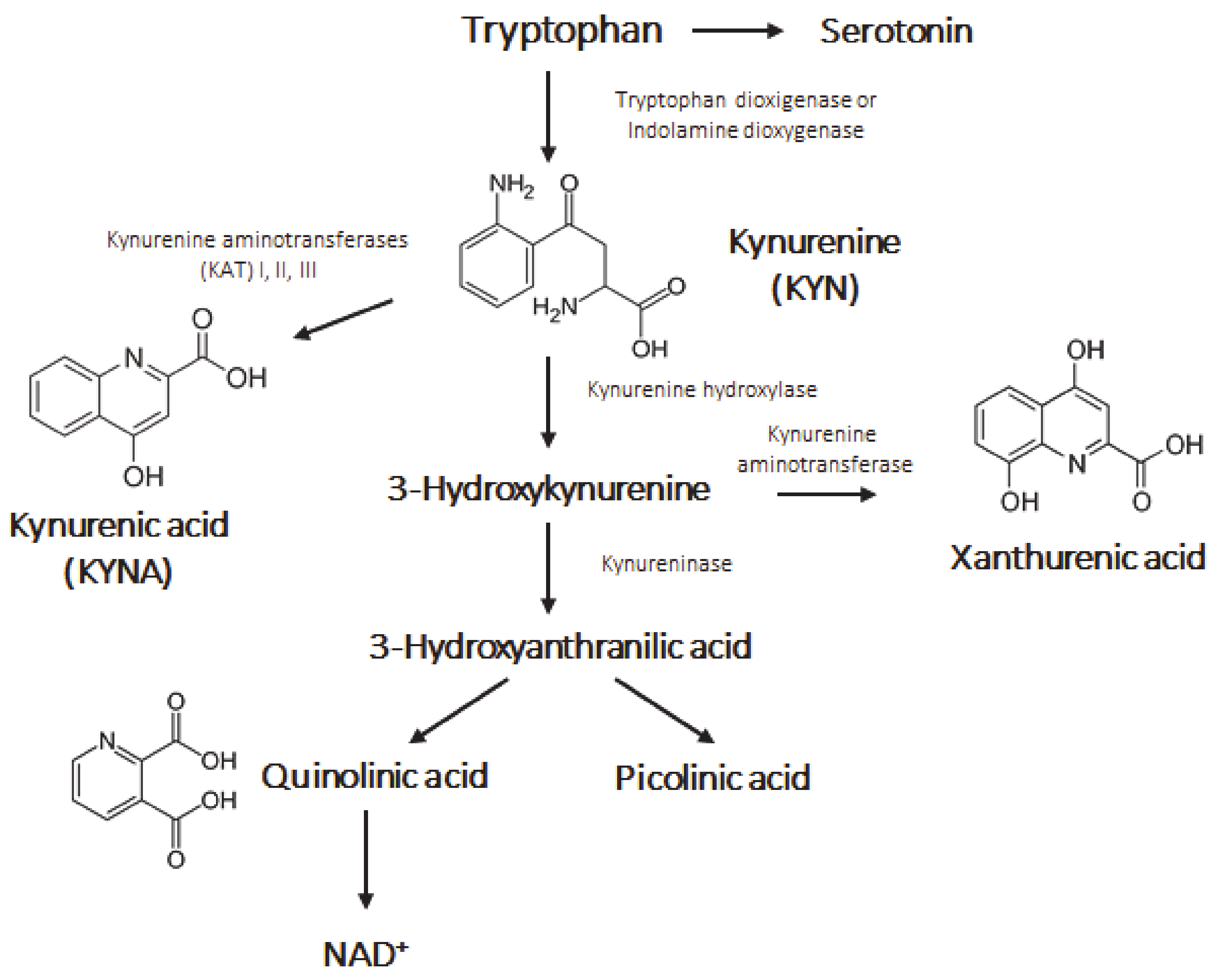
:1. Introduction
2. Materials and Methods
2.1. General Equipment Used during Syntheses
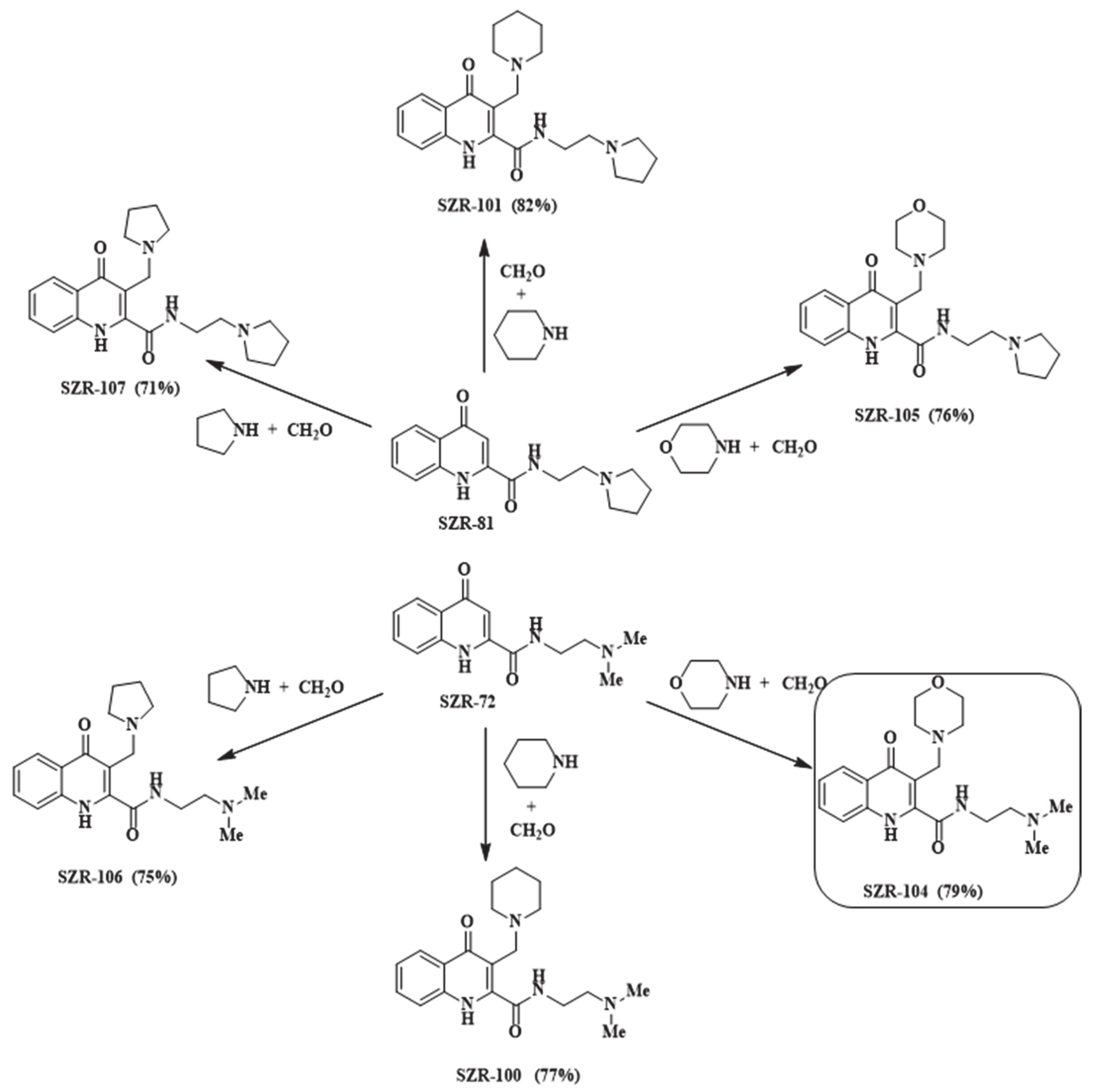
2.2. Synthesis of SZR-Compounds
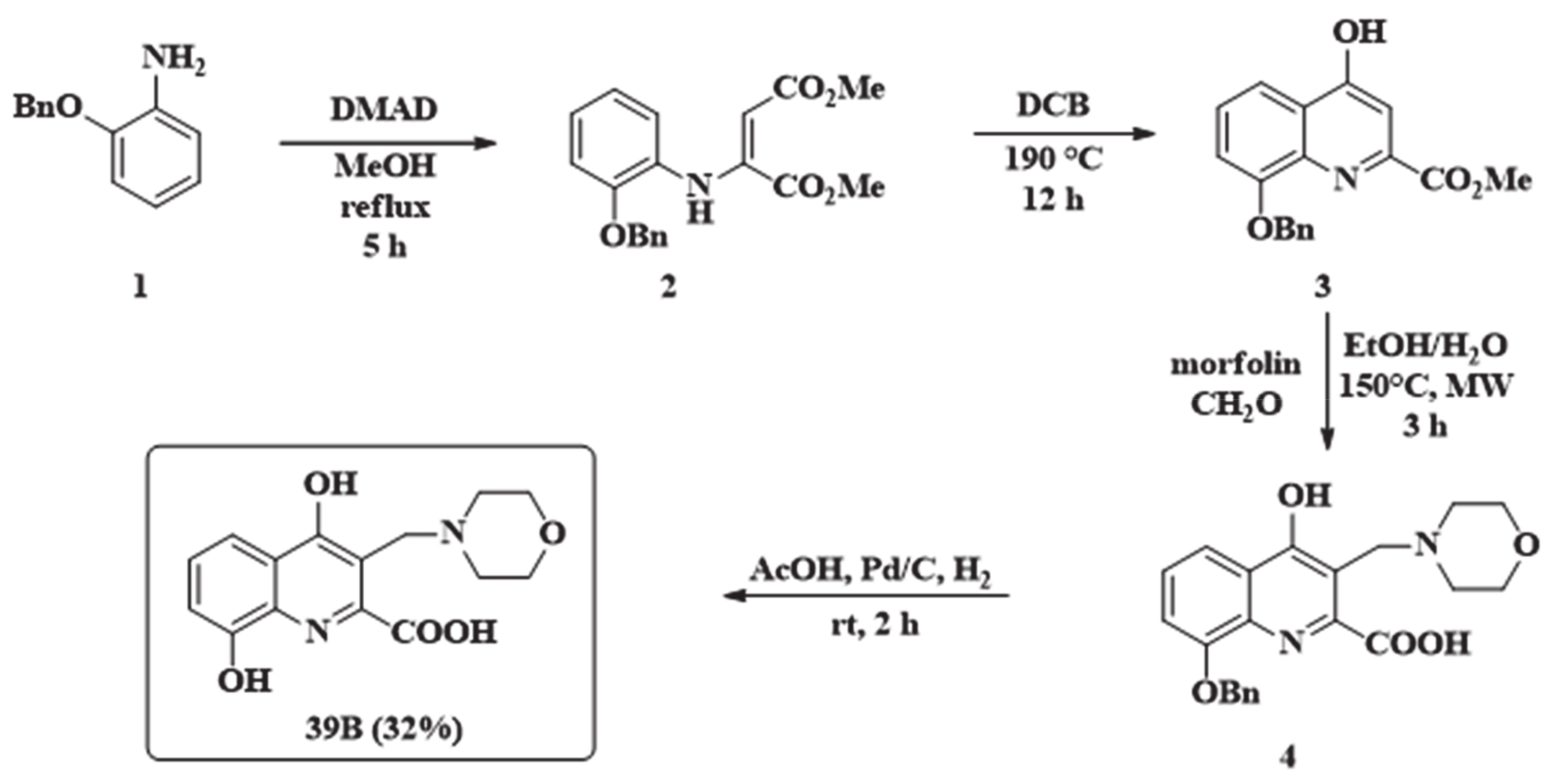
2.3. Synthesis of 8-Hydroxy-3-(Morpholinomethyl)-4-Oxo-1,4-Dihydroquinoline-2-Carboxylic Acid (39B)
2.4. Isolation of Rat Brain Cells
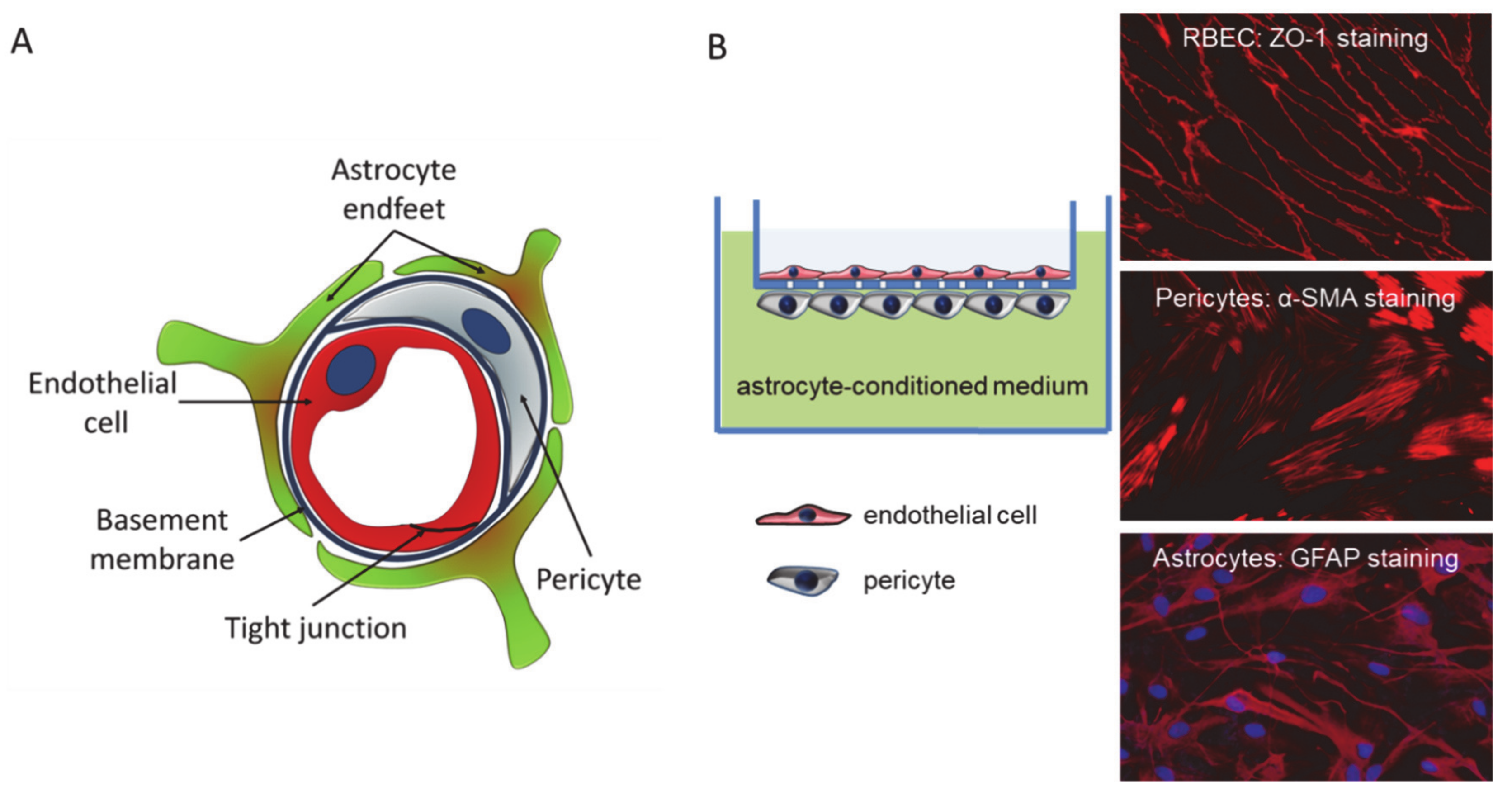
2.5. Construction of the In Vitro Blood–Brain Barrier (BBB) Model
2.6. Permeability Assay
2.7. Concentration Measurement of KYNA Analogues
2.8. Sample Processing for Determination of KYNA, SZR-104, Xanthurenic Acid and 39B Concentrations
2.9. Ultrahigh Performance Liquid Chromatography Coupled to Tandem Mass Spectrometry (UHPLC-MS/MS) Parameters for Determination of KYNA, SZR-104, Xanthurenic Acid and 39B Concentrations
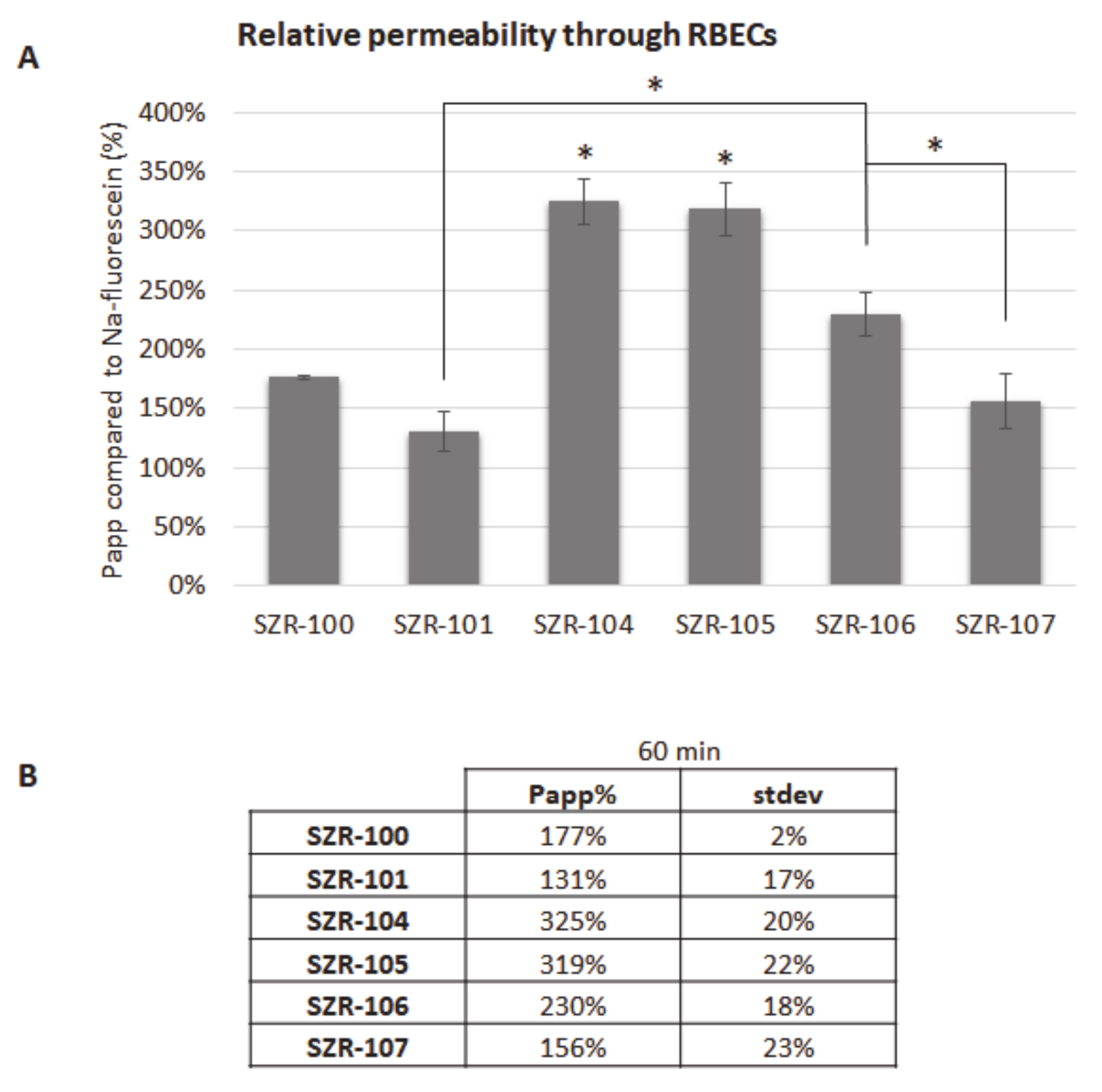
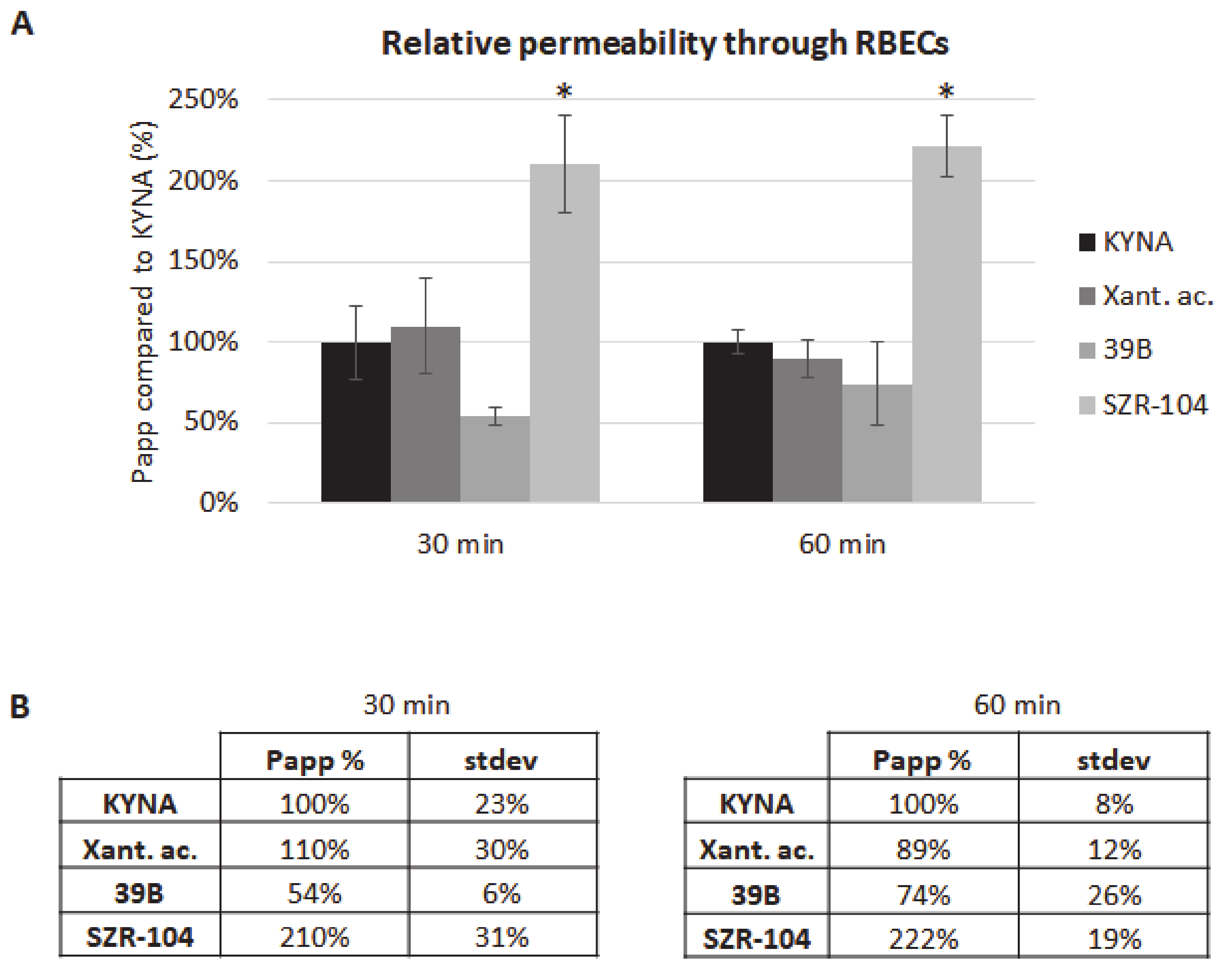
3. Results and Discussion
4. Conclusions
Supplementary Materials
Author Contributions
Funding
Institutional Review Board Statement
Informed Consent Statement
Data Availability Statement
Conflicts of Interest
References
- Rozsa, E.; Robotka, H.; Vecsei, L.; Toldi, J. The Janus-face kynurenic acid. J. Neural Transm. 2008, 115, 1087–1091. [Google Scholar] [CrossRef] [PubMed]
- Vecsei, L.; Szalardy, L.; Fulop, F.; Toldi, J. Kynurenines in the CNS: Recent advances and new questions. Drug Discov. 2013, 12, 64–82. [Google Scholar] [CrossRef] [PubMed]
- Hartai, Z.; Juhasz, A.; Rimanoczy, A.; Janaky, T.; Donko, T.; Dux, L.; Penke, B.; Toth, G.K.; Janka, Z.; Kalman, J. Decreased serum and red blood cell kynurenic acid levels in Alzheimer’s disease. Neurochem. Int. 2007, 50, 308–313. [Google Scholar] [CrossRef] [PubMed]
- Rejdak, K.; Bartosik-Psujek, H.; Dobosz, B.; Kocki, T.; Grieb, P.; Giovannoni, G.; Turski, W.A.; Stelmasiak, Z. Decreased level of kynurenic acid in cerebrospinal fluid of relapsing-onset multiple sclerosis patients. Neurosci. Lett. 2002, 331, 63–65. [Google Scholar] [CrossRef]
- Liu, X.C.; Erhardt, S.; Goiny, M.; Engberg, G.; Mathe, A.A. Decreased levels of kynurenic acid in prefrontal cortex in a genetic animal model of depression. Acta Neuropsychiatr. 2017, 29, 54–58. [Google Scholar] [CrossRef] [Green Version]
- Beal, M.F.; Matson, W.R.; Swartz, K.J.; Gamache, P.H.; Bird, E.D. Kynurenine pathway measurements in Huntington’s disease striatum: Evidence for reduced formation of kynurenic acid. J. Neurochem. 1990, 55, 1327–1339. [Google Scholar] [CrossRef]
- Beal, M.F.; Matson, W.R.; Storey, E.; Milbury, P.; Ryan, E.A.; Ogawa, T.; Bird, E.D. Kynurenic acid concentrations are reduced in Huntington’s disease cerebral cortex. J. Neurol. Sci. 1992, 108, 80–87. [Google Scholar] [CrossRef]
- Jauch, D.; Urbanska, E.M.; Guidetti, P.; Bird, E.D.; Vonsattel, J.P.; Whetsell, W.O., Jr.; Schwarcz, R. Dysfunction of brain kynurenic acid metabolism in Huntington’s disease: Focus on kynurenine aminotransferases. J. Neurol. Sci. 1995, 130, 39–47. [Google Scholar] [CrossRef]
- Krizbai, I.A.; Nyul-Toth, A.; Bauer, H.C.; Farkas, A.E.; Traweger, A.; Hasko, J.; Bauer, H.; Wilhelm, I. Pharmaceutical Targeting of the Brain. Curr. Pharm. Des. 2016, 22, 5442–5462. [Google Scholar] [CrossRef]
- Erhardt, S.; Olsson, S.K.; Engberg, G. Pharmacological manipulation of kynurenic acid: Potential in the treatment of psychiatric disorders. CNS Drugs 2009, 23, 91–101. [Google Scholar] [CrossRef]
- Fukushima, T.; Sone, Y.; Mitsuhashi, S.; Tomiya, M.; Toyo’oka, T. Alteration of kynurenic acid concentration in rat plasma following optically pure kynurenine administration: A comparative study between enantiomers. Chirality 2009, 21, 468–472. [Google Scholar] [CrossRef] [PubMed]
- Moroni, F.; Russi, P.; Lombardi, G.; Beni, M.; Carla, V. Presence of kynurenic acid in the mammalian brain. J. Neurochem. 1988, 51, 177–180. [Google Scholar] [CrossRef] [PubMed]
- Russi, P.; Alesiani, M.; Lombardi, G.; Davolio, P.; Pellicciari, R.; Moroni, F. Nicotinylalanine increases the formation of kynurenic acid in the brain and antagonizes convulsions. J. Neurochem. 1992, 59, 2076–2080. [Google Scholar] [CrossRef] [PubMed]
- Speciale, C.; Wu, H.Q.; Cini, M.; Marconi, M.; Varasi, M.; Schwarcz, R. (R,S)-3,4-dichlorobenzoylalanine (FCE 28833A) causes a large and persistent increase in brain kynurenic acid levels in rats. Eur. J. Pharmacol. 1996, 315, 263–267. [Google Scholar] [CrossRef]
- Varga, N.; Csapo, E.; Majlath, Z.; Ilisz, I.; Krizbai, I.A.; Wilhelm, I.; Knapp, L.; Toldi, J.; Vecsei, L.; Dekany, I. Targeting of the kynurenic acid across the blood–brain barrier by core-shell nanoparticles. Eur. J. Pharm. Sci. Off. J. Eur. Fed. Pharm. Sci. 2016, 86, 67–74. [Google Scholar] [CrossRef] [Green Version]
- Pardutz, A.; Fejes, A.; Bohar, Z.; Tar, L.; Toldi, J.; Vecsei, L. Kynurenines and headache. J. Neural Transm. 2012, 119, 285–296. [Google Scholar] [CrossRef]
- Kortesi, T.; Tuka, B.; Tajti, J.; Bagoly, T.; Fulop, F.; Helyes, Z.; Vecsei, L. Kynurenic Acid Inhibits the Electrical Stimulation Induced Elevated Pituitary Adenylate Cyclase-Activating Polypeptide Expression in the TNC. Front. Neurol. 2017, 8, 745. [Google Scholar] [CrossRef] [Green Version]
- Csati, A.; Edvinsson, L.; Vecsei, L.; Toldi, J.; Fulop, F.; Tajti, J.; Warfvinge, K. Kynurenic acid modulates experimentally induced inflammation in the trigeminal ganglion. J. Headache Pain 2015, 16, 99. [Google Scholar] [CrossRef] [Green Version]
- Lukacs, M.; Warfvinge, K.; Tajti, J.; Fulop, F.; Toldi, J.; Vecsei, L.; Edvinsson, L. Topical dura mater application of CFA induces enhanced expression of c-fos and glutamate in rat trigeminal nucleus caudalis: Attenuated by KYNA derivate (SZR72). J. Headache Pain 2017, 18, 39. [Google Scholar] [CrossRef] [Green Version]
- Greco, R.; Demartini, C.; Zanaboni, A.M.; Redavide, E.; Pampalone, S.; Toldi, J.; Fulop, F.; Blandini, F.; Nappi, G.; Sandrini, G.; et al. Effects of kynurenic acid analogue 1 (KYNA-A1) in nitroglycerin-induced hyperalgesia: Targets and anti-migraine mechanisms. Cephalalgia Int. J. Headache 2017, 37, 1272–1284. [Google Scholar] [CrossRef]
- Zadori, D.; Nyiri, G.; Szonyi, A.; Szatmari, I.; Fulop, F.; Toldi, J.; Freund, T.F.; Vecsei, L.; Klivenyi, P. Neuroprotective effects of a novel kynurenic acid analogue in a transgenic mouse model of Huntington’s disease. J. Neural Transm. 2011, 118, 865–875. [Google Scholar] [CrossRef] [PubMed]
- Lorinczi, B.; Csampai, A.; Fulop, F.; Szatmari, I. Synthesis of New C-3 Substituted Kynurenic Acid Derivatives. Molecules 2020, 25, 937. [Google Scholar] [CrossRef] [PubMed] [Green Version]
- Schmitt, M.K.E.; Macher, J.-P.; Bourguignon, J.-J. Compositions Derived from Quinoline and Quinoxaline, Preparation and Use Thereof. U.S. Patent WO2003010146A1, 6 February 2003. [Google Scholar]
- Datki, Z.; Galik-Olah, Z.; Bohar, Z.; Zadori, D.; Fulop, F.; Szatmari, I.; Galik, B.; Kalman, J.; Vecsei, L. Kynurenic Acid and Its Analogs Are Beneficial Physiologic Attenuators in Bdelloid Rotifers. Molecules 2019, 24, 2171. [Google Scholar] [CrossRef] [PubMed] [Green Version]
- Wilhelm, I.; Fazakas, C.; Krizbai, I.A. In vitro models of the blood–brain barrier. Acta Neurobiol. Exp. 2011, 71, 113–128. [Google Scholar]
- Wilhelm, I.; Krizbai, I.A. In vitro models of the blood–brain barrier for the study of drug delivery to the brain. Mol. Pharm. 2014, 11, 1949–1963. [Google Scholar] [CrossRef] [PubMed]
- Mandi, Y.; Endresz, V.; Mosolygo, T.; Burian, K.; Lantos, I.; Fulop, F.; Szatmari, I.; Lorinczi, B.; Balog, A.; Vecsei, L. The Opposite Effects of Kynurenic Acid and Different Kynurenic Acid Analogs on Tumor Necrosis Factor-alpha (TNF-alpha) Production and Tumor Necrosis Factor-Stimulated Gene-6 (TSG-6) Expression. Front. Immunol. 2019, 10, 1406. [Google Scholar] [CrossRef]
- Demeter, I.; Nagy, K.; Gellert, L.; Vecsei, L.; Fulop, F.; Toldi, J. A novel kynurenic acid analog (SZR104) inhibits pentylenetetrazole-induced epileptiform seizures. An electrophysiological study: Special issue related to kynurenine. J. Neural Transm. 2012, 119, 151–154. [Google Scholar] [CrossRef] [PubMed]


| Compound | Retention Time (min) | Retention Time Window (min) | Precursor Ion (m/z) | Type of Production | Product Ion (m/z) | Collision Energy (eV) | RF Lens (V) |
|---|---|---|---|---|---|---|---|
| SZR-104 | 1.2 | 2.0 | 359.2 | quantifier | 130.1 | 54 | 78 |
| SZR-104 | 1.2 | 2.0 | 359.2 | qualifier | 272.0 | 15 | 78 |
| Xant. ac. | 3.5 | 1.0 | 206.0 | quantifier | 131.9 | 30 | 113 |
| Xant. ac. | 3.5 | 1.0 | 206.0 | qualifier | 160.0 | 20 | 113 |
| KYNA | 4.0 | 1.0 | 190.1 | quantifier | 144.0 | 19 | 55 |
| KYNA | 4.0 | 1.0 | 190.1 | qualifier | 172.0 | 13 | 55 |
| 39B | 6.0 | 1.0 | 305.1 | quantifier | 174.0 | 22 | 80 |
| 39B | 6.0 | 1.0 | 305.1 | qualifier | 261.0 | 10 | 80 |
| SZR-73 | 6.5 | 1.0 | 274.2 | quantifier | 144.0 | 37 | 65 |
| SZR-73 | 6.5 | 1.0 | 274.2 | qualifier | 229.0 | 17 | 65 |
Publisher’s Note: MDPI stays neutral with regard to jurisdictional claims in published maps and institutional affiliations. |
© 2021 by the authors. Licensee MDPI, Basel, Switzerland. This article is an open access article distributed under the terms and conditions of the Creative Commons Attribution (CC BY) license (http://creativecommons.org/licenses/by/4.0/).
Share and Cite
Molnár, K.; Lőrinczi, B.; Fazakas, C.; Szatmári, I.; Fülöp, F.; Kmetykó, N.; Berkecz, R.; Ilisz, I.; Krizbai, I.A.; Wilhelm, I.; et al. SZR-104, a Novel Kynurenic Acid Analogue with High Permeability through the Blood–Brain Barrier. Pharmaceutics 2021, 13, 61. https://0-doi-org.brum.beds.ac.uk/10.3390/pharmaceutics13010061
Molnár K, Lőrinczi B, Fazakas C, Szatmári I, Fülöp F, Kmetykó N, Berkecz R, Ilisz I, Krizbai IA, Wilhelm I, et al. SZR-104, a Novel Kynurenic Acid Analogue with High Permeability through the Blood–Brain Barrier. Pharmaceutics. 2021; 13(1):61. https://0-doi-org.brum.beds.ac.uk/10.3390/pharmaceutics13010061
Chicago/Turabian StyleMolnár, Kinga, Bálint Lőrinczi, Csilla Fazakas, István Szatmári, Ferenc Fülöp, Noémi Kmetykó, Róbert Berkecz, István Ilisz, István A. Krizbai, Imola Wilhelm, and et al. 2021. "SZR-104, a Novel Kynurenic Acid Analogue with High Permeability through the Blood–Brain Barrier" Pharmaceutics 13, no. 1: 61. https://0-doi-org.brum.beds.ac.uk/10.3390/pharmaceutics13010061