
Targeted Endoradiotherapy with Lu2O3-iPSMA/-iFAP Nanoparticles Activated by Neutron Irradiation: Preclinical Evaluation and First Patient Image

 , , , , , , and
Abstract
:1. Introduction
2. Materials and Methods
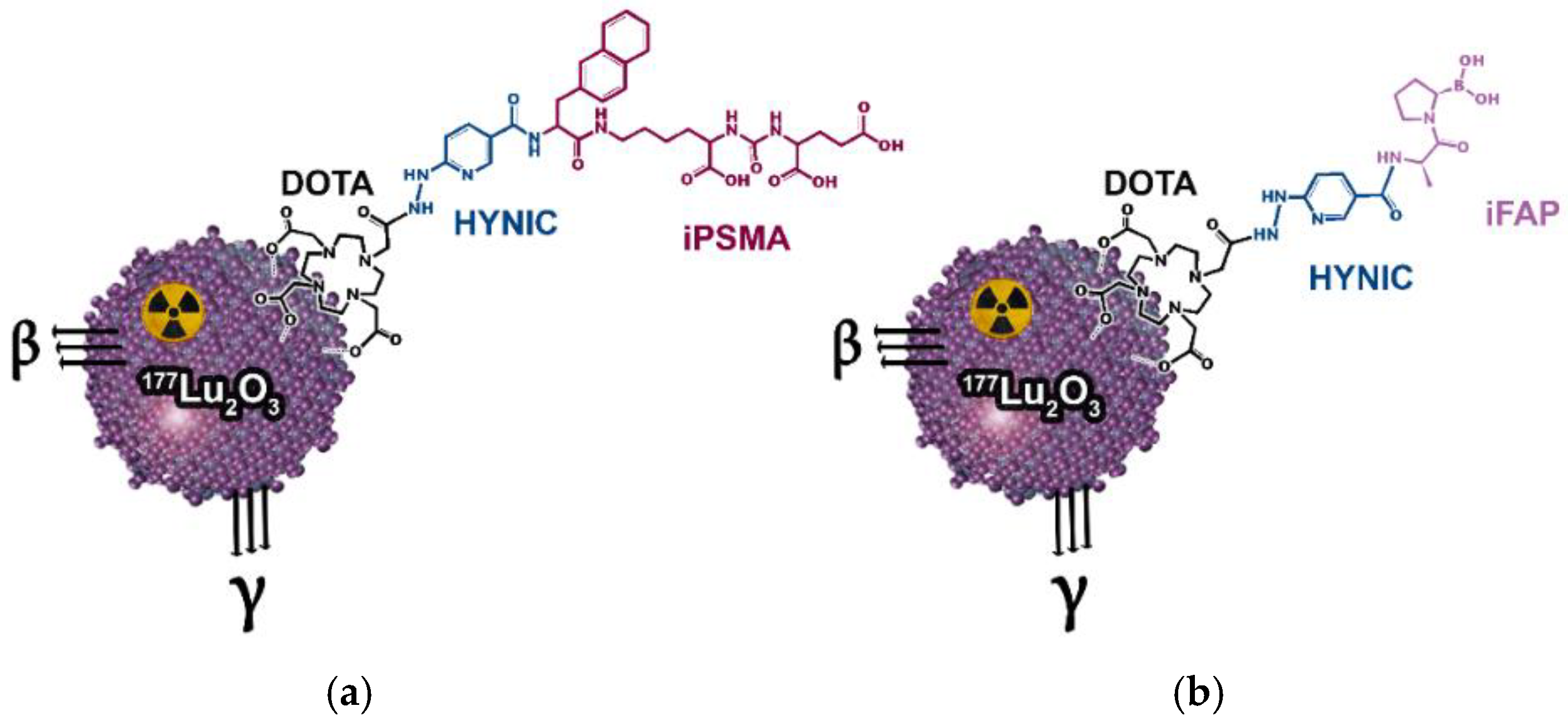
2.1. Synthesis of Lutetium Nanoparticles
2.2. Preparation of 177Lu2O3-iPSMA and 177Lu2O3-iFAP Nanoparticles
2.3. Cell Culture
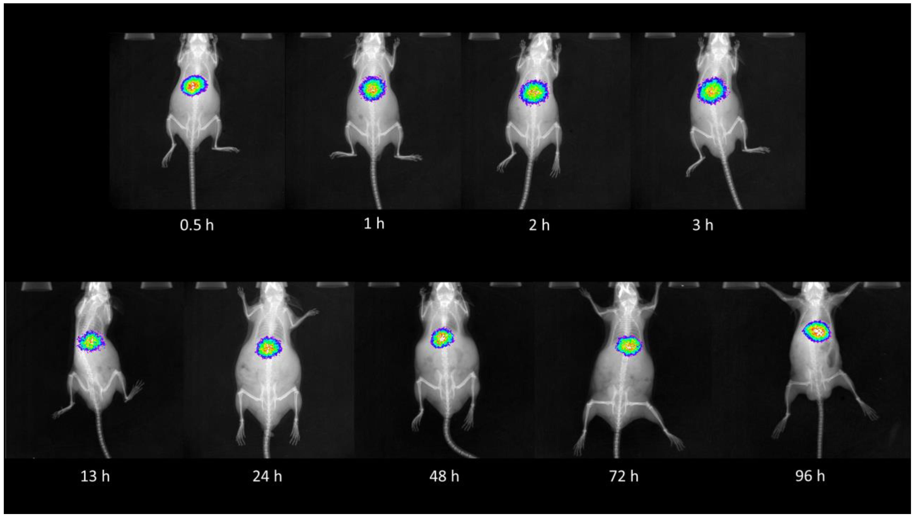
2.4. Bidistribution of 177Lu2O3-iPSMA and 177Lu2O3-iFAP Nanoparticles
2.5. Bidistribution of Nanoparticles in Mice Bearing HCT116 Tumors
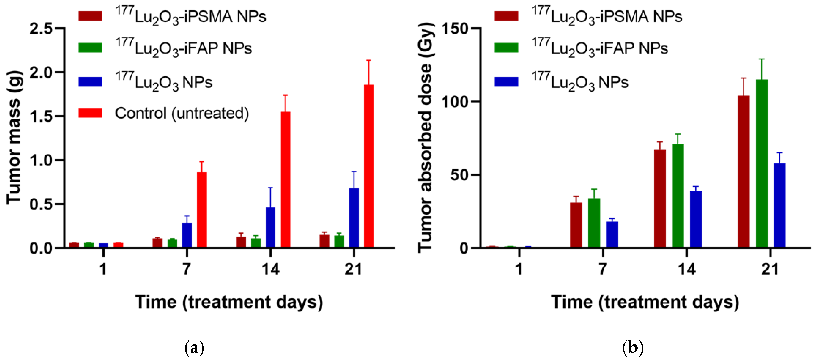
2.6. Therapeutic Protocol
2.7. Metabolic Activity Evaluation
2.8. Creatinine and Liver Enzyme Quantitation
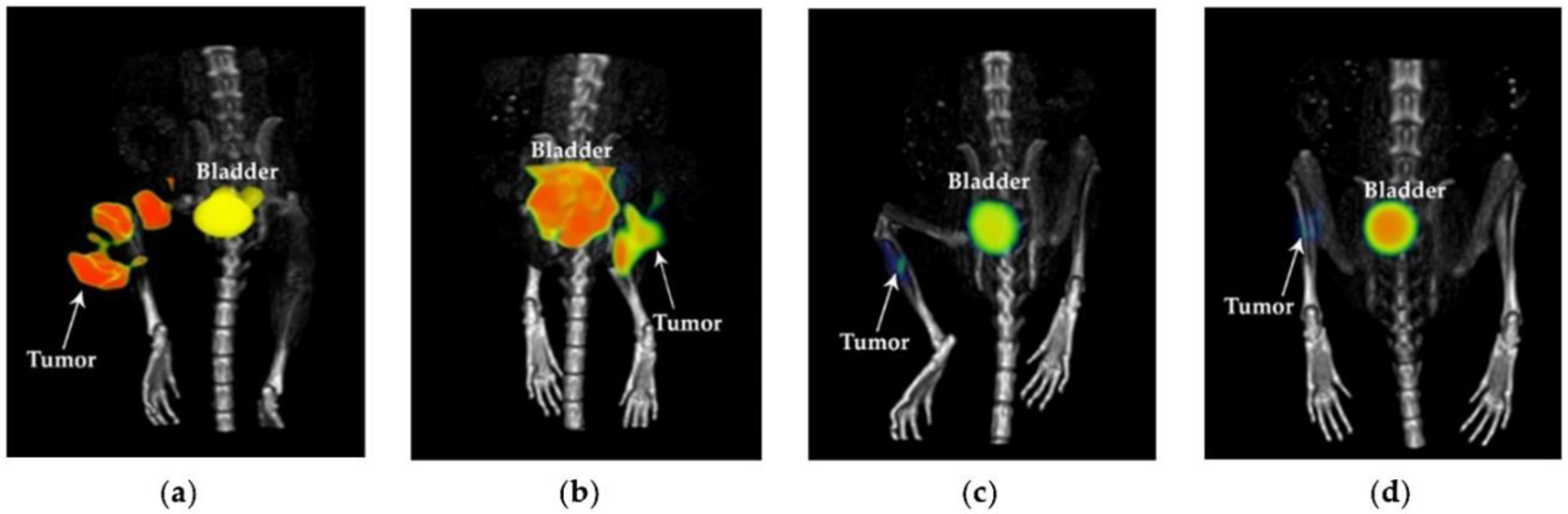
2.9. Clinical Study
2.10. Statistics
3. Results
4. Discussion
5. Conclusions
Author Contributions
Funding
Institutional Review Board Statement
Informed Consent Statement
Data Availability Statement
Acknowledgments
Conflicts of Interest
Appendix A


References
- Cędrowska, E.; Pruszyński, M.; Gawęda, W.; Żuk, M.; Krysiński, P.; Bruchertseifer, F.; Morgenstern, A.; Karageorgou, M.-A.; Bouziotis, P.; Bilewicz, A. Trastuzumab Conjugated Superparamagnetic Iron Oxide Nanoparticles Labeled with 225Ac as a Perspective Tool for Combined α-Radioimmunotherapy and Magnetic Hyperthermia of HER2-Positive Breast Cancer. Molecules 2020, 25, 1025. [Google Scholar] [CrossRef] [Green Version]
- Ferro-Flores, G. Targeted nanomedicines: In the right route towards improved therapies. Curr. Cancer Ther. Rev. 2020, 16, 3–4. [Google Scholar] [CrossRef]
- Ramírez-Nava, G.; Santos-Cuevas, C.; Ferro-Flores, G.; Ocampo-García, B.; Chairez, I.; Gómez-Argumosa, E.; Abundiz-López, L.; García-Pérez, F.O. Hybrid (2D/3D) Dosimetry of Radiolabeled Gold Nanoparticles for Sentinel Lymph Node Detection in Patients with Breast Cancer. Contrast Media Mol. Imaging 2020, 2020, 2728134. [Google Scholar] [CrossRef]
- Silva, F.; Cabral Campello, M.P.; Paulo, A. Radiolabeled Gold Nanoparticles for Imaging and Therapy of Cancer. Materials 2021, 14, 4. [Google Scholar] [CrossRef]
- Sadaghiania, M.S.; Sheikhbahaeia, S.; Wernerab, R.A.; Pientac, K.J.; Pomperac, M.G.; Solnesa, L.B.; Gorinac, M.A.; Nae-Yuh Wang, N.-Y.; Roweac, S.P. A Systematic Review and Meta-analysis of the Effectiveness and Toxicities of Lutetium-177–labeled Prostate-specific Membrane Antigen–targeted Radioligand Therapy in Metastatic Castration-Resistant Prostate Cancer. Eur. Urol. 2021, 80, 82–94. [Google Scholar] [CrossRef]
- Strosberg, J.; Leeuwenkamp, O.; Siddiqui, M.K. Peptide receptor radiotherapy re-treatment in patients with progressive neuroendocrine tumors: A systematic review and meta-analysis. Cancer Treat Rev. 2021, 93, 102141. [Google Scholar] [CrossRef]
- Lim, K.; Kim, H.K.; Le, X.T.; Nguyen, N.T.; Lee, E.S.; Oh, K.T.; Choi, H.G.; Youn, Y.S. Highly Red Light-Emitting Erbium- and Lutetium-Doped Core-Shell Upconverting Nanoparticles Surface-Modified with PEG-Folic Acid/TCPP for Suppressing Cervical Cancer HeLa Cells. Pharmaceutics 2020, 12, 1102. [Google Scholar] [CrossRef]
- Müller, M.; Espinoza, S.; Jüstel, T.; Held, K.D.; Anderson, R.R.; Purschke, M. UVC-Emitting LuPO(4):Pr(3+) Nanoparticles Decrease Radiation Resistance of Hypoxic Cancer Cells. Radiat. Res. 2020, 193, 82–87. [Google Scholar] [CrossRef]
- González-Mancebo, D.; Becerro, A.I.; Corral, A.; Balcerzyk, M.; Ocaña, M. Luminescence and X-ray Absorption Properties of Uniform Eu3+:(H3O)Lu3F10 Nanoprobes. Nanomaterials 2019, 9, 1153. [Google Scholar] [CrossRef] [Green Version]
- Zhang, H.; Ye, K.; Huang, X.; Lin, X.; Ma, L.; Chen, T. Designing lanthanide coordination nanoframeworks as X-ray responsive radiosensitizers for efficient cancer therapy. Inorg. Chem. Front. 2021, 8, 3433–3439. [Google Scholar] [CrossRef]
- Hamson, E.J.; Keane, F.M.; Tholen, S.; Schilling, O.; Gorrell, M.D. Understanding fibroblast activation protein (FAP): Substrates, activities, expression and targeting for cancer therapy. Proteom. Clin. Appl. 2014, 8, 454–463. [Google Scholar] [CrossRef]
- Rajasekaran, A.K.; Anilkumar, G.; Christiansen, J.J. Is prostate-specific membrane antigen a multifunctional protein? Am. J. Physiol. Cell Physiol. 2005, 288, C975–C981. [Google Scholar] [CrossRef] [PubMed] [Green Version]
- Haffner, M.C.; Kronberger, I.E.; Ross, J.S.; Sheehan, C.E.; Zitt, M.; Mühlmann, G.; Ofner, D.; Zelger, B.; Ensinger, C.; Yang, X.J.; et al. Prostate-specific membrane antigen expression in the neovasculature of gastric and colorectal cancers. Hum. Pathol. 2009, 40, 754–761. [Google Scholar] [CrossRef] [PubMed]
- Cuda, T.J.; Riddell, A.D.; Liu, C.; Whitehall, V.L.; Borowsky, J.; Wyld, D.K.; Burge, M.E.; Ahern, E.; Griffin, A.; Lyons, N.J.R.; et al. PET Imaging Quantifying 68Ga-PSMA-11 Uptake in Metastatic Colorectal Cancer. J. Nucl. Med. 2020, 61, 1576–1579. [Google Scholar] [CrossRef] [PubMed]
- Arslan, E.; Ergül, N.; Karagöz, Y.; Gedik, A.A.; Çermik, T.F. Recurrent Brain Metastasis of Triple Negative Breast Cancer with High Uptake in 68Ga-PSMA-11 PET/CT. Clin. Nucl. Med. 2021, 46, e106–e108. [Google Scholar] [CrossRef]
- Tolkach, Y.; Goltz, D.; Kremer, A.; Ahmadzadehfar, H.; Bergheim, D.; Essler, M.; Lam, M.; de Keizer, B.; Fischer, H.P.; Kristiansen, G. Prostate-specific membrane antigen expression in hepatocellular carcinoma: Potential use for prognosis and diagnostic imaging. Oncotarget 2019, 10, 4149–4160. [Google Scholar] [CrossRef] [Green Version]
- Ancira-Cortez, A.; Ferro-Flores, G.; Jiménez-Mancilla, N.; Morales-Avila, E.; Trujillo-Benítez, D.; Ocampo-García, B.; Santos-Cuevas, C.; Escudero-Castellanos, A.; Luna-Gutiérrez, M. Synthesis, chemical and biochemical characterization of Lu2O3-iPSMA nanoparticles activated by neutron irradiation. Mater. Sci. Eng. C Mater. Biol. Appl. 2020, 117, 111335. [Google Scholar] [CrossRef]
- Ancira-Cortez, A.; Trujillo-Benítez, D.; Jiménez-Mancilla, N.; Santos-Cuevas, C.; Morales-Avila, E.; Ferro-Flores, G. Synthesis and physicochemical characterization of Lu and Sm sesquioxide nanoparticles by precipitation-calcination and pulsed laser ablation in liquids. Mat. Chem. Phys. 2021, 275, 125229. [Google Scholar] [CrossRef]
- Trujillo-Benítez, D.; Luna-Gutiérrez, M.; Ferro-Flores, G.; Ocampo-García, B.; Santos-Cuevas, C.; Bravo-Villegas, G.; Morales-Ávila, E.; Cruz-Nova, P.; Díaz-Nieto, L.; García-Quiroz, J.; et al. Design, Synthesis and Preclinical Assessment of 99mTc-iFAP for In Vivo Fibroblast Activation Protein (FAP) Imaging. Molecules 2022, 27, 264. [Google Scholar] [CrossRef]
- Ferro-Flores, G.; Luna-Gutiérrez, M.; Ocampo-García, B.; Santos-Cuevas, C.; Azorín-Vega, E.; Jiménez-Mancilla, N.; Orocio-Rodríguez, E.; Davanzo, J.; García-Pérez, F.O. Clinical translation of a PSMA inhibitor for 99mTc-based SPECT. Nucl. Med. Biol. 2017, 48, 36–44. [Google Scholar] [CrossRef]
- Stabin, M.G.; Sparks, R.B.; Crowe, E. OLINDA/EXM: The second-generation personal computer software for internal dose assessment in nuclear medicine. J. Nucl. Med. 2005, 46, 1023–1027. [Google Scholar] [CrossRef] [PubMed]
- Marks, L.B.; Yorke, E.D.; Jackson, A.; Ten Haken, R.K.; Constine, L.S.; Eisbruch, A.; Bentzen, S.M.; Nam, J.; Deasy, J.O. Use of normal tissue complication probability models in the clinic. Int. J. Radiat. Oncol. Biol. Phys. 2010, 76, S10–S19. [Google Scholar] [CrossRef] [PubMed] [Green Version]
- Imarisio, J.J. Liver Scan Showing Intense Lung Uptake in Neoplasia and Infection. J. Nucl. Med. 1975, 16, 188–190. [Google Scholar] [PubMed]
- Chang, S.W.; Ohara, N. Chronic biliary obstruction induces pulmonary intravascular phagocytosis and endotoxin sensitivity in rats. J. Clin. Investig. 1994, 94, 2009–2019. [Google Scholar] [CrossRef]
- Arneth, B. Tumor Microenvironment. Medicina 2020, 56, 15. [Google Scholar] [CrossRef] [Green Version]
- Vilchis-Juárez, A.; Ferro-Flores, G.; Santos-Cuevas, C. Molecular targeting radiotherapy with cyclo-RGDFK(C) peptides conjugated to 177Lu-labeled gold nanoparticles in tumor-bearing mice. J. Biomed. Nanotechnol. 2014, 10, 393–404. [Google Scholar] [CrossRef]
- Viana, R.D.S.; Costa, L.A.D.M.; Harmon, A.C.; Gomes Filho, M.A.; Falcão, E.H.L.; Vicente, M.G.H.; Junior, S.A.; Mathis, J.M. 177Lu-Labeled Eu-Doped Mesoporous SiO2 Nanoparticles as a Theranostic Radiopharmaceutical for Colorectal Cancer. ACS Appl. Nano Mater. 2020, 3, 8691–8701. [Google Scholar] [CrossRef]
- Mendoza-Nava, H.; Ferro-Flores, G.; Ramírez, F.d.M.; Ocampo-García, B.; Santos-Cuevas, C.; Aranda-Lara, L.; Azorín-Vega, E.; Morales-Avila, E.; Isaac-Olivé, K. 177Lu-Dendrimer Conjugated to Folate and Bombesin with Gold Nanoparticles in the Dendritic Cavity: A Potential Theranostic Radiopharmaceutical. J. Nanomat. 2016, 2016, 1039258. [Google Scholar] [CrossRef] [Green Version]
- Trujillo-Nolasco, M.; Cruz-Nova, P.; Ferro-Flores, G.; Gibbens-Bandala, B.; Morales-Avila, E.; Aranda-Lara, L.; Vargas, M.; Ocampo-García, B. Development of 177Lu-DN(C19)-CXCR4 Ligand Nanosystem for Combinatorial Therapy in Pancreatic Cancer. J. Biomed. Nanotechnol. 2021, 28, 263–278. [Google Scholar] [CrossRef]
- Gibbens-Bandala, B.; Morales-Avila, E.; Ferro-Flores, G.; Santos-Cuevas, C.; Meléndez-Alafort, L.; Trujillo-Nolasco, M.; Ocampo-García, B. 177Lu-Bombesin-PLGA (paclitaxel): A targeted controlled-release nanomedicine for bimodal therapy of breast cancer. Mater. Sci. Eng. C Mater. Biol. Appl. 2019, 105, 110043. [Google Scholar] [CrossRef]
- Sartor, A.O.; Morris, M.J.; Messman, R.; Krause, B.J. Vision: An international, prospective, open-label, multicenter, randomized phase III study of 177Lu-PSMA-617 in the treatment of patients with progressive PSMA-positive metastatic castration-resistant prostate cancer (mCRPC). J. Clin. Oncol. 2020, 39, TPS259. [Google Scholar] [CrossRef]
- Santos-Cuevas, C.; Ferro-Flores, G.; García-Pérez, F.O.; Jiménez-Mancilla, N.; Ramírez-Nava, G.; Ocampo-García, B.; Luna-Gutiérrez, M.; Azorín-Vega, E.; Davanzo, J.; Soldevilla-Gallardo, I. 177Lu-DOTA-HYNIC-Lys(Nal)-Urea-Glu: Biokinetics, Dosimetry, and Evaluation in Patients with Advanced Prostate Cancer. Contrast Media Mol. Imaging 2018, 2018, 5247153. [Google Scholar] [CrossRef] [PubMed] [Green Version]
- Hendlisz, A.; Van den Eynde, M.; Peeters, M.; Maleux, G.; Lambert, B.; Vannoote, J.; De Keukeleire, K.; Verslype, C.; Defreyne, L.; Van Cutsem, E.; et al. Phase III trial comparing protracted intravenous fluorouracil infusion alone or with yttrium-90 resin microspheres radioembolization for liver-limited metastatic colorectal cancer refractory to standard chemotherapy. J. Clin. Oncol. 2010, 28, 3687–3694. [Google Scholar] [CrossRef] [PubMed] [Green Version]
- Gosavi, A.; Puranik, A.D.; Shah, S.; Agrawal, A.; Purandare, N.C.; Shetty, N.; Gala, K.; Kulkarni, S.; Patkar, S.; Goel, M.; et al. Prognostic value of lung shunt fraction in hepatocellular carcinoma and unresectable liver dominant metastatic colorectal cancer undergoing transarterial radioembolisation. Nucl. Med. Commun. 2022, 43, 24–31. [Google Scholar] [CrossRef] [PubMed]


| Organ | Time (h) | |||
|---|---|---|---|---|
| 3 | 48 | 72 | 96 | |
| Heart | 0.11 ± 0.03 | 0.08 ± 0.02 | 0.02 ± 0.01 | 0.01 ± 0.01 |
| Liver | 23.56 ± 2.13 | 18.08 ± 0.94 | 16.87 ± 1.37 | 14.59 ± 1.72 |
| Lung | 0.34 ± 0.32 | 0.21 ± 0.15 | 0.16 ± 0.09 | 0.12 ± 0.05 |
| Pancreas | 0.12 ± 0.04 | 0.04 ± 0.03 | 0.01 ± 0.01 | 0.00 ± 0.00 |
| Spleen | 1.61 ± 0.59 | 1.18 ± 0.91 | 0.99 ± 0.63 | 0.81 ± 0.35 |
| Kidney | 0.85 ± 0.11 | 0.41 ± 0.18 | 0.22 ± 0.07 | 0.19 ± 0.10 |
| Brain | 0.01 ± 0.01 | 0.00 ± 0.00 | 0.00 ± 0.00 | 0.00 ± 0.00 |
| Organ | Time (h) | |||
|---|---|---|---|---|
| 3 | 48 | 72 | 96 | |
| Heart | 0.13 ± 0.03 | 0.05 ± 0.03 | 0.01 ± 0.01 | 0.01 ± 0.01 |
| Liver | 24.98 ± 1.84 | 18.93 ± 2.04 | 17.23 ± 1.41 | 15.01 ± 1.17 |
| Lung | 0.35 ± 0.09 | 0.20 ± 0.11 | 0.14 ± 0.04 | 0.08 ± 0.03 |
| Pancreas | 0.10 ± 0.07 | 0.02 ± 0.01 | 0.00 ± 0.00 | 0.00 ± 0.00 |
| Spleen | 1.52 ± 0.39 | 1.14 ± 0.49 | 0.79 ± 0.39 | 0.75 ± 0.28 |
| Kidney | 0.98 ± 0.10 | 0.52 ± 0.07 | 0.21 ± 0.07 | 0.18 ± 0.08 |
| Brain | 0.00 ± 0.00 | 0.00 ± 0.00 | 0.00 ± 0.00 | 0.00 ± 0.00 |
| Tissue | Time (h) | |||
|---|---|---|---|---|
| 3 | 48 | 72 | 96 | |
| Heart | 0.28 ± 0.11 | 0.19 ± 0.07 | 0.10 ± 0.08 | 0.07 ± 0.02 |
| Lung | 0.30 ± 0.10 | 0.21 ± 0.03 | 0.15 ± 0.04 | 0.11 ± 0.07 |
| Liver | 1.05 ± 0.38 | 0.94 ± 0.21 | 0.88 ± 0.12 | 0.84 ± 0.09 |
| Spleen | 0.39 ± 0.16 | 0.34 ± 0.10 | 0.31 ± 0.09 | 0.28 ± 0.07 |
| Kidney | 0.84 ± 0.19 | 0.63 ± 0.23 | 0.58 ± 0.17 | 0.39 ± 0.21 |
| Tumor (%ID/g) | 80.42 ± 5.87 | 79.32 ± 5.27 | 77.23 ± 6.01 | 75.98 ± 5.41 |
| Tissue | Time (h) | |||
|---|---|---|---|---|
| 3 | 48 | 72 | 96 | |
| Heart | 0.22 ± 0.09 | 0.18 ± 0.09 | 0.14 ± 0.07 | 0.05 ± 0.02 |
| Lung | 0.26 ± 0.11 | 0.19 ± 08 | 0.16 ± 0.05 | 0.10 ± 0.04 |
| Liver | 1.18 ± 0.70 | 1.10 ± 0.42 | 0.83 ± 0.22 | 0.81 ± 0.13 |
| Spleen | 0.40 ± 0.14 | 0.35 ± 0.12 | 0.32 ± 0.11 | 0.26 ± 0.09 |
| Kidney | 0.91 ± 0.17 | 0.78 ± 0.15 | 0.62 ± 0.13 | 0.50 ± 0.18 |
| Tumor (%ID/g) | 83.87 ± 5.33 | 81.21 ± 5.64 | 78.58 ± 3.81 | 77.32 ± 4.25 |
| Tissue | Time (h) | |||
|---|---|---|---|---|
| 3 | 48 | 72 | 96 | |
| Heart | 0.27 ± 0.06 | 0.21 ± 0.08 | 0.16 ± 0.07 | 0.11 ± 0.04 |
| Lung | 0.29 ± 0.13 | 0.25 ± 0.11 | 0.20 ± 0.14 | 0.18 ± 0.05 |
| Liver | 1.69 ± 0.97 | 1.40 ± 0.52 | 1.36 ± 0.45 | 1.29 ± 0.12 |
| Spleen | 0.55 ± 0.28 | 0.49 ± 0.25 | 0.40 ± 0.21 | 0.38 ± 0.14 |
| Kidney | 0.98 ± 0.31 | 0.95 ± 0.29 | 0.88 ± 0.43 | 0.83 ± 0.34 |
| Tumor (%ID/g) | 64.12 ± 4.98 | 57.25 ± 4.56 | 53.58 ± 3.25 | 49.52 ± 3.31 |
| Treatment Group | SUV | Creatinine (mg/dL) | Aspartate Aminotransferase (AST) (IU/L) | Alanine Aminotransferase (ALT) (IU/L) | Lactate Dehydrogenase (LDH) (IU/L) |
|---|---|---|---|---|---|
| 177Lu2O3-iPSMA | 0.421 ± 0.092 | 0.208 ± 0.032 | 152 ± 13 | 71 ± 6 | 292 ± 37 |
| 177Lu2O3-iFAP | 0.375 ± 0.104 | 0.197 ± 0.042 | 137 ± 16 | 65 ± 7 | 285 ± 44 |
| 177Lu2O3 | 1.821 ± 0.891 | 0.186 ± 0.051 | 142 ± 13 | 69 ± 5 | 294 ± 39 |
| Control | 2.654 ± 0.742 | 0.193 ± 0.061 | 148 ± 15 | 74 ± 8 | 302 ± 42 |
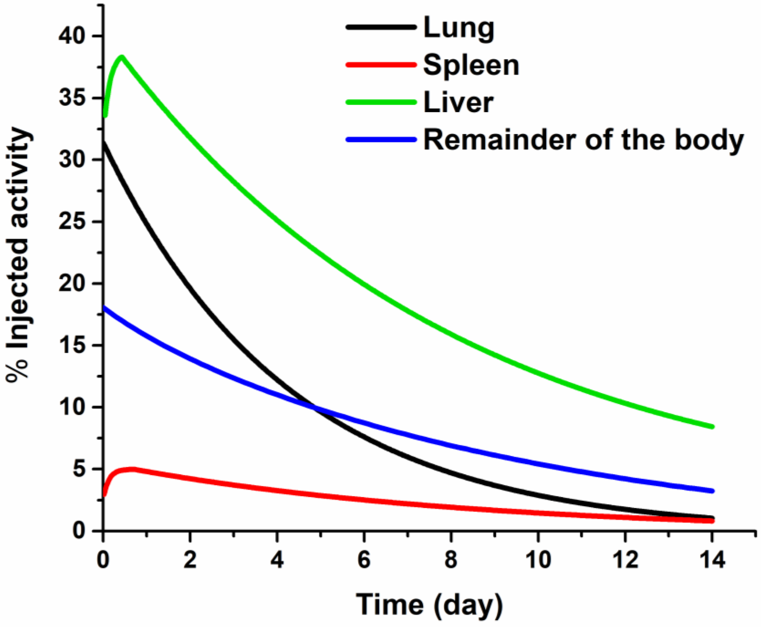
| Organ | Biokinetic Model | (MBq h/MBq) |
|---|---|---|
| Liver | 86.59 | |
| Lung | 43.33 | |
| Spleen | 9.56 | |
| Remainder of the body | 32.60 |
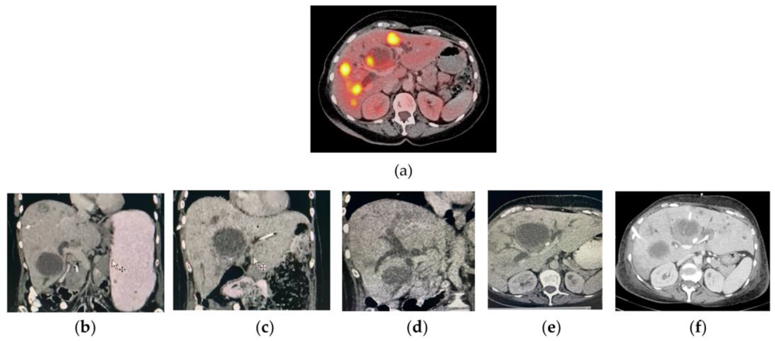
| Liver Metastasis (Lesion Number) | Volume (cm3) | (MBq h/MBq) | Dose (Gy) (per 1350 MBq) |
|---|---|---|---|
| L1 | 8.44 | 6.73 | 84.52 |
| L2 | 6.12 | 3.96 | 68.32 |
| L3 | 7.70 | 3.09 | 42.48 |
| L4 | 9.26 | 4.23 | 48.38 |
| L5 | 10.27 | 5.91 | 60.95 |
| L6 | 3.01 | 2.76 | 95.87 |
| L7 | 9.70 | 5.69 | 62.02 |
| L8 | 3.47 | 2.59 | 78.10 |
| L9 | 17.53 | 9.46 | 57.21 |
| L10 | 2.58 | 2.70 | 109.10 |
| L11 | 1.42 | 2.28 | 201.61 |
| L12 | 7.76 | 5.22 | 70.66 |
| L13 | 12.47 | 6.03 | 50.94 |
| L14 | 2.15 | 1.71 | 82.29 |
| Target Organ | Absorbed Doses |
|---|---|
| (Gy) | |
| Adrenals | 1.18 × 10− × 10−01 |
| Brain | 4.29 × 10−02 |
| Breasts | 7.05 × 10−02 |
| Gallbladder wall | 1.49 × 10−01 |
| Lower large intestine wall | 4.81 × 10−02 |
| Small intestine | 6.37 × 10−02 |
| Stomach wall | 9.79 × 10−02 |
| Upper laege intestine wall | 8.29 × 10−02 |
| Heart | 1.29 × 10−01 |
| Kidneys | 1.08 × 10−01 |
| Liver | 3.21 × 10+00 |
| Lungs | 3.42 × 10+00 |
| Muscle | 7.22 × 10−02 |
| Ovaries | 5.97 × 10−02 |
| Pancreas | 1.43 × 10−01 |
| Red marrow | 5.36 × 10−02 |
| Osteogenic cells | 1.94 × 10−01 |
| Skin | 5.81 × 10−02 |
| Spleen | 3.78 × 10+00 |
| Thymus | 7.52 × 10−02 |
| Thyroid | 5.86 × 10−02 |
| Urinary bladder wall | 5.61 × 10−02 |
| Uterus | 5.90 × 10−02 |
| Total body | 2.82 × 10−01 |
Publisher’s Note: MDPI stays neutral with regard to jurisdictional claims in published maps and institutional affiliations. |
© 2022 by the authors. Licensee MDPI, Basel, Switzerland. This article is an open access article distributed under the terms and conditions of the Creative Commons Attribution (CC BY) license (https://creativecommons.org/licenses/by/4.0/).
Share and Cite
Luna-Gutiérrez, M.; Ocampo-García, B.; Jiménez-Mancilla, N.; Ancira-Cortez, A.; Trujillo-Benítez, D.; Hernández-Jiménez, T.; Ramírez-Nava, G.; Hernández-Ramírez, R.; Santos-Cuevas, C.; Ferro-Flores, G. Targeted Endoradiotherapy with Lu2O3-iPSMA/-iFAP Nanoparticles Activated by Neutron Irradiation: Preclinical Evaluation and First Patient Image. Pharmaceutics 2022, 14, 720. https://0-doi-org.brum.beds.ac.uk/10.3390/pharmaceutics14040720
Luna-Gutiérrez M, Ocampo-García B, Jiménez-Mancilla N, Ancira-Cortez A, Trujillo-Benítez D, Hernández-Jiménez T, Ramírez-Nava G, Hernández-Ramírez R, Santos-Cuevas C, Ferro-Flores G. Targeted Endoradiotherapy with Lu2O3-iPSMA/-iFAP Nanoparticles Activated by Neutron Irradiation: Preclinical Evaluation and First Patient Image. Pharmaceutics. 2022; 14(4):720. https://0-doi-org.brum.beds.ac.uk/10.3390/pharmaceutics14040720
Chicago/Turabian StyleLuna-Gutiérrez, Myrna, Blanca Ocampo-García, Nallely Jiménez-Mancilla, Alejandra Ancira-Cortez, Diana Trujillo-Benítez, Tania Hernández-Jiménez, Gerardo Ramírez-Nava, Rodrigo Hernández-Ramírez, Clara Santos-Cuevas, and Guillermina Ferro-Flores. 2022. "Targeted Endoradiotherapy with Lu2O3-iPSMA/-iFAP Nanoparticles Activated by Neutron Irradiation: Preclinical Evaluation and First Patient Image" Pharmaceutics 14, no. 4: 720. https://0-doi-org.brum.beds.ac.uk/10.3390/pharmaceutics14040720