
The Formulation of Curcumin: 2-Hydroxypropyl-β-cyclodextrin Complex with Smart Hydrogel for Prolonged Release of Curcumin
 , , , , , , , and
Abstract
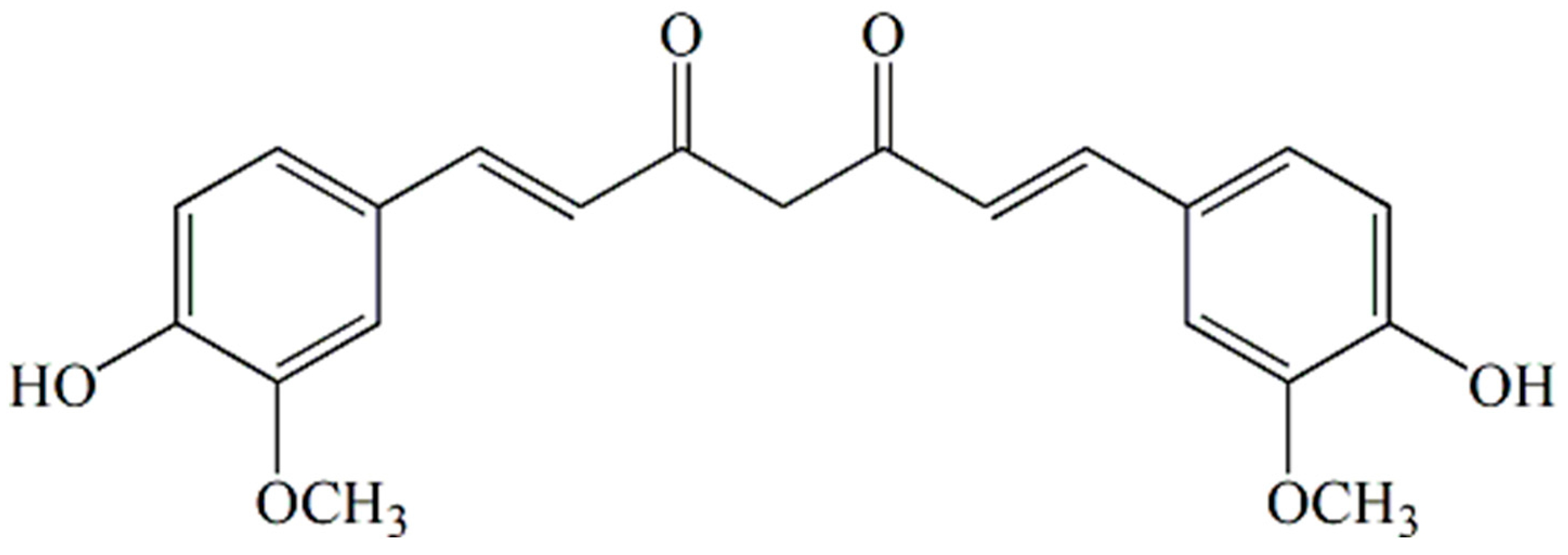
:1. Introduction
2. Materials and Methods
2.1. Reagents
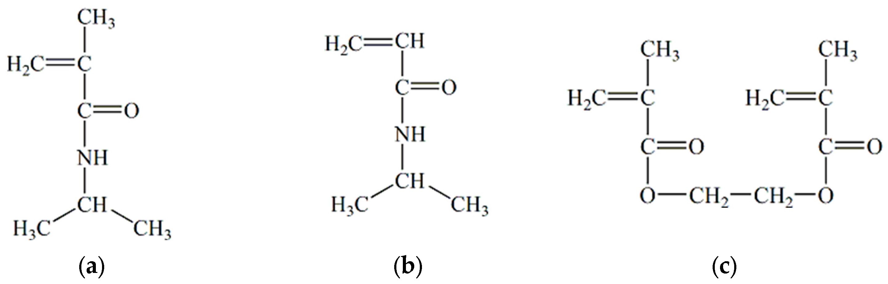
2.2. Synthesis of Hydrogels
2.3. Lyophilization of Gels
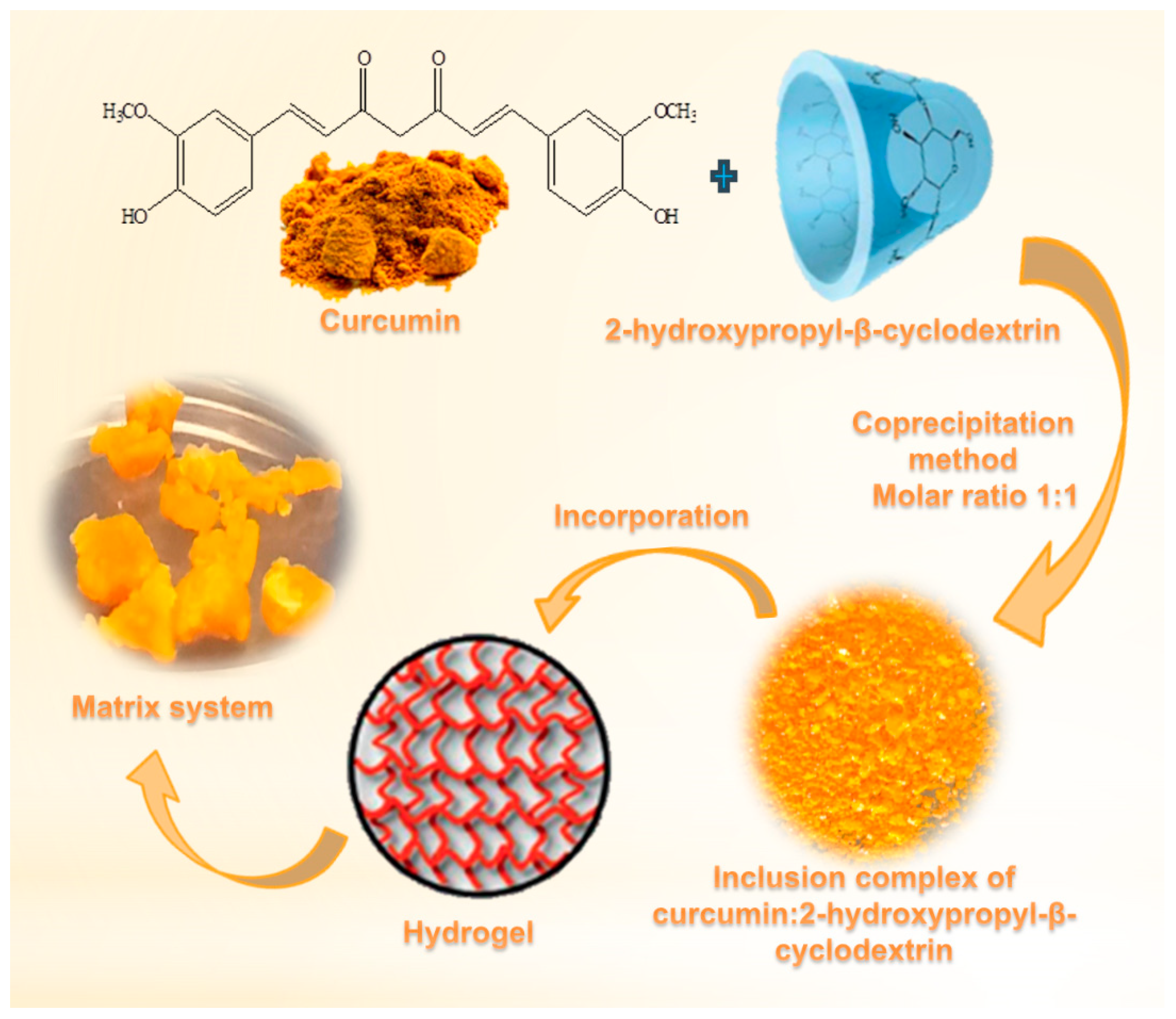
2.4. Obtaining of the Complex
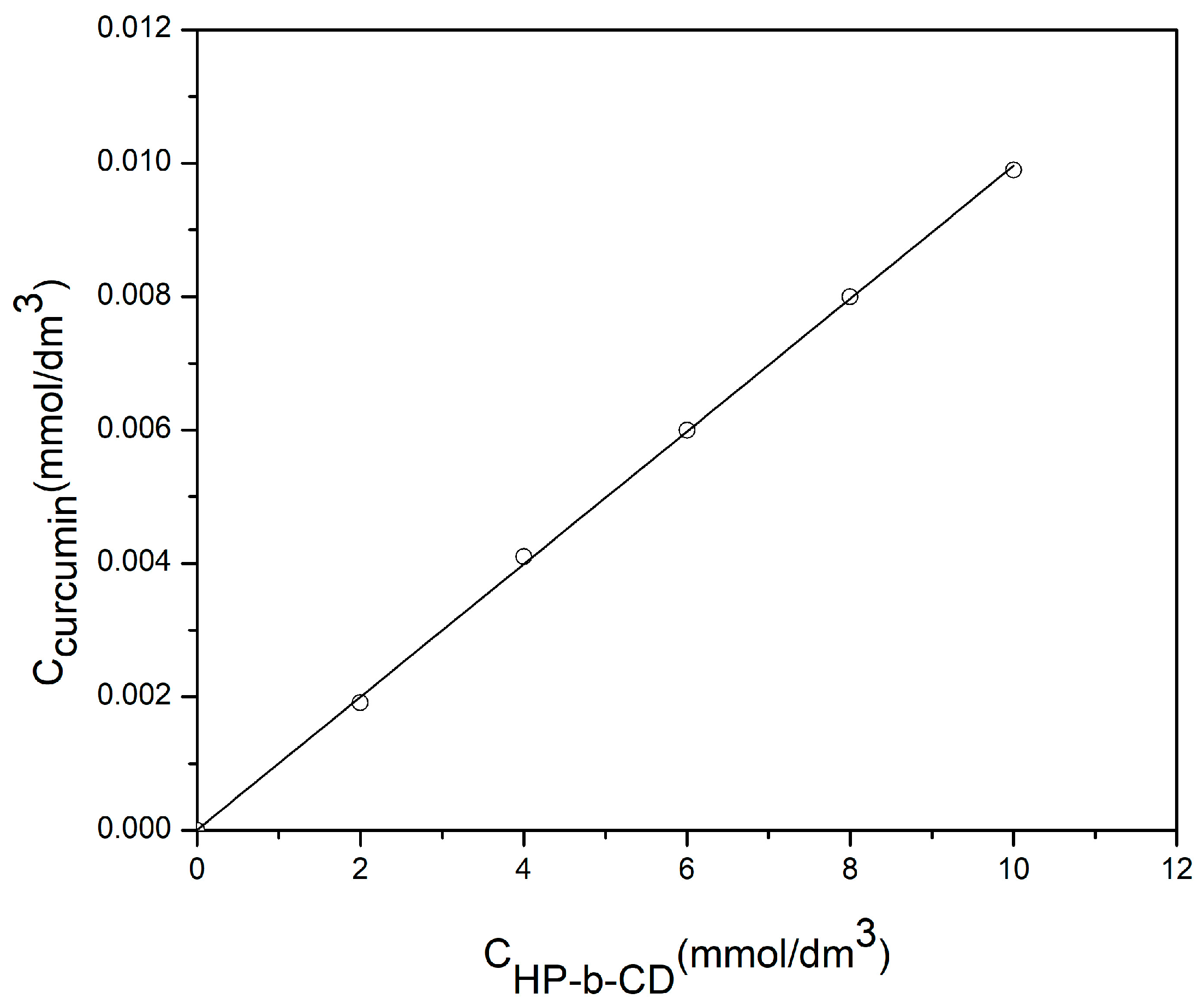
2.5. Phase Solubility
2.6. Incorporation of Complex of Curcumin: 2-Hydroxypropyl-β-cyclodextrin into p(NiPMAm/NiPAm) Gels
2.7. The Release of Curcumin from Matrix System
2.8. Determination of the Concentration of Some Compounds by Using High Pressure Liquid Chromatography (HPLC)
2.9. Swelling of Hydrogels
2.10. Fourier Transform Infrared Spectroscopy (FTIR)
2.11. Scanning Electron Microscopy (SEM)
2.12. Differential Scanning Calorimetry (DSC)
2.13. X-ray Diffraction (XRD)
2.14. Nuclear Magnetic Resonance (1H-NMR)
3. Results
3.1. Phase Solubility
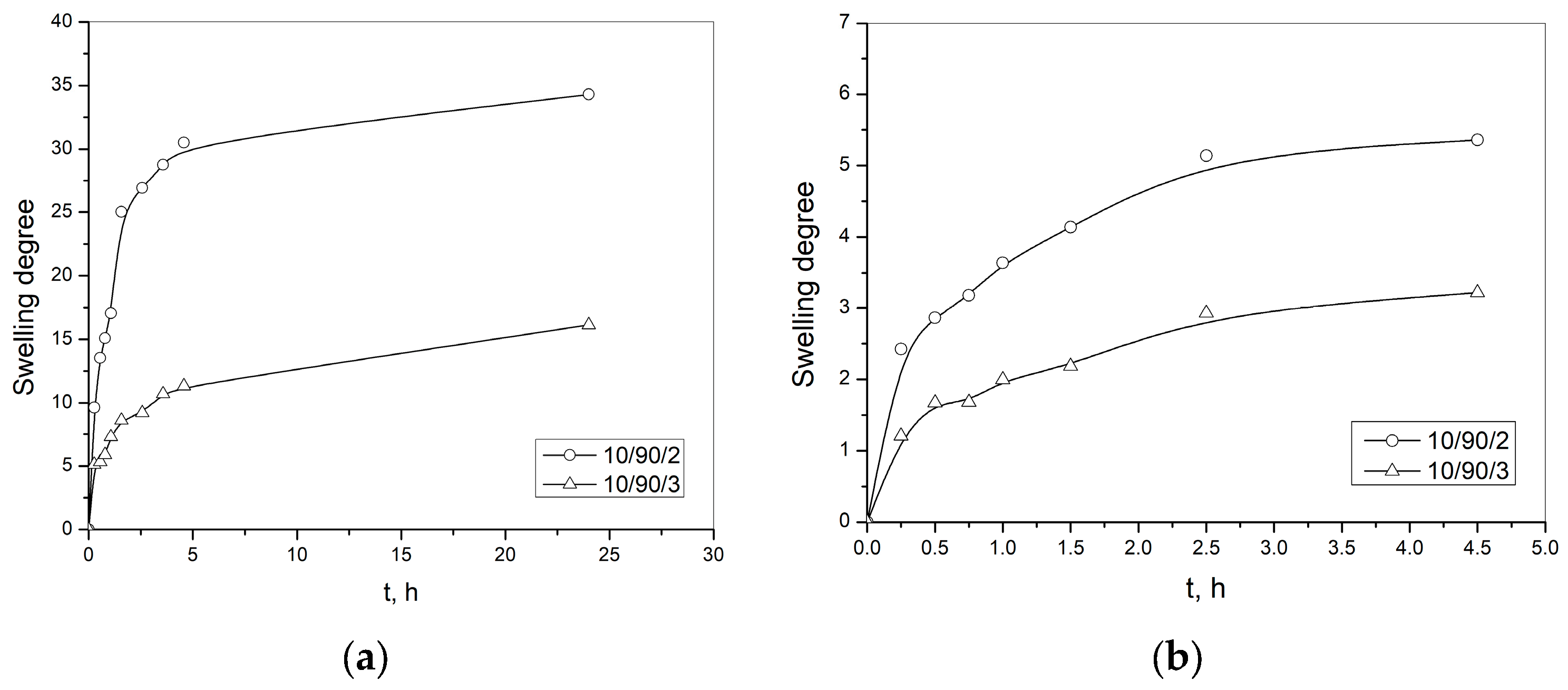
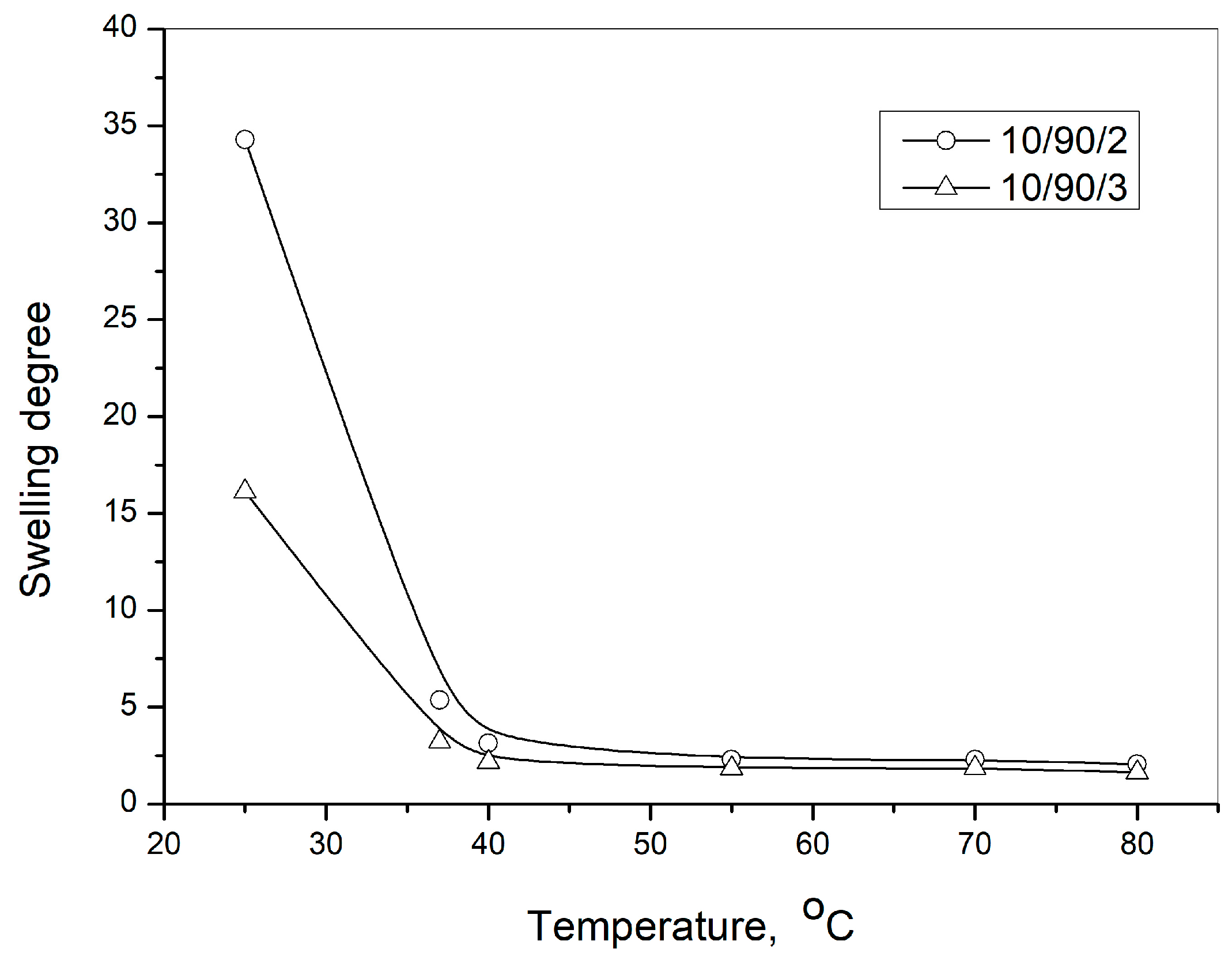
3.2. The Swelling
3.3. Residual Monomers Analysis
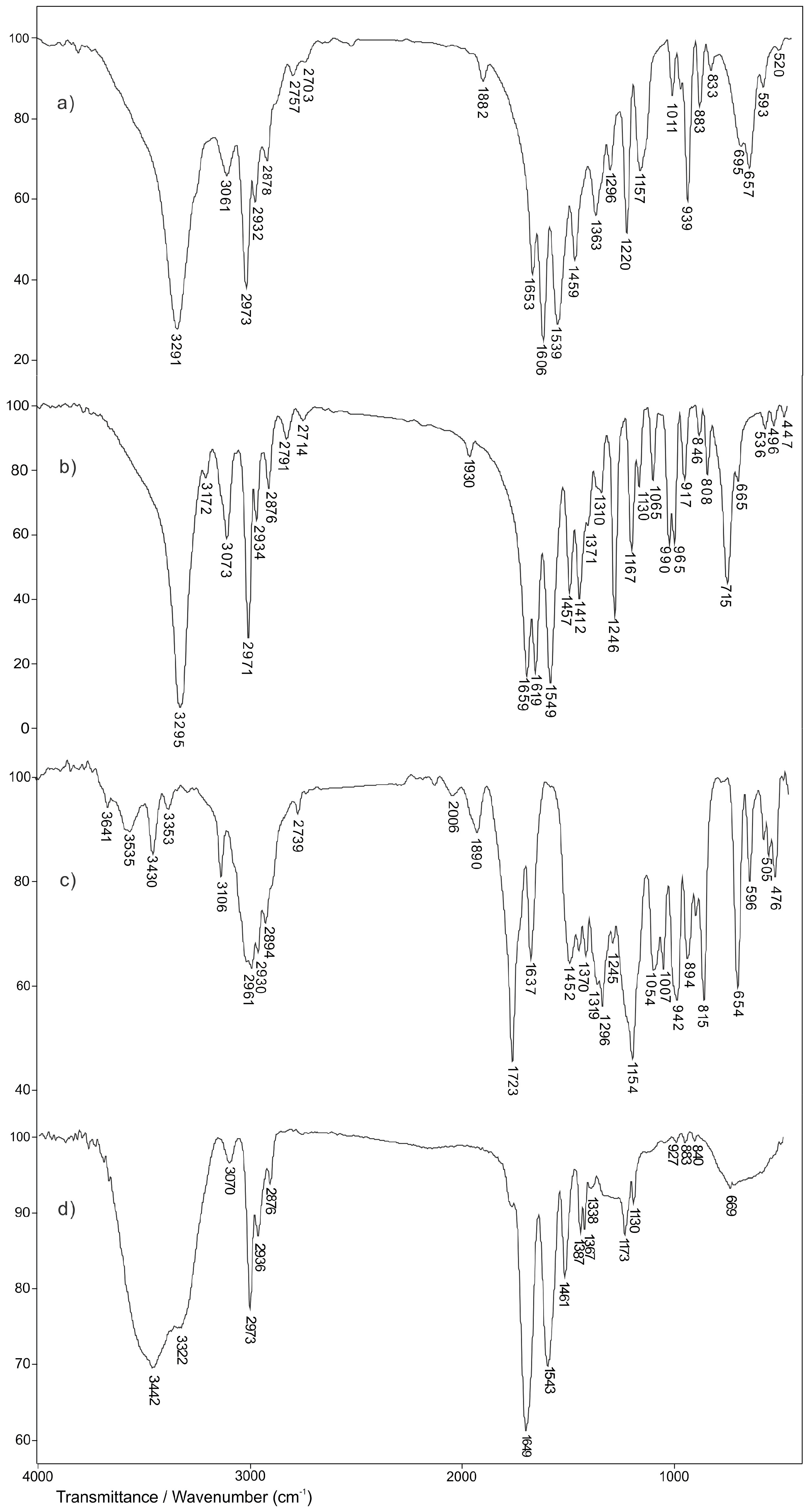
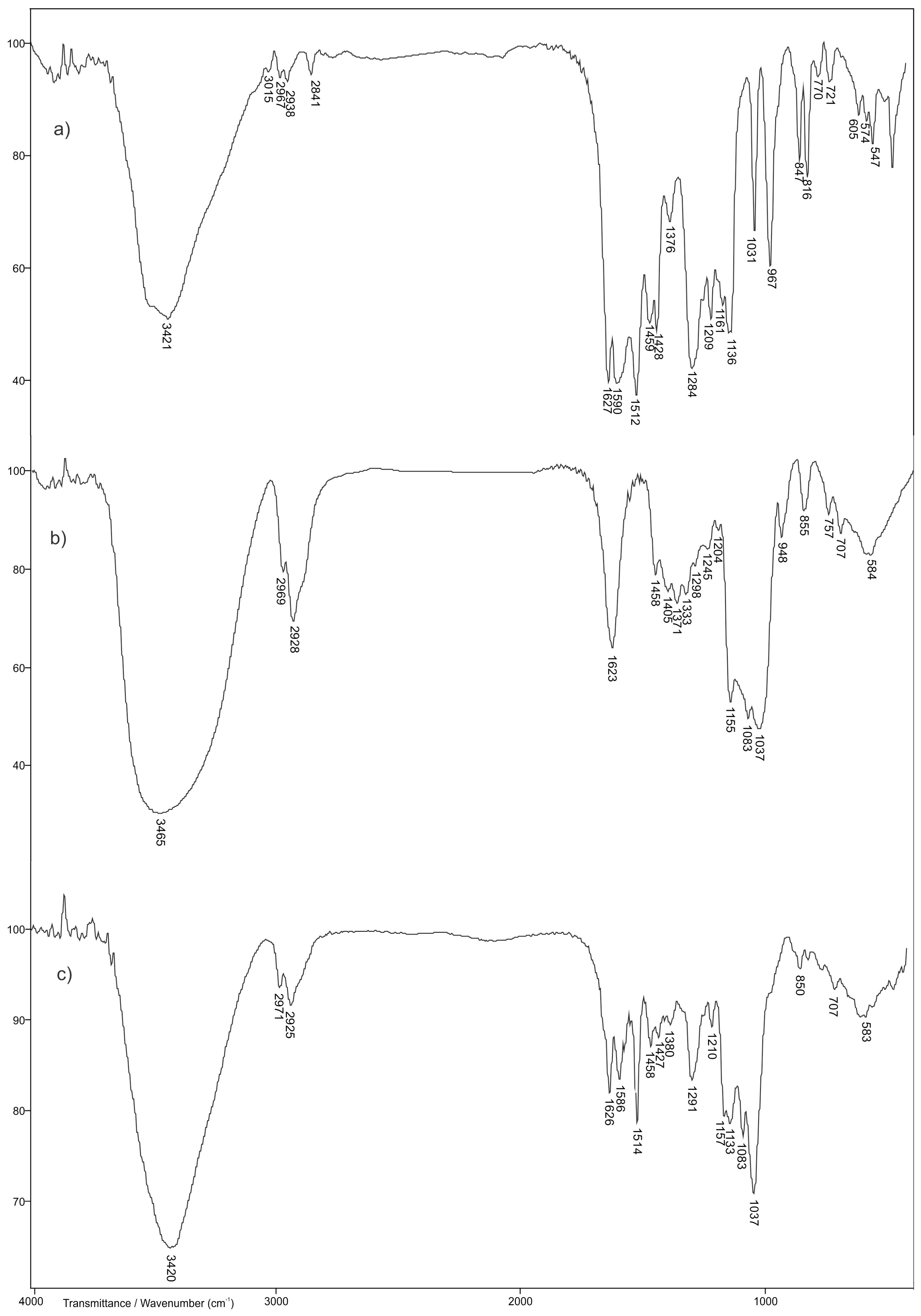
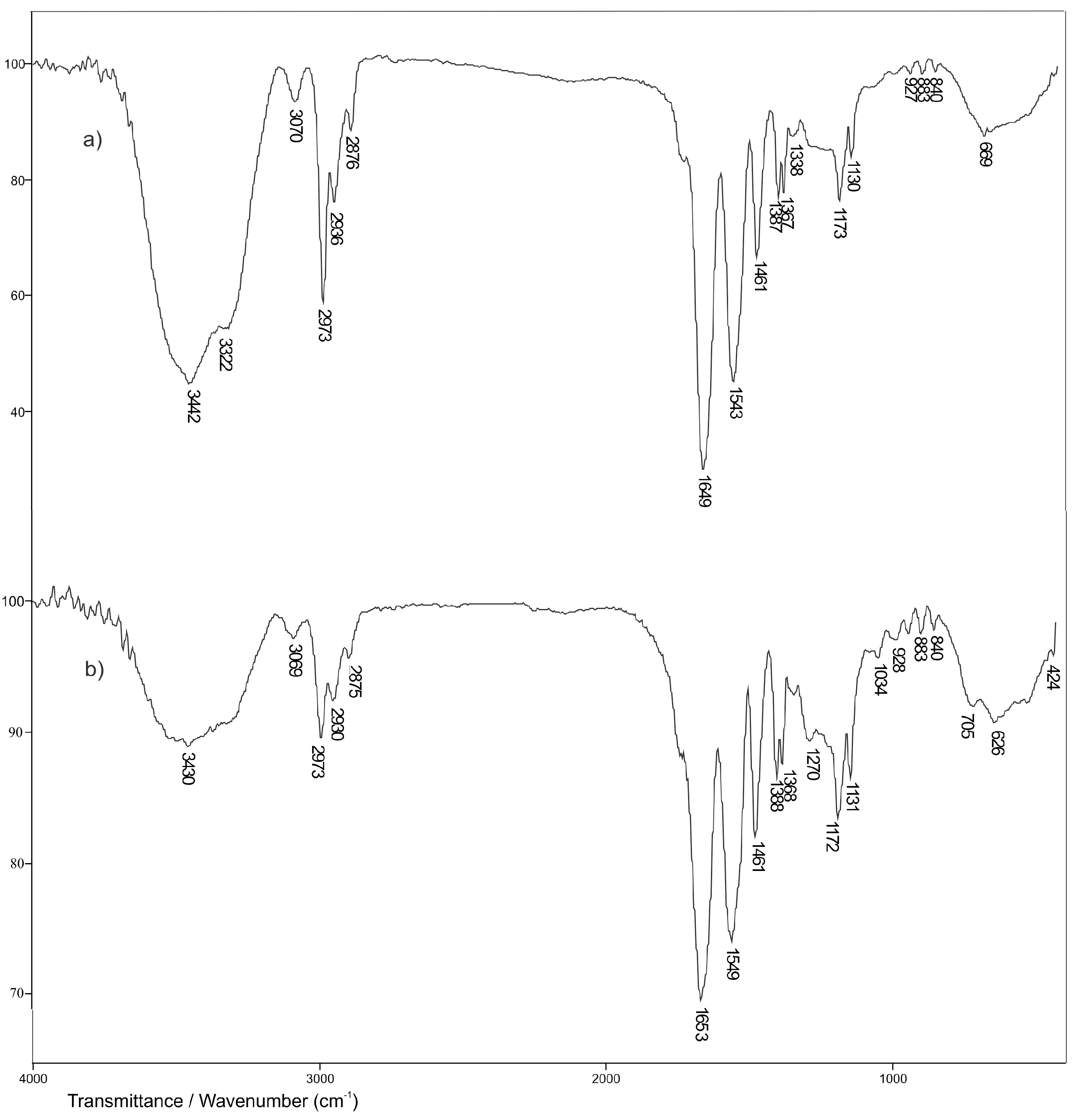
3.4. FTIR Spectroscopy Analysis
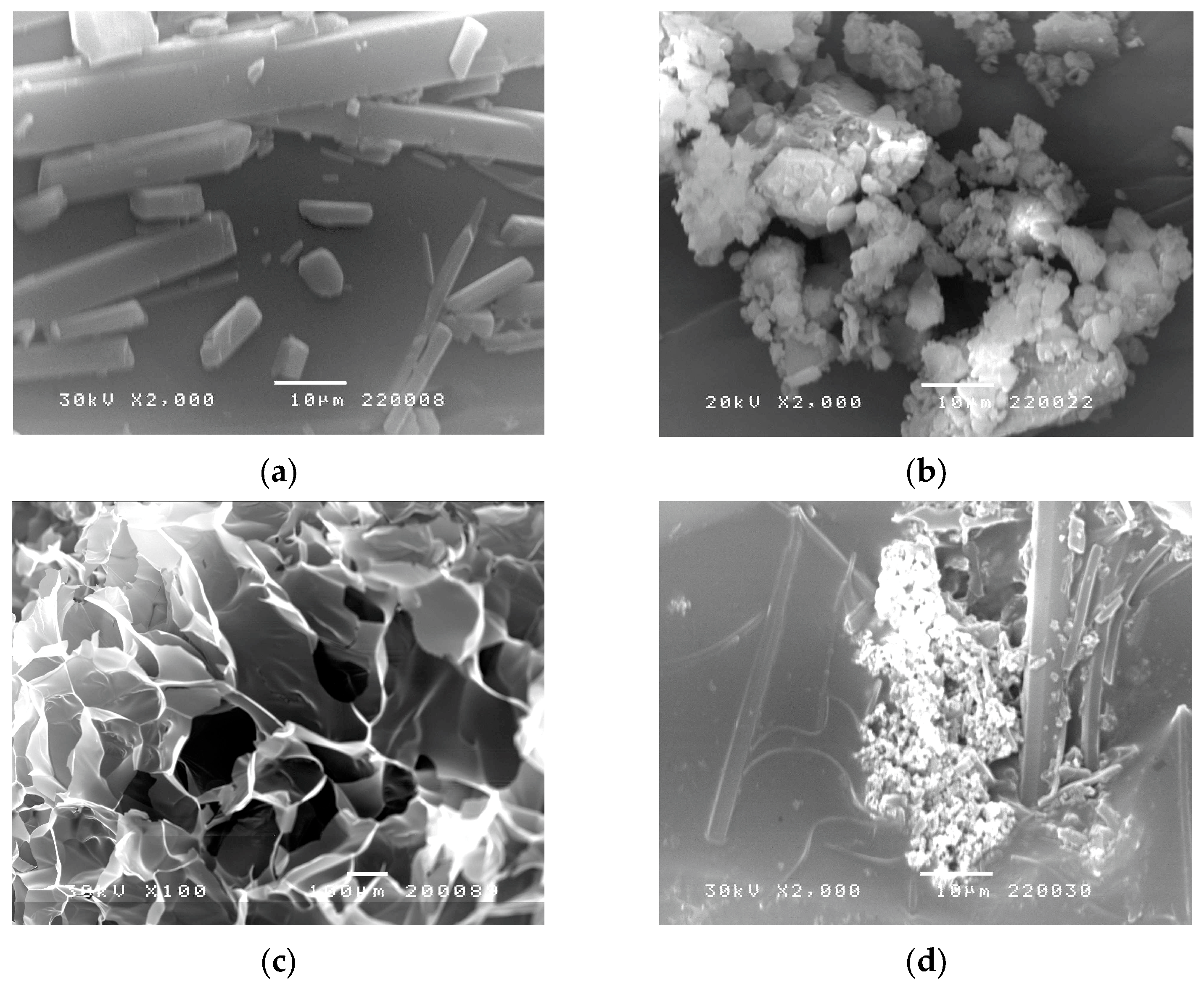
3.5. Scanning Electron Microscopy (SEM)
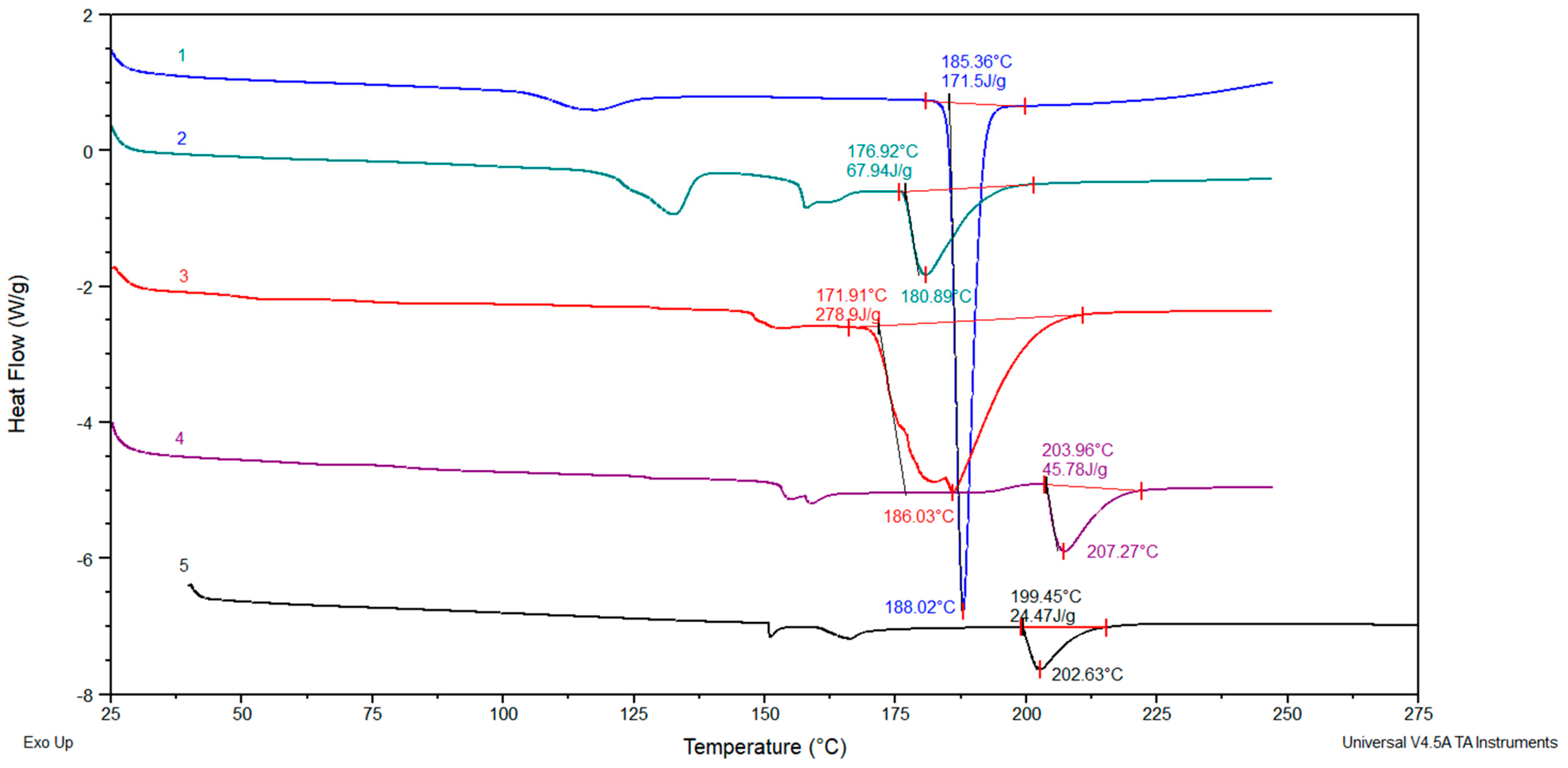
3.6. Differential Scanning Calorimetry (DSC)
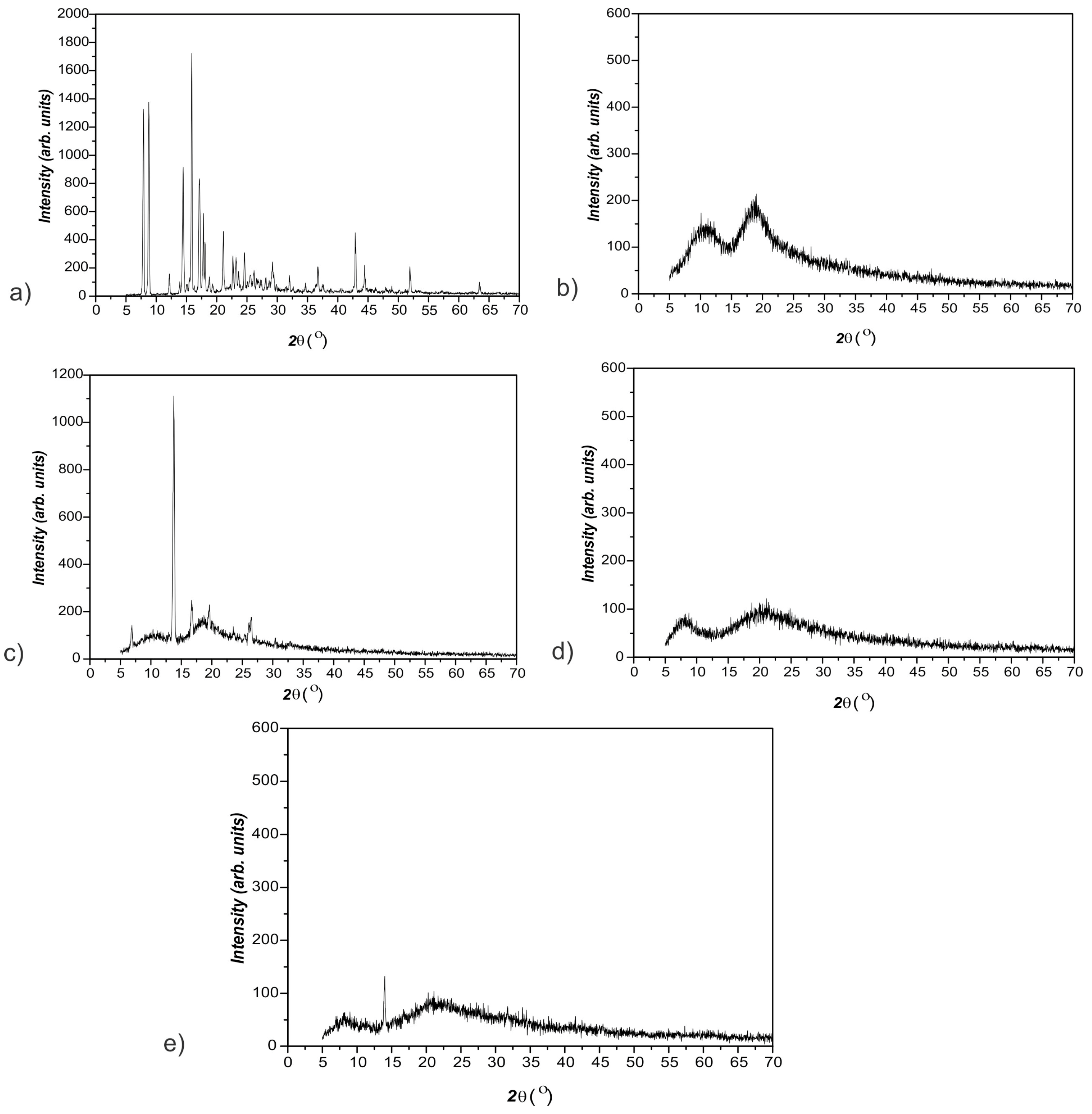
3.7. X-ray Diffraction (XRD)
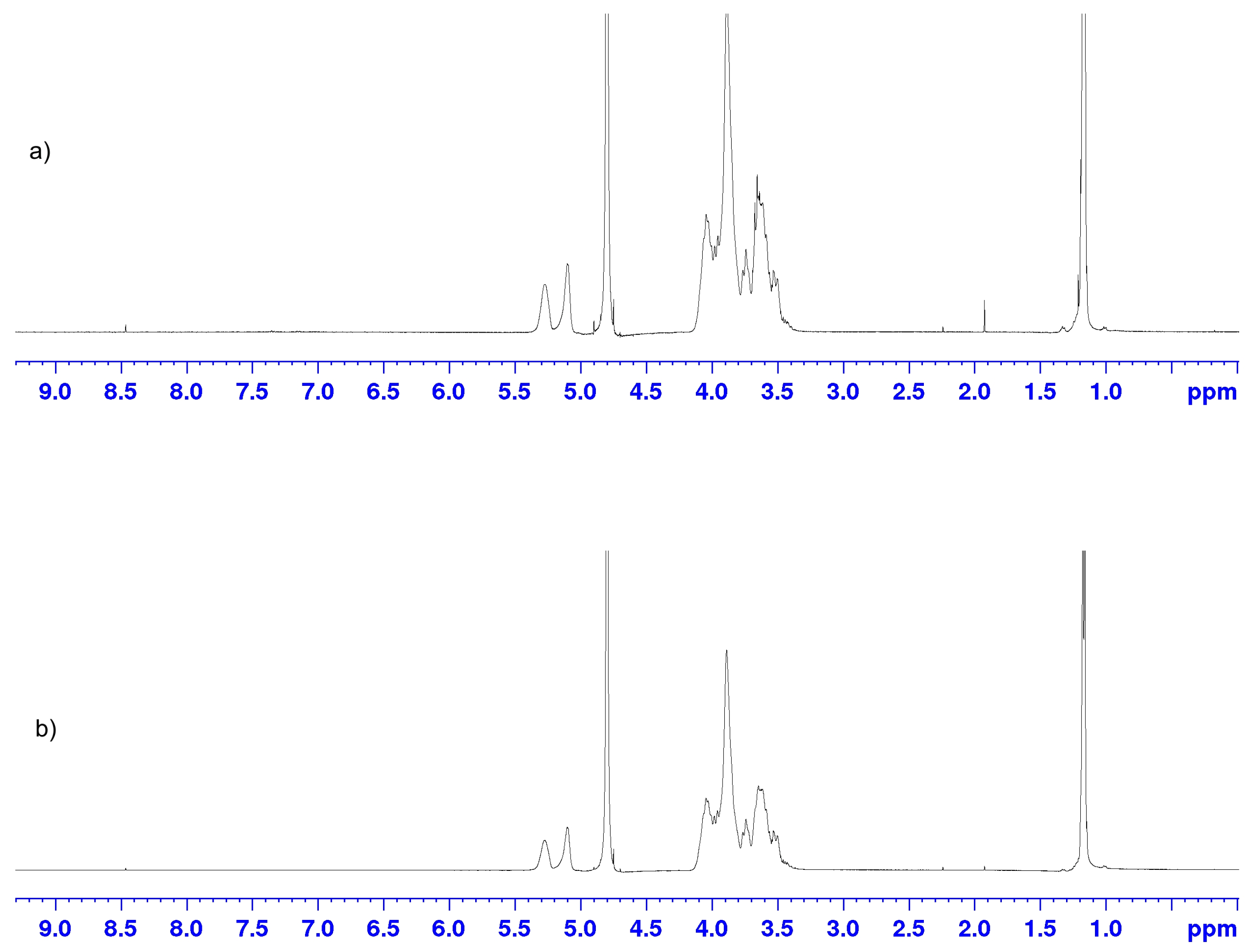
3.8. Nuclear Magnetic Resonance (1H-NMR)
3.9. The Loading Efficiency of Curcumin into the p(NiPMAm/NiPAm) Hydrogel
3.10. In Vitro Release of Curcumin from p(NiPMAm/NiPAm) Gels
4. Conclusions
5. Patents
Author Contributions
Funding
Institutional Review Board Statement
Informed Consent Statement
Data Availability Statement
Acknowledgments
Conflicts of Interest
References
- Xia, Z.H.; Chen, W.B.; Shi, L.; Jiang, X.; Li, K.; Wang, Y.X.; Liu, Y.Q. The underlying mechanisms of curcumin inhibition of hyperglycemia and hyperlipidemia in rats fed a high-fat diet combined with STZ treatment. Molecules 2020, 25, 271. [Google Scholar] [CrossRef] [PubMed] [Green Version]
- Trigo-Gutierrez, J.K.; Vega-Chacón, Y.; Soares, A.B.; Mima, E.G.D.O. Antimicrobial activity of curcumin in nanoformulations: A comprehensive review. Int. J. Mol. Sci. 2021, 22, 7130. [Google Scholar] [CrossRef] [PubMed]
- Kheiripour, N.; Plarak, A.; Heshmati, A.; Asl, S.S.; Mehri, F.; Ebadollahi-Natanzi, A.; Ranjbar, A.; Hosseini, A. Evaluation of the hepatoprotective effects of curcumin and nanocurcumin against paraquat-induced liver injury in rats: Modulation of oxidative stress and Nrf2 pathway. J. Biochem. Mol. Toxicol. 2021, 35, e22739. [Google Scholar] [CrossRef] [PubMed]
- González-Ortega, L.A.; Acosta-Osorio, A.A.; Grube-Pagola, P.; Palmeros-Exsome, C.; Cano-Sarmiento, C.; García-Varela, R.; García, H.S. Anti-inflammatory activity of curcumin in gel carriers on mice with atrial edema. J. Oleo Sci. 2020, 69, 123–131. [Google Scholar] [CrossRef] [Green Version]
- Belhan, S.; Yıldırım, S.; Huyut, Z.; Özdek, U.; Oto, G.; Algül, S. Effects of curcumin on sperm quality, lipid profile, antioxidant activity and histopathological changes in streptozotocin-induced diabetes in rats. Andrologia 2020, 52, e13584. [Google Scholar] [CrossRef]
- Mirzaei, H.; Bagheri, H.; Ghasemi, F.; Khoi, J.M.; Pourhanifeh, M.H.; Heyden, Y.V.; Mortezapour, E.; Nikdasti, A.; Jeandet, P.; Khan, H.; et al. Anti-cancer activity of curcumin on multiple myeloma. Anti Cancer Agents Med. Chem. 2021, 21, 575–586. [Google Scholar] [CrossRef]
- Loutfy, S.A.; Elberry, M.H.; Farroh, K.Y.; Mohamed, H.T.; Mohamed, A.A.; Mohamed, E.B.; Faraag, A.H.I.; Mousa, S.A. Antiviral Activity of Chitosan Nanoparticles Encapsulating Curcumin Against Hepatitis C Virus Genotype 4a in Human Hepatoma Cell Lines. Int. J. Nanomed. 2022, 17, 2891–2892. [Google Scholar] [CrossRef]
- Mai, N.N.S.; Nakai, R.; Kawano, Y.; Hanawa, T. Enhancing the solubility of curcumin using a solid dispersion system with hydroxypropyl-β-cyclodextrin prepared by grinding, freeze-drying, and common solvent evaporation methods. Pharmacy 2020, 8, 203. [Google Scholar] [CrossRef]
- Witika, B.A.; Makoni, P.A.; Matafwali, S.K.; Mweetwa, L.L.; Shandele, G.C.; Walker, R.B. Enhancement of biological and pharmacological properties of an encapsulated polyphenol: Curcumin. Molecules 2021, 26, 4244. [Google Scholar] [CrossRef]
- Rahmani, A.H.; Alsahli, M.A.; Aly, S.M.; Khan, M.A.; Aldebasi, Y.H. Role of curcumin in disease prevention and treatment. Adv. Biomed. Res. 2018, 7, 1–9. [Google Scholar] [CrossRef]
- Khosropanah, M.H.; Dinarvand, A.; Nezhadhosseini, A.; Haghighi, A.; Hashemi, S.; Nirouzad, F.; Dehghani, H. Analysis of the antiproliferative effects of curcumin and nanocurcumin in MDA-MB231 as a breast cancer cell line. Iran J. Pharm. Res. 2016, 15, 231. [Google Scholar]
- Arya, P.; Raghav, N. In-vitro studies of Curcumin-β-cyclodextrin inclusion complex as sustained release system. J. Mol. Struct. 2021, 1228, 129774. [Google Scholar] [CrossRef]
- Mangolim, C.S.; Moriwaki, C.; Nogueira, A.C.; Sato, F.; Baesso, M.L.; Neto, A.M.; Matioli, G. Curcumin–β-cyclodextrin inclusion complex: Stability, solubility, characterisation by FT-IR, FT-Raman, X-ray diffraction and photoacoustic spectroscopy, and food application. Food Chem. 2014, 153, 361–370. [Google Scholar] [CrossRef] [Green Version]
- Jáfar, M.H.; Kamal, N.N.S.N.M.; Hui, B.Y.; Kamaruzzaman, M.F.; Zain, N.N.M.; Yahaya, N.; Raoov, M. Inclusion of Curcumin in β-cyclodextrins as Potential Drug Delivery System: Preparation, Characterization and Its Preliminary Cytotoxicity Approaches. Sains Malays 2018, 47, 977–989. [Google Scholar] [CrossRef]
- Jantarat, C.; Sirathanarun, P.; Ratanapongsai, S.; Watcharakan, P.; Sunyapong, S.; Wadu, A. Curcumin-Hydroxypropyl-β-Cyclodextrin Inclusion Complex Preparation Methods: Effect of Common Solvent Evaporation, Freeze Drying, and pH Shift on Solubility and Stability of Curcumin. Trop. J. Pharm. Res. 2014, 13, 1215. [Google Scholar] [CrossRef]
- Rashidzadeh, H.; Rezaei, S.J.T.; Zamani, S.; Sarijloo, E.; Ramazani, A. pH-sensitive curcumin conjugated micelles for tumor triggered drug delivery. J. Biomater. Sci. 2021, 32, 320–336. [Google Scholar] [CrossRef]
- Odeh, F.; Nsairat, H.; Alshaer, W.; Alsotari, S.; Buqaien, R.; Ismail, S.; Awidi, A.; Bawab, A.A. Remote loading of curcumin-in-modified β-cyclodextrins into liposomes using a transmembrane pH gradient. RSC Adv. 2019, 9, 37148–37161. [Google Scholar] [CrossRef] [Green Version]
- Tai, K.; Rappolt, M.; Mao, L.; Gao, Y.; Li, X.; Yuan, F. The stabilization and release performances of curcumin-loaded liposomes coated by high and low molecular weight chitosan. Food Hydrocoll. 2020, 99, 105355. [Google Scholar] [CrossRef]
- Md Saari, N.H.; Chua, L.S.; Hasham, R.; Yuliati, L. Curcumin-loaded nanoemulsion for better cellular permeation. Sci. Pharm. 2020, 88, 44. [Google Scholar] [CrossRef]
- Wei, Z.; Lin, Q.; Yang, J.; Long, S.; Zhang, G.; Wang, X. Fabrication of novel dual thermo-and pH-sensitive poly (N-isopropylacrylamide-N-methylolacrylamide-acrylic acid) electrospun ultrafine fibres for controlled drug release. Mater. Sci. Eng. C 2020, 115, 111050. [Google Scholar] [CrossRef]
- Karpkird, T.; Khunsakorn, R.; Noptheeranuphap, C.; Midpanon, S. Inclusion complexes and photostability of UV filters and curcumin with beta-cyclodextrin polymers: Effect on cross-linkers. J. Incl. Phenom. Macrocycl. Chem. 2018, 91, 37–45. [Google Scholar] [CrossRef]
- Chen, J.; Qin, X.; Zhong, S.; Chen, S.; Su, W.; Liu, Y. Characterization of Curcumin/Cyclodextrin Polymer Inclusion Complex and Investigation on Its Antioxidant and Antiproliferative Activities. Molecules 2018, 23, 1179. [Google Scholar] [CrossRef] [PubMed] [Green Version]
- Chen, J.; Li, J.; Fan, T.; Zhong, S.; Qin, X.; Li, R.; Gao, J.; Liang, Y. Protective effects of curcumin/cyclodextrin polymer inclusion complex against hydrogen peroxide-induced LO2 cells damage. Food Sci. Nutr. 2022, 10, 1649–1656. [Google Scholar] [CrossRef] [PubMed]
- Celebioglu, A.; Uyar, T. Fast-dissolving antioxidant curcumin/cyclodextrin inclusion complex electrospun nanofibrous webs. Food Chem. 2020, 317, 126397. [Google Scholar] [CrossRef] [PubMed]
- Aytac, Z.; Uyar, T. Core-shell nanofibers of curcumin/cyclodextrin inclusion complex andpolylactic acid: Enhanced water solubility and slow release of curcumin. Int. J. Pharm. 2017, 518, 177–184. [Google Scholar] [CrossRef]
- Rezaei, A.; Nasirpour, A. Evaluation of Release Kinetics and Mechanisms of Curcumin and Curcumin-β-Cyclodextrin Inclusion Complex Incorporated in Electrospun Almond Gum/PVA Nanofibers in Simulated Saliva and Simulated Gastrointestinal Conditions. Bionanoscience 2019, 9, 438–445. [Google Scholar] [CrossRef] [Green Version]
- Zhang, Y. Enhancing Antidepressant Effect of Poloxamer/Chitosan Thermosensitive Gel Containing Curcumin-Cyclodextrin Inclusion Complex. Int. J. Polym. Sci. 2018, 2018, 3041417. [Google Scholar] [CrossRef]
- Kasapoglu-Calik, M.; Ozdemir, M. Synthesis and controlled release of curcumin-β-cyclodextrin inclusion complex from nanocomposite poly(N-isopropylacrylamide/sodium alginate) hydrogels. J. App. Polym. Sci. 2019, 136, 47544. [Google Scholar] [CrossRef]
- Yang, Z.; Liu, J.; Lu, Y. Doxorubicin and CD CUR inclusion complex co loaded in thermosensitive hydrogel PLGA PEG PLGA localized administration for osteosarcoma. Int. J. Oncol. 2020, 57, 433–444. [Google Scholar] [CrossRef]
- Duse, L.; Agel, M.R.; Pinnapireddy, S.R.; Schäfer, J.; Selo, M.A.; Ehrhardt, C.; Bakowsky, U. Photodynamic therapy of ovarian carcinoma cells with curcumin-loaded biodegradable polymeric nanoparticles. Pharmaceutics 2019, 11, 282. [Google Scholar] [CrossRef] [Green Version]
- Purpura, M.; Lowery, R.P.; Wilson, J.M.; Mannan, H.; Münch, G.; Razmovski-Naumovski, V. Analysis of different innovative formulations of curcumin for improved relative oral bioavailability in human subjects. Eur. J. Nutr. 2018, 57, 929–938. [Google Scholar] [CrossRef] [Green Version]
- Zhang, M.; Zhuang, B.; Du, G.; Han, G.; Jin, Y. Curcumin solid dispersion-loaded in situ hydrogels for local treatment of injured vaginal bacterial infection and improvement of vaginal wound healing. J. Pharm. Pharmacol. 2019, 71, 1044–1054. [Google Scholar] [CrossRef]
- Shefa, A.A.; Sultana, T.; Park, M.K.; Lee, S.Y.; Gwon, J.; Lee, B. Curcumin incorporation into an oxidized cellulose nanofiber-polyvinyl alcohol hydrogel system promotes wound healing. Mater. Des. 2020, 186, 108313. [Google Scholar] [CrossRef]
- Zhao, Y.; Liu, J.G.; Chen, W.M.; Yu, A.C. Efficacy of thermosensitive chitosan/β-glycerophosphate hydrogel loaded with β-cyclodextrin-curcumin for the treatment of cutaneous wound infection in rats. Exp. Ther. Med. 2018, 14, 1304–1313. [Google Scholar] [CrossRef] [Green Version]
- Ayar, Z.; Shafieian, M.; Mahmoodi, N.; Sabzevari, O.; Hassannejad, Z. A rechargeable drug delivery system based on pNIPAM hydrogel for the local release of curcumin. J. Appl. Polym. Sci. 2021, 138, 51167. [Google Scholar] [CrossRef]
- Zielinska, A.; Alves, H.; Marques, V.; Durazzo, A.; Lucarini, M.; Alves, T.; Morsink, M.; Willemen, N.; Eder, P.; Chaud, M.; et al. Properties, Extraction Methods, and Delivery Systems for Curcumin as a Natural Source of Beneficial Health Effects. Medicina 2020, 56, 336. [Google Scholar] [CrossRef]
- Sun, M.; Su, X.; Ding, B.; He, X.; Liu, X.; Yu, A.; Lou, H.; Zhai, G. Advances in nanotechnology-based delivery systems for curcumin. Nanomedicine 2012, 7, 1. [Google Scholar] [CrossRef]
- Ilić-Stojanović, S.; Nikolić, L.; Nikolić, V.; Ristić, I.; Budinski-Simendić, J.; Kapor, A.; Nikolić, G.M. The Structure Characterization of Thermosensitive Poly(N-isopropylacrylamide-co-2-hydroxypropyl methacrylate) Hydrogel. Polym. Int. 2014, 63, 973–981. [Google Scholar] [CrossRef]
- Ilic-Stojanovic, S.; Urosevic, M.; Nikolic, L.; Petrovic, D.; Stanojevic, J.; Najman, S.; Nikolic, V. Intelligent Hydrogels Poly(N-Isopropylmethacrylamide): Synthesis, Structure Characterization, Swelling Properties and their Radiation Decomposition. Polymers 2020, 12, 1112. [Google Scholar] [CrossRef]
- Zdravković, A.; Nikolić, L.; Ilić-Stojanović, S.; Nikolić, V.; Najman, S.; Mitić, Ž.; Ćirić, A.; Petrović, S. Removal of heavy metal ions from aqueous solutions by hydrogels based on N-isopropylacrylamide and acrylic acid. Polym. Bull. 2018, 75, 4797–4821. [Google Scholar] [CrossRef] [Green Version]
- Ilić-Stojanović, S.; Nikolić, L.; Nikolić, V.; Petrović, S.; Najman, S.; Mitić, Ž.; Oro, V. Semi-crystalline copolymer hydrogels as smart drug carriers: In vitro thermo-responsive naproxen release. Pharmaceutics 2021, 13, 1–22. [Google Scholar] [CrossRef] [PubMed]
- Higuchi, T.; Connors, K. Phase solubility techniques. Adv. Anal. Chem. Instrum. 1965, 7, 117–212. [Google Scholar]
- Bajpai, S.K. Swelling–deswelling behavior of poly(acrylamide-co-maleic acid) hydrogels. J. Appl. Polym. Sci. 2001, 80, 2782–2789. [Google Scholar] [CrossRef]
- Ritger, P.L.; Peppas, N.A. A simple equation for description of solute release II. Fickian and anomalous release from swellable devices. J. Control. Release 1987, 5, 37–42. [Google Scholar] [CrossRef]
- Wang, J.; Wu, W.; Lin, Z. Kinetics and thermodynamics of the water sorption of 2-hydroxyethyl methacrylate/styrene copolymer hydrogels. J. Appl. Polym. Sci. 2008, 109, 3018–3023. [Google Scholar] [CrossRef]
- Hansen, C.M. The significance of the surface condition in solutions to the diffusion equation: Explaining “anomalous” sigmoidal, Case II, and Super Case II absorption behavior. Eur. Polym. J. 2010, 46, 651–662. [Google Scholar] [CrossRef]
- Heskins, M.; Guillet, J.E. Solution Properties of Poly(N-isopropylacrylamide). J. Macromol. Sci. Pure Appl. Chem. 1968, 2, 1441–1455. [Google Scholar] [CrossRef]
- Rwei, S.P.; Anh, T.H.N.; Chiang, W.Y.; Way, T.F.; Hsu, Y.J. Synthesis and drug delivery application of thermo-and pH-sensitive hydrogels: Poly (β-CD-co-N-isopropylacrylamide-co-IAM). Materials 2016, 9, 1003. [Google Scholar] [CrossRef] [Green Version]
- Tang, X.L.; Guo, S.M.; Liu, Z.D.; Tang, R.Z.; Pang, J.Y.; Chen, Y. Preparation of thermo-sensitive poly (N-isopropylacrylamide) film using KHz alternating current Dielectric barrier discharge. Adv. Eng. Res. 2018, 120, 598–602. [Google Scholar]
- Kundu, S.; Nithiyanantham, U. In situ formation of curcumin stabilized shape-selective Ag nanostructures in aqueous solution and their pronounced SERS activity. RSC Adv. 2013, 3, 25278–25290. [Google Scholar] [CrossRef]
- Safie, N.E.; Ludin, N.A.; Súait, M.S.; Hamid, N.H.; Sepeai, S.; Ibrahim, M.A.; Teridi, M.A.M. Preliminary study of natural pigments photochemical properties of curcuma longa l and lawsonia inermis l. as tio 2 photoelectrode sensitizer. Malays. J. Anal. Sci. 2015, 19, 1243–1249. [Google Scholar]
- Ismail, E.H.; Sabry, D.Y.; Mahdy, H.; Khalil, M.M.H. Synthesis and Characterization of some Ternary Metal Complexes of Curcumin with 1, 10-phenanthroline and their Anticancer Applications. J. Sci. Res. 2014, 6, 509–519. [Google Scholar] [CrossRef] [Green Version]
- Valand, N.N.; Patel, M.B.; Menon, S.K. Curcumin-p-sulfonatocalix [4] resorcinarene (p-SC [4]R) interaction: Thermo-physico chemistry, stability and biological evaluation. RSC Adv. 2015, 5, 8739–8752. [Google Scholar] [CrossRef]
- Ali, M.S.; Pandit, V.; Jain, M.; Dhar, K.L. Mucoadhesive microparticulate drug delivery system of curcumin against Helicobacter pylori infection: Design, development and optimization. J. Adv. Pharm. Technol. Res. 2014, 5, 48–56. [Google Scholar] [CrossRef]
- Mattos de Silva, M.R.; Santos, E.P.; Barros, R.C.S.A.; Garcia, S.; Albuquerque, M.G.; Oliveira, J.S.C.; Sader, M.S. The development of a new complexation technique of hydrocortisone acetate with 2-hydroxypropyl-β-cyclodextrin: Preparation and characterization. J. Anal. Pharm. Res. 2018, 7, 1–5. [Google Scholar] [CrossRef] [Green Version]
- Wang, J.; Cao, Y.; Sun, B.; Wang, C. Physicochemical and release characterisation of garlic oil-β-cyclodextrin inclusion complexes. Food Chem. 2011, 127, 1680–1685. [Google Scholar] [CrossRef]
- Nikolić, V.D.; Ilić-Stojanović, S.S.; Nikolić, L.B.; Cakić, M.D.; Zdravković, A.S.; Kapor, A.J.; Popsavin, M.M. Photostability of piroxicam in the inclusion complex with 2-hydroxypropyl-β-cyclodextrin. Hem. Ind. 2014, 68, 107–116. [Google Scholar] [CrossRef] [Green Version]
- Rachmawati, H.; Edityaningrum, C.A.; Mauludin, R. Molecular inclusion complex of curcumin–β-cyclodextrin nanoparticle to enhance curcumin skin permeability from hydrophilic matrix gel. AAPS Pharmscitech 2013, 14, 1303–1312. [Google Scholar] [CrossRef] [Green Version]
- Li, N.; Wang, N.; Wu, T.; Qiu, C.; Wang, X.; Jiang, S.; Wang, T. Preparation of curcumin-hydroxypropyl-β-cyclodextrin inclusion complex by cosolvency-lyophilization procedure to enhance oral bioavailability of the drug. Drug Dev. Ind. Pharm. 2018, 44, 1966–1974. [Google Scholar] [CrossRef]
- Costa, P.; Lobo, J.M.S. Modeling and comparison of dissolution profiles. Eur. J. Pharm. Sci. 2001, 13, 123–133. [Google Scholar] [CrossRef]
- Đorđević, S.; Isailović, T.; Cekić, N.; Vuleta, G.; Savić, S. Parenteralne nanoemulzije diazepama-fizičkohemijska karakterizacija i in vitro ispitivanje brzine oslobađanja. Arh. Farm. 2016, 66, 24–41. [Google Scholar]
- Zhang, Y.; Huo, M.; Zhou, J.; Zou, A.; Li, W.; Yao, C.; Xie, S. DDSolver: An add-in program for modeling and comparison of drug dissolution profiles. AAPS J. 2010, 12, 263–271. [Google Scholar] [CrossRef] [PubMed] [Green Version]


| Temperature, °C | Sample | n | k, min1/n | D, cm2/min |
|---|---|---|---|---|
| 25 | 10/90/2 | 0.82 | 0.092 | 1.65 ∙ 10−5 |
| 10/90/3 | 0.60 | 0.127 | 3.17 ∙ 10−5 | |
| 37 | 10/90/2 | 0.49 | 0.231 | 1.05 ∙ 10−4 |
| 10/90/3 | 0.63 | 0.164 | 5.26 ∙ 10−5 |
| Sample | The Content of Residual Monomers in Sample, % | ||
|---|---|---|---|
| NiPMAm | NiPAm | EGDM | |
| 10/90/2 | 0.11 | 0.28 | 0.07 |
| 10/90/3 | 0.13 | 0.30 | 0.08 |
| Type of Proton | Chemical Shifts Values δ | Δδ | |
|---|---|---|---|
| 2-HP-β-CD | Complex | ||
| CH3 | 1.1692 | 1.1678 | +0.0014 |
| H-C1 | 5.1005 | 5.0993 | +0.0012 |
| H-C2 | 5.2732 | 5.2743 | −0.0011 |
| Sample | η of Curcumin (%) |
|---|---|
| 10/90/2 | 78.35 |
| 10/90/3 | 67.58 |
| Kinetic Model | Parameter | Sample 10/90/2 | Sample 10/90/3 |
|---|---|---|---|
| Higuchi 𝐹 = 𝑘𝐻∙𝑡1/2 | kH | 9.218 | 3.350 |
| R2 | −0.918 | −1.147 | |
| AIC | 97.98 | 74.811 | |
| Korsmeyer–Peppas 𝐹 = 𝑘𝐾𝑃∙𝑡𝑛 | kKP | 33.247 | 12.499 |
| n | 0.074 | 0.063 | |
| R2 | 0.984 | 0.996 | |
| AIC | 41.036 | −2.927 | |
| Baker–Lonsdale 3/2[1−(1−𝐹/100)2/3]−𝐹/100 =𝑘 𝐵𝐿∙𝑡 | kBL | 0.002 | 0 |
| R2 | −0.636 | −1.054 | |
| AIC | 96.078 | 74.27 |
Disclaimer/Publisher’s Note: The statements, opinions and data contained in all publications are solely those of the individual author(s) and contributor(s) and not of MDPI and/or the editor(s). MDPI and/or the editor(s) disclaim responsibility for any injury to people or property resulting from any ideas, methods, instructions or products referred to in the content. |
© 2023 by the authors. Licensee MDPI, Basel, Switzerland. This article is an open access article distributed under the terms and conditions of the Creative Commons Attribution (CC BY) license (https://creativecommons.org/licenses/by/4.0/).
Share and Cite
Nikolić, L.; Urošević, M.; Nikolić, V.; Gajić, I.; Dinić, A.; Miljković, V.; Rakić, S.; Đokić, S.; Kesić, J.; Ilić-Stojanović, S.; et al. The Formulation of Curcumin: 2-Hydroxypropyl-β-cyclodextrin Complex with Smart Hydrogel for Prolonged Release of Curcumin. Pharmaceutics 2023, 15, 382. https://0-doi-org.brum.beds.ac.uk/10.3390/pharmaceutics15020382
Nikolić L, Urošević M, Nikolić V, Gajić I, Dinić A, Miljković V, Rakić S, Đokić S, Kesić J, Ilić-Stojanović S, et al. The Formulation of Curcumin: 2-Hydroxypropyl-β-cyclodextrin Complex with Smart Hydrogel for Prolonged Release of Curcumin. Pharmaceutics. 2023; 15(2):382. https://0-doi-org.brum.beds.ac.uk/10.3390/pharmaceutics15020382
Chicago/Turabian StyleNikolić, Ljubiša, Maja Urošević, Vesna Nikolić, Ivana Gajić, Ana Dinić, Vojkan Miljković, Srđan Rakić, Sanja Đokić, Jelena Kesić, Snežana Ilić-Stojanović, and et al. 2023. "The Formulation of Curcumin: 2-Hydroxypropyl-β-cyclodextrin Complex with Smart Hydrogel for Prolonged Release of Curcumin" Pharmaceutics 15, no. 2: 382. https://0-doi-org.brum.beds.ac.uk/10.3390/pharmaceutics15020382