Article

Essential Oils of Sage, Rosemary, and Bay Laurel Inhibit the Life Stages of Oomycete Pathogens Important in Aquaculture

1
Faculty of Food Technology and Biotechnology, University of Zagreb, Pierottijeva 6, 10 000 Zagreb, Croatia
2
Faculty of Chemistry and Technology, University of Split, Ruđera Boškovića 35, 21 000 Split, Croatia
*
Author to whom correspondence should be addressed.
Submission received: 29 July 2021 / Revised: 11 August 2021 / Accepted: 13 August 2021 / Published: 15 August 2021
(This article belongs to the Special Issue Chemical Composition and Antimicrobial Activity of Essential Oils)

Abstract

:
Saprolegnia parasitica, the causative agent of saprolegniosis in fish, and Aphanomyces astaci, the causative agent of crayfish plague, are oomycete pathogens that cause economic losses in aquaculture. Since toxic chemicals are currently used to control them, we aimed to investigate their inhibition by essential oils of sage, rosemary, and bay laurel as environmentally acceptable alternatives. Gas Chromatography–Mass Spectrometry (GC–MS) analysis showed that the essential oils tested were rich in bioactive volatiles, mainly monoterpenes. Mycelium and zoospores of A. astaci were more sensitive compared to those of S. parasitica, where only sage essential oil completely inhibited mycelial growth. EC50 values (i.e., concentrations of samples at which the growth was inhibited by 50%) for mycelial growth determined by the radial growth inhibition assay were 0.031–0.098 µL/mL for A. astaci and 0.040 µL/mL for S. parasitica. EC50 values determined by the zoospore germination inhibition assay were 0.007–0.049 µL/mL for A. astaci and 0.012–0.063 µL/mL for S. parasitica. The observed inhibition, most pronounced for sage essential oil, could be partly due to dominant constituents of the essential oils, such as camphor, but more likely resulted from a synergistic effect of multiple compounds. Our results may serve as a basis for in vivo experiments and the development of environmentally friendly methods to control oomycete pathogens in aquaculture.

Graphical Abstract

1. Introduction

Saprolegnia parasitica and Aphanomyces astaci (Oomycota) cause significant economic losses in freshwater aquaculture: S. parasitica affects salmonid hatcheries and farms [1], and A. astaci crayfish rearing facilities [2,3,4]. Saprolegnia parasitica is parasitic to salmonid fishes such as Oncorhynchus mykiss and Salmo salar and infects all developmental stages [5,6,7]. It can also infect crayfish species, especially when the animals are injured, and its pathogenicity towards Astacus astacus, Pacifastacus leniusculus, and Procambarus clarkii has been confirmed by infection experiments [8]. Saprolegnia-infected fish develop a cottony mycelium on the gills and injured skin, while the infected eggs usually die as a result of hyphal rupture of the chorionic membrane and subsequent osmotic shock [9]. Aphanomyces astaci is a causative agent of crayfish plague. Its hyphae penetrate the cuticle of crayfish and spread throughout the tissues, leading to the development of a fatal disease [10,11]. It is mainly known for its destructive effect on native European crayfish species, while North American crayfish are considered carriers of the pathogen but can still succumb to the disease under certain stressful conditions, such as in a high-density farming environment [12,13]. Therefore, A. astaci may pose a serious threat to the crayfish farming industry on a global scale, both to native (e.g., Astacus astacus) and invasive (e.g., Procambarus clarkii and Cherax quadricarinatus) crayfish [2,4,14].
Diseases in freshwater aquaculture caused by oomycetes were previously treated successfully with malachite green, but its use is no longer allowed in the EU [15] and the USA [16] due to its teratogenic and carcinogenic properties [17,18,19]. Other chemicals currently used worldwide are also not sustainable solutions. In fact, formalin poses a health risk to fish farm workers who handle it [20] and to fish consumers, as it leaves residues in the fish [21]. Other agents such as bronopol, copper sulfate, and peracetic acid are less toxic but still pose a serious threat to the aquatic biota [22,23,24].
The toxicity of chemical agents used for oomycete control in fisheries prompts the search for environmentally friendly alternatives that should be effective against pathogenic oomycetes but also safe for operators, animals, and the environment. In this context, it has been increasingly shown that alcoholic extracts [25,26,27,28,29,30] and essential oils [31,32,33,34,35,36,37,38,39] of selected plants can inhibit pathogenic oomycetes in vitro, with essential oils generally being more potent than alcoholic extracts [38]. In particular, plants from the Lauraceae and Lamiaceae families, such as Thymbra spicata and Cinnamomum zeylanicum, have been reported to inhibit pathogenic freshwater oomycetes, mostly using the mycelium and zoospores of Saprolegnia spp. as models [32,34,36,37,38,40]. In comparison, the inhibitory activity of essential oils and their major components against Aphanomyces spp. is much less studied [28,40]. So far, there are no reports on the inhibitory activity of essential oils against A. astaci, but some essential oils showed good inhibitory activity against the phylogenetically related plant pathogen A. euteiches and the fish pathogen A. invadans [28,40].
The aim of this study was to test, for the first time, the inhibition of the oomycete pathogens S. parasitica and A. astaci by the essential oils of sage (Salvia officinalis), bay laurel (Laurus nobilis), and rosemary (Rosmarinus officinalis), which have previously shown potent antifungal and antimicrobial activity [41,42,43]. We analyzed the volatile composition of the essential oils and then tested the sensitivity of the alternative stages of the oomycete life cycle (i.e., mycelium and zoospores, as infectious stage) to essential oils.

2. Results

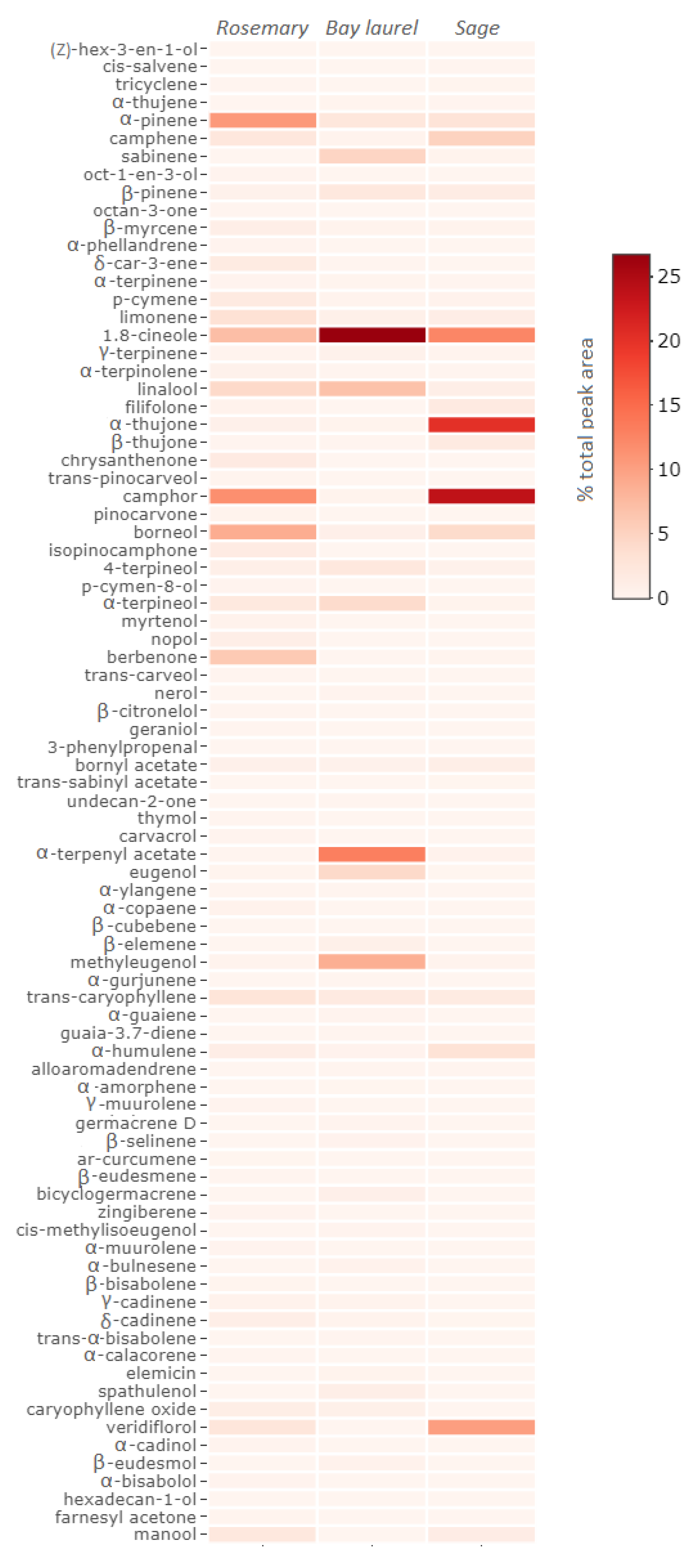
2.1. Chemical Composition of Essential Oils

The chemical composition of essential oils was analyzed by Gas Chromatography–Mass Spectrometry (GC–MS) (Figure 1, Table S1, Supplementary material). All essential oils were rich in volatile components, with the highest number of compounds identified in rosemary (65 compounds, 90% of total GC peak area), followed by bay laurel (53 compounds, 93% of total GC peak area), and eventually sage essential oil (35 compounds, 98% of total GC peak area). The major detected compounds in all three essential oils were monoterpenes, such as camphor (11.7%), α-pinene (10.8%), 1,8-cineole (7.3%), borneol (8.9%), and linalool (4.4%) in rosemary essential oil, then 1,8-cineole (26.8%), α-terphenyl acetate (13.2%), linalool (6.9%), sabinene (4.9%), and α-terpineol (4.1%) in bay laurel essential oil, and camphor (23.9%), α-thujone (20.3%), 1,8-cineole (12.5%), and camphene (5.2%) in sage essential oil. Some essential oils were also rich in sesquiterpenes, such as berbenone (6.1%), trans-caryophyllene (2.8%), and veridiflorol (2.6%) in rosemary essential oil, and veridiflorol (10.3%) and α-humulene (3.2%) in sage essential oil, while in bay laurel essential oil, a compound classified as a phenylpropane derivate, eugenol, was also identified in significant quantity (4.4% of total peak area).

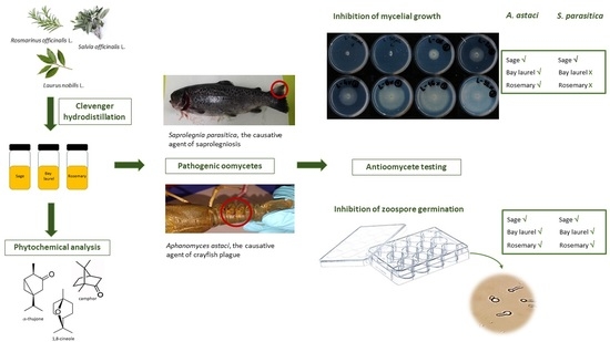
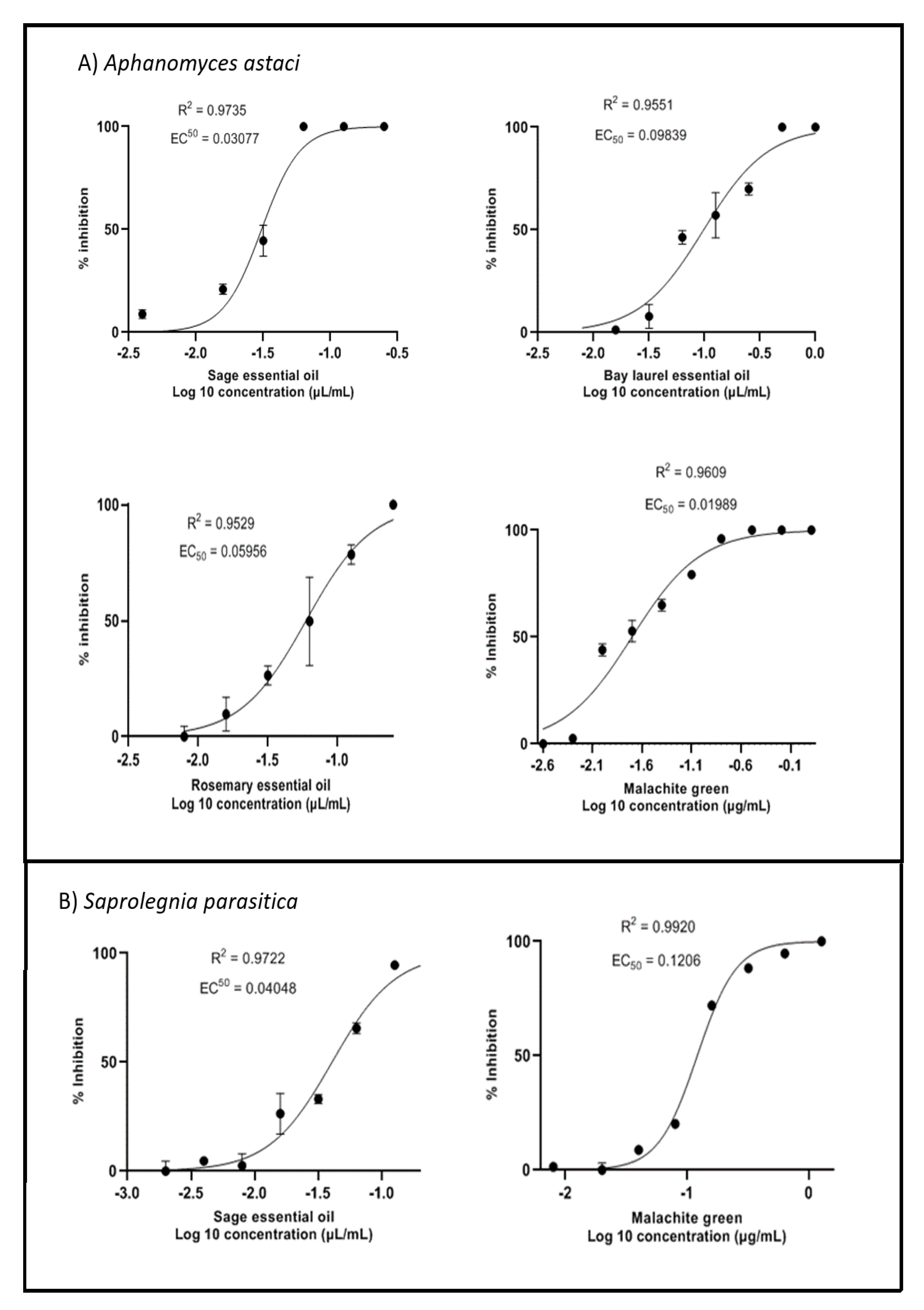
2.2. Inhibition of Mycelial Growth

All essential oils tested inhibited mycelial growth of A. astaci (Figure 2, Table 1). Sage essential oil was the most effective, with an EC50 value (i.e., the concentration of the sample at which the mycelial growth was inhibited by 50%) two times lower than that of rosemary and three times lower than that of bay laurel (EC50 values were 0.03 µL/mL, 0.06 µL/mL, and 0.10 µL/mL, respectively). In the case of S. parasitica, only sage essential oil showed a significant inhibitory effect on mycelial growth (EC50 = 0.04 µL/mL), while rosemary and bay laurel essential oils showed no such effect, and 100% inhibition could not be achieved at the concentrations tested, so EC50 values could not be calculated (Figure 2, Table 1). Thus, the mycelium of A. astaci was more sensitive to essential oils of selected Mediterranean plants than the mycelium of S. parasitica: the EC50 value for the effect of sage essential oil on the mycelium of S. parasitica was higher than the corresponding EC50 value for A. astaci (0.04 vs. 0.03 µL/mL), while the effect of rosemary and bay laurel essential oils was significantly more pronounced towards A. astaci (data for S. parasitica not shown). Finally, the mycelium of S. parasitica was six times more resistant to malachite green (the EC50 value of malachite green for A. astaci was 0.02 µg/mL, while for S. parasitica it was 0.12 µg/mL).

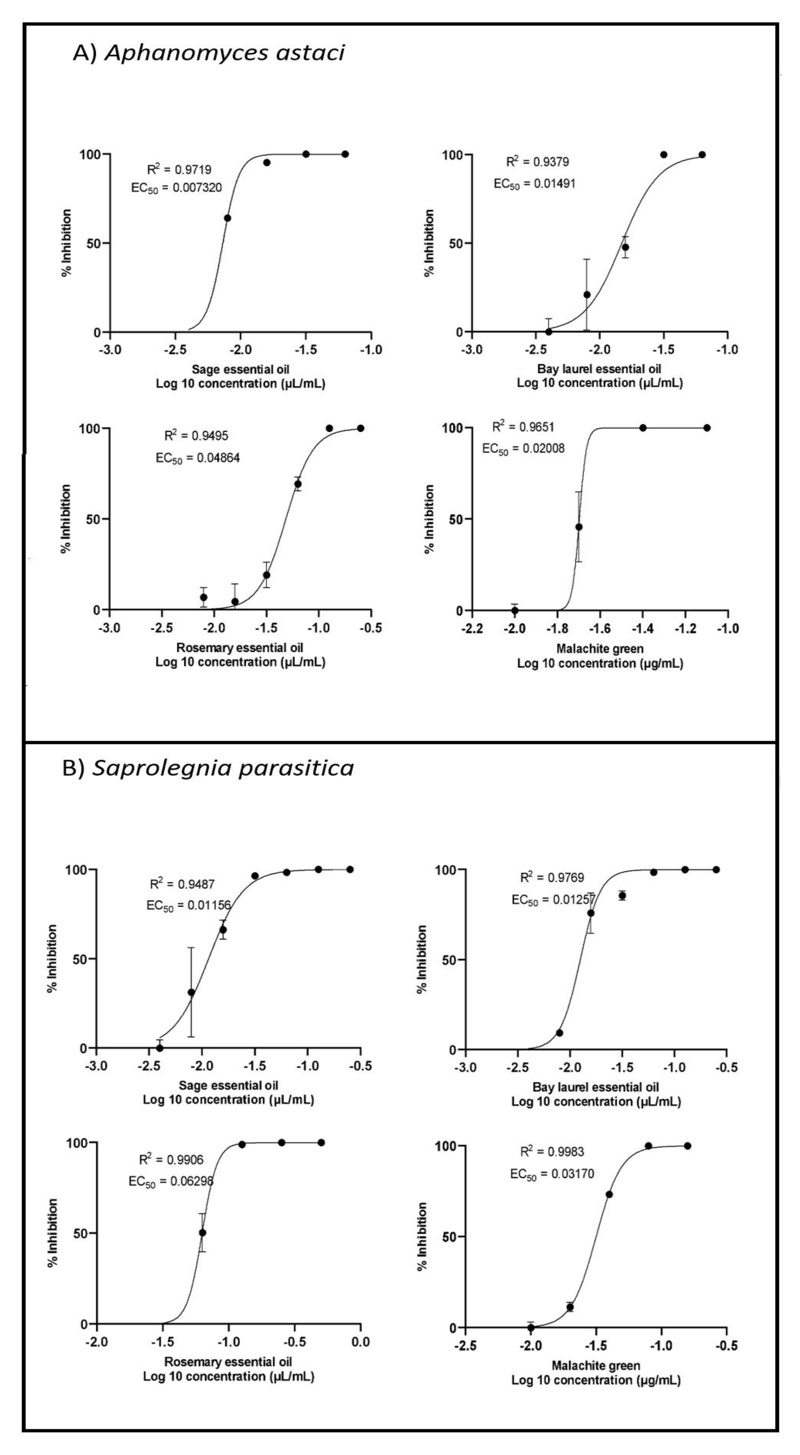
2.3. Inhibition of Zoospore Germination

All essential oils inhibited the germination of zoospores of A. astaci and S. parasitica, with EC50 values ranging from 0.007 µL/mL to 0.063 µL/mL (Figure 3, Table 1). Among the essential oils tested, sage essential oil showed the strongest potency in inhibiting zoospore germination, followed by bay laurel essential oil and finally rosemary essential oil. In addition, the zoospores of A. astaci were slightly more sensitive to rosemary and sage essential oils (and malachite green) than the zoospores of S. parasitica, while the sensitivity of zoospores of both species to bay laurel essential oil was similar. When comparing the sensitivity of different life stages of pathogenic oomycetes, the zoospores were 4–6 times more sensitive than the mycelium to the effect of the tested samples. The only exception to this trend was the effect of rosemary essential oil and malachite green on the life stages of A. astaci, for which similar concentrations were required to inhibit zoospore germination and mycelial growth.

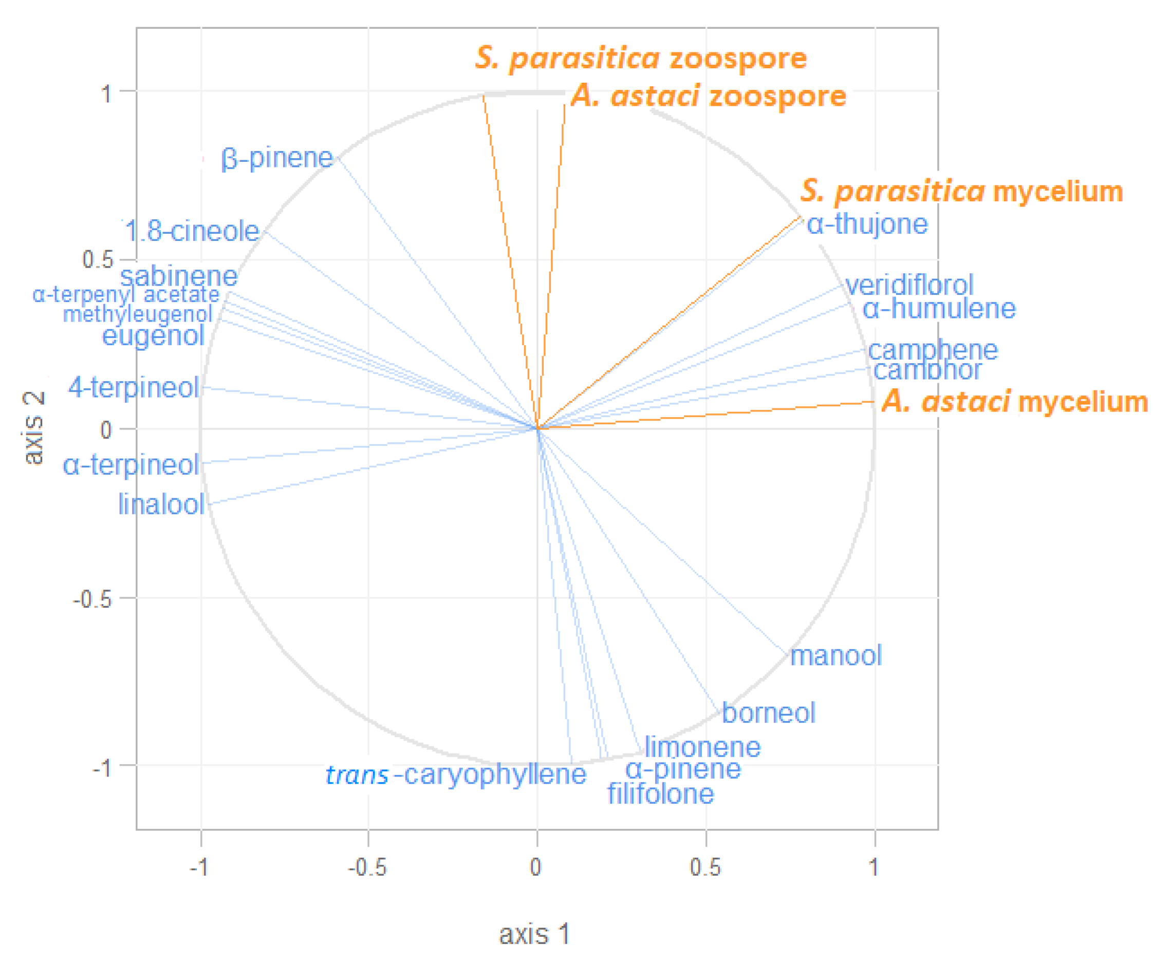
2.4. Correlation of the Observed Inhibitory Effects and Representation of Different Volatiles in the Essential Oils Studied

The multivariate partial least-squares regression (PLS-R2) technique was used to investigate the relationship between the observed inhibitory effects (EC50 values for mycelial growth and zoospore inhibition) and the representation of different volatiles in the essential oils of sage, rosemary, and bay laurel. The relationship between the predictor variables (different volatiles) and the response variables (EC50 values) is visually represented in the form of a correlation radar (Figure 4). The results showed that camphor, camphene, α-humulene, α-thujone, and veridiflorol were positively correlated with the inhibition of mycelial growth and moderately correlated with the inhibition of zoospore germination of both pathogens. In addition, β-pinene and 1,8-cineole were positively correlated with the inhibition of zoospore germination of S. parasitica.

3. Discussion

We demonstrated for the first time the inhibitory potential of sage, bay laurel, and rosemary essential oils, as natural substances rich in volatile bioactive constituents, against mycelial growth and zoospore germination of two oomycete pathogens important in aquaculture, S. parasitica and A. astaci.
The essential oils tested showed significant inhibition of mycelial growth of A. astaci and, in the case of sage essential oil, inhibition of mycelial growth of S. parasitica. This is the first study to report the inhibitory effect of sage essential oil on the mycelium of S. parasitica, although the existing literature indicates the strong potential of plants from the Lamiaceae and Lauraceae families to inhibit the mycelial growth of S. parasitica [32,36,37,44]. However, it should be noted that previously reported inhibitory concentrations were up to three orders of magnitude higher than those determined in this study (i.e., 0.1–100 µL/mL compared to ~0.05 µL/mL). This may be partially explained by methodological differences, as the inhibitory concentrations for the disk diffusion assay (usually reported in the literature) are generally higher than the inhibitory concentrations for the agar dilution assay used here [45]. It is also possible that sage essential oil has stronger bioactive properties than previously tested plants from the Lamiaceae and Lauraceae families, such as oregano (Origanum onites), thyme (Thymbra spicata), savory (Satureja cuneifolia), and cinnamon (Cinnamomum verum) [32,36,44,46]. Moreover, this is the first report of the inhibitory effect of essential oils against A. astaci. Nevertheless, some authors reported a good inhibitory effect of tea tree oil against the mycelial growth of the fish pathogen A. invadans [28] and of 38 essential oils, including rosemary essential oil and essential oils from some other Lauraceae and Lamiaceae plants, against the mycelial growth of the plant pathogen A. euteiches [40].
Most of the existing studies focused on the inhibition of mycelial growth, while there are very few reports on the inhibition of zoospores, which are the infective stage [35,39]. Our study shows a good inhibitory activity of the tested essential oils against zoospore germination of A. astaci and S. parasitica, with EC50 values ranging from 0.007 to 0.063 µL/mL and sage essential oil being the most potent. Similarly, the essential oils of Mentha longifolia and Thymus daenensis (Lamiaceae) completely inhibited the germination of S. parasitica zoospores, but at much higher concentrations of 2.5 and 5 µL/mL, respectively [39]. This discrepancy can be partly explained by different protocols used but nevertheless indicates the promising properties of the essential oils tested here, especially sage.
We observed differences between species in sensitivity to the effects of the essential oils tested. The mycelium of A. astaci was much more sensitive than the mycelium of S. parasitica, and the same trend was observed for zoospores, although less pronounced. The higher sensitivity of mycelium and zoospores of Aphanomyces spp. compared to those of Saprolegnia spp. has been reported previously. Namely, an absolute ethanol extract of Cassia fistula (Fabaceae) inhibited mycelial growth of S. parasitica and S. diclina at 2000 µg/mL, compared to 500 µg/mL required for the inhibition of A. invadans, and similar values were obtained for zoospore germination [26].
The inhibitory effect of the essential oils tested differed markedly between oomycete life cycle stages, with zoospores being 1.2–6.5 times more sensitive than mycelium. For example, essential oils of bay laurel and rosemary showed strong inhibitory activity against zoospore germination of S. parasitica but had only a weak effect on mycelial growth. Previous studies comparing the sensitivity of mycelium and zoospores of pathogenic oomycetes to various compounds showed different results [26,27,35,47,48]. For example, the EC50 values of propamocarb hydrochloride for the inhibition of mycelial growth were two or more orders of magnitude higher than the EC50 values for the germination of zoospores of different isolates of Phytophthora nicotianae [47]. In another study, the concentrations of an absolute ethanol extract of Cassia fistula required to inhibit zoospore germination of S. parasitica, S. diclina, and A. invadans were similar to the concentrations required to inhibit mycelial growth [26]. Finally, the concentrations of Laureliopsis philippiana essential oil from bark and leaf required to inhibit zoospore formation of S. parasitica and S. australis were equal to or higher than the concentrations required to inhibit mycelial growth [35]. All of these studies, including the results presented here, suggest that some compounds are more potent zoospore inhibitors (such as the essential oils of sage, rosemary, and bay laurel), while others preferentially target the mycelium, or both life stages. This likely reflects differences in the mode of action of different compounds on zoospores and mycelium, as well as different detoxification mechanisms in different life stages of oomycetes, but further studies, including transcriptomic and proteomic analyses, are needed to clarify this.
The observed inhibitory activities of the essential oils could be attributed to their rich content of bioactive volatiles (mainly monoterpenes, such as camphor, and sesquiterpenes) and the synergistic activities of numerous minor compounds. The chemical composition of the tested essential oils was similar to those previously reported by our group [49] and in other studies [50,51,52,53,54]. Sage essential oil showed the strongest inhibitory potential on both pathogens and both life cycle stages. As indicated by PLS-R2 analysis, its anti-oomycete activity could be attributed to some of its major constituents, camphor, α-thujone, veridiflorol, camphene, and α-humulene, which were absent or present at low levels in other essential oils. In addition, β-pinene and 1,8-cineole (present in significant amounts in both sage and bay laurel essential oils) were positively correlated with the inhibition of S. parasitica zoospore germination, explaining the similar EC50 values of these two essential oils for S. parasitica zoospore germination (EC50 0.012 and 0.013 µL/mL, respectively). Some of these compounds were previously reported to exhibit good anti-oomycete [44,55] and antifungal [56,57,58,59] activity. For example, camphor (up to 38.06 µg/mL) progressively slowed down the mycelial growth of S. parasitica and S. delica, while thujone and β-pinene (500 and 1000 µg/mL, respectively) inhibited the mycelial growth of S. parasitica [44,55]. Moreover, α-thujone and camphor have potent antifungal activity against Fusarium graminearum, F. culmorum, and Schizosaccharomyces pombe, which is mainly explained by the induction of oxidative stress and subsequent apoptotic cell death, but also by a decrease in genomic stability and epigenetic changes [56,57,58,59]. Thus, the high camphor content in sage essential oil probably contributed significantly to the observed inhibitory effects. The mechanism underlying the inhibition of oomycetes by camphor remains to be investigated but could be due to oxidative stress-mediated apoptosis, similar to the data obtained for fungal cells. However, it should be noted that it has previously been shown that the synergistic effect of many compounds present in essential oils is stronger than that of any single compound [35,44], and this was probably the case here.
Based on the obtained results, we propose that the essential oils of wild Mediterranean plants, especially sage, could be used as an ecologically acceptable method to control A. astaci and S. parasitica in aquaculture. This will require in vivo experiments to test the applicability of these essential oils either by dietary supplementation or by bathing the eggs and animals in essential oil suspensions. Previous studies suggest that the application of some essential oils and plant extracts may be useful in controlling Saprolegnia and Aphanomyces spp. infections in eggs and/or adults. For example, repeated incubation of S. parasitica-infected rainbow trout eggs with some essential oils, such as those of Zataria multiflora and Satureja cuneifolia at concentrations of 5 ppm or higher, resulted in an increase in hatching rate [34,36]. In addition, dietary supplementation with various essential oils, including sage essential oil and plant extracts, improved the immune response, fatty acid utilization, and growth performance of rainbow trout [60,61] and even conferred resistance to A. invadans infection in Indian major carp (Labeo rohita) [62]. In addition, immersion of infected fish in water containing 1% aqueous leaf extract of Azadirachta indica for 5 min daily for 24 days resulted in gradual healing of induced lesions in Chana striata [63]. However, some of the current constraints to large-scale application of essential oils, such as high extraction costs and variations in the composition of essential oils obtained from plants from different locations and seasons, have yet to be addressed. Production costs could be reduced by including inexpensive pretreatments prior to hydrodistillation that improve essential oils yields [49], while the quality of stock solutions could be standardized by optimizing growing conditions and harvest timing and by using genetic engineering [64,65]. Therefore, further experiments are needed as an extension of the results presented here to open the prospect of developing an ecologically acceptable control of S. parasitica and A. astaci infections through the application of essential oils. In particular, the possible toxicity of the effective concentrations of essential oils to fish/crayfish, as well as the possible rejection of the essential oil-supplemented feed due to their strong taste and odor [66], must be excluded.

4. Conclusions

The essential oils of Mediterranean wild plants are rich in bioactive volatiles, and in this study we demonstrated their potent activity against the pathogens A. astaci and S. parasitica. Thus, our results open a perspective for an environmentally friendly and sustainable control of diseases caused by oomycetes in salmonid and crayfish aquaculture through the application of essential oils. The inhibitory effect was particularly pronounced for sage essential oil, and some of its most abundant components, such as camphor, may be the major inhibitory molecules, although a synergistic effect of many minor components is also likely. Following our results, further studies are needed to develop protocols for the administration of essential oils as egg/animal baths or feed supplements and to standardize the production of essential oils on a large scale.

5. Materials and Methods

5.1. Microorganisms

Two oomycete pathogens of freshwater animals used were: Aphanomyces astaci (Schikora, 1906) strain B, PsI genotype (isolate PEC 8), and Saprolegnia parasitica Coker isolate CBS 223.65. Aphanomyces astaci was provided by F. Grandjean (University of Poitiers, Poitiers, France) and belonged to genotype PsI, which has been shown to exhibit marked virulence, particularly against native European crayfish species such as the noble crayfish Astacus astacus [67,68]. Saprolegnia parasitica CBS 223.65 is a reference strain isolated in the Netherlands from northern pike (Esox lucius) and was provided by R. Galuppi (University of Bologna, Bologna, Italy). Microorganisms were maintained in the laboratory at 18 °C on PG1 solid medium supplemented with ampicillin and oxolinic acid [69].

5.2. Plant Material and Essential Oil Isolation

Fresh leaves were collected from wild plants of rosemary (Rosmarinus officinalis), sage (Salvia officinalis), and bay laurel (Laurus nobilis) in the south Mediterranean region of Croatia in August 2020 and air-dried at room temperature (20 ± 2 °C) for one week. Dry leaves were packed in polyethylene bags and kept in a dark, dry, and cool place. Before being used for the extractions, all leaves were separated from branches and stems, and the bay laurel leaves were cut into four pieces each. The plant material was then ground using a household blender (Tefal) for 20 s into fine powder. The essential oils were isolated by hydrodistillation with reflux extraction pretreatment from 20 g of ground plant material mixed with 250 mL of purified water according to a previously developed protocol [49], yielding 0.2, 0.5, and 0.3 mL of oil/g of dry plant material of rosemary, sage, and bay laurel, respectively. For subsequent testing of anti-oomycete activity, the essential oils were diluted 1:9 in 96% ethanol. These 100 µL/mL stock solutions were then further diluted while performing the inhibition assays described in Section 5.4.

5.3. Gas Chromatography–Mass Spectrometry (GC-MS) Analysis

GC–MS analyses were carried out with an Agilent Technologies (Palo Alto, CA, USA) gas chromatograph model 7890 A equipped with a mass-selective detector (MSD) model 5975 C (Agilent Technologies, Palo Alto, CA, USA) and an HP-5MS 5% phenyl-methylpolysiloxane capillary column (30 m × 0.25 mm, 0.25 μm film thickness). In brief, the injector and detector temperatures were 250 °C and 300 °C, respectively; the column temperature was held at 70 °C for 2 min, was then increased from 70 to 200 °C at 3 °C/min, and was finally held at 200 °C for 18 min; 1.0 μL of the sample was injected using split mode (split ratio 1:50). Helium was used as a carrier gas (1.0 mL/min). The MSD (EI mode) was operated at 70 eV, and the scan range was set to 30–350 amu. Identification of volatile constituents was based on the comparison of their retention indices (RIs), determined relative to the retention times of a homologous series of n-alkanes (C9–C25), with those reported in the literature, and their mass spectra with those of authentic compounds available in our laboratories or those listed in NIST 17 (D-Gaithersburg, MD, USA) and Wiley W9N08 (Wiley, New York, NY, USA). Relative concentrations of components were calculated by the area normalization method without considering response factors.

5.4. Testing of Anti-Oomycete Activity of Essential Oils

5.4.1. Inhibition of Mycelial Growth

The inhibitory effect of sage, bay laurel, and rosemary essential oils on mycelial growth of S. parasitica and A. astaci was tested using a radial growth inhibition assay followed by the determination of EC50 values (i.e., concentrations of samples at which mycelial growth is inhibited by 50%), as previously described [48], with some modifications. Malachite green, a known oomycete inhibitor [70], was used as a positive control (stock solution was 512 µg/mL in distilled water). Briefly, 100 µL of each sample was dissolved in 10 mL of molten PG1 medium and poured into 550 mm radius Petri dishes. In control plates, solvents were added in place of the samples, i.e., ethanol was used as a negative control for essential oils, and distilled water for malachite green. Up to eight twofold dilutions were tested for each sample, with initial concentrations of 1 µL/mL for essential oils and 1.28 µg/mL for malachite green. Each plate was inoculated by placing a 5 mm agar plug containing mycelium (taken from the edge of an actively growing mycelial mat) in the center and incubated at 18 °C. There was no statistically significant difference in the growth of the test species in the presence of ethanol or distilled water (t-test; p > 0.05). Three biological replicates were performed for each oomycete species, sample, and concentration. The assay was terminated after the mycelium in the negative control plates reached the end of the Petri dish, i.e., after five days for A. astaci and after two days for S. parasitica. Next, two perpendicular measurements of mycelial radius were taken for each plate and averaged, and the agar plug size was subtracted to obtain the final measurements of radial growth. Measurements from the sample plates were subtracted from the negative control measurements and converted to percent inhibition.

5.4.2. Inhibition of Zoospore Germination

Sporulation of A. astaci and S. parasitica was induced by washing a grown mycelium incubated at 18 °C with sterile stream water, as described in detail in previous studies [67,71]. Zoospores were counted in the Thoma chamber using a light microscope (Zeiss Primo Star, Carl Zeiss, Oberkochen, Germany) at 100× magnification. The final concentration of zoospores was approximately 100,000 zoospores/mL for A. astaci and 80,000 zoospores/mL for S. parasitica.
To assess the effect of essential oils on the germination of zoospores of A. astaci and S. parasitica, we used previously described protocols with some modifications [48,71,72]. Up to six twofold dilutions of the test samples were used, with starting concentrations of 0.125 µL/mL for essential oils and 2.54 µg/mL for malachite green. To induce germination of A. astaci zoospores, 2 mL of stream water with the addition of CaCl2 (11.1 g/L) was mixed with 20 µL of the essential oil samples and 2 mL of water with zoospores and incubated in 12-well plates at room temperature for 16 h. In the case of S. parasitica, zoospores were vortexed for 45 s, then 2 mL of water with zoospore suspension was mixed with 2 mL of fresh PG1 liquid medium and 20 µL of essential oil samples and incubated for 1 h at 18 °C. After incubation, samples were photographed using an inverted microscope (Carl Zeiss, Oberkochen, Germany) at 200× magnification. Three replicates were made for each species, sample, and dilution. The percentage of germinating spores was determined by counting at least 200 spores in three to four randomly selected objective fields. A spore with a germ tube of at least one cyst diameter in length was counted as germinated. The germination percentage in the wells of negative controls to which solvents were added instead of samples was 33.4% and 97.5% for A. astaci and S. parasitica, respectively, in agreement with previously published data [71,72,73]. The germination percentages obtained for the different samples were converted to percent germination inhibition values.

5.5. Statistical Analysis

The heatmap of the volatile chemical composition of the essential oils of rosemary, bay laurel, and sage obtained by GC–MS was generated using the heatmaply package in R v. 3.2.0 [74] and the default methods for calculating the distance matrix (“euclidean”).
To estimate EC50 values for inhibition of mycelial growth and zoospore germination (i.e., concentrations of samples at which mycelial growth/zoospore germination was 50% inhibited), compound concentrations were log-transformed, data normalized, and nonlinear regression with curve fitting (by least squares) was performed using GraphPad Prism version 9.
To investigate the effects of different volatiles on the inhibitory potential of sage, rosemary, and bay laurel essential oils against A. astaci and S. parasitica, the partial least-squares regression (PLS-R2) approach was used. The response variables were the EC50 values for mycelial growth and zoospore germination, while the volatile bioactive compounds detected in the essential oils with at least 2% of the total GC–MS peak area served as predictors. PLS-R2 analysis was performed using the package “plsdepot” [75] in the program R v. 3.2.0 [74].

Supplementary Materials

The following are available online at https://0-www-mdpi-com.brum.beds.ac.uk/article/10.3390/plants10081676/s1, Table S1: Volatile composition of essential oils determined by GC–MS.

Author Contributions

Conceptualization, A.B.; methodology, A.M., M.D., D.G., D.P., I.J., Z.M. and A.B.; validation, A.M., A.B. and I.J.; formal analysis, D.G.; investigation, A.M., D.P., M.D., Z.M. and I.J.; resources, A.B.; writing—original draft preparation, A.M.; writing—review and editing, A.B.; visualization, A.M. and D.G.; supervision, A.B.; project administration, A.B. and D.G.; funding acquisition, A.B. All authors have read and agreed to the published version of the manuscript.

Funding

This research was funded by the project “Interactions of freshwater pathogenic oomycetes and the environment” (InteractOomyc, UIP-2017-05-6267), Croatian Science Foundation.

Acknowledgments

The authors are thankful to F. Grandjean and R. Galuppi for providing the A. astaci and S. parasitica isolates and to Ivana Čakarić and Ana Kozina for help in the laboratory work.

Conflicts of Interest

The authors declare no conflict of interest.

References

  1. Van den Berg, A.H.; McLaggan, D.; Diéguez-Uribeondo, J.; van West, P. The impact of the water moulds Saprolegnia diclina and Saprolegnia parasitica on natural ecosystems and the aquaculture industry. Fungal Biol. Rev. 2013, 27, 33–42. [Google Scholar] [CrossRef]
  2. Harlioǧlu, M.M. The harvest of the freshwater crayfish Astacus leptodactylus Eschscholtz in Turkey: Harvest history, impact of crayfish plague, and present distribution of harvested populations. Aquac. Int. 2008, 16, 351–360. [Google Scholar] [CrossRef]
  3. Holdich, D.M. A review of astaciculture: Freshwater crayfish farming. Aquat. Living Resour. 1993, 6, 307–317. [Google Scholar] [CrossRef]
  4. Souty-Grosset, C.; Reynolds, J.D. Current ideas on methodological approaches in European crayfish conservation and restocking procedures. Knowl. Manag. Aquat. Ecosyst. 2009, 1, 394–395. [Google Scholar] [CrossRef]
  5. Sarowar, M.N.; Hossain, M.J.; Nasrin, T.; Naznin, T.; Hossain, Z.; Rahman, M.M. Molecular identification of oomycete species affecting aquaculture in Bangladesh. Aquac. Fish. 2019, 4, 105–113. [Google Scholar] [CrossRef]
  6. Stueland, S.; Hatai, K.; Skaar, I. Morphological and physiological characteristics of Saprolegnia spp. strains pathogenic to Atlantic salmon, Salmo salar L. J. Fish Dis. 2005, 28, 445–453. [Google Scholar] [CrossRef]
  7. Thoen, E.; Evensen, Ø.; Skaar, I. Pathogenicity of Saprolegnia spp. to Atlantic salmon, Salmo salar L., eggs. J. Fish Dis. 2011, 34, 601–608. [Google Scholar] [CrossRef]
  8. Diéguez-Uribeondo, J.; Cerenius, L.; Söderhäll, K. Saprolegnia parasitica and its virulence on three different species of freshwater crayfish. Aquaculture 1994, 120, 219–228. [Google Scholar] [CrossRef]
  9. Liu, Y.; De Bruijn, I.; Jack, A.L.; Drynan, K.; van Den Berg, A.H.; Thoen, E.; Sandoval-Sierra, V.; Skaar, I.; van West, P.; Diéguez-Uribeondo, J.; et al. Deciphering microbial landscapes of fish eggs to mitigate emerging diseases. ISME J. 2014, 8, 2002–2014. [Google Scholar] [CrossRef]
  10. Edsman, L.; Nyström, P.; Sandström, A.; Stenberg, M.; Kokko, H.; Tiitinen, V.; Makkonen, J.; Jussila, J. Eroded swimmeret syndrome in female crayfish Pacifastacus leniusculus associated with Aphanomyces astaci and Fusarium spp. infections. Dis. Aquat. Organ. 2015, 112, 219–228. [Google Scholar] [CrossRef]
  11. Kokko, H.; Koistinen, L.; Harlioǧlu, M.M.; Makkonen, J.; Aydin, H.; Jussila, J. Des populations turques d’écrevisses à pattes grêles (Astacus leptodactylus) productives porteuses d’Aphanomyces astaci. Knowl. Manag. Aquat. Ecosyst. 2012, 404, 12. [Google Scholar] [CrossRef] [Green Version]
  12. Aydin, H.; Kokko, H.; Makkonen, J.; Kortet, R.; Kukkonen, H.; Jussila, J. The signal crayfish is vulnerable to both the As and the PsI-isolates of the crayfish plague. Knowl. Manag. Aquat. Ecosyst. 2014, 413, 03. [Google Scholar] [CrossRef] [Green Version]
  13. Sandström, A.; Andersson, M.; Asp, A.; Bohman, P.; Edsman, L.; Engdahl, F.; Nyström, P.; Stenberg, M.; Hertonsson, P.; Vrålstad, T.; et al. Population collapses in introduced non-indigenous crayfish. Biol. Invasions 2014, 16, 1961–1977. [Google Scholar] [CrossRef]
  14. Edgerton, B.F.; Evans, L.H.; Stephens, F.J.; Overstreet, R.M. Review article: Synopsis of freshwater crayfish diseases and commensal organisms. Aquaculture 2002, 206, 57–135. [Google Scholar] [CrossRef] [Green Version]
  15. EC. Council Regulation 2377/90/EEC of 26 June 1990 on laying down a Community procedure for the establishment of maximum residue limits of veterinary medicinal products in foodstuff of animal origin. Off. J. Eur. Commun. 1990, 224, 1–12. [Google Scholar]
  16. Marking, L.L.; Rach, J.J.; Schreier, T.M. American Fisheries Society Evaluation of Antifungal Agents for Fish Culture. Progress. Fish-Culturist 1994, 56, 225–231. [Google Scholar] [CrossRef]
  17. Meyer, F.P.; Jorgenson, T.A. Teratological and Other Effects of Malachite Green on Development of Rainbow Trout and Rabbits. Trans. Am. Fish. Soc. 1983, 112, 818–824. [Google Scholar] [CrossRef]
  18. Panandiker, A.; Fernandes, C.; Rao, K.V.K. The cytotoxic properties of malachite green are associated with the increased demethylase, aryl hydrocarbon hydroxylase and lipid peroxidation in primary cultures of Syrian hamster embryo cells. Cancer Lett. 1992, 67, 93–101. [Google Scholar] [CrossRef]
  19. Srivastava, S.; Sinha, R.; Roy, D. Toxicological effects of malachite green. Aquat. Toxicol. 2004, 66, 319–329. [Google Scholar] [CrossRef]
  20. Wooster, G.; Martinez, C.; Bowser, P.; O’Hara, D. Human Health Risks Associated with Formalin Treatments Used in Aquaculture: Initial Study. N. Am. J. Aquac. 2005, 67, 111–113. [Google Scholar] [CrossRef]
  21. Norliana, S.; Abdulamir, A.S.; Abu Bakar, F.; Salleh, A.B. The health risk of formaldehyde to human beings. Am. J. Pharmacol. Toxicol. 2009, 4, 98–106. [Google Scholar] [CrossRef]
  22. Cui, N.; Zhang, X.; Xie, Q.; Wang, S.; Chen, J.; Huang, L.; Qiao, X.; Li, X.; Cai, X. Toxicity profile of labile preservative bronopol in water: The role of more persistent and toxic transformation products. Environ. Pollut. 2011, 159, 609–615. [Google Scholar] [CrossRef]
  23. Jacob, A.P.; Culver, D.A.; Lanno, R.P.; Voigt, A. Ecological impacts of fluridone and copper sulphate in catfish aquaculture ponds. Environ. Toxicol. Chem. 2016, 35, 1183–1194. [Google Scholar] [CrossRef] [PubMed]
  24. Jussila, J.; Toljamo, A.; Makkonen, J.; Kukkonen, H.; Kokko, H. Practical disinfection chemicals for fishing and crayfishing gear against crayfish plague transfer. Knowl. Manag. Aquat. Ecosyst. 2014, 413, 02. [Google Scholar] [CrossRef] [Green Version]
  25. Afzali, S.F.; Wong, W.L. In vitro screening of Sonneratia alba extract against the oomycete fish pathogen, Aphanomyces invadans. Iran. J. Fish. Sci. 2017, 16, 1333–1340. [Google Scholar] [CrossRef]
  26. Borisutpeth, M.; Kanbutra, P.; Weerakhun, S.; Wada, S.; Hatai, K. In vitro antifungal activity of Cassia fistula L. against selected pathogenic water molds. Int. J. Phytomed. 2014, 6, 237–242. [Google Scholar] [CrossRef]
  27. Borisutpeth, P.; Kanbutra, P.; Hanjavanit, C.; Chukanhom, K.; Funaki, D.; Hatai, K. Effects of Thai Herbs on the Control of Fungal Infection in Tilapia Eggs and the Toxicity to the Eggs. Aquac. Sci. 2009, 57, 475–482. [Google Scholar] [CrossRef]
  28. Campbell, R.E.; Lilley, J.H.; Taukhid; Panyawachira, V.; Kanchanakhan, S. In vitro screening of novel treatments for Aphanomyces invadans. Aquac. Res. 2001, 32, 223–233. [Google Scholar] [CrossRef]
  29. Pagliarulo, C.; Sateriale, D.; Scioscia, E.; De Tommasi, N.; Colicchio, R.; Pagliuca, C.; Scaglione, E.; Jussila, J.; Makkonen, J.; Salvatore, P.; et al. Growth, survival and spore formation of the pathogenic aquatic oomycete Aphanomyces astaci and Fungus fusarium avenaceum are inhibited by Zanthoxylum rhoifolium bark extracts in vitro. Fishes 2018, 3, 12. [Google Scholar] [CrossRef] [Green Version]
  30. Tandel, R.S.; Chadha, N.K.; Dash, P.; Sawant, P.B.; Pandey, N.N.; Chandra, S.; Bhat, R.A.H.; Thakuria, D. An in-vitro study of Himalayan plant extracts against oomycetes disease Saprolegniasis in rainbow trout (Oncorhynchus mykiss). J. Environ. Biol. 2021, 42, 1008–1018. [Google Scholar] [CrossRef]
  31. Caruana, S.; Yoon, G.H.; Freeman, M.A.; Mackie, J.A.; Shinn, A.P. The efficacy of selected plant extracts and bioflavonoids in controlling infections of Saprolegnia australis (Saprolegniales; Oomycetes). Aquaculture 2012, 358–359, 146–154. [Google Scholar] [CrossRef]
  32. Gormez, O.; Diler, O. In vitro antifungal activity of essential oils from Tymbra, Origanum, Satureja species and some pure compounds on the fish pathogenic fungus, Saprolegnia parasitica. Aquac. Res. 2014, 45, 1196–1201. [Google Scholar] [CrossRef]
  33. Hoskonen, P.; Heikkinen, J.; Eskelinen, P.; Pirhonen, J. Efficacy of clove oil and ethanol against Saprolegnia sp. and usability as antifungal agents during incubation of rainbow trout Oncorhynchus mykiss (Walbaum) eggs. Aquac. Res. 2015, 46, 581–589. [Google Scholar] [CrossRef]
  34. Khosravi, A.R.; Shokri, H.; Sharifrohani, M.; Mousavi, H.E.; Moosavi, Z. Evaluation of the antifungal activity of Zataria multiflora, Geranium herbarium, and Eucalyptus camaldolensis essential oils on Saprolegnia parasitica-infected rainbow trout (Oncorhynchus mykiss) eggs. Foodborne Pathog. Dis. 2012, 9, 674–679. [Google Scholar] [CrossRef] [Green Version]
  35. Madrid, A.; Godoy, P.; González, S.; Zaror, L.; Moller, A.; Werner, E.; Cuellar, M.; Villena, J.; Montenegro, I. Chemical characterization and anti-oomycete activity of Laureliopsis philippianna essential oils against Saprolegnia parasitica and S. australis. Molecules 2015, 20, 8033–8047. [Google Scholar] [CrossRef] [Green Version]
  36. Metin, S.; Diler, O.; Didinen, B.I.; Terzioglu, S.; Gormez, O. In vitro and in vivo antifungal activity of Satureja cuneifolia ten essential oil on Saprolegnia parasitica strains isolated from rainbow trout (Oncorhynchus mykiss, Walbaum) eggs. Aquac. Res. 2015, 46, 1396–1402. [Google Scholar] [CrossRef]
  37. Nardoni, S.; Najar, B.; Fronte, B.; Pistelli, L.; Mancianti, F. In vitro activity of essential oils against Saprolegnia parasitica. Molecules 2019, 24, 1270. [Google Scholar] [CrossRef] [Green Version]
  38. Pirbalouti, A.G.; Taheri, M.; Raisee, M.; Bahrami, H.R.; Abdizadeh, R. In vitro antifungal activity of plant extracts on Saprolegnia parasitica from cutaneous lesions of rainbow trout (Oncorhynchus mykiss) eggs. J. Food Agric. Environ. 2009, 7, 94–96. [Google Scholar]
  39. Saleh, M.; Soltani, M.; Islami, H.R. In vitro antifungal activity of some essential oils against some filamentous fungi of rainbow trout (Oncorhynchus mykiss) eggs. AACL Bioflux 2015, 8, 367–380. [Google Scholar]
  40. Parikh, L.; Agindotan, B.O.; Burrows, M.E. Antifungal Activity of Plant-Derived Essential Oils on Pathogens of Pulse Crops. Plant Dis. 2021. [Google Scholar] [CrossRef]
  41. Jiang, Y.; Wu, N.; Fu, Y.J.; Wang, W.; Luo, M.; Zhao, C.J.; Zu, Y.G.; Liu, X.L. Chemical composition and antimicrobial activity of the essential oil of Rosemary. Environ. Toxicol. Pharmacol. 2011, 32, 63–68. [Google Scholar] [CrossRef]
  42. Özçakmak, S.; Muhammet, D.; Azime, Y. Antifungal activity of lemon balm and sage essential oils on the growth of ochratoxigenic Penicillium verrucosum. Afr. J. Microbiol. Res. 2012, 6, 3079–3084. [Google Scholar] [CrossRef] [Green Version]
  43. Fidan, H.; Stefanova, G.; Kostova, I.; Stankov, S.; Damyanova, S.; Stoyanova, A.; Zheljazkov, V.D. Chemical Composition and Antimicrobial Activity of Laurus nobilis L. Essential oils from Bulgaria. Molecules 2019, 24, 804. [Google Scholar] [CrossRef] [PubMed] [Green Version]
  44. Tampieri, M.P.; Galuppi, R.; Carelle, M.S.; Macchioni, F.; Cioni, P.L.; Morelli, I. Effect of selected essential oils and pure compounds on Saprolegnia parasitica. Pharm. Biol. 2003, 41, 584–591. [Google Scholar] [CrossRef] [Green Version]
  45. Klancnik, A.; Guzej, B.; Kolar, M.H.; Abramovic, H.; Mozina, S.S. In vitro antimicrobial and antioxidant activity of commercial rosemary extract formulations. J. Food Prot. 2009, 72, 1174–1752. [Google Scholar] [CrossRef]
  46. Stević, T.; Berić, T.; Šavikin, K.; Soković, M.; Gođevac, D.; Dimkić, I.; Stanković, S. Antifungal activity of selected essential oils against fungi isolated from medicinal plant. Ind. Crops Prod. 2014, 55, 116–122. [Google Scholar] [CrossRef]
  47. Hu, J.; Hong, C.; Stromberg, E.L.; Moorman, G.W. Effects of propamocarb hydrochloride on mycelial growth, sporulation, and infection by Phytophthora nicotianae isolates from Virginia nurseries. Plant Dis. 2007, 91, 414–420. [Google Scholar] [CrossRef]
  48. Lawrence, S.A.; Armstrong, C.B.; Patrick, W.M.; Gerth, M.L. High-throughput chemical screening identifies compounds that inhibit different stages of the Phytophthora agathidicida and Phytophthora cinnamomi life cycles. Front. Microbiol. 2017, 8, 1340. [Google Scholar] [CrossRef] [PubMed] [Green Version]
  49. Miljanović, A.; Bielen, A.; Grbin, D.; Marijanović, Z.; Andlar, M.; Rezić, T.; Roca, S.; Jerković, I.; Vikić-Topić, D.; Dent, M. Effect of enzymatic, ultrasound, and reflux extraction pretreatments on the chemical composition of essential oils. Molecules 2020, 25, 4818. [Google Scholar] [CrossRef] [PubMed]
  50. Boulila, A.; Hassen, I.; Haouari, L.; Mejri, F.; Amor, I.B.; Casabianca, H.; Hosni, K. Enzyme-assisted extraction of bioactive compounds from bay leaves (Laurus nobilis L.). Ind. Crops Prod. 2015, 74, 485–493. [Google Scholar] [CrossRef]
  51. Hatipoglu, S.D.; Zorlu, N.; Dirmenci, T.; Goren, A.C.; Ozturk, T.; Topcu, G. Determination of volatile organic compounds in fourty five salvia species by thermal desorption-GC-MS technique. Rec. Nat. Prod. 2016, 10, 659–700. [Google Scholar]
  52. Hosni, K.; Hassen, I.; Chaâbane, H.; Jemli, M.; Dallali, S.; Sebei, H.; Casabianca, H. Enzyme-assisted extraction of essential oils from thyme (Thymus capitatus L.) and rosemary (Rosmarinus officinalis L.): Impact on yield, chemical composition and antimicrobial activity. Ind. Crops Prod. 2013, 47, 291–299. [Google Scholar] [CrossRef]
  53. Olmedo, R.H.; Asensio, C.M.; Grosso, N.R. Thermal stability and antioxidant activity of essential oils from aromatic plants farmed in Argentina. Ind. Crops Prod. 2015, 69, 21–28. [Google Scholar] [CrossRef]
  54. Russo, A.; Formisano, C.; Rigano, D.; Senatore, F.; Delfine, S.; Cardile, V.; Rosselli, S.; Bruno, M. Chemical composition and anticancer activity of essential oils of Mediterranean sage (Salvia officinalis L.) grown in different environmental conditions. Food Chem. Toxicol. 2013, 55, 42–47. [Google Scholar] [CrossRef] [PubMed]
  55. Tedesco, P.; Beraldo, P.; Massimo, M.; Fioravanti, M.L.; Volpatti, M.; Dirks, R.; Galuppi, R. Comparative Therapeutic Effects of Natural Compounds Against Saprolegnia spp. (Oomycota) and Amyloodinium ocellatum (Dinophyceae). Front. Vet. Sci. 2020, 7, 83. [Google Scholar] [CrossRef] [PubMed]
  56. Teker, T.; Sefer, Ö.; Gazdağlı, A.; Yörük, E.; Varol, G.İ.; Albayrak, G. α-Thujone exhibits an antifungal activity against F. graminearum by inducing oxidative stress, apoptosis, epigenetics alterations and reduced toxin synthesis. Eur. J. Plant Pathol. 2021, 160, 611–622. [Google Scholar] [CrossRef]
  57. Gazdağlı, A.; Sefer, Ö.; Yörük, E.; Varol, G.İ.; Teker, T.; Albayrak, G. Investigation of Camphor Effects on Fusarium graminearum and F. culmorum at Different Molecular Levels. Pathogens 2018, 7, 90. [Google Scholar] [CrossRef] [PubMed] [Green Version]
  58. Agus, H.H.; Sengoz, C.O.; Yilmaz, S. Oxidative stress-mediated apoptotic cell death induced by camphor in sod1-deficient Schizosaccharomyces pombe. Toxicol. Res. 2019, 8, 216–226. [Google Scholar] [CrossRef] [Green Version]
  59. Agus, H.H.; Kok, G.; Derinoz, E.; Oncel, D.; Yilmaz, S. Involvement of Pca1 in ROS-mediated apoptotic cell death induced by alpha-thujone in the fission yeast (Schizosaccharomyces pombe). FEMS Yeast Res. 2020, 20, foaa022. [Google Scholar] [CrossRef]
  60. Sönmez, A.Y.; Bilen, S.; Albayrak, M.; Yilmaz, S.; Biswas, G.; Hisar, O.; Yanik, T. Effects of Dietary Supplementation of Herbal Oils Containing 1,8-cineole, Carvacrol or Pulegone on Growth Performance, Survival, Fatty Acid Composition, and Liver and Kidney Histology of Rainbow Trout (Oncorhynchus mykiss) Fingerlings. Turk. J. Fish. Aquat. Sci. 2015, 15, 813–819. [Google Scholar] [CrossRef]
  61. Sari, A.B.; Ustuner-Aydal, O. Antioxidant and Immunostimulant Effects of Origanum Minutiflorum O. Schwarz Et. Ph Davis in Rainbow Trout. Fresenius Environ. Bull. 2018, 27, 1013–1021. [Google Scholar]
  62. Yogeshwari, G.; Jagruthi, C.; Anbazahan, S.M.; Mari, L.S.S.; Selvanathan, J.; Arockiaraj, J.; Dhayanithi, N.B.; Ajithkumar, T.T.; Balasundaram, C.; Ramasamy, H. Herbal supplementation diet on immune response in Labeo rohita against Aphanomyces invadans. Aquaculture 2015, 437, 351–359. [Google Scholar] [CrossRef]
  63. Harikrishnan, R.; Balasundaram, C.; Bhuvaneswari, R. Restorative effect of Azadirachta indicab aqueous leaf extract dip treatment on haematological parameter changes in Cyprinus carpio (L.) experimentally infected with Aphanomyces invadans fungus. J. Appl. Ichthyol. 2005, 21, 410–413. [Google Scholar] [CrossRef]
  64. Pavela, R.; Benelli, G. Essential oils as ecofriendly biopesticides? Challenges and constraints. Trends Plant Sci. 2016, 21, 1000–1007. [Google Scholar] [CrossRef]
  65. Lange, B.M.; Croteau, R. Genetic engineering of essential oil production in mint. Curr. Opin. Plant Biol. 1999, 2, 139–144. [Google Scholar] [CrossRef]
  66. Stevanović, Z.D.; Bošnjak-Neumüller, J.; Pajić-Lijaković, I.; Raj, J.; Vasiljević, M. Essential oils as feed additives—Future perspectives. Molecules 2018, 23, 1717. [Google Scholar] [CrossRef] [Green Version]
  67. Makkonen, J.; Jussila, J.; Kortet, R.; Vainikka, A.; Kokko, H. Differing virulence of Aphanomyces astaci isolates and elevated resistance of noble crayfish Astacus astacus against crayfish plague. Dis. Aquat. Organ. 2012, 102, 129–136. [Google Scholar] [CrossRef] [PubMed] [Green Version]
  68. Jussila, J.; Kokko, H.; Kortet, R.; Makkonen, J. Aphanomyces astaci PsI-genotype isolates from different Finnish signal crayfish stocks show variation in their virulence but still kill fast. Knowl. Manag. Aquat. Ecosyst. 2013, 411, 10. [Google Scholar] [CrossRef] [Green Version]
  69. Unestam, T. Studies on the Crayfish Plague Fungus Aphanomyces astaci I. Some Factors Affecting Growth in vitro. Physiol. Plant. 1965, 18, 483–505. [Google Scholar] [CrossRef]
  70. Zahran, E.; Noga, E.J. Evidence for synergism of the antimicrobial peptide piscidin 2 with antiparasitic and antioomycete drugs. J. Fish Dis. 2010, 33, 995–1003. [Google Scholar] [CrossRef]
  71. Diéguez-Uribeondo, J.; Cerenius, L.; Söderhäll, K. Repeated zoospore emergence in Saprolegnia parasitica. Mycol. Res. 1994, 98, 810–815. [Google Scholar] [CrossRef]
  72. Häll, L.; Unestam, T. The effect of fungicides on survival of the crayfish plague fungus, Aphanomyces astaci, Oomycetes, growing on fish scales. Mycopathologia 1980, 72, 131–134. [Google Scholar] [CrossRef] [PubMed]
  73. Svensson, E.; Unestam, T. Differectial Induction of Zoospore Encystment and Germination in Aphanomyces astaci, Oomycetes. Physiol. Plant. 1975, 35, 210–216. [Google Scholar] [CrossRef]
  74. R Development Core Team. R: A Language and Environment for Statistical Computing; R Development Core Team: Vienna, Austria, 2017. [Google Scholar]
  75. Sanchez, G. R Package Plsdepot PLS Regression 1. 2012, pp. 1–13. Available online: https://cran.r-project.org/web/packages/plsdepot/plsdepot.pdf (accessed on 15 May 2021).
Figure 1. Heatmap of the volatile chemical composition of rosemary, bay laurel, and sage essential oils, as determined by Gas Chromatography–Mass Spectrometry (GC–MS).
Plants 10 01676 g001
Figure 2. Mycelial growth inhibition curves of A. astaci and S. parasitica treated with sage, bay laurel, and rosemary essential oils. Non-linear regression curve fitting to estimate EC50 values (i.e., concentrations of samples at which mycelial growth was inhibited by 50%) for S. parasitica treated with rosemary and bay laurel essential oils could not be performed, since 100% inhibition was not achieved even with the highest tested concentrations. Mean values ± standard error (n = 3) are presented.
Plants 10 01676 g002
Figure 3. Zoospore germination inhibition curves of A. astaci and S. parasitica treated with sage, bay laurel, and rosemary essential oils. Mean values ± standard error (n = 3) are presented.
Plants 10 01676 g003
Figure 4. Partial least-squares regression (PLS-R2) radar of correlation. The orange lines represent the EC50 values for inhibition of mycelial growth and zoospore germination (response variables), while the blue lines represent volatile bioactive compounds present in the essential oils (predictors). Variables placed on the same side of the square within the circle are positively correlated, while those on the opposite side are negatively correlated.
Plants 10 01676 g004
Table 1. EC50 values for inhibition of mycelial growth and zoospore germination of A. astaci and S. parasitica by rosemary, sage, and bay laurel essential oils.
EC50 for Mycelium
Growth (µL/mL)
EC50 for Zoospore
Germination (µL/mL)
A. astaciS. parasiticaA. astaciS. parasitica
Rosemary essential oil
Sage essential oil
Bay laurel essential oil
0.060N.D. *0.0490.063
0.0310.0400.0070.012
0.098N.D. *0.0150.013
µg/mLµg/mL
Malachite green (pos. control)0.0200.1200.0200.032
* N.D.: EC50 values could not be determined since 100% inhibition was not achieved with the highest concentration tested.
Publisher’s Note: MDPI stays neutral with regard to jurisdictional claims in published maps and institutional affiliations.

