Article

Phytochemical Profiling and Assessment of Anticancer Activity of Leptocarpha rivularis Extracts Obtained from In Vitro Cultures

1
Instituto de Ciencias Biomédicas, Universidad Autónoma de Chile, Santiago 8910339, Chile
2
Instituto de Investigación Agropecuarias, INIA–La Platina, Avda. Santa Rosa, Santiago 11610, Chile
3
Departamento de Química, Universidad Técnica Federico Santa María, Avenida España 1680, Valparaíso 2340000, Chile
4
Grupo Química y Bioquímica Aplicada en Biotecnología, Instituto de Ciencias Químicas Aplicadas, Facultad de Ingeniería, Universidad Autónoma de Chile, Llano Subercaseaux 2801, San Miguel, Santiago 8910339, Chile
*
Authors to whom correspondence should be addressed.
Submission received: 27 January 2022 / Revised: 14 February 2022 / Accepted: 15 February 2022 / Published: 18 February 2022
(This article belongs to the Topic Frontiers in Phytochemicals)

Abstract

:
Plant cell culture is a source of plant material from which bioactive metabolites can be extracted. In this work, the in vitro propagation of Leptocarpha rivularis, an endemic Chilean shrub with anticancer activity, is described. Different media were tested and optimized for the introduction, propagation, and rooting steps of the micropropagation process. At the end of this process, 83% of plants were successfully acclimatized under greenhouse conditions. Callus induction from the internodal stem segment was performed using various combinations of phytohormones. Green-colored, friable, and non-organogenic callus was generated with a callus induction index higher than 90%. The chemical composition of extracts and callus, obtained from clonal plants, was assessed and the results indicate that the phytochemical profiles of extracts from micropropagated plants are like those found for plants collected from natural habitats, leptocarpine (LTC) being the major component. However, no LTC was detected in callus extract. HeLa and CoN cells, treated with LTC or extract of micropropagated plants, exhibit important diminution on cell viability and a drastic decrease in gene expression of IL-6 and mmp2, genes associated with carcinogenic activity. These effects are more important in cancer cells than in normal cells. Thus, micropropagated L. rivularis could be developed as a potential source of efficient antiproliferative agents.

1. Introduction

Leptocarpha rivularis DC, the common name of which is “Palo negro”, is an endemic, monotypic and dioecious species that belongs to the Asteraceae family. It grows mainly in the southern part of Chile (between VII and X regions) [1], and it has been known and used for centuries as folk medicine by the Mapuche people [2]. Previous phytochemical studies of L. rivularis have shown that extracts obtained from leaves, bark, and flowers exhibit antioxidant, hypoglycemic, and anticancer effects [3,4,5,6,7]. These biological activities have been attributed to the presence of sesquiterpene lactones, i.e., leptocarpine (LTC) as the major component and small amounts of rivularin and the 17,18-dihydroleptocarpin derivative [8,9,10,11]. It has also been described that LTC presents important cytotoxic effects on several cancer cell lines by inducing apoptosis [10], and inhibitory activity on specific growth factors in osteosarcoma [11].
Thus, the demonstrated anti-cancer properties of L. rivularis have prompted a huge increase in its consumption as alternative medicine, and therefore a high demand for harvesting it from its natural ecological niche has been produced. Even though thus far there is no information to suggest overexploitation, some attempts at the vegetative propagation of L. rivularis have been reported [12], and in vitro clonal micropropagation has also recently been described as an interesting alternative to grow it under controlled laboratory conditions [13].
Considering that the extraction and isolation of bioactive metabolites are afforded with low yields and that the relative concentrations of active compounds are mainly determined by the geographical location where plant material is collected, it becomes crucial to have a constant and permanent supply of plant material [14,15]. Thus, with this aim we used plant cell culture, a well-known platform that has been used to produce bioactive natural products [16,17,18], to develop a successful in vitro propagation protocol. Briefly, clonal explants were produced from in vitro culture of L. rivularis tissue. Then, a culture media was formulated for clonal propagation and plant material generated exclusively from in vitro culture was obtained. In addition, callus formation (callogenesis) of L. rivularis was induced from this in vitro-produced plant material. Callus is a mass of undifferentiated cells without organization, which develops naturally in a plant because of a wound, and can stop plant development, and differentiate or multiply themselves indefinitely [19,20]. As callus do not photosynthesize, they lack lignified structures, which facilitates the extraction and identification of metabolites [21,22,23]. Therefore, micropropagation of L. rivularis, is intended to avoid the massive pruning and obtention of LTC compound from plant material generated exclusively from in vitro culture. The phytochemical profiles and biological activities of extracts obtained from callus and clonal explants were compared with extracts of plants collected from their natural ecological niche.

2. Results and Discussion

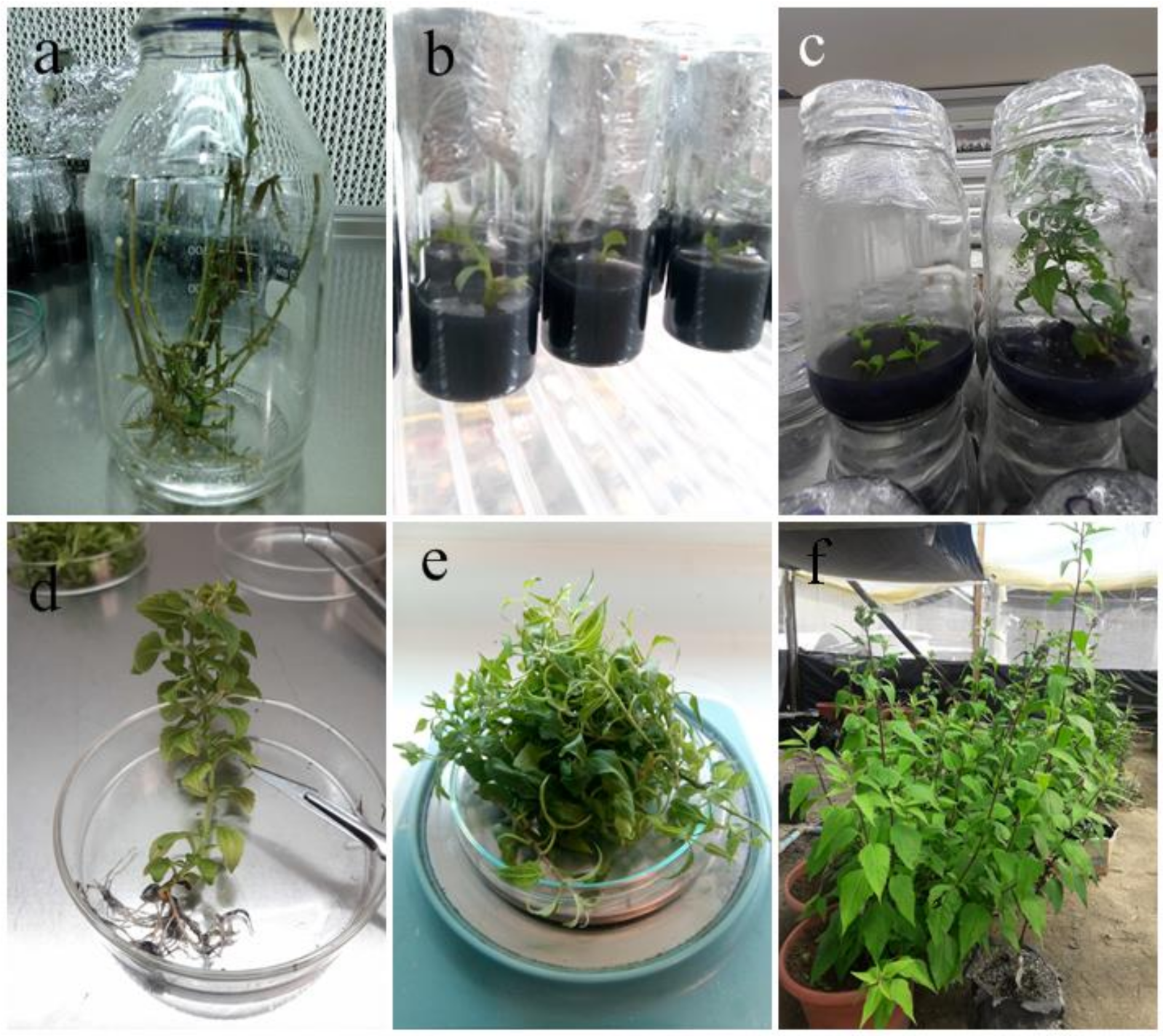
2.1. In Vitro Micropropagation of Leptocarpha Rivularis

The in vitro propagation platform for the micropropagation of L. rivularis involves three different stages, namely, introduction (I), propagation (P) and rooting (R). Plant cuttings, having at least two axillary buds each, were used for the in vitro culture introduction of plant material. These cuttings were cultured by 20–30 days in different semi-solid culture medium (Murashige and Skoog, MS) supplemented with 3% (w/v) of sucrose, ascorbic acid (0.05 g/L), charcoal (1 g/L), and different hormone combinations (see Table 1).
After two weeks (I1), lateral buds sprouted, and the development of leaf blade was observed. The formulation of this medium was adjusted for the propagation stage (Table 1). In this step, explants elongated their stems, allowing micropropagation due to the abundant number of lateral shoots that they generated, considerably increasing their plant mass. A growth index of 2.0 ± 0.5 and 3.5 ± 0.5 shoots per explants was observed for introduction and propagation, respectively. Explants in P3 medium rooted spontaneously when cultured over 60 days. For rooting, explants were transferred to R1 medium for 30 days. In both mediums, 75% of the explants generated a fascicular root, which allowed obtaining 83% of successfully acclimatized plants under greenhouse conditions (Table 1, Figure 1). The explant survival rate of in vitro culture was 89.6 ± 2.7 and 95.0 ± 5.0 for propagation and rooting, respectively. These values were measured 30 days after disinfection and the beginning of spontaneous rooting, and denote the means of three independent assays with n = 130.
After two months of subculture, the introduced plant material of L. rivularis remained free from exogenous contamination. Thus, half of the obtained explants were kept for propagation, whereas the other half was used for the generation of callus from leaves and internodal segments. The medium was renewed every 45 days, obtaining an average multiplication rate of n = 3. The development of protocols for the implementation of clonal micropropagation platforms in semisolid medium, using media formulated in this work, allowed obtaining L. rivularis explants that completed the growth and development processes in a normal way, successfully acclimatizing under greenhouse conditions and developing fertile reproductive structures. It is worth mentioning that the main difference between our results and the protocol developed by Dorta et al. [13] resides in the composition of the culture medium used in different steps of the in vitro clonal micropropagation platform of Leptocarpha rivularis. For example, here, mixtures of hormones (BAP and IBA) and MS (P3 and R1) were used as medium for both propagation and rooting. On the other hand, Dorta et al. used MSG/IBA medium for propagation and a free hormone MSG medium for rooting. In both cases, rooting was induced with yields over 70%. Overall, the percentage of successfully developed plants growing under greenhouse conditions is in the range of 83 to 90% for both protocols.

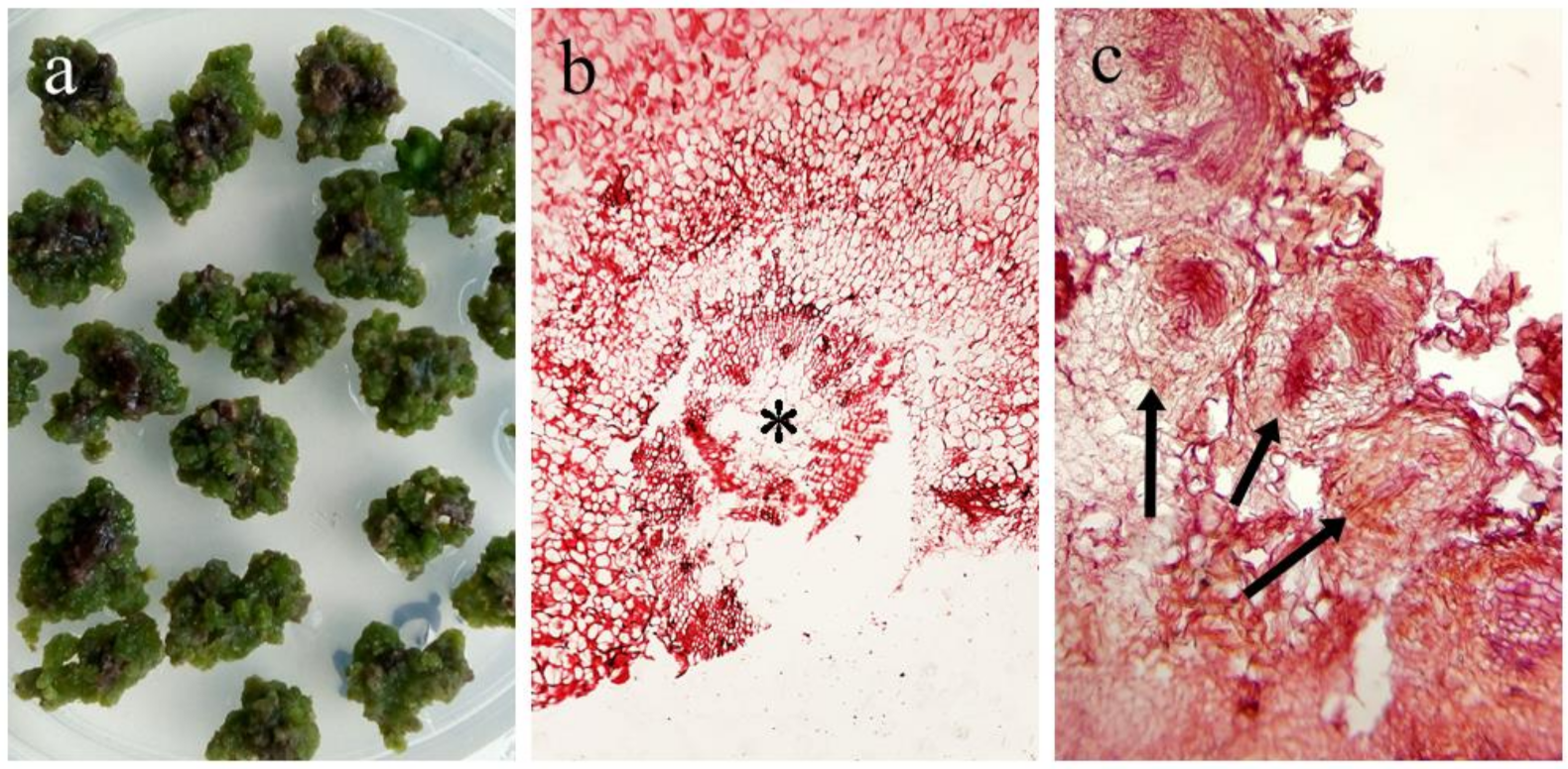
2.2. Callus Induction

Callus induction from the internodal stem segment of in vitro-generated L. rivularis was explored. As there is no previous information, different hormone combinations, proposed for species of the Asteraceae family [21,22], were tested until successful callus induction from the internodal stem segment was performed. Callus induction index (CIF) and other characteristics such as color, texture and differentiation potential were used to determine the callus formation response (Table 2).
The data in Table 2 indicate that efficient induced callogenesis is obtained in C7 and C8 mediums, i.e., having a CIF higher than 90%. These CIF values are 2–3 times larger than those previously reported for similar systems [21,24]. As result of this process, a green-colored, friable, and non-organogenic callus was generated in both medium. This is an interesting result, because it has been proposed that the development stage and tissue differentiation are as important as hormone combination to determine callus generation feasibility [26,27,28,29]. Thus, reprogramming the cell cycle in highly specialized adult cells should be a very complex process, which means that callus generation from leaves of L. rivularis is quite a difficult task. A plausible explanation for this high efficiency in callus induction is the use of cytokines instead of auxins, unlike other species belonging to the Asteraceae family [28].
Histological analyses of these calluses are shown in Figure 2.
The photography in Figure 2c confirms the generation of undifferentiated cells grouped in round-shaped structures known as meristemoids. These are groups of adult cells that, under hormone stimuli, de-differentiate into mesenchymal cells. This group of cells are self-renewing, adopt a spiral shape and generate a large cell and smaller meristemoid that maintains its self-renewal activity, which ultimately leads to callus structure formation. Calluses doubled in mass every 60 days and were kept in culture for up to six months. Subsequently, an oxidative process began, which stopped cell multiplication and finally led to tissue death.

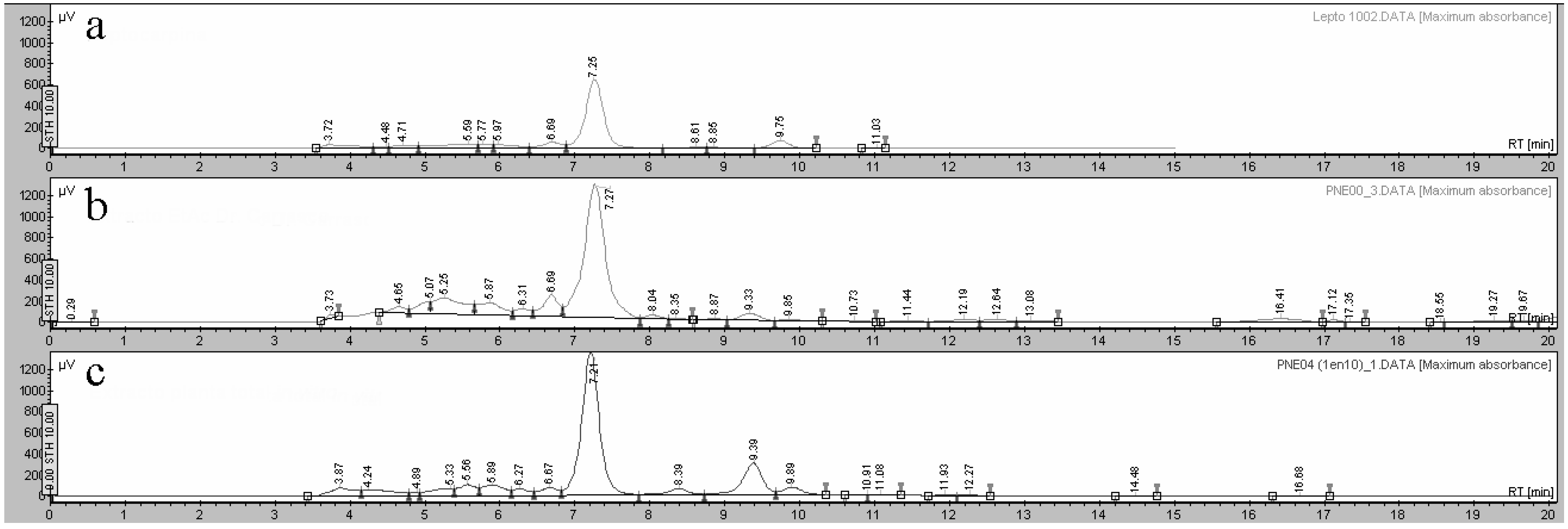
2.3. Phytochemical Analysis of Extracts

The chemical composition of extracts of L. rivularis has been extensively studied and it is well established that they contain sesquiterpene lactones, of which LTC is the major component [5,6,30,31]. On the other hand, as the phytochemical composition of plants propagated under in vitro conditions could vary due to controlled cultivation conditions, it becomes necessary to determine the composition of extracts obtained from propagated explants and callus generated in vitro. Thus, the phytochemical composition of extracts obtained from plants collected from their natural habitat, callus, and clonal explants was characterized by HPLC and using a pure sample of LTC as standard. The comparison of the obtained chromatographic profiles is shown in Figure 3.
Interestingly, similar phytochemical compositions were observed for extracts obtained from plants and micropropagated explants, and LTC was shown to be the major component in both extracts. In addition, LTC was not detected in callus extract.

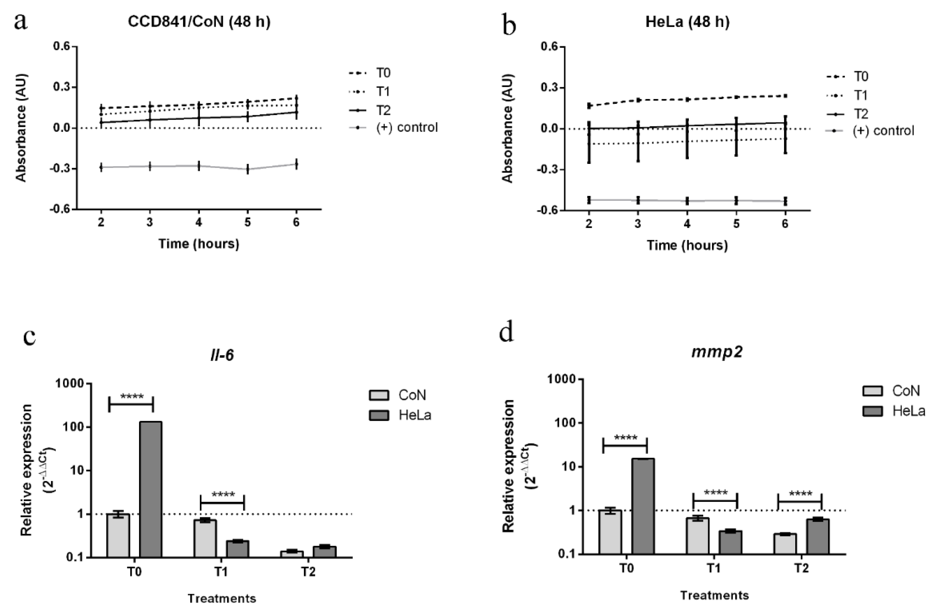
2.4. Asseessment of Biological Activity of In Vitro Plant Extracts

In previous work, it was shown that both LTC and extracts of L. rivularis exhibit antiproliferative activity against different cancer cell lines [5,6,8,10]. In this work, the cytotoxicity of extracts, obtained from in vitro-propagated plants of L. rivularis, was evaluated by using cancer and normal cell lines, namely, HeLa (cervical adenocarcinoma) and CCD841/CoN (colon epithelium non-cancer cell line), respectively. Cell viability was evaluated using the Resazurin colorimetric assay, which is an indirect method that measures cellular metabolic capacity variations. As result of cell metabolism, resazurin is reduced by oxide-reductases found in the mitochondria of viable cells and, consequently, a color change or fluorescence emission can be detected [32,33]. For cells growing under normal conditions, the absorbance can be taken as proportional to cell density and therefore a decrease in absorbance implies a decrease in cell viability.
In this work, changes in resazurin absorbance were determined for untreated cells (T0), cells treated with LTC (T1), L. rivularis extract (T2), and DMSO at lethal concentration (positive control).
Negative changes in absorbance were obtained for both of the cell lines submitted to treatment with a high concentration of DMSO (see Figure 4a,b). These negative values indicate that all metabolic process were stopped due to cell death.
On the other hand, cells treated with LTC (1.2 ppm) showed a statistically significant decrease in cell metabolism as compared with untreated cells T0 (0.1% DMSO). Interestingly, the decrease in cell viability after 48 h of exposure to LTC treatment was higher for HeLa than CoN cells. For cancer cells, negative absorbance values were measured, suggesting that a lethal level of LTC was reached, whereas for CoN cells and at the same LTC concentration only a decrease in their metabolic activity was observed. This result is in line with previous studies that have shown that LTC acts specifically on tumor cells of different origin [8,10,11]. Finally, treatment with the extract of clonal explants (T2, 12 ppm) induced a decrease in metabolism in both cell lines but without reaching lethal levels. The data in Figure 4b suggest that a lethal level could be reached at a higher extract concentration. It is worth mentioning that T1 and T2 concentrations were chosen based on the IC50 values previously reported for different cancer lines [6,10], and therefore the concentration producing the highest antiproliferative effect can still be optimized. However, these results confirm that LTC is the most active metabolite in L. rivularis extract, and that clonal plant extracts could be considered as potential antiproliferative agents.
To delve into possible LTC mechanisms of action, we have analyzed the expression of Il-6 and mmp2 genes, which encode interleukine-6 and matrix metalloproteinase 2, respectively. It is well established that IL-6 regulates immune response and cancer cell proliferation [34,35,36,37], whereas MMP-2 promotes cell metastasis processes [38].
In Figure 4c,d is plotted, in a logarithmic scale, the relative fold change expression of Il-6 and mmp2 in CoN and HeLa cells after 48 h of treatment with LTC (T1) and the extract of clonal explants (T2). Measurements were performed at 48 h because gene expression reaches a peak after this cell culture period. Our results show that, in the absence of any treatment (T0, 0.1% DMSO), the expression levels of both genes are higher in cancer cells than in normal cells, which is in line with what has been previously reported [39]. Interestingly, LTC (T1) induced a drastic decrease in the expression of both genes in HeLa cells, i.e., the ratio of relative expression, T0/T1, is 720 and 45 for Il-6 and mmp2, respectively. However, LTC had a much lower effect on CoN cells, i.e., T0/T1 values were 1.4 and 1.5 for Il-6 and mmp2 expression, respectively. In other words, these results confirm that LTC effects are somehow more heavily expressed on cancer cell lines than in normal cells. Even though there are no studies of the effect of LTC on the expression of these two marker genes, it has been reported that the antitumor activity of natural compounds and extracts is related to the decreasing expression of genes encoding IL-6 and MMP-2 [40,41,42,43]. It has been shown that the inhibition of IL-6 occurs by the interruption of the autocrine signal dependent on the IL-6/STAT3 system. STAT3 and NF-κβ are transcription factors that are activated and maintain a signaling crosstalk in the tumor environment. Both mechanisms induce tumor cell proliferation, survival, invasion, and chemo resistance [44,45,46]. On the other hand, the LTC-induced decrease in mmp2 expression implies that LTC should reduce metastasis in addition to antiproliferative and apoptosis effects, attributed to the inhibition of the NF-κβ signaling pathway [10].
Finally, the data shown in Figure 4c,d indicate that T2 reduces gene expression in cancer cells in a similar way. The ratio of the relative fold change expression of Il-6 and mmp2 (T0/T2) is approximately 720 and 24 for HeLa cells, and 7 and 3.5 for HeLa and CoN cells, respectively. However, the most striking result is the strong effect of T2 on normal cells, which takes the ratio T0/T2 values far from the unity observed for LTC. In other words, it turns out that T2 affects normal cells in a way that LTC does not. This difference could be attributed to other active compounds that are present in the extract at lower concentrations. Overall, this tendency indicates a greater effect of T2 on gene expression in HeLa than in CoN cells, but it is worth mentioning that any decrease in the expression of these genes in cancer or normal cell lines should help to prevent or avoid cancer proliferation and metastasis. Thus, we think that clonal extract of L. rivularis is a promising alternative for developing anticancer agents.

3. Materials and Methods

3.1. In Vitro Micropropagation Platform for Leptocarpha Rivularis

Plant material was donated by a private nursery, located in O’Higgins region, Chile. Non-lignified cuttings measuring 0.3 cm wide by 10 cm in length were used for in vitro culture. Introduction of plant material was carried out using a standard disinfection protocol; briefly, plant material was washed for 20 min with commercial soap and one hour with commercial antifungal agents in doses recommended by the manufacturers, and sterilized for 10 min using a 10% sodium hypochlorite solution. Then, cuttings were sectioned into 2 cm fragments, trying to conserve two axillary buds in each, and transferred to semi-solid medium, which was formulated in this research. To induce axillary buds to sprout, the introduction medium was used for 20–30 days. The medium was used for the propagation and elongation of explants. This culture medium must be renewed every 30–45 days. The extension for over 60 days in this medium generates the rooting of explants. All micropropagation stages were carried out at constant temperature, 24 ± 2 °C, relative humidity, ranging from 30 to 50%, with photosynthetic photon flux density (PPFD) at 40 μmol m−2 s−1, provided by cool white fluorescent tube lights (Philips, Kolkata, India), and a photoperiod of 16/8 h light/dark.

3.2. Callus Induction of Leptocarpha Rivularis

The callogenesis process was tested using various combinations of phytohormones considering several concentrations and information referring to callogenesis processes in the genus Asteraceae (see Table 2), observing the effect of these on callus induction of material from lateral leaves and internodal segments obtained from plantlets multiplied in vitro. The induction conditions used considered periods of darkness for 30 days and subsequently, a photoperiod regime of 16 h light/8 h darkness (16/8), or an immediate period of 16/8, without going through the 30 days in darkness. In addition, variables such as temperature (23 ± 2 °C) and humidity (50–70% RH) remained constant. According to the structure’s evolution, media renovation proceeded every 2 to 3 months. Callus formation response was determined according to callus induction index (CIF) [47]. Variables were volume (wet weight/dry weight), callification time, morphology (friable, compact, oxidized), and the presence or absence of organogenesis or other structures [25] (Table 2). Histological sections were performed using widely described protocols [48]. Structures fixed in a FAA solution were embedded in paraffin and subsequently, cuts between 8 to 10 µm were made using microtome (Microm HM325, ThermoFisher, Waltham, MA, USA). These were stained with Safranin-O according to published protocols [49].

3.3. Extraction and Composition Analysis of Leptocarpha Rivularis Extracts

Extracts of L. rivularis were obtained from plant material generated under in vitro conditions, i.e., total plant and callus extracts. Extract obtained from plants collected from their natural ecological niche was provided by Dr. Héctor Carrasco. Briefly, clonal plantlets without rooting and callus originated from internodal segments were dehydrated at room temperature for 5 days (plants) or by lyophilization for 48 h (callus) in a freeze dryer (Ílshin Biobase, Dongducheon-si, Korea). Dried material was macerated in ethyl acetate (EtAc) for two days, and the solvent was removed under reduced pressure at 50 °C using a rotary evaporator (Buchi, Essen, Germany). Phytochemical profiles of all extracts were determined by high-performance chromatography (HPLC). Chromatograms were obtained with an HPLC instrument (Jasco, Tokyo, Japan) formed by a quaternary pump (PU-2089, Jasco) coupled with a high-sensitivity diode array detector (MD-2010, Jasco), covering from 195 nm to 650 nm with 1 nm of minimum wavelength interval. Semi-polar extract samples (20 uL) were injected onto a C18 column (Kromasill, KR100-5-C18 4.6 × 150 mm). Acetonitrile (ACN):water mixtures of increasing polarity (80%, 70%, 60%) were used as mobile phase at a constant flow of 0.5 mL/min (50 MPa max.). Pure LTC, isolated and characterized in previous work, was used as standard.

3.4. Biological Activity of the Total Extracts of Leptocarpha Rivularis In Vitro

Biological activity of LTC was analyzed by means of a cell viability assay and quantification of expression of genes associated with cellular processes of cell proliferation. HeLa ATTCC® CCL2™ (cervical adenocarcinoma) and CCD841/CoN (colon epithelium non-cancer cell line) cell lines were used. Tests were performed under standard culture conditions, and Dulbecco’s modified Eagle’s medium (DMEM, Gibco, Waltham, MA, USA) with no added antibiotics and supplemented with 10% inactive fetal bovine serum was used as basal medium under controlled conditions of CO2 (5%) and temperature (37 °C) optimal for both cell lines. For cell viability assay, Resazurin colorimetric assay (Sigma-Aldrich, St. Louis, MO, USA) was used according to the supplier’s recommendations. This assay was performed as follows: in 96-well plates, 103 cells/well were seeded with 200 uL of basal medium for 24 h. Subsequently, basal medium was replaced with basal medium containing LTC (T1, 1.2 ppm) or total extract from in vitro plant material (T2, 12 ppm) [6], which were dissolved in 0.1% DMSO. Negative control was wells containing culture medium with the same volume of 0.1% DMSO added (T0). DMSO at higher concentration (10%) was used as the positive control to induce cell death, because it has been shown that doses greater than 1% induce cell death [50]. Finally, 100 uL of resazurin was added to each well, and maintained under cell culture conditions for an incubation period of 2 h. Color change, induced by a reduction in resazurin, was quantified by measuring absorbance at 530 nm with an Infinite 200 PRO (TECAN, Municipality, Switzerland) at 2, 3, 4, 5, and 6 h after incubation. This assay was carried out at 24, 48 and 72 h after treatment application and each well was measured in triplicate.
For gene expression assays, 105 cells/mL were seeded in 35 mm plates (p6) using cell culture media and under the same culture conditions as those previously described. The material was collected after 48 h of exposure to different treatments using cell lysis solution from Total RNA extraction kit (Macherey-Nagel, Düren, Germany) and extraction was carried out according to the supplier’s instructions. RNA amount was determined using an Infinite 200 PRO spectrophotometer. Gene expression analysis was performed using quantitative real-time polymerase chain reaction (qRT-PCR) using Light Cycler 96 equipment (Roche, Basel, Switzerland). RNA transcription and amplification was completed using Takyon™ One-Step MasterMix (Eurogentec, Seraing, Belgium). Reactions were carried out in triplicate, using previously described primers for carcinogenic marker genes, metalloproteinase-2 enzyme (mmp2) and interleukin-6 cytokine (Il-6), together with hypoxanthine phosphoribosyltransferase-1 enzyme (hprt-1), corresponding to the endogenous gene of constitutive expression [51,52,53]. Ct values for each treatment were normalized by the expression of the hprt-1 gene, and quantified according to the 2−ΔΔCt methodology [54]. Statistical analyses for the viability test and to determine the changes in gene expression were performed using GraphPad Prism 6 software with a two-way ANOVA and subsequently, mean values with less significant differences of p < 0.05 were considered according to Sidak’s multiple comparisons test.

4. Conclusions

The clonal micropropagation of L. rivularis and callus induction from the internodal segment of in vitro-generated explants were successfully accomplished. The chemical composition of in vitro plant extracts and callus was determined by HPLC and compared with extracts obtained from adult plants grown in natural habitats. Interestingly, the results indicate that both kind of extracts have a similar chemical composition, and that LTC is not present in callus extracts. On the other hand, the cell viability and gene expression of Il-6 and mmp2, genes associated with carcinogenic activity, were measured for HeLa and CoN cells, in the presence and absence of LTC and the extract of micropropagated L. rivularis plants. The results show that LTC and the extract exhibit comparative antiproliferative activity, i.e., the diminution of cell viability is more important in cancer cells than in normal cells. Similar behavior was observed for the decrease in gene expression. This effect has not been previously evidenced for L. rivularis and suggests the existence of an alternative mechanism of action for their extract and LTC, namely one that directly affects NF-κβ activity and the regulation of the cell cycle, and another affecting the activity of metalloproteinases, and therefore inhibiting the metastasis process of cancer cells.

Author Contributions

Conceptualization, J.R.; methodology, J.R., C.R.-K., G.A. and L.E.; validation, J.R., E.S.-M., L.E., H.C. and A.F.O.; formal analysis, J.R.; investigation, J.R.; resources, J.R., E.S.-M. and H.C.; writing—original draft preparation, J.R.; writing—review and editing, J.R., H.C. and A.F.O.; supervision, J.R.; project administration, J.R.; funding acquisition, J.R. All authors have read and agreed to the published version of the manuscript.

Funding

This study was funded by ANID FONDECYT postdoctoral fellowship (project no. 3180267).

Institutional Review Board Statement

Not applicable.

Informed Consent Statement

Not applicable.

Data Availability Statement

Not applicable.

Acknowledgments

The authors thank Álvaro Sequeida for comments, suggestions, and English translation of the manuscript. We are grateful to Juan Hormazabal and Julio Tapia for providing the cellular lines used in this study, HeLa and CCD841/CoN, respectively. J.R. thanks Catalina Alvarez and Gustavo Romo for access to the facilities where in vitro experiments were carried out.

Conflicts of Interest

The authors declare no conflict of interest.

References

  1. Zuloaga, F.; Belgrano, M. The Catalogue of Vascular Plants of the Southern Cone and the Flora of Argentina: Their contribution to the World Flora. Rodriguésia 2015, 66, 989–1024. [Google Scholar] [CrossRef] [Green Version]
  2. Obando, M. Scientific evidence of the medicinal properties of plants for use in intercultural medicine in Chile. Bol. Latinoam. Caribe Plantas Med. Aromat. 2020, 19, 207. [Google Scholar]
  3. Uquiche, E.; Garcés, F. Recovery and antioxidant activity of extracts from leptocarpha rivularis by supercritical carbon dioxide extraction. J. Supercrit. Fluids 2016, 110, 257–264. [Google Scholar] [CrossRef]
  4. Jimenez-Gonzalez, A.; Quispe, C.; Borquez, J.; Sepulveda, B.; Riveros, F.; Areche, C.; Nagles, E.; Garcia-Beltran, O.; Simirgiotis, M.J. UHPLC-ESI-ORBITRAP-MS analysis of the native Mapuche medicinal plant palo negro (Leptocarpha rivularis DC.-Asteraceae) and evaluation of its antioxidant and cholinesterase inhibitory properties. J. Enzyme Inhib. Med. Chem. 2018, 33, 936–944. [Google Scholar] [CrossRef]
  5. Montenegro, I.; Moreira, J.; Ramirez, I.; Dorta, F.; Sanchez, E.; Alfaro, J.F.; Valenzuela, M.; Jara-Gutierrez, C.; Munoz, O.; Alvear, M.; et al. Chemical Composition, Antioxidant and Anticancer Activities of Leptocarpha rivularis DC Flower Extracts. Molecules 2021, 26, 67. [Google Scholar] [CrossRef] [PubMed]
  6. Olea, A.F.; Villena, J.; Moller, A.; Martinez, R.; Carrasco, H. Enhancement of cytotoxic activity by encapsulation in pluronic polymer micelles: Leptocarpha rivularis extracts against human cancer cell lines. J. Chil. Chem. Soc. 2019, 64, 4437–4440. [Google Scholar] [CrossRef] [Green Version]
  7. Alvarez-Contreras, C.; Martinez, R.; Cubillos, V. Efecto Hipoglicemiante de la Infusión de Leptocarpha rivularis: Estudio en Ratas Diabéticas Tipo II por Inducción con Aloxano; Editorial Académica Española: Chisinau, Moldava, 2018. [Google Scholar]
  8. Martinez, R.; Kesternich, V.; Gutierrez, E.; Dolz, H.; Mansilla, H. Conformational-Analysis and biological activity of leptocarpin and leptocarpin acetate. Planta Med. 1995, 61, 188–189. [Google Scholar] [CrossRef] [PubMed]
  9. Martinez, R.; Kesternich, V.; Carrasco, H.; Alvarez-Contreras, C.; Montenegro, C.; Ugarte, R.; Gutierrez, E.; Moreno, J.; Garcia, C.; Werner, E.; et al. Synthesis and conformational analysis of leptocarpin derivatives. Influence of modification of the oxirane ring on leptocarpin’s cytotoxic activity. J. Chil. Chem. Soc. 2006, 51, 1010–1014. [Google Scholar] [CrossRef]
  10. Bosio, C.; Tomasoni, G.; Martinez, R.; Olea, A.F.; Carrasco, H.; Villena, J. Cytotoxic and apoptotic effects of leptocarpin, a plant-derived sesquiterpene lactone, on human cancer cell lines. Chem. Biol. Inter. 2015, 242, 415–421. [Google Scholar] [CrossRef] [PubMed]
  11. Zheng, S.W.; Wan, W.G.; Miao, H.X.; Tang, R.; Wang, B.; Huang, Q.Z.; Liu, W.L.; Zheng, J.P.; Chen, C.Q.; Zhong, H.B.; et al. Leptocarpin Suppresses Proliferation, Migration, and Invasion of Human Osteosarcoma by Targeting Type-1 Insulin-Like Growth Factor Receptor (IGF-1R). Med. Sci. Monit. 2017, 23, 4132–4140. [Google Scholar] [CrossRef] [Green Version]
  12. Cordero, C. Propagación Vegetativa de Leptocarpha rivularis DC, Especie Nativa de Uso Medicinal. Bachelor’s Thesis, Pontificia Universidad Católica de Valparaíso, Valparaíso, Chile, 2000. [Google Scholar]
  13. Dorta, F.; Polanco, V.; Ramirez, I.; Seeger, M.; Madrid, A.; Montenegro, I. In vitro propagation of Leptocarpha rivularis, a native medicinal plant. In Vitro Cell. Dev. Biol. Plant 2020, 56, 827–832. [Google Scholar] [CrossRef]
  14. Bairu, M.W.; Amoo, S.O.; Van Staden, J. Comparative phytochemical analysis of wild and in vitro-derived greenhouse-grown tubers, in vitro shoots and callus-like basal tissues of Harpagophytum procumbens. S. Afr. J. Bot. 2011, 77, 479–484. [Google Scholar] [CrossRef] [Green Version]
  15. Mangoale, R.M.; Afolayan, A.J. Comparative Phytochemical Constituents and Antioxidant Activity of Wild and Cultivated Alepidea amatymbica Eckl & Zeyh. Biomed. Res. Int. 2020, 2020, 5808624. [Google Scholar] [PubMed]
  16. Mulabagal, V.; Tsay, H.-S. Plant Cell Cultures-An Alternative and Efficient Source for the Production of Biologically Important Secondary Metabolites. Int. J. Appl. Sci. Eng. 2004, 2, 29–48. [Google Scholar]
  17. Ochoa-Villarreal, M.; Howat, S.; Hong, S.; Jang, M.O.; Jin, Y.-W.; Lee, E.-K.; Loake, G.J. Plant cell culture strategies for the production of natural products. BMB Rep. 2016, 49, 149–158. [Google Scholar] [CrossRef] [PubMed]
  18. Bhojwani, S.S.; Dantu, P.K. Production of Industrial Phytochemicals. In Plant Tissue Culture: An Introductory Text; Springer: New Delhi, India, 2013; pp. 275–286. [Google Scholar]
  19. Espinosa-Leal, C.A.; Puente-Garza, C.A.; Garcia-Lara, S. In vitro plant tissue culture: Means for production of biological active compounds. Planta 2018, 248, 1–18. [Google Scholar] [CrossRef] [PubMed]
  20. Efferth, T. Biotechnology Applications of Plant Callus Cultures. Engineering 2019, 5, 50–59. [Google Scholar] [CrossRef]
  21. Benitez-Garcia, I.; Vanegas-Espinoza, P.E.; Melendez-Martinez, A.J.; Heredia, F.J.; Paredes-Lopez, O.; Del Villar-Martinez, A.A. Callus culture development of two varieties of Tagetes erecta and carotenoid production. Electron. J. Biotechnol. 2014, 17, 107–113. [Google Scholar] [CrossRef] [Green Version]
  22. Nielsen, E.; Temporiti, M.E.E.; Cella, R. Improvement of phytochemical production by plant cells and organ culture and by genetic engineering. Plant Cell Rep. 2019, 38, 1199–1215. [Google Scholar] [CrossRef]
  23. Mukhia, S.; Mandal, P.; Singh, D.K.; Singh, D. Comparison of pharmacological properties and phytochemical constituents of in vitro propagated and naturally occurring liverwort Lunularia cruciata. BMC Complement. Altern. Med. 2019, 19, 181. [Google Scholar] [CrossRef] [Green Version]
  24. Voronova, O. Callus formation and plant regeneration in sunflower (Helianthus L., asteraceae) in vitro tissue culture. In Proceedings of the 19th International Sunflower Conference, Edirne, Turkey, 29 May–3 June 2016; p. 211. [Google Scholar]
  25. Ikeuchi, M.; Sugimoto, K.; Iwase, A. Plant Callus: Mechanisms of Induction and Repression. Plant Cell 2013, 25, 3159–3173. [Google Scholar] [CrossRef] [PubMed] [Green Version]
  26. Sugimoto, K.; Meyerowitz, E.M. Regeneration in Arabidopsis tissue culture. Methods Mol. Biol. 2013, 959, 265–275. [Google Scholar]
  27. Osman, N.I.; Jaafar Sidik, N.; Awal, A. Effects of variations in culture media and hormonal treatments upon callus induction potential in endosperm explant of Barringtonia racemosa L. Asian Pac. J. Trop. Biomed. 2016, 6, 143–147. [Google Scholar] [CrossRef] [Green Version]
  28. Feher, A. Callus, Dedifferentiation, Totipotency, Somatic Embryogenesis: What These Terms Mean in the Era of Molecular Plant Biology? Front Plant. Sci. 2019, 10, 536. [Google Scholar] [CrossRef] [PubMed] [Green Version]
  29. Bhatia, S. Chapter 2-Plant tissue culture. In Modern Applications of Plant Biotechnology in Pharmaceutical Sciences; Bhatia, S., Sharma, K., Dahiya, R., Bera, T., Eds.; Academic Press: Boston, MA, USA, 2015; pp. 31–107. [Google Scholar]
  30. Martinez, R.; Ayamante, B.; Nunez-Alarcon, J.A.; de Vivar, A.R. Leptocarpin and 17,18-dihydroleptocarpin, two new heliangolides from Leptocarpha rivularis. Phytochemistry 1979, 18, 1527–1528. [Google Scholar] [CrossRef]
  31. Martinez, R.; Kesternich, V.; Carrasco, H.; Bustos, C.; Fernandez, S. Structure, conformation and biological activity studies on rivularin, a new heliangolide isolated from Leptocarpha rivularis. Bol. Soc. Chil. Quim. 1998, 43, 7–12. [Google Scholar]
  32. Präbst, K.; Engelhardt, H.; Ringgeler, S.; Hübner, H. Basic Colorimetric Proliferation Assays: MTT, WST, and Resazurin. Methods Mol. Biol. 2017, 1601, 1–17. [Google Scholar] [PubMed]
  33. Rampersad, S.N. Multiple applications of Alamar Blue as an indicator of metabolic function and cellular health in cell viability bioassays. Sensors 2012, 12, 12347–12360. [Google Scholar] [CrossRef] [PubMed]
  34. Badache, A.; Hynes, N.E. Interleukin 6 inhibits proliferation and, in cooperation with an epidermal growth factor receptor autocrine loop, increases migration of T47D breast cancer cells. Cancer Res. 2001, 61, 383–391. [Google Scholar]
  35. Paduch, R.; Walter-Croneck, A.; Zdzisińska, B.; Szuster-Ciesielska, A.; Kandefer-Szerszeń, M. Role of reactive oxygen species (ROS), metalloproteinase-2 (MMP-2) and interleukin-6 (IL-6) in direct interactions between tumour cell spheroids and endothelial cell monolayer. Cell Biol. Int. 2005, 29, 497–505. [Google Scholar] [CrossRef]
  36. Roomi, M.W.; Monterrey, J.C.; Kalinovsky, T.; Rath, M.; Niedzwiecki, A. In vitro modulation of MMP-2 and MMP-9 in human cervical and ovarian cancer cell lines by cytokines, inducers and inhibitors. Oncol. Rep. 2010, 23, 605–614. [Google Scholar] [PubMed]
  37. Luo, Y.; Zheng, S.G. Hall of Fame among Pro-inflammatory Cytokines: Interleukin-6 Gene and Its Transcriptional Regulation Mechanisms. Front. Immunol. 2016, 7, 604. [Google Scholar] [CrossRef] [PubMed]
  38. Kessenbrock, K.; Plaks, V.; Werb, Z. Matrix metalloproteinases: Regulators of the tumor microenvironment. Cell 2010, 141, 52–67. [Google Scholar] [CrossRef] [PubMed] [Green Version]
  39. Li, L.; Cheng, F.W.; Wang, F.; Jia, B.; Luo, X.; Zhang, S.Q. The activation of TLR7 regulates the expression of VEGF, TIMP1, MMP2, IL-6, and IL-15 in Hela cells. Mol. Cell Biochem. 2014, 389, 43–49. [Google Scholar] [CrossRef]
  40. Zhang, J.; Wang, R.; Cheng, L.; Xu, H. Celastrol inhibit the proliferation, invasion and migration of human cervical HeLa cancer cells through down-regulation of MMP-2 and MMP-9. J. Cell Mol. Med. 2021, 25, 5335–5338. [Google Scholar] [CrossRef]
  41. Suradej, B.; Sookkhee, S.; Panyakaew, J.; Mungkornasawakul, P.; Wikan, N.; Smith, D.R.; Potikanond, S.; Nimlamool, W. Kaempferia parviflora Extract Inhibits STAT3 Activation and Interleukin-6 Production in HeLa Cervical Cancer Cells. Int. J. Mol. Sci. 2019, 20, 4226. [Google Scholar] [CrossRef] [Green Version]
  42. Shih, Y.W.; Wu, P.F.; Lee, Y.C.; Shi, M.D.; Chiang, T.A. Myricetin suppresses invasion and migration of human lung adenocarcinoma A549 cells: Possible mediation by blocking the ERK signaling pathway. J. Agric. Food Chem. 2009, 57, 3490–3499. [Google Scholar] [CrossRef] [PubMed]
  43. Shi, X.; Luo, X.; Chen, T.; Guo, W.; Liang, C.; Tang, S.; Mo, J. Naringenin inhibits migration, invasion, induces apoptosis in human lung cancer cells and arrests tumour progression in vitro. J. Cell Mol. Med. 2021, 25, 2563–2571. [Google Scholar] [CrossRef]
  44. Yoon, S.; Woo, S.U.; Kang, J.H.; Kim, K.; Shin, H.J.; Gwak, H.S.; Park, S.; Chwae, Y.J. NF-κB and STAT3 cooperatively induce IL6 in starved cancer cells. Oncogene 2012, 31, 3467–3481. [Google Scholar] [CrossRef] [PubMed] [Green Version]
  45. McFarland, B.C.; Hong, S.W.; Rajbhandari, R.; Twitty, G.B., Jr.; Gray, G.K.; Yu, H.; Benveniste, E.N.; Nozell, S.E. NF-κB-induced IL-6 ensures STAT3 activation and tumor aggressiveness in glioblastoma. PLoS ONE 2013, 8, e78728. [Google Scholar] [CrossRef]
  46. Morgan, E.L.; Macdonald, A. Autocrine STAT3 activation in HPV positive cervical cancer through a virus-driven Rac1-NFκB-IL-6 signalling axis. PLoS Pathog. 2019, 15, e1007835. [Google Scholar] [CrossRef] [PubMed] [Green Version]
  47. Ghasempour, H.; Soheilikhah, Z.; Zebarjadi, Z.; Ghasempour, S.; Karimi, N. In vitro micro propagation, callus induction and shoot regeneration in safflower L. cv. Lesaf. Iran. J. Plant Physiol. 2014, 4, 999–1004. [Google Scholar]
  48. Soukup, A.; Tylová, E. Essential methods of plant sample preparation for light microscopy. In Plant Cell Morphogenesis. Methods in Molecular Biology; Cvrčková, F., Žárský, V., Eds.; Humana Press: Totowa, NY, USA, 2019; Volume 1080, pp. 1–23. [Google Scholar]
  49. Johansen, D.A. Plant Microtechnique. Nature 1941, 147, 222. [Google Scholar]
  50. Verheijen, M.; Lienhard, M.; Schrooders, Y.; Clayton, O.; Nudischer, R.; Boerno, S.; Timmermann, B.; Selevsek, N.; Schlapbach, R.; Gmuender, H.; et al. DMSO induces drastic changes in human cellular processes and epigenetic landscape in vitro. Sci. Rep. 2019, 9, 4641. [Google Scholar] [CrossRef] [Green Version]
  51. Partridge, J.J.; Madsen, M.A.; Ardi, V.C.; Papagiannakopoulos, T.; Kupriyanova, T.A.; Quigley, J.P.; Deryugina, E.I. Functional analysis of matrix metalloproteinases and tissue inhibitors of metalloproteinases differentially expressed by variants of human HT-1080 fibrosarcoma exhibiting high and low levels of intravasation and metastasis. J. Biol. Chem. 2007, 282, 35964–35977. [Google Scholar] [CrossRef] [Green Version]
  52. Cheng, X.; Folco, E.J.; Shimizu, K.; Libby, P. Adiponectin induces pro-inflammatory programs in human macrophages and CD4+ T cells. J. Biol. Chem. 2012, 287, 36896–36904. [Google Scholar] [CrossRef] [Green Version]
  53. Estrada-Jiménez, T.; Millán-Pérez Peña, L.; Flores-Mendoza, L.; Sedeño-Monge, V.; Santos-López, G.; Rosas-Murrieta, N.; Reyes-Carmona, S.; Terán-Cabanillas, E.; Hernández, J.; Herrera-Camacho, I.; et al. Upregulation of the Suppressors of Cytokine Signaling 1 and 3 Is Associated with Arrest of Phosphorylated-STAT1 Nuclear Importation and Reduced Innate Response in Denguevirus-Infected Macrophages. Viral Immunol. 2016, 29, 95–104. [Google Scholar] [CrossRef]
  54. Livak, K.J.; Schmittgen, T.D. Analysis of Relative Gene Expression Data Using Real-Time Quantitative PCR and the 2−ΔΔCT Method. Methods 2001, 25, 402–408. [Google Scholar] [CrossRef]
Figure 1. In vitro clonal micro propagation platform of Leptocarpha rivularis. (a) Stick with dormant shoot sterilization; (b) in vitro culture establishment; (c) propagation protocol for in vitro explants; (d) complete “adult” plant generated by in vitro culture. Uses for this material will be (e) semipolar extraction or (f) explant acclimation under greenhouse conditions.
Plants 11 00546 g001
Figure 2. Histological analysis of L. rivularis callus induced under in vitro culture conditions. (a) Callus structures; (b) cells at the contact surface 21 days after callus induction, * shows an initial internodal section and following dedifferentiation; (c) arrows indicate meristemoids formation.
Plants 11 00546 g002
Figure 3. HPLC chromatograms obtained for: (a) LTC pure compound; (b) extract of L. rivularis plants collected from natural habitat; (c) extract of micropropagated explants of L. rivularis.
Plants 11 00546 g003
Figure 4. Biological activity of in vitro plant extracts. Resazurin colorimetric assay of: (a) control cell line, CoN; (b) HeLa cancer cell line. Relative fold change expression: (c) IL-6, (d) mmp2. All data represent the means of three independent replicates ± SD (n = 9). The significance of difference was analyzed by Sidak’s multiple comparisons test (p < 0.05); asterisks indicate significant differences compared with negative control under the same treatment conditions (**** p < 0.0001).
Plants 11 00546 g004
Table 1. Medium composition for in vitro clonal micropropagation platform of Leptocarpha rivularis. Murashige and Skoog medium, MS, with 3% (w/v) of sucrose, ascorbic acid (0.05 g/L), charcoal (1 g/L) and different hormone combinations. Hormones included are: 1-naphthaleneacetic acid (NAA), 6-benzylaminopurine (BAP), indole-3-butyric acid (IBA) and gibberellic acid (GA3). * Indicates best medium protocol.
Introduction (I)Propagation (P)Rooting (R)
I1I2I3I4I5 *P1 *P2P3R1
Hormones (µM)BAP2.200.440.443.504.404.404.404.403.50
IBA 0.490.490.490.490.49 0.490.49
NAA 0.27
GA3 0.140.140.14 0.140.14
Table 2. Medium composition for Leptocarpha rivularis callus induction. Different combinations of auxins and cytokines were used: 1-naphthaleneacetic acid (NAA), 6-benzylaminopurine (BAP), isopentenyl adenine (2ip), kinetin (KIN), thidiazuron (TDZ), zeatin (Z). n.c.i.: no callus induction.
Hormones
Induction MediaRefAuxinsConc. (µM)CytokinesConc. (µM)CIF (%) * (Mean ± S.E.)Plant OrganCallus Charact
C0 0leaf, intermodal sectionn.c.i.
C1 NAA2.7TDZ4.50leafn.c.i.
C2 NAA2.7KIN4.60leafn.c.i.
C3 NAA2.7KIN2.333.3 ± 1.2leafcompact, rooting
C4 NAA5.5KIN2.30leafn.c.i.
C5 NAA5.5KIN2.30leafn.c.i.
C6[24]NAA5.5BAP4.40rootn.c.i.
C6[24]NAA5.5BAP4.436.1 ± 2.6leafcompact, greenish
C7 NAA2.7BAP4.494.4 ± 1.5internodal sectionfriable, green
C8 NAA5.5BAP4.491.8 ± 1.3internodal sectionfriable, green
C9[21]2,4D4.9BAP4.40leafn.c.i.
C10[21]2,4D9.8BAP8.827.8 ± 3.2leaf, rootcompact, brown
C11[21]2,4D13.5BAP8.80leafn.c.i.
C12 BAP-KIN 0leaf, intermodal sectionn.c.i.
C13 BAP-Z 2.7 ± 0.6leafcompact, rooting
C14 2ip 0leafn.c.i.
* CIF (%) = number of explants forming calluses/(total number of explants × 100%). Callus description following Ikeuchi et al. 2013 [25].
Publisher’s Note: MDPI stays neutral with regard to jurisdictional claims in published maps and institutional affiliations.

