Article

Polyphenol Profile and Pharmaceutical Potential of Quercus spp. Bark Extracts

by
Hosam O. Elansary
1,2,3,*,
Agnieszka Szopa
4,
Paweł Kubica
4,
Halina Ekiert
4,
Mohamed A. Mattar
5,
Mohamed A. Al-Yafrasi
1,
Diaa O. El-Ansary
6,
Tarek K. Zin El-Abedin
5 and
Kowiyou Yessoufou
3
1
Plant Production Department, College of Food and Agriculture Sciences, King Saud University, Riyadh 11451, Saudi Arabia
2
Floriculture, Ornamental Horticulture, and Garden Design Department, Faculty of Agriculture (El-Shatby), Alexandria University, Alexandria 21526, Egypt
3
Department of Geography, Environmental Management, and Energy Studies, University of Johannesburg, APK campus, Johannesburg 2092, South Africa
4
Department of Pharmaceutical Botany, Medical College, Jagiellonian University, ul. Medyczna 9, 30-688 Kraków, Poland
5
Department of Agricultural Engineering, College of Food and Agriculture Sciences, King Saud University, Riyadh 11451, Saudi Arabia
6
Precision Agriculture Laboratory, Department of Pomology, Faculty of Agriculture (El-Shatby), Alexandria University, Alexandria 21526, Egypt
*
Author to whom correspondence should be addressed.
Submission received: 13 October 2019 / Revised: 2 November 2019 / Accepted: 6 November 2019 / Published: 9 November 2019
(This article belongs to the Special Issue Bioactive Compounds in Plants)

Abstract

:
Targeted profiling of polyphenols in trees may reveal valuable sources of natural compounds with major applications in pharmacology and disease control. The current study targeted the profiling of polyphenols using HPLC-DAD in Quercus robur, Q. macrocarpa and Q. acutissima bark extracts. Free radical scavenging of each extract was investigated using antioxidant assays. Antimicrobial activities against a wide spectrum of bacteria and fungi were explored, as well as anticancer activities against different cancer cell lines. The HPLC-DAD analyses revealed the availability of several polyphenols in high amounts, including ellagic acid (in Q. robur) and caffeic acid (in Q. macrocarpa) in all three species. The bioactivity assay revealed high antioxidant activity in Q. robur compared to that of the other species, as well as phenolic standards. The three oak bark extracts showed clear antibacterial activities against most bacteria tested, with the highest antibacterial activities in the extracts of Q. robur. In addition, the three extracts showed higher antibacterial activities against Pseudomonas aeruginosa, Micrococcus flavus, and Escherichia coli compared to that of other bacteria. There were strong antifungal activities against some fungi, such as Aspergillus flavus, Penicillium funiculosum, and Penicillium ochrochloron. There were also noticeable anticancer activities against MCF-7, HeLa, Jurkat, and HT-29 cell lines, with the highest anticancer activity in the extracts of Q. robur. This is the first study that reveals not only novel sources of important polyphenols (e.g., ellagic acid) in Q. robur, Q. macrocarpa and Q. acutissima bark but also their anticancer activities against diverse cancer cell lines.

1. Introduction

Tree barks are widely used in traditional medicine to treat several diseases because of their medicinal properties grounded in the presence of phenolic compounds that can have antioxidant, antimicrobial, and anti-inflammatory activities [1,2,3]. The bark of Quercus species in particular is receiving increased attention because of its diverse traditional medicinal uses, its abundance and the low price of its wood residues, such as bark [4]. The genus Quercus, belonging to the Fagaceae family, contains trees that are distributed worldwide, with an estimated 450 species [5,6]. There are differences in their morphological appearance and chemical composition.
The best-known species in Europe is Quercus robur L. (known as common oak). This plant occurs naturally in Europe, Asia, and North America and is used in traditional medicine for the treatment of diarrhea and inflammation [7]. The bark of Q. robur is listed in the official database of pharmaco-therapeutic plants by the European Medicine Agency [8]. The European Pharmacopoeia [9,10] referred to the raw plant material of Q. robur as the cut and dried bark of young branches and lateral shoots, which contain a minimal amount of 3% tannins expressed as pyrogallol and calculated with reference to the dried herbal substance. Q. robur bark contains a high amount of tannins (hydrolyzable and condensed tannins) (8%–20%). These tannins are composed of either galloyl esters and their derivatives (gallotannins, ellagitannins, and complex tannins) or oligomeric and polymeric proanthocyanidins and can possess different interflavanyl couplings and substitution patterns (condensed tannins) [11,12]. However, there is no information in the literature regarding the phenolic profile of bark extracts of this species. Although it was reported that Q. robur barks from Poland have strong antioxidant activities [2], the phenolic composition of these barks and the respective bioactivities of their compounds remain unexplored.
Furthermore, Quercus acutissima Carruth. (sawtooth oak) is another naturally occurring species, native to eastern and southern regions of Asia and naturalized in some eastern regions of North America. The fruits are not preferable as food for cattle because of their poor taste, and the wood is of low quality. However, the use of Q. acutissima as a medicinal plant has been mentioned in traditional Asian medicine, especially bark extracts which are used in the treatment of skin disorders, and some studies have confirmed the effectiveness of these extracts in this regard (i.e., in the treatment of skin disorders) [13,14].
As far as the species Quercus macrocarpa Michx. (bur oak) is concerned, it is native to North America. This species is included in the Red List Species Program by the International Union for Conservation of Nature and Natural Resources as a species of least concern [15]. Q. macrocarpa is a valuable species for cultivation with high drought tolerance [16]. Little is known about the chemical composition and possible bioactivity of its bark.
Overall, the Quercus genus is distributed worldwide, with the traditional use of potentially bioactive raw material in specific regions [8,17]; however, information from experimental studies regarding the bioactivity (e.g., anticancer activity) of the bark is limited. In this study, the polyphenol profile of three Quercus spp. (Q. acutissima, Q. macrocarpa, and Q. robur) was evaluated for the first time by HPLC-DAD analysis. Moreover, the antioxidant, antibacterial, antifungal, and anticancer activities were explored using different antioxidant methods, a wide spectrum of bacteria and fungi as well as different human cancer cell lines.

2. Results

2.1. Targeted Profiling of Biologically Active Metabolites

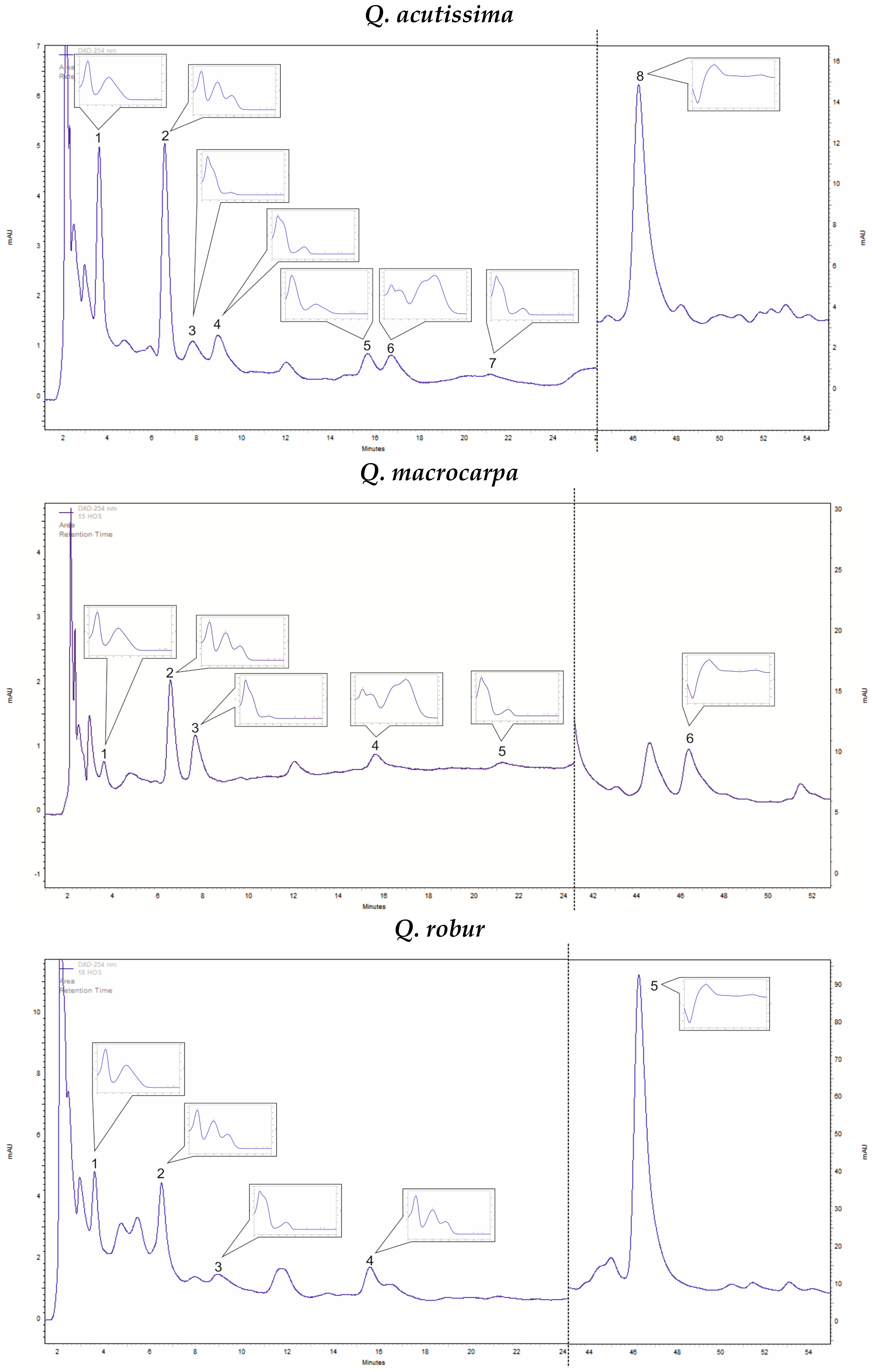
2.1.1. Quercus Acutissima

In Q. acutissima methanolic bark extracts, only four phenolic acids (caffeic acid, ellagic acid, gallic acid, and protocatechuic acid) were confirmed out of the 21 screened (Table 1). The dominant compound was ellagic acid (13.50 mg 100 g−1 DW), followed by gallic acid (7.09 mg 100 g−1 DW). The amounts of protocatechuic and caffeic acids were lower. Out of the five analyzed catechin derivatives in the bark extracts, four were detected (catechin, epicatechin, epigallocatechin, and epigallocatechin gallate; Table 1). Their amounts were comparable and ranged from 8.31 to 12.91 mg 100 g−1 DW. Quantitatively the dominant compounds were epicatechin (12.66 mg 100 g−1 DW) and epigallocatechin (12.66 mg 100 g−1 DW) (Figure 1).

2.1.2. Quercus Macrocarpa

In Q. macrocarpa bark extracts, a very high amount of caffeic acid was detected at 100.58 mg 100 g−1 DW (Table 1). Other phenolic acids, including ellagic, protocatechuic, and gallic acid, were also confirmed, but in much lower amounts of 5.07, 3.36, and 0.87 mg 100 g−1 DW, respectively. Two catechin derivatives were found: epicatechin – 11.00 mg 100 g−1 DW and epigallocatechin – 10.15 mg 100 g−1 DW (Table 1 and Figure 1).

2.1.3. Quercus Robur

In Q. robur bark extracts, four phenolic acids (out of 21 compounds) were detected: ellagic acid, gallic acid, protocatechuic acid, and vanillic acid. The dominant compound was ellagic acid (97.82 mg 100 g−1 DW), whereas the amounts of the other compounds were lower (gallic acid—8.23 mg 100 g−1 DW, protocatechuic acid—6.96 mg 100 g−1 DW, and vanillic acid—2.61 mg 100 g−1 DW). In the studied extracts, a high amount of catechin was estimated at 44.52 mg 100 g−1 DW (Table 1 and Figure 1).

2.2. Antioxidant Activities

The antioxidant activities of the bark extracts of the three species as well as ellagic and caffeic acids are shown in Table 2. Q. robur showed significantly higher antioxidant activities by means of the DPPH (IC50, 3.0 µg mL−1), β-carotene bleaching (IC50, 3.3 µg mL−1), FRAP (IC50, 3.8 mM TEAC g−1 extract) assays compared to other species. Q. macrocarpa exhibited higher antioxidant activities than Q. acutissima. Furthermore, Q. robur antioxidant activities were comparable to standard antioxidants (BHT). The antioxidant activities of ellagic and caffeic acids were comparable to those of Q. robur and Q. macrocarpa, respectively.

2.3. Antibacterial Activities

The antibacterial activities of the bark extracts of Q. acutissima, Q. macrocarpa, Q. robur as well as ellagic and caffeic acids using the micro-dilution methods are shown in Table 3. The three extracts exhibited clear antibacterial activities against most species of microorganism studied. The MIC values ranged between 0.04 and 0.29 mg mL−1, whereas the MBC ranged between 0.11 and 0.66 mg mL−1. The response of the bacterial species to the extracts used varied among species. The highest antibacterial activities were found for the extracts of Q. robur compared to those of the other two species. The three extracts exhibited higher antibacterial activities against Pseudomonas aeruginosa, M. flavus and E. coli compared to other bacterial species. Further, their antibacterial activities were comparable to those of antibiotics. The antibacterial activities of phenolic standards of the ellagic and caffeic acids were comparable and higher than those of Q. robur and Q. macrocarpa extracts, respectively.

2.4. Antifungal Activities

The extracts were screened for their antifungal activities against several fungi, as shown in Table 4. The MIC ranged between 0.16 and 2 mg mL−1, whereas the MFC ranged between 0.23 and 3.61 mg mL−1. There were obvious antifungal activities against some fungi, including A. flavus, Penicillium funiculosum, and Penicillium ochrochloron. However, A. ochraceus and A. niger, as well as C. albicans, showed slight resistance to the extracts. The activities of the extracts were comparable to commercial reagents in most cases. The antifungal activities of phenolic standards of the ellagic and caffeic acids were comparable to those of Q. robur and Q. macrocarpa extracts, respectively.

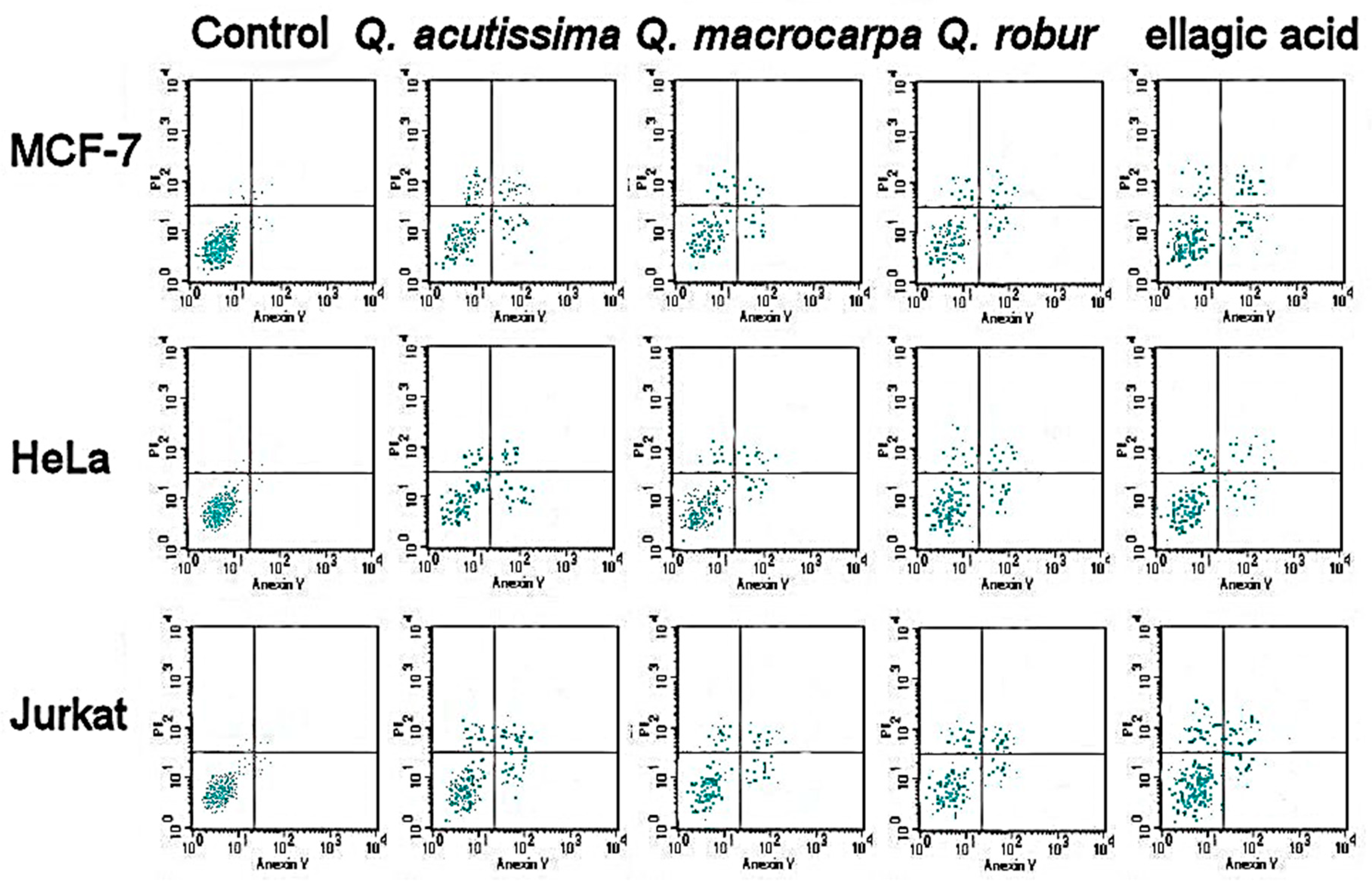
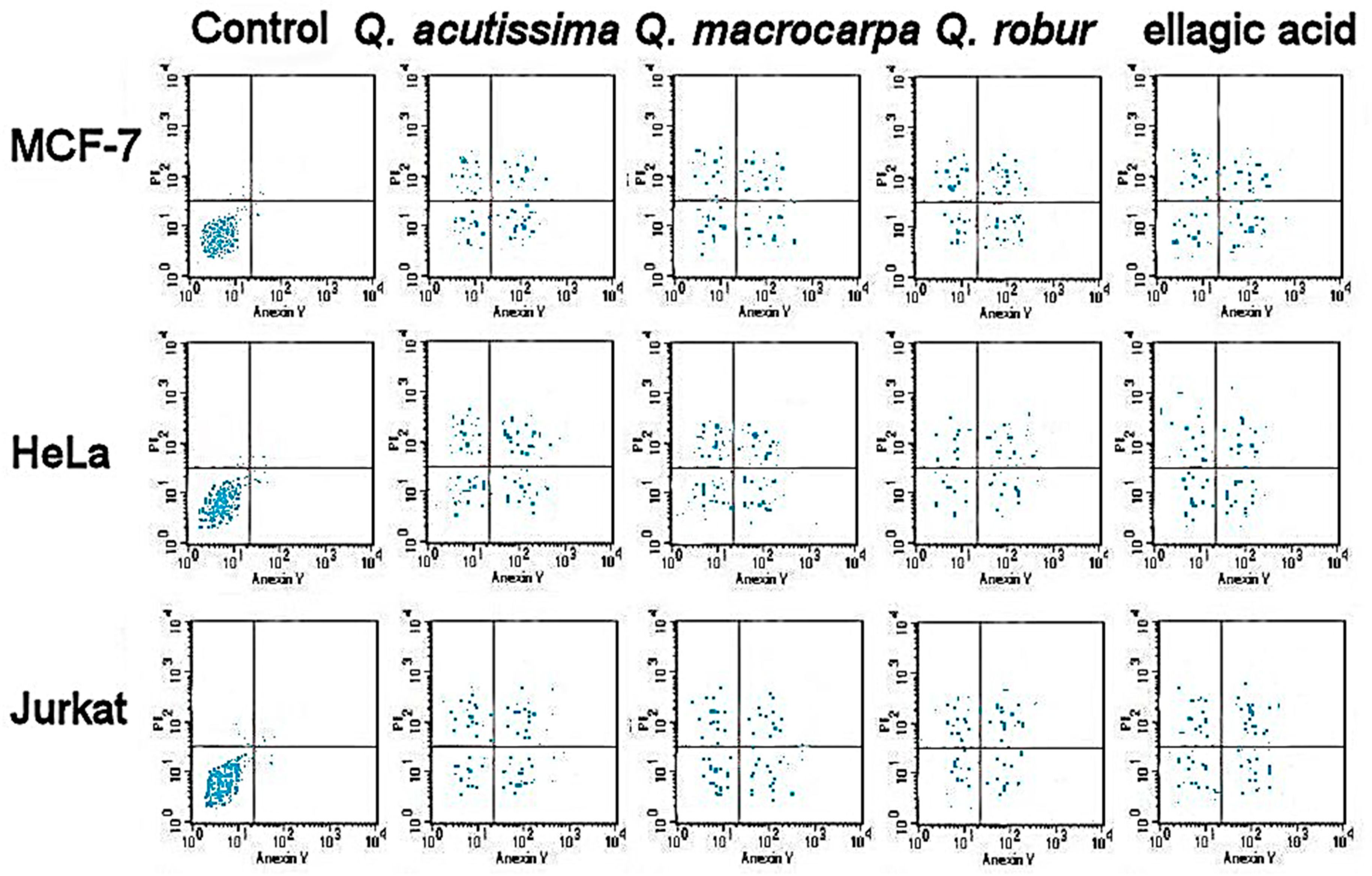
2.5. Anticancer Activities

The bark extracts were screened for their anticancer activities against different cancer cell lines, as shown in Table 5. There were obvious anticancer activities against MCF-7, HeLa, Jurkat, and HT-29 cell lines. The highest anticancer activity was found in the extracts of Q. robur compared to that of Q. macrocarpa and Q. acutissima. Only Q. robur exhibited anticancer activity against T24. The anticancer activities of phenolic standards of the ellagic and caffeic acids were comparable to those of Q. robur and Q. macrocarpa extracts, respectively. The apoptotic assay revealed the accumulation of necrotic as well as both early and late apoptotic cells in different treatments in a dose dependent manner (Figure 2 and Figure 3).

3. Discussion

The HPLC-DAD analyses of methanolic extracts of the bark of three Quercus species indicated that specific phenolic acids and catechin derivatives were the major active ingredients. Three phenolic acids were common in all three bark extracts (ellagic acid, gallic acid, and protocatechuic acid) and they are benzoic acid derivatives. Ellagic acid and gallic acid are known derivatives produced by tannin hydrolyses, typical for the Quercus species [2,12].
Interestingly, extremely high amounts of ellagic acid were found in Q. robur bark extract (97.82 mg 100 g−1 DW) that were 7-fold that of Q. acutissima and 17-fold that of Q. macrocarpa bark extracts (Table 1). In Q. acutissima and Q. macrocarpa bark extracts, there were noticeable amounts of caffeic acid. In Q. macrocarpa, the caffeic acid content was −100.58 mg 100 g−1 DW (23-fold that of Q. acutissima) (Table 1). In Q. robur bark extract, vanillic acid was detected (Table 1). This phenolic acid was not detected in other Quercus species. In the Quercus species, the most often studied bioactive metabolite content is that of leaf and needle extracts of Q. robur [18]. A previous study documented some phenolic acids, including p-hydroxybenzoic acid, vanillic acid, gallic acid, syringic acid, ferulic acid, and o- and p-coumaric acids. For Q. acutissima, gentisic acid (phenolic acid) was confirmed in the extracts of fresh acorns [19]. However, in the available literature, no information regarding phenolic acid estimation in Q. macrocarpa or in the bark extracts of the three Quercus species was found. The latter is important because the cortex is recognized as the raw material of oaks. This study demonstrated differences in secondary metabolite composition among the examined cortex extracts and is the first to document the phenolic acid profiles in these materials.
The detected phenolic acids in the studied extracts are very important from a pharmacological and economic point of view. For example, gallic acid has antibacterial, hypoglycemic, anticancer, and antimutagenic activities [20,21]. In agreement with the current study, ellagic acid is known for strong antioxidative, antiproliferative, and anticancer properties [22,23]. Protocatechuic acid has antifungal, antibacterial, antiviral, anti-inflammatory, antiatherosclerotic, antiulcer, and anticancer properties [20,24,25]. Vanillic acid also exhibits antioxidant and hepatoprotective actions [26,27]. The presence of these compounds, in addition to tannins, contributes to the pharmacological activities of these raw materials.
The Quercus cortex is recognized in phototherapy as a valuable plant raw material because of its extremely high tannin content [10]. In the current study, the chromatographic analyses detected catechin and some derivatives, epicatechin, epigallocatechin, and epigallocatechin gallate, in bark extracts (Table 1). The presence of these compounds was confirmed in the Q. robur bark extract [28]. However, little is known about secondary metabolites polyphenolic compositions in the bark extracts of Q. acutissima and Q. macrocarpa. Only the presence of catechin was previously described in Q. acutissima [13,19]. Our study has contributed to the greater understanding of the tannin composition of Q. acutissima and Q. macrocarpa.
Q. robur exhibited significantly higher antioxidant activities by means of the DPPH and β-carotene bleaching assays compared to that of the other species and had activities comparable to that of standard antioxidants. Such important antioxidant activities are primarily attributable to the major bioactive compound, which is ellagic acid. High antioxidant activities were described for ellagic acid [29]. In agreement with our results, a study [2] reported strong antioxidant activities in Q. robur bark from Poland; however, they did not detect the phenolic profile of those trees. A recent investigation on Q. robur and Q. petraea leaves, twigs, and acorns from Serbia revealed strong antioxidant activities [5]. Q. macrocarpa had higher antioxidant activities than Q. acutissima and this can be explained by the extremely high amount of caffeic acid in Q. macrocarpa. Caffeic acid is known for antioxidant, antibacterial, and antifungal activities [30], which is in agreement with the results of the current study.
The three extracts showed antibacterial activities against most bacteria species studied and the highest antibacterial activities were found in the extracts of Q. robur as compared to that of the other two species. A previous report on Q. robur from Finland documented some antibacterial activity of the bark extract on S. aureus and C. albicans using the agar diffusion method [31]. In the current study, strong antibacterial activities were found against Pseudomonas aeruginosa, M. flavus, and E. coli and moderate activities against other bacterial species. The work on Q. robur bark bioactivities is relatively limited, but other species, such as Q. cortex, have revealed some antibacterial activities against Chromobacterium violaceum [32]. These strong antibacterial activities might be attributed to ellagic acid, which has some antibacterial activities against certain bacteria, such as Streptococcus mutans, Streptococcus sanguis, and Streptococcus salivarius [33]. Additionally, the catechin-rich sources revealed in this study have obvious strong antibacterial and antifungal activities and are comparable to other genera. Green tea (Camellia sinensis) are flavan-3-ols that have moderate to strong antibacterial activities against Gram-positive and Gram-negative bacterial species [34]. Furthermore, green tea polyphenols have been associated with some antifungal effects against C. albicans [35]. In the current study, obvious antifungal activities were found against A. flavus, Penicillium funiculosum, and Penicillium ochrochloron, as well as moderate activities against C. albicans. Such effects are mainly attributable to specific polyphenols such as ellagic acid, which are flavan-3-ols, and caffeic acid. These polyphenols might be the major component of the raw material as in the bark of oaks.
There were anticancer activities against MCF-7, HeLa, Jurkat, and HT-29 cell lines and the highest activities were found in the extracts of Q. robur compared to that of Q. macrocarpa and Q. acutissima. A previous report documented that Q. petraea (stem bark) and Q. robur (leaf) extracts have potent inhibitory activities against LoVo colon, PC3 prostate, and U373 glioblastoma cancer cell lines, but they did not describe the active ingredients in the extracts [36]. Furthermore, the use of Q. robur, Q. macrocarpa, and Q. acutissima bark extract as an anticancer agent is novel. The anticancer activities are mainly attributed to major constituents, such as the ellagic acid found in this study. Also, flavan-3-ols have previously shown anticancer activities [37]. The oak barks selected for the current study are valuable sources of anticancer, antioxidant, and antimicrobial natural compounds.

4. Materials and Methods

4.1. Plant Material and Sample Preparation

The outer bark of Q. acutissima, Q. macrocarpa, and Q. robur (Fagaceae family) were sampled from the University of Guelph Arboretum in Guelph, Ontario, Canada, identified by Hosam Elansary, and then vouchered at the University of Guelph and at Alexandria University (Hosam000975–2018). The Quercus spp. bark samples were dried by lyophilization (Labconco, USA) and then powdered. The dried pulverized plant samples of 0.5 g DW (dry weight) each, in 3 replications, were put in 15 mL tubes and subjected to extraction with 10 mL methanol (Chempur, Poland) by sonication (2 × 30 min at 30 °C) in an ultrasonic bath (Sonic-2, POLSONIC; ultrasonic power 2 × 100W, 40 kHz, water bath dimensions 150 × 135 × 100 mm). The extracts were filtered using Whatman paper and left in crystallizers to evaporate methanol at room temperature. The dry residue was dissolved in 2 mL of methanol (Merck, HPLC grade purity) [38]. Then the samples were stored at −80 °C for future bioassays. For bioassays, the methanol was totally removed by evaporating the methanol in a rotary evaporator. Analytical/HPLC grade chemicals were used (Sigma Aldrich, Germany) for the bioassays. The bacterial and fungal cultures were obtained from the Department of Floriculture and Ornamental Horticulture, Faculty of Agriculture, Alexandria, Egypt. Cell cultures of breast adenocarcinoma (MCF-7), cervical adenocarcinoma (HeLa), T-cell lymphoblast like (Jurkat), colon adenocarcinoma (HT-29), urinary bladder carcinoma (T24), and HEK-293 (human normal cells) were purchased from American Type Culture Collection (ATCC).

4.2. Chemicals

The following standards were used for phenolic acid quantification: benzoic acid and its derivatives (3,4-dihydroxyphenylacetic, ellagic, gallic, gentisic, p-hydroxybenzoic, protocatechuic, salicylic, syringic, and vanillic acids); cinnamic acid and its derivatives (caffeic, o-coumaric, m-coumaric, p-coumaric, ferulic, hydrocaffeic, isoferulic, and sinapic acids); and depsides (chlorogenic, neochlorogenic, and rosmarinic acids). To quantify the flavonoids, aglycone (kaempferol, luteolin, myricetin, quercetin and rhamnetin) and glycoside (apigetrin, cynaroside, hyperoside, isoquercetin, quercitrin, robinin, rutin, trifolin, vitexin) standards were used. To quantify the catechin derivatives, epicatechin, epicatechin gallate, epigallocatechin, epigallocatechin gallate and catechin were used. All the substances were acquired from Sigma-Aldrich, Germany.

4.3. Analyses of Phenolic Compounds

Analyses of the bark methanolic extracts were performed by a HPLC method [39,40] using the Merck-Hitachi liquid chromatograph (LaChrom Elite) with a DAD detector L-2455. The Purospher RP-18e (250 × 4 mm; 5 μm, Merck) column was used and the temperature was set to 25 °C. The mobile phase consisted of A—methanol, B—methanol: 0.5% acetic acid 1:4 (v/v). The flow rate was 1 mL/min, the gradient was as follows: 100% B for 0–20 min; 100–80% B for 20–35 min; 80–60% B for 35–55 min; 60–0% B for 55–70 min; 0% B for 70–75 min; 0–100% B for 75–80 min; 100% B for 80–90 min. The injection volume was 20 µL and the compounds of interest were detected at 254 nm. The applied HPLC method was previously validated by our group [39]. The tested parameters were the following: accuracy; precision at three levels of standard substance concentrations in solution, 50%, 100%, and 150%; linearity; limit of detection (LOD); and limit of quantification (LOQ) [39]. Identification of compounds was performed either by comparison with UV spectra and retention times of reference substances or using co-chromatography (Figure 1). The compounds were quantified using the calibration curves method [38,40,41].

4.4. Antioxidant Activity

DPPH, β-carotene bleaching [42] and ferric reducing antioxidant power (FRAP) [43] assays were used to determine the antioxidant activities of the bark extracts. For DPPH, the samples were incubated for 30 min, and then a wavelength of 517 nm was used to measure absorbance. During the β-carotene bleaching assay, the wavelength of 470 nm was used to determine the absorbance. The amount of the sample (IC50 in µg/mL) that scavenged 50% of the DPPH/ β-carotene bleaching solutions was determined by plotting the inhibition percent against extract concentration. A standard antioxidant was used (butylated hydroxytoluene, BHT) as a positive control and the inhibition concentration of each sample was compared with that of the BHT and blank. The FRAP reagent was prepared as described in previous studies (e.g., [43]) using TPTZ (tripyridyl triazine, Sigma-Aldrich, Berlin, Germany). Aliquots (100 μL) of bark extracts or Trolox (Sigma-Aldrich, Berlin, Germany) were added to FRAP reagent (3 mL), mixed, incubated for half an hour at 37 °C and the absorbance was measured at 593 nm. Aqueous solutions of known serial concentrations of Trolox (0–0.5 Mmol/L) were used for the calibration. Two sets of triplicate replications were conducted for all experiments.

4.5. Antibacterial Activity

Antibacterial activities of bark extracts were screened against Bacillus cereus (ATCC 14579), Escherichia coli (ATCC 35210), Listeria monocytogenes (clinical isolate), Micrococcus flavus (ATCC 10240), Pseudomonas aeruginosa (ATCC 27853), and Staphylococcus aureus (ATCC 6538) using the micro-dilution method [44]. Microtiter plates (96-well) containing a serial concentration of bark extract in each well mixed with bacterial inoculum (1.0 × 10⁴ CFU per well) in 100 μL tryptic soy broth were incubated at 37 °C for 24 h in a rotary shaker. The minimum inhibitory concentration (MIC) was defined as the lowest concentration of plant extract that exhibited no visible growth using a binocular microscope and was determined following the incubation period of the microtiter plates. The minimum bactericide concentration (MBC), which was defined as the lowest concentration that caused no visible growth and indicated the killing of 99.5% of the inoculum, was determined using serial subculturing of bark extracts (2 μL). A wavelength of 655 nm was used to determine the optical density in a spectrophotometer. A positive control was used (streptomycin, 0.01–10 mg/mL), as well as a negative one (DMSO, 1%).

4.6. Antifungal Activity

The antifungal activities of bark extracts were determined using a variety of infectious and economically important fungi, including Aspergillus flavus (ATCC 9643), A. ochraceus (ATCC 12066), A. niger (ATCC 6275), Candida albicans (ATCC 12066), Penicillium ochrochloron (ATCC 48663), and Penicillium funiculosum (ATCC 56755). The microdilution method was employed in this assay [45] using bark extract (2 μL) mixed with broth malt medium and the fugal inoculum (spore suspension concentration of 1.0 × 10⁵) in microtiter plates. The plates were incubated at 28 °C for 72 h in a rotary shaker; then the MIC was determined as the lowest concentration inhibiting fungal growth at the binocular microscopic level. The minimum fungicidal concentration (MFC) was defined as the minimum concentration showing no visible growth and indicating the killing of 99.5% of the original inoculum. MFC was determined using serial sub-cultivations of the bark extracts (2 µL) added to 100 µL of broth and inoculum, and then incubated at 28 °C for 72 h. A positive control was used (ketoconazole, 1–3500 µg/mL).

4.7. Anticancer Activities

Cytotoxic activities of the bark extracts were tested on MCF-7, HeLa, Jurkat, HT-29, and T24, as well as HEK-293 (human normal cells) following the MTT method [46]. Briefly, cells were grown in 75 cm2 flasks in MEM with 10% FBS, 17.8 mM NaHCO3, 0.1 mM non-essential amino acids, and 1 mM sodium pyruvate. They were seeded into 96-well plates at a density of 4 × 10−4 per well, left overnight in 270 µL medium, and incubated at 37 °C, 5% CO2. Steri-filtered bark extracts were added to the culture media in microtiter plates. Five doses of bark extract were used to reach a final concentration of 50, 100, 200, 300, and 400 µg/mL culture media. Samples were solubilized in DMSO (1%). Untreated cells were considered negative controls and vinblastine sulfate and taxol were used as positive controls. After the incubation of the culture media for 2 days at 37 °C and 5% CO2, PBS washing was performed to remove traces of the extract and the medium was supplied by 12 mM MTT dissolved in PBS. Dissolved in isoprobanol, 0.04 N HCl was mixed in each well, allowed to sit for 40 min, and the absorbance was determined at a 570 nm wavelength using a microplate reader (Thermo, MA, USA). The percentage of activity inhibition was calculated in triplicate:
% Inhibition = (Abs. 570 nm control‒Abs. 570 nm sample)/Abs. 570 nm control × 100. Furthermore, IC50 values were obtained by plotting the percentage of cell viability against extract concentration and expressed in µg/mL. The IC30 and IC50 were used to show the dose dependent apoptotic cell population using flow cytometry (FAC Scan, Becton Dickinson, Iowa, USA) following [46,47,48].

4.8. Statistical Analyses

The least significance difference (LSD) was determined using SPSS software (version 22.0). The quantitative results of chromatographic analyses were expressed in mg 100 g−1 dry weight (DW) as the mean ± standard deviation (SD) of three series of experiments.

5. Conclusions

This is the first study to profile polyphenols in Q. robur, Q. macrocarpa, and Q. acutissima bark extracts, as well as their bioactivities as antioxidants, antibacterial, antifungal, and anticancer materials. The study revealed the availability of several polyphenols in the three species. The bioactivity assay revealed high antioxidant activity in Q. robur compared to that of the other species. The three oak bark extracts showed clear antibacterial activities against most bacteria tested. The highest antibacterial activities were found in the extracts of Q. robur and the three extracts showed higher antibacterial activities against Pseudomonas aeruginosa, M. flavus, and E. coli compared to activities against other bacteria. There were strong antifungal activities against some fungi, such as A. flavus, Penicillium funiculosum, and Penicillium ochrochloron. There were anticancer activities against MCF-7, HeLa, Jurkat, and HT-29 cell lines. The highest anticancer activity was found in the extracts of the Q. robur compared to that of Q. macrocarpa and Q. acutissima. The use of Q. robur, Q. macrocarpa, and Q. acutissima bark extract as an anticancer agent is novel and is attributed to specific phenols such as ellagic acid. The oak bark used in this study are valuable sources of antioxidant, antimicrobial, and anticancer compounds.

Author Contributions

Conceptualization, H.O.E., A.S. and P.K.; methodology, H.O.E., A.S., M.A.M., M.A.A.-Y., H.E. and P.K. X.X.; software, H.O.E., A.S., M.A.M., M.A.A.-Y., H.E. and P.K.; validation, H.O.E., A.S., M.A.M., M.A.A.-Y., H.E., K.Y., D.O.E.-A., T.K.Z.E.-A. and P.K.; formal analysis, H.O.E., A.S., M.A.M., M.A.A.-Y., H.E., K.Y., D.O.E.-A., T.K.Z.E.-A. and P.K.; investigation, H.O.E., A.S., M.A.M., M.A.A.-Y., H.E. and P.K.; resources, H.O.E., A.S., M.A.M., M.A.A.-Y., H.E. and P.K.; data curation, H.O.E., A.S., M.A.M., M.A.A.-Y., K.Y., D.O.E.-A., T.K.Z.E.-A., H.E. and P.K.; writing—original draft preparation, H.O.E., A.S., M.A.M., M.A.A.-Y., K.Y., D.O.E.-A., T.K.Z.E.-A., H.E. and P.K.; writing—review and editing, H.O.E., A.S., M.A.M., M.A.A.-Y., H.E. and P.K.; visualization, H.O.E., A.S., M.A.M., M.A.A.-Y., H.E. and P.K.; funding acquisition, H.O.E. and A.S.

Funding

The study was funded by the Deanship of Scientific Research at King Saud University through research group No (RG-1440-12).

Conflicts of Interest

The authors declare no conflict of interest.

Abbreviations

HPLC-DADHigh-Performance Liquid Chromatography with Diode-Array Detection
Q. acutissimaQuercus acutissima
Q. macrocarpaQuercus macrocarpa
Q. roburQuercus robur;
MCF-7cell cultures of breast adenocarcinoma;
HeLacell cultures of cervical adenocarcinoma;
Jurkatcell cultures of T-cell lymphoblast like;
HT-29cell cultures of colon adenocarcinoma
T24urinary bladder carcinoma;
ATCCAmerican Type Culture Collection;
BHTbutylated hydroxytoluene
DPPH2,2-Diphenyl-1-picrylhydrazyl

References

  1. Salem, M.Z.M.; Elansary, H.O.; Elkelish, A.A.; Zeidler, A.; Ali, H.M.; EL-Hefny, M.; Yessoufou, K. In vitro Bioactivity and Antimicrobial Activity of Picea abies and Larix decidua Wood and Bark Extracts. Bioresources 2016, 11, 9421–9437. [Google Scholar] [CrossRef]
  2. Dróżdż, P.; Pyrzynska, K. Assessment of polyphenol content and antioxidant activity of oak bark extracts. Eur. J. Wood Wood Prod. 2018, 76, 793–795. [Google Scholar] [CrossRef]
  3. Vong, A.T.; Chong, H.W.; Lim, V. Preliminary Study of the Potential Extracts from Selected Plants to Improve Surface Cleaning. Plants 2018, 7, 17. [Google Scholar] [CrossRef] [PubMed]
  4. Bouras, M.; Chadni, M.; Barba, F.J.; Grimi, N.; Bals, O.; Vorobiev, E. Optimization of microwave-assisted extraction of polyphenols from Quercus bark. Ind. Crops Prod. 2015, 77, 590–601. [Google Scholar] [CrossRef]
  5. Sánchez-Burgos, J.A.; Ramírez-Mares, M.V.; Larrosa, M.M.; Gallegos-Infante, J.A.; González-Laredo, R.F.; Medina-Torres, L.; Rocha-Guzmán, N.E. Antioxidant, antimicrobial, antitopoisomerase and gastroprotective effect of herbal infusions from four Quercus species. Ind. Crops Prod. 2013, 42, 57–62. [Google Scholar] [CrossRef]
  6. Barta, C.E.; Bolander, B.; Bilby, S.R.; Brown, J.H.; Brown, R.N.; Duryee, A.M.; Edelman, D.R.; Gray, C.E.; Gossett, C.; Haddock, A.G.; et al. In Situ Dark Adaptation Enhances the Efficiency of DNA Extraction from Mature Pin Oak (Quercus palustris) Leaves, Facilitating the Identification of Partial Sequences of the 18S rRNA and Isoprene Synthase (IspS) Genes. Plants 2017, 6, 52. [Google Scholar] [CrossRef] [PubMed]
  7. Ahmed, M.; Fatima, H.; Qasim, M.; Gul, B.; Ihsan ul, H. Polarity directed optimization of phytochemical and in vitro biological potential of an indigenous folklore: Quercus dilatata Lindl. ex Royle. BMC Complement. Altern. Med. 2017, 17, 386. [Google Scholar] [CrossRef] [PubMed]
  8. Committee on Herbal Medicinal Products. Assessment report on Quercus robur L., Quercus petraea (Matt.) Liebl., Quercus pubescens Willd., cortex; European Medicines Agency: London, UK, 2010; EMA/HMPC/3206/2009. [Google Scholar]
  9. European Directorate for the Quality of Medicines. Schisandra fruit in European Pharmacopoeia. In European Pharmacopoeia 9.0; EDQM: Strasburg, France, 2017; p. 1514. [Google Scholar]
  10. Shikov, A.N.; Pozharitskaya, O.N.; Makarov, V.G.; Wagner, H.; Verpoorte, R.; Heinrich, M. Medicinal Plants of the Russian Pharmacopoeia; their history and applications. J. Ethnopharmacol. 2014, 154, 481–536. [Google Scholar] [CrossRef] [PubMed] [Green Version]
  11. Niemetz, R.; Gross, G.G. Enzymology of gallotannin and ellagitannin biosynthesis. Phytochemistry 2005, 66, 2001–2011. [Google Scholar] [CrossRef] [PubMed]
  12. Bobinac, M.T.; Batos, B.; Miljkovic, D.; Radulovic, S. Polycyclism and Phenological Variability in the Common Oak (Quercus Robur L.). Archi. Biol. Sci. 2012, 64, 97–105. [Google Scholar] [CrossRef]
  13. Tanaka, N.; Shimomura, K.; Ishimaru, K. Tannin production in callus cultures of Quercus acutissima. Phytochemistry 1995, 40, 1151–1154. [Google Scholar] [CrossRef]
  14. Koseki, J.; Matsumoto, T.; Matsubara, Y.; Tsuchiya, K.; Mizuhara, Y.; Sekiguchi, K.; Nishimura, H.; Watanabe, J.; Kaneko, A.; Hattori, T.; et al. Inhibition of Rat 5α-Reductase Activity and Testosterone-Induced Sebum Synthesis in Hamster Sebocytes by an Extract of Quercus acutissima Cortex. Evid. Based Complement. Altern. Med. 2015, 2015, 9. [Google Scholar] [CrossRef] [PubMed]
  15. International Union for Conservation of Nature. The IUCN Red List of Threatened Species, Version 2019-2; IUCN: Grand, Swizerland, 2019; Volume 2, Available online: https://www.iucnredlist.org/ (accessed on 13 October 2019).
  16. Tang, Z.; Kozlowski, T. Some physiological and morphological responses of Quercusmacrocarpa seedlings to flooding. Can. J. For. Res. 2011, 12, 196–202. [Google Scholar] [CrossRef]
  17. Jaradat, N.A.; Zaid, A.N.; Al-Ramahi, R.; Alqub, M.A.; Hussein, F.; Hamdan, Z.; Mustafa, M.; Qneibi, M.; Ali, I. Ethnopharmacological survey of medicinal plants practiced by traditional healers and herbalists for treatment of some urological diseases in the West Bank/Palestine. BMC Complement. Alternat. Med. 2017, 17, 255. [Google Scholar] [CrossRef] [PubMed]
  18. Kuiters, A.T.; Sarink, H.M. Leaching of phenolic compounds from leaf and needle litter of several deciduous and coniferous trees. Soil Biol. Biochem. 1986, 18, 475–480. [Google Scholar] [CrossRef]
  19. Ishimaru, K.; Nonaka, G.-I.; Nishioka, I. Phenolic glucoside gallates from quercus mongolica and q. acutissima. Phytochemistry 1987, 26, 1147–1152. [Google Scholar] [CrossRef]
  20. Khadem, S.; Marles, R.J. Monocyclic Phenolic Acids; Hydroxy- and Polyhydroxybenzoic Acids: Occurrence and Recent Bioactivity Studies. Molecules 2010, 15, 7985–8005. [Google Scholar] [CrossRef] [PubMed]
  21. Navarro, M.; Moreira, I.; Arnaez, E.; Quesada, S.; Azofeifa, G.; Alvarado, D.; Monagas, M.J. Proanthocyanidin characterization, antioxidant and cytotoxic activities of three plants commonly used in traditional medicine in Costa Rica: Petiveria alliaceae L., Phyllanthus niruri L. and Senna reticulata Willd. Plants 2017, 6, 50. [Google Scholar] [CrossRef] [PubMed]
  22. Seeram, N.P.; Adams, L.S.; Henning, S.M.; Niu, Y.; Zhang, Y.; Nair, M.G.; Heber, D. In vitro antiproliferative, apoptotic and antioxidant activities of punicalagin, ellagic acid and a total pomegranate tannin extract are enhanced in combination with other polyphenols as found in pomegranate juice. J. Nutr. Biochem. 2005, 16, 360–367. [Google Scholar] [CrossRef] [PubMed] [Green Version]
  23. Navarro, M.; Moreira, I.; Arnaez, E.; Quesada, S.; Azofeifa, G.; Vargas, F.; Alvarado, D.; Chen, P. Flavonoids and Ellagitannins Characterization, Antioxidant and Cytotoxic Activities of Phyllanthus acuminatus Vahl. Plants 2017, 6, 62. [Google Scholar] [CrossRef] [PubMed]
  24. Kedzierska, M.; Olas, B.; Wachowicz, B.; Glowacki, R.; Bald, E.; Czernek, U.; Szydłowska-Pazera, K.; Potemski, P.; Piekarski, J.; Jeziorski, A. Effects of the commercial extract of aronia on oxidative stress in blood platelets isolated from breast cancer patients after the surgery and various phases of the chemotherapy. Fitoterapia 2012, 83, 310–317. [Google Scholar] [CrossRef] [PubMed]
  25. Kakkar, S.; Bais, S. A Review on Protocatechuic Acid and Its Pharmacological Potential. ISRN Pharmacol. 2014, 2014, 952943. [Google Scholar] [CrossRef] [PubMed]
  26. Brand-Williams, W.; Cuvelier, M.E.; Berset, C. Use of a free radical method to evaluate antioxidant activity. LWT Food Sci. Technol. 1995, 28, 25–30. [Google Scholar] [CrossRef]
  27. Itoh, A.; Isoda, K.; Kondoh, M.; Kawase, M.; Watari, A.; Kobayashi, M.; Tamesada, M.; Yagi, K. Hepatoprotective Effect of Syringic Acid and Vanillic Acid on CCl4-Induced Liver Injury. Biol. Pharm. Bull. 2010, 33, 983–987. [Google Scholar] [CrossRef] [PubMed] [Green Version]
  28. Kuliev, Z.A.; Vdovin, A.D.; Abdullaev, N.D.; Makhmatkulov, A.B.; Malikov, V.M. Study of the catechins and proanthocyanidins of Quercus robur. Chem. Nat. Compd. 1997, 33, 642–652. [Google Scholar] [CrossRef]
  29. Kilic, I.; Yeşiloğlu, Y.; Bayrak, Y. Spectroscopic studies on the antioxidant activity of ellagic acid. Spectrochim. Acta Part. A Mol. Biomol. Spectrosc. 2014, 130, 447–452. [Google Scholar] [CrossRef] [PubMed]
  30. Pinho, F.V.S.d.A.; da Cruz, L.C.; Rodrigues, N.R.; Waczuk, E.P.; Souza, C.E.; Coutinho, H.D.; da Costa, J.G.; Athayde, M.L.; Boligon, A.A.; Franco, J.L.; et al. Phytochemical Composition, Antifungal and Antioxidant Activity of Duguetia furfuracea A. St.-Hill. Oxidative Med. Cell. Longev. 2016, 2016, 7821051. [Google Scholar] [CrossRef] [PubMed]
  31. Andrenšek, S.; Simonovska, B.; Vovk, I.; Fyhrquist, P.; Vuorela, H.; Vuorela, P. Antimicrobial and antioxidative enrichment of oak (Quercus robur) bark by rotation planar extraction using ExtraChrom®. Int. J. Food Microbiol. 2004, 92, 181–187. [Google Scholar] [CrossRef] [PubMed]
  32. Deryabin, D.G.; Tolmacheva, A.A. Antibacterial and Anti-Quorum Sensing Molecular Composition Derived from Quercus cortex (Oak bark) Extract. Molecules 2015, 20, 17093–17108. [Google Scholar] [CrossRef] [PubMed]
  33. De, R.; Sarkar, A.; Ghosh, P.; Ganguly, M.; Karmakar, B.C.; Saha, D.R.; Halder, A.; Chowdhury, A.; Mukhopadhyay, A.K. Antimicrobial activity of ellagic acid against Helicobacter pylori isolates from India and during infections in mice. J. Antimicrob. Chemother. 2018, 73, 1595–1603. [Google Scholar] [CrossRef] [PubMed]
  34. Taylor, P.W.; Hamilton-Miller, J.M.T.; Stapleton, P.D. Antimicrobial properties of green tea catechins. Food Sci. Technol. Bull. 2005, 2, 71–81. [Google Scholar] [CrossRef] [PubMed]
  35. Steinmann, J.; Buer, J.; Pietschmann, T.; Steinmann, E. Anti-infective properties of epigallocatechin-3-gallate (EGCG), a component of green tea. Br. J. Pharmacol. 2013, 168, 1059–1073. [Google Scholar] [CrossRef] [PubMed]
  36. Frédérich, M.; Marcowycz, A.; Cieckiewicz, E.; Mégalizzi, V.; Angenot, L.; Kiss, R. In vitro anticancer potential of tree extracts from the Walloon Region forest. Planta Med. 2009, 75, 1634–1637. [Google Scholar] [CrossRef] [PubMed]
  37. Yang, C.S.; Wang, H.; Chen, J.X.; Zhang, J. Effects of Tea Catechins on Cancer Signaling Pathways. Enzymes 2014, 36, 195–221. [Google Scholar] [PubMed] [Green Version]
  38. Szopa, A.; Kokotkiewicz, A.; Kubica, P.; Banaszczak, P.; Wojtanowska-Krośniak, A.; Krośniak, M.; Marzec-Wróblewska, U.; Badura, A.; Zagrodzki, P.; Bucinski, A.; et al. Comparative analysis of different groups of phenolic compounds in fruit and leaf extracts of Aronia sp.: A-melanocarpa, A-arbutifolia, and A. xprunifolia and their antioxidant activities. Eur. Food Res. Technol. 2017, 243, 1645–1657. [Google Scholar] [CrossRef]
  39. Sulkowska-Ziaja, K.; Maślanka, A.; Szewczyk, A.; Muszyńska, B. Physiologically Active Compounds in Four Species of Phellinus. Nat. Prod. Commun. 2017, 12, 363–366. [Google Scholar] [CrossRef] [PubMed]
  40. Szopa, A.; Kokotkiewicz, A.; Bednarz, M.; Luczkiewicz, M.; Ekiert, H. Studies on the accumulation of phenolic acids and flavonoids in different in vitro culture systems of Schisandra chinensis (Turcz.) Baill. using a DAD- HPLC method. Phytochem. Lett. 2017, 20, 462–469. [Google Scholar] [CrossRef]
  41. Szopa, A.; Ekiert, H.; Szewczyk, A.; Fugas, E. Production of bioactive phenolic acids and furanocoumarins in in vitro cultures of Ruta graveolens L. and Ruta graveolens ssdivaricata (Tenore) Gams. under different light conditions. Plant. Cell Tissue Organ. Culture 2012, 110, 329–336. [Google Scholar] [CrossRef]
  42. Elansary, H.O.; Yessoufou, K.; Abdel-Hamid, A.M.E.; El-Esawi, M.A.; Ali, H.M.; Elshikh, M.S. Seaweed Extracts Enhance Salam Turfgrass Performance during Prolonged Irrigation Intervals and Saline Shock. Front. Plant. Sci. 2017, 8, 830. [Google Scholar] [CrossRef] [PubMed]
  43. Ferreira, J.P.A.; Miranda, I.; Sousa, V.B.; Pereira, H. Chemical composition of barks from Quercus faginea trees and characterization of their lipophilic and polar extracts. PLoS ONE 2018, 13, e0197135. [Google Scholar] [CrossRef] [PubMed]
  44. Elansary, H.O.; Szopa, A.; Kubica, P.; Ekiert, H.; Ali, H.M.; Elshikh, M.S.; Abdel-Salam, E.M.; El-Esawi, M.; El-Ansary, D.O. Bioactivities of traditional medicinal plants in Alexandria. Evid. Based Complement. Altern. Med. 2018, 2018, 1463579. [Google Scholar] [CrossRef] [PubMed]
  45. Elansary, H.O.; Abdel-Hamid, A.M.E.; Mahmoud, E.A.; Al-Mana, F.A.; El-Ansary, D.O.; Zin Elabedin, T.K.A. Heuchera Creme Brulee and Mahogany medicinal value under water stress and oligosaccharide (COS) treatment. Evid. Based Complement. Alternat. Med. 2019, 2019, 4242359. [Google Scholar] [CrossRef] [PubMed]
  46. Elansary, H.O.; Szopa, A.; Kubica, P.; Al-Mana, F.A.; Mahmoud, E.A.; Zin Elabedin, T.K.A.; Mattar, M.A.; Ekiert, H. Phenolic Compounds of Catalpa speciosa, Taxus cuspidata, and Magnolia acuminata have antioxidant and anticancer activity. Molecules 2019, 24, 412. [Google Scholar] [CrossRef] [PubMed]
  47. Elansary, H.O.; Abdelgaleil, S.A.M.; Mahmoud, E.A.; Yessoufou, K.; Elhindi, K.; El-Hendawy, S. Effective antioxidant, antimicrobial and anticancer activities of essential oils of horticultural aromatic crops in Northern Egypt. BMC Complement. Altern. Med. 2018, 18, 214. [Google Scholar] [CrossRef] [PubMed]
  48. Yessoufou, K.; Elansary, H.O.; Mahmoud, E.A.; Skalicka-Woźniak, K. Antifungal, antibacterial and anticancer activities of Ficus drupacea L. stem bark extract and biologically active isolated compounds. Ind. Crops Prod. 2015, 74, 752–758. [Google Scholar] [CrossRef]
Figure 1. Representative of HPLC-DAD (λ = 254 nm) chromatograms of Quercus ssp. bark extracts. Q. acutissima; 1—Gallic acid, 2—Protocatechuic acid, 3—Epigallocatechin, 4—Catechin, 5—Epigallocatechin gallate, 6—Caffeic acid, 7—Epicatechin, 8—Ellagic acid. Q. macrocarpa; 1—Gallic acid, 2—Protocatechuic acid, 3—Epigallocatechin, 4—Caffeic acid, 5—Epicatechin, 6—Elagic acid. Q. robur; 1—Gallic acid, 2—Protocatechuic acid, 3—Catechin, 4—Vanillic acid, 5—Ellagic acid.
Plants 08 00486 g001
Figure 2. Apoptotic cell population (IC30) using flow cytometry.
Plants 08 00486 g002
Figure 3. Apoptotic cell population (IC50) using flow cytometry.
Plants 08 00486 g003
Table 1. The phenolic acids and catechin derivatives compositions of Q. acutissima, Q. macrocarpa and Q. robur outer bark extracts.
Quercus SpeciesCompoundtRλmaxAmount [mg 100 g−1] DW
Q. acutissimaCatechin8.96214, 27810.52 ± 1.87
Caffeic acid16.71218, 236, 3234.30 ± 0.05
Ellagic acid46.2225313.50 ± 2.84
Epicatechin21.15213, 27812.66 ± 2.97
Epigallocatechin7.8021412.91 ± 1.91
Epigallocatechin gallate15.63215, 2748.31 ± 0.03
Gallic acid3.61220, 2717.09 ± 0.59
Protocatechuic acid6.55220, 259, 2945.39 ± 0.76
Q. macrocarpaCaffeic acid15.61218, 236, 323100.58 ± 18.02
Ellagic acid46.182535.07 ± 0.05
Epicatechin21.32213, 27811.00 ± 0.34
Epigalloctechin7.9021410.15 ± 0.32
Gallic acid3.58220, 2710.87 ± 0.03
Protocatechuic acid6.54220, 259, 2943.36 ± 0.02
Q. roburCatechin 8.95214, 27844.52 ± 5.64
Ellagic acid46.2225397.82 ± 1.74
Gallic acid3.59220, 2718.23 ± 0.39
Protocatechuic acid6.51220, 259, 2946.96 ± 1.14
Vanillic acid15.59219, 260, 2932.61 ± 0.15
Table 2. DPPH and β-carotene bleaching acid of Q. acutissima, Q. macrocarpa and Q. robur outer bark extracts as well as phenol standards. Values are expressed as mean of triplicate determinations ± SD.
DPPH Free Radical Scavenging Activity
(IC50, µg mL−1)
β-Carotene-linoleic Acid Assay (IC50, µg mL−1)FRAP
(IC50, mM TEAC/g extract)
Q. acutissima4.5 ± 0.1a4.9 ± 0.1a5.4 ± 0.1a
Q. macrocarpa3.7 ± 0.1b4.1 ± 0.1b4.5 ± 0.1b
Q. robur3.0 ± 0.1c3.3 ± 0.1c3.8 ± 0.1d
ellagic acid3.0 ± 0.1c3.4 ± 0.1c3.7 ± 0.1d
caffeic acid3.2 ± 0.1c3.7 ± 0.1c4.1 ± 0.1c
BHT2.9 ± 0.1c3.2 ± 0.1c-
Trolox--3.5 ± 0.1e
Values with different letters within a column indicates significant differences (p = 0.05). TEAC: Trolox equivalents antioxidant.
Table 3. Minimum inhibitory (MIC) and bactericidal concentration (MBC) of Q. acutissima, Q. macrocarpa and Q. robur outer bark extracts (mg mL−1) as well as phenolic standards.
P. aeruginosa
(ATCC 27853)
MIC
MBC
B. cereus
(ATCC 14579)
MIC
MBC
L. monocytogenes
(Clinical Isolate)
MIC
MBC
E. coli
(ATCC 35210)
MIC
MBC
M. flavus
(ATCC 10240)
MIC
MBC
S. aureus
(ATCC 6538)
MIC
MBC
Q. acutissima0.09 ± 0.010.17 ± 0.010.27 ± 0.020.17 ± 0.010.17 ± 0.010.23 ± 0.01
0.18 ± 0.020.37 ± 0.030.66 ± 0.030.32 ± 0.020.41 ± 0.030.46 ± 0.01
Q. macrocarpa0.07 ± 0.010.16 ± 0.010.29 ± 0.010.13 ± 0.010.14 ± 0.010.22 ± 0.01
0.15 ± 0.010.35 ± 0.030.62 ± 0.020.29 ± 0.020.34 ± 0.030.44 ± 0.02
Q. robur0.05 ± 0.010.11 ± 0.010.25 ± 0.010.10 ± 0.010.10 ± 0.010.23 ± 0.02
0.11 ± 0.010.27 ± 0.020.53 ± 0.030.21 ± 0.020.20 ± 0.020.45 ± 0.01
ellagic acid0.04 ± 0.010.09 ± 0.010.23 ± 0.010.09 ± 0.010.09 ± 0.010.20 ± 0.01
0.10 ± 0.010.22 ± 0.010.49 ± 0.020.19 ± 0.030.18 ± 0.010.41 ± 0.03
caffeic acid0.06 ± 0.010.13 ± 0.010.27 ± 0.010.11 ± 0.010.13 ± 0.010.20 ± 0.01
0.13 ± 0.010.29 ± 0.010.58 ± 0.030.25 ± 0.010.30 ± 0.020.41 ± 0.03
Streptomycin0.08 ± 0.010.07 ± 0.030.14 ± 0.010.12 ± 0.010.11 ± 0.010.19 ± 0.01
0.16 ± 0.010.15 ± 0.010.29 ± 0.030.27 ± 0.010.21 ± 0.020.32 ± 0.01
Table 4. Minimum inhibitory (MIC) and fungicidal concentration (MFC) of Q. acutissima, Q. macrocarpa and Q. robur outer bark extracts (mg mL−1) as well as phenolic standards.
Aspergillus flavus
MIC
MFC
Aspergillus ochraceus
MIC
MFC
Aspergillus niger
MIC
MFC
Candida albicans
MIC
MFC
Penicillium funiculosum
MIC
MFC
Penicillium ochrochloron
MIC
MFC
Q. acutissima0.24 ± 0.010.26 ± 0.020.21 ± 0.010.40 ± 0.020.38 ± 0.020.25 ± 0.01
0.51 ± 0.030.57 ± 0.020.41 ± 0.020.86 ± 0.030.69 ± 0.030.52 ± 0.02
Q. macrocarpa0.22 ± 0.020.24 ± 0.030.21 ± 0.010.34 ± 0.030.29 ± 0.030.21 ± 0.02
0.43 ± 0.010.48 ± 0.020.40 ± 0.030.76 ± 0.030.68 ± 0.030.43 ± 0.03
Q. robur0.19 ± 0.020.26 ± 0.010.16 ± 0.010.31 ± 0.010.26 ± 0.010.16 ± 0.01
0.40 ± 0.020.53 ± 0.030.35 ± 0.020.62 ± 0.030.63 ± 0.030.33 ± 0.03
ellagic acid0.15 ± 0.010.22 ± 0.030.13 ± 0.010.30 ± 0.030.23 ± 0.020.12 ± 0.01
0.33 ± 0.030.45 ± 0.030.28 ± 0.010.61 ± 0.030.51 ± 0.030.25 ± 0.01
caffeic acid0.20 ± 0.010.22 ± 0.010.20 ± 0.010.32 ± 0.010.27 ± 0.010.20 ± 0.03
0.40 ± 0.010.45 ± 0.010.38 ± 0.010.64 ± 0.030.62 ± 0.020.42 ± 0.01
KTZ0.21 ± 0.010.21 ± 0.010.12 ± 0.010.20 ± 0.012.00 ± 0.100.21 ± 0.01
0.41 ± 0.010.42 ± 0.020.23 ± 0.010.42 ± 0.013.61 ± 0.030.42 ± 0.01
Table 5. In vitro antiproliferative activity [IC50 (µg/mL)] of Q. acutissima, Q. macrocarpa and Q. robur outer bark extracts as well as phenolic standards on cancer cell lines.
MCF-7HeLaJurkatHT-29T24HEK-293
Q. acutissima52.14 ± 2.162.4 ± 2.346.2 ± 2.3173.11 ± 6.7˃400˃400
Q. macrocarpa43.54 ± 1.354.1 ± 2.142.5 ± 1.2149.24 ± 3.7˃400˃400
Q. robur22.10 ± 1.231.42 ± 1.028.4 ± 2.799.8 ± 2.1290.28˃400
ellagic acid20.23 ± 1.029.33 ± 1.327.1 ± 1.694.5 ± 1.9273.31˃400
caffeic acid40.31 ± 1.950.5 ± 2.838.85 ± 1.8131.32 ± 4.1˃400˃400
Vinblastine sulfate2.6 ± 0.080.1 ± 0.0721.0 ± 0.565.12 ± 3.151.4 ± 2.5
Taxol0.09 ± 0.008

