
Vitamin Compatibility with the Marek’s Disease Vaccine †
by
 ,
,
Seyed Abolghasem Fatemi
1,* ,
,
Christopher J. Williams
2,
Joshua Deines
2 and
Edgar David Peebles
1 1
Department of Poultry Science, Mississippi State University, Mississippi State, MS 39762, USA
2
Zoetis Animal Health, Research Triangle Park, Durham, NC 27703, USA
*
Author to whom correspondence should be addressed.
†
This publication is a contribution of the Mississippi Agriculture and Forestry Experiment Station. This material is based upon work that is supported by the National Institute of Food and Agriculture, U.S. Department of Agriculture, Hatch project under accession number 1011797. Use of trade names in this publication does not imply endorsement by Mississippi Agricultural and Forestry Experiment Station of these products, nor similar ones not mentioned.
Poultry 2023, 2(4), 442-448; https://0-doi-org.brum.beds.ac.uk/10.3390/poultry2040033
Submission received: 9 May 2023
/
Revised: 9 July 2023
/
Accepted: 17 July 2023
/
Published: 25 September 2023
Abstract
:In ovo injection of the Marek’s disease (MD) vaccine (MDV) has been widely practiced in commercial US hatcheries. However, the MDV is very sensitive and may not be compatible with some nutrients when administered together by in ovo injection. When individually administered by in ovo injection, L-Ascorbic acid (L-AA) and 25-hydroxyvitamin D3 (25OHD3) have previously exhibited very promising results on the post-hatch physiological and immunological characteristics of broilers subjected to stressful commercial conditions. However, the compatibility of the MDV with these vitamins has not been previously explored. Their compatibility must first be established before their combined administration by in ovo injection can be considered. Therefore, the objective in this study was to determine the compatibility of the MDV with various levels of 25OHD3 or L-AA. The treatments employed were MDV-alone, MDV in combination with 0.6 (low) or 2.4 (high) μg doses of 25OHD3, or MDV in combination with 1.2 (low) or 12 (high) mg doses of L-AA. The live and dead ratio of primary chick embryo fibroblast cells infected by the MD virus (CEF-MDV) in each treatment was determined every 30 min for 2 h. The L-AA at both the low and high doses resulted in a 70% death of CEF-MDV within 1 h, but either dose of the 25OHD3 exhibited only an approximate 5% lower CEF-MDV survival as compared to those in the MDV-alone treatment. Therefore, it is suggested that the two designated doses of 25OHD3 have the potential to be effectively combined with the MDV for subsequent administration by in ovo injection.
1. Introduction
Currently, vaccination is the common method in the modern commercial poultry industry to control infectious diseases, including infectious bursal disease (IBD), infectious bronchitis (IB), Marek’s disease (MD), Newcastle disease (ND), and infectious laryngotracheitis (ILT) [1]. Effective immunization involving optimum levels of humoral and cellular immune responses is realized between 5 days and 2 weeks after vaccination [2]. Furthermore, this process can be accelerated by in ovo vaccination [3]. In ovo vaccination has emerged to promote early immunity [3,4]. Currently, in ovo vaccination has been developed to protect chickens against MD, ND, IBD, HPAI, and coccidiosis. In addition, the current use of in ovo vaccination has been largely limited to broiler hatching eggs. However, among the aforementioned in ovo vaccines, the MD vaccine (MDV) that is most commonly used in US hatcheries contains HVT alone, as the backbone virus in association with the MD virus, or as a combinatorial recombinant vaccine such as HVT-IBD, HVT-ND, and HVT-ILT [5]. It has been reported that in ovo injection of the MDV at 18 days of incubation (doi) results in an approximate 90% level of immune protection [4], an improvement in intestinal histomorphology [6], and an increase in the expression of genes associated with humoral immunity [7] in early post-hatch broilers.
The in ovo administration of various nutrients [3] has been shown to promote subsequent growth in broiler embryos [8] and post-hatch chicks [9]. The second metabolite of vitamin D, 25-hydroxyvitamin D3 (25OHD3), is involved in many physiological and metabolic pathways such as bone development [10], embryonic growth [11], muscle development [12], and innate and adaptive immunity [13]. In previous research, it has been shown that the in ovo administration of various levels of 25OHD3 at 18 doi as compared to commercial diluent resulted in an increase in the bone quality [14] and breast meat yield [15] and an enhancement in the small intestine morphology [16], immunity [16], and live performance [15] of broilers. Furthermore, L-Ascorbic acid (L-AA) is well known as an antioxidant and immunocompetence agent. Also, as compared to a non-injected control treatment, the combination of 0.4 μg of 25OHD3 with 6 μg of vitamin K3 has been shown to increase the humoral immunity, weight gain, and feed intake of 42-day-old broilers [17]. The in ovo administration of different doses of L-AA at 17 doi has resulted in an improvement in the antioxidant capacity [18], inflammatory response [19,20], and post-hatch live performance [17,21] of broilers. The in ovo injection of 0.6 µg of 25OHD3 combined with commercial diluent resulted in an increase in the hatchability of live embryonated embryos [22] and the subsequent bone quality of post-hatch broilers [14]. Furthermore, immunity [16,23,24], breast meat yield [25], and live performance [15,25] were improved in Ross 708 broilers in response to 2.4 µg of 25OHD3 as compared to non-injected or diluent control groups. However, in comparison to non-injected or diluent-injected control groups, there were no beneficial effects on hatch and post-hatch variables when lower than 0.3 µg or higher than 2.4 µg of 25OHD3 was in ovo injected [24,25]. As compared to saline or non-injected control treatments, L-AA at 12 mg resulted in an increase in the hatchability [26], body weight gain [20], and antioxidant capacity [16] of Ross 708 broilers, while there were no positive effects on their hatching process or post-hatch live performance when administered below 1.2 mg [17,26]. Moreover, as compared to a non-injected treatment, the amniotic in ovo administration of 6 mg of L-AA at 15 doi has been shown to improve the intestinal histomorphology and bone quality of 7-day-old broilers that were fasted for 36 h post hatch [27]. In addition, as compared to a sham treatment group, the in ovo injection of 3 mg of L-AA in the amino at 15 doi resulted in an increase in the expression of genes linked to heat stress control in 16 and 18 doi broilers [28]. Furthermore, amniotic in ovo feeding of 3 mg of L-AA at 15 doi has been shown to increase enzymatic antioxidant activity at 42 days of post hatch age, and result in an increase in humoral immunity at 1, 21, and 42 days of post hatch age [29].
Thus, it may be beneficial to combine MDV with multifunctional nutrients for efficient simultaneous delivery of each to benefit from their individual effects and possibly promote MDV efficacy. However, an evaluation of the effects of various nutrients that are candidates for in ovo administration on the functional properties of the MDV, including the viability of the chick embryo fibroblasts that constitute it, has not been previously reported. It has been previously demonstrated that the MD virus has the capability of transforming primary chick embryo fibroblasts [30]. Therefore, the objective of this study is to explore potential doses of 25OHD3 and L-AA that might be compatible with the commercial MDV by exhibiting negligible effects on the viability of primary chick embryo fibroblast cells infected by the MD virus (CEF-MDV).
2. Materials and Methods
2.1. Injection Solution Precreation
A water-soluble form of 25OHD3 (ROVIMIX® Hy-D® 1.25%; DSM Nutritional Products Inc., Parsippany, NJ, USA) was used. The 25OHD3 solution concentrations (0.6 (low) or 2.4 (high) μg/mL) that were used were prepared according to the procedures of Fatemi et al. [31,32]. A powder form of L-AA suitable for cell culture (Sigma-Aldrich Inc., St. Louis, MO, USA) was used. The solution concentrations of L-AA (1.2 (low) and 12 (high) mg/mL) were prepared according to the method described by Zhang et al. [17]. The 25OHD3 and L-AA solutions were prepared in commercial MDV diluent (Merial Co., Athens, GA, USA). Finally, according to the procedure described by Williams [4] and Gimeno et al. [7], a full dose of the CEF-MDV, possessing HVT serotype 3 and the FC-126 strain of the MD virus which is commercially available from licensed serial release (Merial, Inc. Duluth, GA, USA), was added to each in ovo injection solution that contained one of the treatment doses of 25OHD3 or L-AA. Approximately 25,000 chick embryo fibroblast cells from a standard lineage were delivered in each dose of the CEF-MDV (Merial, Inc. Duluth, GA, USA). The pH of each treatment solution was also measured using an Orion Star™ A211 analyzer (Thermo Fisher Scientific, Waltham, MA, USA) before compatibility analysis was performed.
2.2. MDV Integrity (Live/Dead CEF-MDV Ratio)
After thorough mixing, five 1 mL volumes of each solution were transferred to a cell culture plate designated for that particular treatment. Solution samples were kept at 4 °C for MDV integrity determinations every 30 min over a 2 h period (5 total determinations; 0, 30, 60, 90, and 120 min). At each 30 min time interval, 5 random 50 ul samples were taken using a single needle and syringe from each of the 5 individual solutions (1 mL total volume) belonging to a common cell culture plate designated for that particular treatment. Subsequently, each individual 1 mL sample taken from the culture plates was directly placed into a 5 mL cell culture tube designated for that specific treatment. Therefore, based on the original concentrations of the 25OHD3 and L-AA used in the 1 mL sample in each cell culture tube, the amounts of 25OHD3 tested in the low and high dose treatments were 0.6 and 2.4 ug, respectively, and the amounts of L-AA tested in the low and high dose treatments were 1.2 and 12 mg, respectively. The respective abbreviated treatment designations for the diluent and low and high doses of 25OHD3 and L-AA were: Diluent, 25OHD3-0.6, 25OHD3-2.4, L-AA-1.2, and L-AA-12. At each of the 5 time intervals, a 100 uL volume of each sample was mixed with 10 uL of Trypan blue dye (Sigma-Aldrich Inc., St. Louis, MO, USA) in a small capped conical serum vial for 5 min. A 20 uL volume of dyed sample was inserted, using controlled capillary action, under a cover slip mounted on a glass side. A live/dead CEF-MDV ratio was determined by the use of a Cellometer AutoT-4 device (Nexcelom Bioscience, Lawrence, MA, USA), and the CEF-MDV survival percentage in each solution was calculated. Because ruptured or dead CEF-MDV exhibited an increased absorption of dye, differentiation between live and dead CEF-MDV concentrations was detected. Only 20–25% of chick embryo fibroblasts are infected with HVT in the MDV. However, the viability of both infected and non-infected chick embryo fibroblasts in the MDV was detected. The data for this study were generated by a Cellometer AutoT-4 device, and the values indicated the percentages of live cells. The device did not provide replication at each individual time point to allow for statistical analysis. Statistical analysis could only be performed if the data were pooled across time. Because the timing of the responses to treatment was important, the authors elected to plot the progression of cell viability with time so that the results could be reported more meaningfully and with more relevance and accuracy. Thus, the extracted information for a Cellometer AutoT-4 device was plotted to illustrate the percentages of live cells without conducting a statistical comparison between treatments, which allowed us to numerically compare the treatments.
3. Results
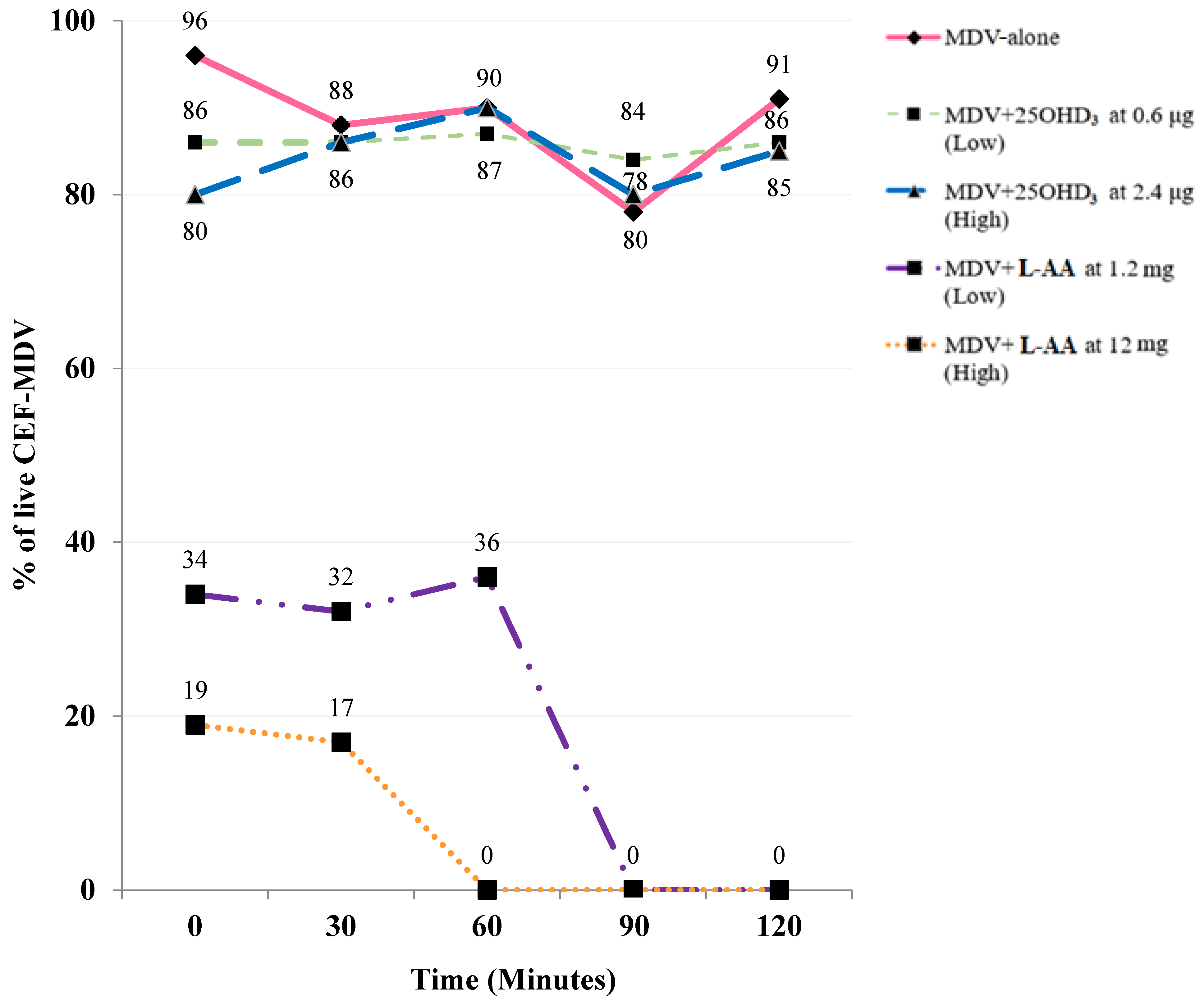
The pH values of the treatment solutions were: Diluent = 6.91; 25OHD3-0.6 = 6.91; 25OHD3-2.4 = 6.90; L-AA-1.2 = 4.33; and L-AA-12 = 2.55. The compatibility results of the MDV with the two doses of 25OHD3 and L-AA are illustrated in Figure 1. The low or high levels of L-AA in combination with MDV resulted in a 65 to 80% respective death of CEF-MDV after 30 min, which remained low during the total 2 h period. However, both levels of 25OHD3 over the 2 h period had similar effects to that of MDV-alone, in which the percentage of viable CEF-MDV in the MDV-alone, MDV+ 25OHD3-0.6, and 25OHD3-2.4 treatments were 88, 86, and 84%, respectively.
4. Discussion
To date, the viability of CEF-MDV upon its exposure to various vitamins has not been evaluated. Without statistical comparison, the plots of the progression of cell viability with time for each treatment clearly indicated that at the levels of inclusion used, L-AA had a detrimental effect on the viability of CEF-MDV. However, low and high levels of the vitamin D source (25OHD3) did not show significant negative effects on CEF-MDV viability, indicating that 25OHD3 at the doses tested may be suitable for in ovo application in combination with MDV. The negative impact of L-AA may be linked to its acidic characteristics and the high level of sensitivity of the MDV to drastic changes in pH. Therefore, despite the buffering capacity of the MDV diluent, a lowering of the pH of the MDV solution may have decreased CEF-MDV viability. In addition, the lowest dose of L-AA used in the current study was 1.2 mg, which is 500 times more than the highest dose of 25OHD3 (2.4 μg) that was tested. The hatch and post-hatch performance results of broilers have not been promising when they were administered concentrations of L-AA lower than 1.2 mg/mL by in ovo injection [18]. This was the basis for choosing a 1.2 mg/mL concentration of L-AA as the low-level dose in this study.
Vitamin D metabolites, including vitamin D3 and 25OHD3, are normally required to be suspended in ethanol to be soluble for in ovo administration [14]. However, in this study, the 25OHD3 source was water soluble and did not require alcohol suspension. There are several solutions that have been used to suspend various nutrients for in ovo injection purposes. These have included distilled water [33], saline [18,19,20,21,26], ethanol [17,34], soybean oil [35], and glycerol [36]. Nevertheless, commercial diluent has been used in more recent studies involving the in ovo injection of 25OHD3 [15,16,23,24,25,32]. Furthermore, CEF-MDV can only survive in commercial diluent, and the diluent is easily accessible. In the current study, both vitamin solution concentrations were prepared in a commercial diluent to which the full dose of MDV was added. Thus, commercial diluent should be used in further studies investigating the combinatorial administration of MDV and vitamins.
5. Conclusions
In conclusion, different doses of two bioactive vitamins (L-AA and 25OHD3) were separately combined with MDV in commercial diluent in order to determine the compatibility of MDV with the aforementioned vitamins. The compatibility results reported in this study using plots of cell viability demonstrated that both low and high doses of L-AA had negative effects on CEF-MDV survival. However, low and high doses of 25OHD3 exhibited minimal detrimental effects on CEF-MDV survival, indicating that 25OHD3 is a potential candidate for in ovo administration at 18 doi in combination with the MDV.
Author Contributions
Conceptualization, S.A.F., J.D. and E.D.P.; methodology, S.A.F., C.J.W. and J.D.; software, S.A.F.; validation, S.A.F. and E.D.P.; formal analysis, S.A.F.; investigation, S.A.F.; resources, E.D.P.; data curation, S.A.F. and J.D.; writing—original draft preparation, S.A.F.; writing—review and editing, S.A.F., C.J.W., J.D. and E.D.P.; visualization, S.A.F. and E.D.P.; supervision, S.A.F.; project administration, S.A.F.; funding acquisition, E.D.P. All authors have read and agreed to the published version of the manuscript.
Funding
This research was supported by the United States Department of Agriculture (USDA agreement no. 58-6064-9-016), Zoetis Animal Health Co., DSM Nutritional Products Inc., and Boehringer Ingelheim.
Institutional Review Board Statement
No animals or humans were used in this experiment; therefore, no institutional review board was necessary.
Informed Consent Statement
Not applicable.
Data Availability Statement
Not applicable.
Acknowledgments
The authors express their appreciation for the assistance provided by Eric Nixon and the Zoetis team of scientists. Our appreciation is also extended to Rodney Johnson for invaluable assistance.
Conflicts of Interest
The authors declare no conflict of interest.
References
- Hein, R.; Koopman, R.; García, M.; Armour, N.; Dunn, J.R.; Barbosa, T.; Martinez, A. Review of poultry recombinant vector vaccines. Avian Dis. 2021, 65, 438–452. [Google Scholar] [CrossRef] [PubMed]
- Negash, T.; Al-Garib, S.O.; Gruys, E. Comparison of in ovo and post-hatch vaccination with particular reference to infectious bursal disease. A review. Vet. Q. 2004, 26, 76–87. [Google Scholar] [CrossRef] [PubMed]
- Razib, D.; Pravin, M.; Rajesh, J. In ovo feeding as a tool for improving performance and gut health of poultry: A review. Front. Vet. Sci. 2021, 8, 754246. [Google Scholar]
- Sharma, J.M. Introduction to poultry vaccines and immunity. Adv. Anim. Vet. Sci. 1999, 41, 481–493. [Google Scholar]
- Van Hulten, M.C.W.; Cruz-Coy, J.; Gergen, L.; Pouwels, H.; Ten Dam, G.B.; Verstegen, I.; de Groof, A.; Morsey, M.; Tarpey, I. Efficacy of a turkey herpesvirus double construct vaccine (HVT-ND-IBD) against challenge with different strains of Newcastle disease, infectious bursal disease and Marek’s disease viruses. Avian Pathol. 2021, 50, 18–30. [Google Scholar] [CrossRef]
- Peebles, E.D.; Barbosa, T.M.; Cummings, T.S.; Gerard, P.D.; Williams, C.J.; Wilson, F.D. Comparative effects of in ovo versus subcutaneous administration of the Marek’s disease vaccine and pre-placement holding time on the intestinal villus to crypt ratios of Ross 708 broilers during early post-hatch development. Poult. Sci. 2019, 98, 712–716. [Google Scholar] [CrossRef]
- Gimeno, I.M.; Glaize, A.; Cortes, A.L. Effect of Marek’s disease vaccines on interferon and toll like receptors when administered in ovo. Vet. Immunol. Immunopathol. 2018, 201, 62–66. [Google Scholar] [CrossRef]
- Uni, Z.; Ferket, P.R.; Tako, E.; Kedar, O. In ovo feeding improves energy status of late-term chicken embryos. Poult. Sci. 2005, 84, 764–770. [Google Scholar] [CrossRef]
- Uni, Z.; Ferket, P.R. Methods for early nutrition and their potential. Worlds Poult. Sci. J. 2004, 60, 101–111. [Google Scholar] [CrossRef]
- Fritts, C.A.; Waldroup, P.W. Effect of source and level of vitamin D on live performance and bone development in growing broilers. J. Appl. Poult. Res. 2003, 12, 45–52. [Google Scholar] [CrossRef]
- Narbaitz, R.; Tsang, C.P.; Grunder, A.A. Effects of vitamin D deficiency in the chicken embryo. Calcif. Tissue Int. 1987, 40, 109–113. [Google Scholar] [CrossRef] [PubMed]
- Vignale, K.; Greene, E.S.; Caldas, J.V.; England, J.; Boonsinchai, N.; Sodsee, P.; Pollock, E.D.; Dridi, S.; Coon, C.N. 25-Hydroxycholecalciferol enhances male broiler breast meat yield through the mTOR pathway. J. Nutr. 2015, 145, 855–863. [Google Scholar] [CrossRef]
- Adams, J.S.; Hewison, M. Unexpected actions of vitamin D: New perspectives on the regulation of innate and adaptive immunity. Nat. Clin. Pract. Endocrinol. Metab. 2008, 42, 80–90. [Google Scholar] [CrossRef] [PubMed]
- Bello, A.; Hester, P.Y.; Gerard, P.D.; Zhai, W.; Peebles, E.D. Effects of commercial in ovo injection of 25-hydroxycholecalciferol on bone development and mineralization in male and female broilers. Poult. Sci. 2014, 93, 2734–2739. [Google Scholar] [CrossRef] [PubMed]
- Fatemi, S.A.; Elliott, K.E.C.; Bello, A.; Durojaye, O.A.; Zhang, H.; Alqhtani, A.H.; Peebles, E.D. Effects of the in ovo injection of vitamin D3 and 25-hydroxyvitamin D3 in Ross 708 broilers subsequently fed commercial or calcium and phosphorus-restricted diets. I. performance, carcass characteristics, and incidence of woody breast myopathy. Poult. Sci. 2021, 100, 101220. [Google Scholar] [CrossRef]
- Fatemi, S.A.; Elliott, K.E.C.; Bello, A.; Macklin, K.S.; Peebles, E.D. Effects of the in ovo injection of vitamin D3 and 25-hydroxyvitamin D3 in Ross 708 broilers subsequently challenged with coccidiosis: II. Immunological and inflammatory responses and small intestine histomorphology. Animals 2022, 12, 1027. [Google Scholar] [CrossRef]
- Abbasi, T.; Shakeri, M.; Zaghari, M.; Kohram, H. Growth performance parameters, bone calcification and immune response of in ovo injection of 25-hydroxycholecalciferol and vitamin K3 in male ross 308 broilers. Theriogenology 2017, 90, 260–265. [Google Scholar] [CrossRef]
- Zhang, H.; Elliott, K.E.C.; Durojaye, O.A.; Fatemi, S.A.; Schilling, M.W.; Peebles, E.D. Effects of in ovo injection of L-ascorbic acid on growth performance, carcass composition, plasma antioxidant capacity, and meat quality in broiler chickens. Poult Sci. 2019, 98, 3617–3625. [Google Scholar] [CrossRef]
- El-Senousey, H.K.; Chen, B.; Wang, J.Y.; Atta, A.M.; Mohamed, F.R.; Nie, Q.H. In ovo injection of ascorbic acid modulates antioxidant defense system and immune gene expression in newly hatched local Chinese yellow broiler chicks. Poult. Sci. 2018, 97, 425–429. [Google Scholar] [CrossRef]
- Mousstaaid, A.; Fatemi, S.A.; Levy, A.W.; Purswell, J.L.; Olanrewaju, H.A.; Baughman, B.; McNulty, K.; Gerard, P.D.; Peebles, E.D. Effects of the in ovo administration of L-ascorbic acid on tissue L-ascorbic acid concentrations, systemic inflammation, and tracheal histomorphology of Ross 708 Broilers subjected to elevated levels of atmospheric ammonia. Poultry 2023, 2, 158–173. [Google Scholar] [CrossRef]
- Mousstaaid, A.; Fatemi, S.A.; Elliott, K.E.C.; Levy, A.W.; Miller, W.W.; Olanrewaju, H.A.; Purswell, J.L.; Gerard, P.D.; Peebles, E.D. Effects of the in ovo administration of L-ascorbic acid on the performance and incidence of corneal erosion in Ross 708 broilers subjected to elevated levels of atmospheric ammonia. Animals 2023, 13, 399. [Google Scholar] [CrossRef] [PubMed]
- Bello, A.; Zhai, W.; Gerard, P.D.; Peebles, E.D. Effects of the commercial in ovo injection of 25-hydroxycholecalciferol on the hatchability and hatching chick quality of broilers. Poult. Sci. 2013, 92, 2551–2559. [Google Scholar] [CrossRef] [PubMed]
- Fatemi, S.A.; Elliott, K.E.C.; Bello, A.; Zhang, H.; Peebles, E.D. Effects of the in ovo injection of vitamin D3 and 25-hydroxyvitamin D3 in Ross 708 broilers subsequently fed commercial or calcium and phosphorous-restricted diets: II. Immunity and small intestine morphology. Poult. Sci. 2021, 100, 101240. [Google Scholar] [CrossRef] [PubMed]
- Fatemi, S.A.; Macklin, K.S.; Zhang, L.; Mousstaaid, A.; Poudel, S.; Poudel, I.; Peebles, E.D. Improvement in the immunity- and vitamin D3 activity-related gene expression of coccidiosis-challenged Ross 708 broilers in response to the in ovo injection of 25-hydroxyvitamin D3. Animals 2022, 12, 2517. [Google Scholar] [CrossRef]
- Fatemi, S.A.; Alqhtani, A.H.; Elliott, K.E.C.; Bello, A.; Zhang, H.; Levy, A.W.; Peebles, E.D. Improvement in the performance and inflammatory reaction of Ross 708 broilers in response to the in ovo injection of 25-hydroxyvitamin D3. Poult. Sci. 2021, 100, 138–146. [Google Scholar] [CrossRef]
- Zhang, H.; Elliott, K.E.C.; Durojaye, O.A.; Fatemi, S.A.; Peebles, E.D. Effects of in ovo-administration of L-ascorbic acid on broiler hatchability and its influence on the effects of pre-placement holding time on broiler quality characteristics. Poult. Sci. 2018, 97, 1941–1947. [Google Scholar] [CrossRef]
- Soltani, T.; Salarmoini, M.; Afsharmanesh, M.; Tasharrofi, S. The effects of in ovo injection of ascorbic acid on hatchability, growth performance, intestinal morphology, and tibia breaking strength in 36h post hatch fasted broiler chickens. Poult. Sci. J. 2019, 7, 43–49. [Google Scholar]
- Zhu, Y.F.; Bodinga, M.B.; Zhou, J.H.; Zhu, L.Q.; Cao, Y.L.; Ren, Z.Z.; Yang, X.J. Effects of in ovo injection of vitamin C on heat shock protein and metabolic genes expression. Animal 2020, 14, 360–367. [Google Scholar] [CrossRef]
- Zhu, Y.F.; Li, S.Z.; Sun, Q.Z.; Yang, X.J. Effect of in ovo feeding of vitamin C on antioxidation and immune function of broiler chickens. Animal 2019, 13, 1927–1933. [Google Scholar] [CrossRef]
- Buranathai, C.; Rodriguez, J.; Grose, C. Transformation of primary chick embryo fibroblasts by Marek’s disease vaccine. Virology 1997, 239, 20–35. [Google Scholar] [CrossRef]
- Fatemi, S.A.; Elliott, K.E.C.; Bello, A.; Durojaye, O.A.; Zhang, H.J.; Peebles, E.D. The effects of in ovo-injected vitamin D3 sources on the eggshell temperature and early post-hatch performance of Ross 708 broilers. Poult. Sci. 2020, 99, 1357–1362. [Google Scholar] [CrossRef] [PubMed]
- Fatemi, S.A.; Elliott, K.E.C.; Bello, A.; Durojaye, O.; Zhang, H.; Turner, B.; Peebles, E.D. Effects of source and level of in ovo-injected vitamin D3 on the hatchability and serum 25-hydroxycholecalciferol concentrations of Ross 708 broilers. Poult. Sci. 2020, 99, 3877–3884. [Google Scholar] [CrossRef] [PubMed]
- Zhai, W.; Gerard, P.D.; Pulikanti, R.; Peebles, E.D. Effects of in ovo injection of carbohydrates on embryonic metabolism, hatchability, and subsequent somatic characteristics of broiler hatchlings. Poult. Sci. 2011, 90, 2134–2143. [Google Scholar] [CrossRef]
- Xu, H.; Hu, Z.; Lu, Y.; Jiang, Y.; Li, D.; Lie, B.; Du, R.; Lei, B.; Yang, C.; Zhang, Z.; et al. Improvement in the early growth, immune system and tibia development of broilers in response to the in ovo injection of 25-hydroxyvitamin D3. J. Appl. Poult. Res. 2023, 51, 265–275. [Google Scholar] [CrossRef]
- Hayakawa, T.; Shiraishi, J.; Ohta, Y. Effects of in ovo vitamin D3 injection on subsequent growth of broilers. J. Poult. Sci. 2019, 56, 220–223. [Google Scholar] [CrossRef]
- Mansour, D.S.; El-Senosi, Y.A.; Mohamed, M.I.; Amer, M.M.; Elaroussi, M.A. Effects of injecting vitamin D3 or an active metabolite in ovo on chick embryonic development and calcium homeostasis. W. J. Pharm. Pharm. Sci. 2017, 6, 1454–1467. [Google Scholar]
Figure 1.
Percentage of live primary chick embryo fibroblast cells infected by the Marek′s disease virus (CEF-MDV) every 30 min (0, 30, 60, 90, and 120 min) when the MD vaccine (MDV) was combined with low (1.2 mg) and high (12 mg) doses of L-ascorbic acid (L-AA) or low (0.6 μg) and high (2.4 μg) doses of 25-hydroxyvitamin D3 (25OHD3) during a 2 h period.

Disclaimer/Publisher’s Note: The statements, opinions and data contained in all publications are solely those of the individual author(s) and contributor(s) and not of MDPI and/or the editor(s). MDPI and/or the editor(s) disclaim responsibility for any injury to people or property resulting from any ideas, methods, instructions or products referred to in the content. |
© 2023 by the authors. Licensee MDPI, Basel, Switzerland. This article is an open access article distributed under the terms and conditions of the Creative Commons Attribution (CC BY) license (https://creativecommons.org/licenses/by/4.0/).
Share and Cite
MDPI and ACS Style
Fatemi, S.A.; Williams, C.J.; Deines, J.; Peebles, E.D. Vitamin Compatibility with the Marek’s Disease Vaccine. Poultry 2023, 2, 442-448. https://0-doi-org.brum.beds.ac.uk/10.3390/poultry2040033
AMA Style
Fatemi SA, Williams CJ, Deines J, Peebles ED. Vitamin Compatibility with the Marek’s Disease Vaccine. Poultry. 2023; 2(4):442-448. https://0-doi-org.brum.beds.ac.uk/10.3390/poultry2040033
Chicago/Turabian StyleFatemi, Seyed Abolghasem, Christopher J. Williams, Joshua Deines, and Edgar David Peebles. 2023. "Vitamin Compatibility with the Marek’s Disease Vaccine" Poultry 2, no. 4: 442-448. https://0-doi-org.brum.beds.ac.uk/10.3390/poultry2040033