
Infrared Spectroscopy Studies of Aluminum Oxide and Metallic Aluminum Powders, Part I: Thermal Dehydration and Decomposition


The Applied Research Laboratory, The Pennsylvania State University, University Park, PA 16804, USA
*
Author to whom correspondence should be addressed.
Powders 2022, 1(1), 47-61; https://0-doi-org.brum.beds.ac.uk/10.3390/powders1010005
Submission received: 9 February 2022
/
Revised: 28 February 2022
/
Accepted: 3 March 2022
/
Published: 10 March 2022
(This article belongs to the Special Issue Feature Papers in Powders)
Abstract
:In this work, we study three aluminum oxides (alpha, gamma, boehmite) and various oxidized metallic aluminum powders to observe their dehydration and decomposition behavior using in situ diffuse reflectance infrared Fourier transform spectroscopy (DRIFTS) and scanning electron microscopy (SEM). We find that a temperature increase to the aluminum oxides (aluminas) reduces physically adsorbed water molecules to reveal the presence of hydroxyl groups. All three aluminas contained bridged hydroxyls located at 3670 cm−1; we found additional surface hydroxyls, which varied based on the oxidation state of the aluminum atom. Oxidized metallic aluminum powders that were aged resulted in similar behavior; however, the results differed depending on the method of aging. We find that naturally aged aluminum (NA-Al) powders with heavy oxidation in the form of the tri-hydroxide decomposed and did not reveal any detectable surface hydroxyl peaks. When aged using artificial methods (AA-Al), we find both surface hydroxyls, including bridged hydroxyls at 3670, 3700, and 3730 cm−1, and a remaining boehmite-like surface. These results show that metallic aluminum powders can be tailored for specific applications, regardless of age. It also elucidates different ways to pre-process the powders to control the surface oxide layer, corroborated by comparison with the models oxides studied herein.
1. Introduction
Metallic aluminum powders have a wide variety of engineering applications, including catalysis [1,2,3], propulsion [4,5,6], pigments [7,8], and as foaming agents [9]. Aluminum continues to find innovative uses and is prominent in the continuing evolution of additive manufacturing [10,11,12]. The particle surface properties are fundamental to understanding how the powders will perform in filling, spray, and composite applications. To this end, we study the surface properties and thermal behavior of both alumina and metallic aluminum powders to better understand their as-received and dried state behavior. In our work, the alumina powders will serve as a reference for the study of the more complex surfaces found on the metallic aluminum powder surfaces using infrared spectroscopy (IR).
For aluminum powders specifically, the oxidized and hydrated surface layers can influence the powder packing, increase agglomeration, and be detrimental to the mechanical properties of fabricated parts. Recent work on alumina coatings has included characterization, including X-ray diffraction (XRD), atomic force microscopy (AFM), and energy dispersive spectroscopy (EDS) [13]. We hope to fill an important knowledge gap to help researchers identify the initial state, dehydration, and decomposition properties of aluminum powders in wide variety of states using a simplistic IR setup. This allows for the easy identification of the oxide (Al2O3), oxy-hydroxide (AlO(OH)), and hydroxide (Al(OH)3) phases present on different powders in terms of age and water exposure. Subsequently, we study the dehydration and decomposition behavior to see how the phases change as a result of applied heat. Similar studies have been completed for various types of aluminas; however, to the best of our knowledge we have not seen a study performed in situ with aluminum powders [14]. The ability to prepare aluminum powder surfaces for improved chemical receptivity is of interest to a wide variety of industrial, medical, and academic applications where the surface state is critical for the final product properties [15]. This is especially relevant in additive manufacturing where the powder is typically recycled [16].
Both aluminum and alumina powders are hygroscopic, leading to the formation of chemisorbed surface hydroxyls and accumulated physisorbed water, which can inhibit or change their surface reactivity. After exposure to atmospheric water, the surface is covered with hydroxyl groups which differ depending on the coordination of the Al atom, the state of the oxygen, and other hydrogen bonding contributions. Physisorbed water molecules can form two hydroxyl groups on a coordinational-unsaturated (cus) Al surface atom and subsequently create sites for molecular water physisorption. The distribution of hydroxyl types varies depending on the crystalline phase, face, and aging (i.e., exposure to atmospheric water and temperature). There are several models that have been developed over the years that describe the structure of these surface hydroxyls, most notably Peri [17,18], Tsyganenko [19], Knözinger’s [20], Busca [21], and Morterra [2,8,22,23,24]. We use these models to identify the surface hydroxyls observed in each IR spectrum to help build an encompassing picture of the dehydration behavior of both alumina and metallic aluminum powder particles. In this two part series, we also use probe molecules to show how these types of surface hydroxyls interact with organosilanes.
Aging of the aluminum particle surface happens over time and the resulting phases are a direct result of the storage conditions [25,26,27]. The tri-hydroxide phase changes the surface morphology by preferentially forming needles and triangular units on the particle surface, which have lowered reactivity and influence flow behavior by increasing the chances of mechanically inter-locking between particles. Thermal treatment has been utilized to decompose these phases into the more reactive phase boehmite and to liberate the hydrogen bond surface hydroxyls. This is particularly relevant to metallurgical processing and additive manufacturing, where the surface hydroxides and oxides can interfere with the final product [28]; additionally, surface aging can interfere with flow properties impeding proper powder distribution [15].
Previous work in our research group has included the surface analysis of aluminum powders under ambient conditions [29,30]. In this contribution, we report the thermal behavior of metallic aluminum particles using model, high surface area alumina powders as reference materials. Moreover, we demonstrate this behavior using in situ DRIFTS with the ability to track the behavior. Using this method, we can take advantage of the different IR absorbance peaks related to each phase to follow the behavior of the powders.
2. Materials and Methods
2.1. Materials
Ethanol (Pharmco), Boehmite (50 nm, Sasol), -alumina (300 nm, Electron Microscopy Sciences, Hatfield, PA, USA), -alumina (50 nm, Aldrich), and aluminum powder (20 m, Valimet H15) were all used as received. Additional characterization data are provided in Appendix A. Distilled water was utilized for immersion studies. Ethanol (99.5 (w/w), <0.1 water (w/w), KOPTEC, PA, USA) was used as received for all rinsing procedures.
2.2. Powder Preparations
The as-received aluminum powder (AR-Al) was not modified prior to analysis. Naturally aged (NA-Al) aluminum samples were retrieved from ambient storage conditions after ten years of aging under ambient storage and in air. The samples experienced temperature fluctuation during storage, however, were not exposed to extreme high or low temperatures or humidity.
A relative humidity (RH) chamber was used to investigate the effect high temperature and humidity conditions on the accelerated aging process (AA-Al). Powdered aluminum (10 g, Valimet) was loaded in to the bottom of a 50 mL beaker and placed in a humidity chamber at 85 °C and 85 percent RH for 1 h. The samples were agitated to prevent localized surface exposure. The resulting sample was rinsed with ethanol and dried under N2 prior to analysis.
The water-immersed (WI-Al) sample was prepared using powdered aluminum (Valimet, H15) in distilled water at a concentration of 100 g/50 mL at 70 °C and allowed to stir for 1 day. These conditions were previously studied by Hart for the preparation of boehmite films. Subsequently, the sample was rinsed three times with ethanol and dried in a chamber under N2 before the analyses were completed.
2.3. Scanning Electron Microscopy (SEM)
The powder samples were imaged using a Verios G-4 scanning electron microscope (ThermoFisher Scientific, Hillsboro, OR, USA) at various magnification levels. A small amount of powder was mounted on typical SEM stubs using carbon tape and subsequently coated with 5 nm iridium to reduce charging.
2.4. Diffuse Reflectance Infrared Fourier Transform Spectroscopy (DRIFTS), Ambient and Heating Experiments
All of the spectra collected in this manuscript were measured on a Fourier transform infrared spectrophotometer (Bruker, Vertex V70). We used the Praying Mantis® accessory (Harrick Scientific, Pleasantville, NY, USA), which enabled us to use a reactor chamber capable of heating the sample. The detector used was Mercury-Cadmium-Telluride (MCT) and the reaction chamber utilized CaF2 windows with an 900 cm−1 cutoff. The measurements were collected at 6 cm−1 resolution and 400 scans. The samples were heated in situ through the sample cup, which was increased by a temperature controller with a 110 V proportional-interval-derivative (PID) control (Harrick Scientific). The temperature was increased sequentially at a step size of 10 °C or as noted on the figures. The chamber was continuously purged with argon to remove atomospheric water and oxygen.
3. Results and Discussion
3.1. Aluminas: Boehmite, Gamma, Alpha
The dehydration of the model aluminas was studied to understand the nature of the surface composition and to identify the possible surface hydroxyls present on each surface. The infrared spectra presented in this section were collected in the following manner: the sample was heated and equilibrated at surface temperatures of 100, 200, and 230 °C, under a constant Ar purge and ambient pressure in the in situ DRIFTS cell. We selected the high temperature of 230 °C so that any tri-hydroxide content would decompose and that we would be above the water desorption temperature of 100 °C. All spectra were collected on the same sample and referenced to dried KBr. The spectral contribution of water vapor was accounted for by subtraction of the gas phase water vapor spectrum.
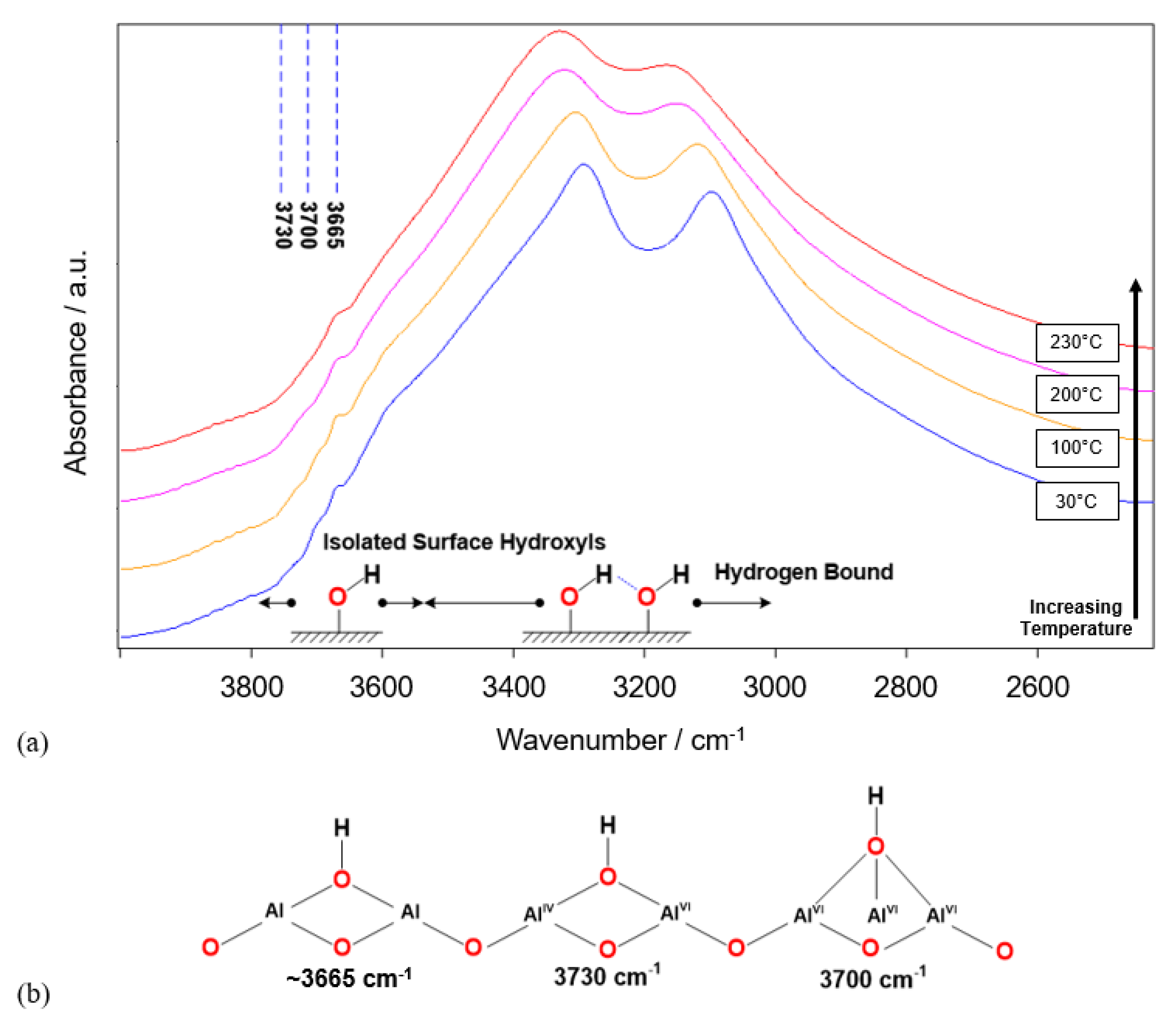
Figure 1 shows the IR spectra for the dehydration of boehmite as a function of temperature where the spectra are presented from highest temperature to the lowest. At ambient temperature, the two modes for the bulk, layered hydroxyls appear at 3290 and 3096 cm−1 for the asymmetric and symmetric modes, respectively. Heating to 100 °C shows a small shift and increase in the surface hydroxyl region. Upon heating to 200 °C, there is continued shifting and broadening of the associated hydroxyl region, attenuation, and appearance of surface hydroxyl absorbance peaks due to the loss of physisorbed water. When the sample hit the maximum temperature of 230 °C, there is a small amount of blue shifting of the associated hydroxyls (2 cm−1) and increased broadening. The vibrational frequencies of the associated hydroxyls shift due to the increase in –OH bond lengths as a result of the increase in temperature and relaxation of the lattice structure [31]. The surface hydroxyl groups liberated by dehydration are of the bridged and tri-bridged by Morterra’s model and appear at 3730, 3700, and 3665 cm−1 as shown in Figure 1.
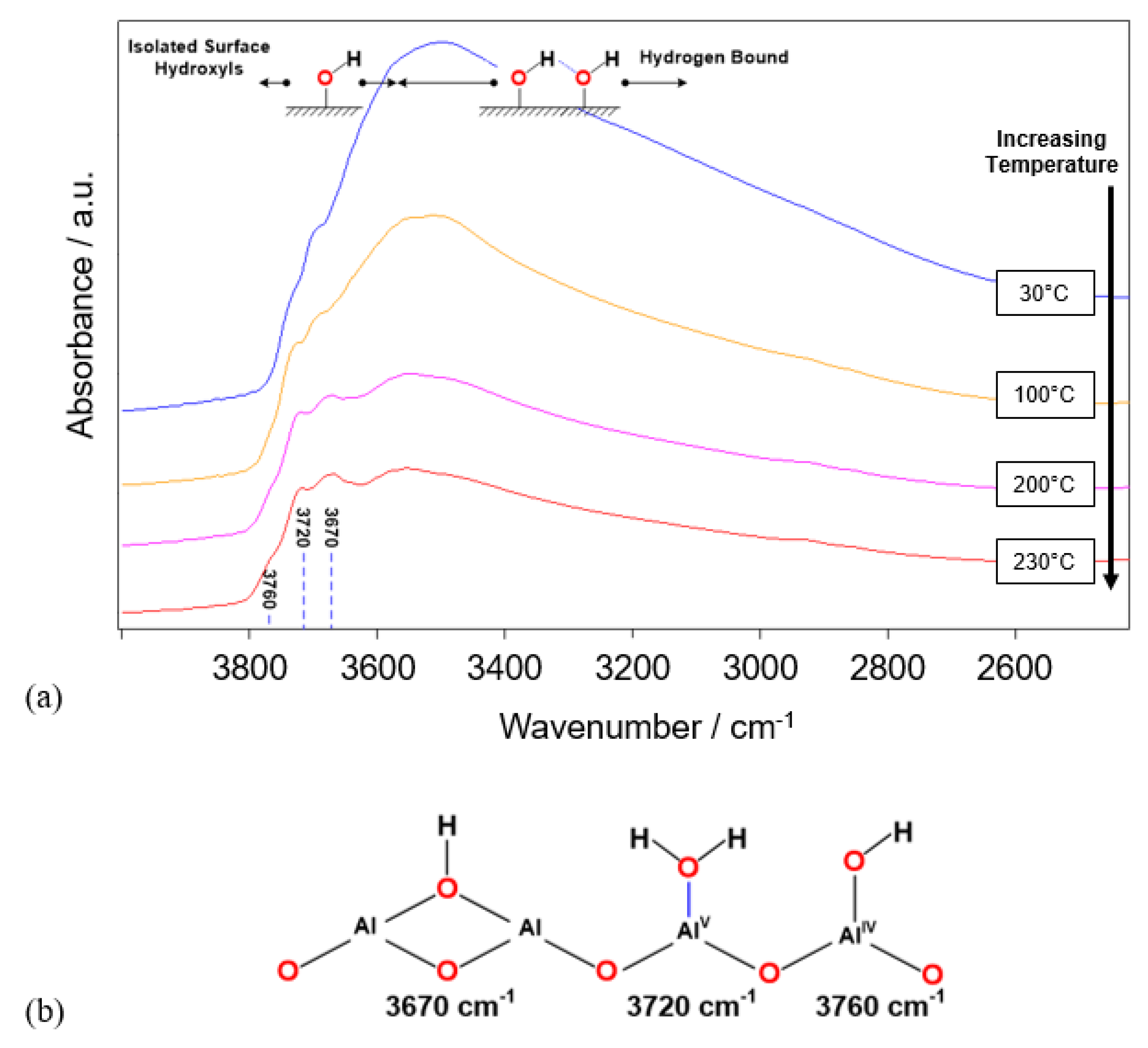
Figure 2 shows the corresponding in situ dehydration process of -alumina presented from lowest to highest temperature. Heating to 100 °C shows a decrease in the intensity of the broad –OH feature at 3500 cm−1 corresponding to the loss of molecular water, which makes the surface hydroxyl peaks visible at 3760, 3720, and 3670 cm−1. Increased heating to 200 °C shows continued loss of absorbance in the board –OH feature. At 230 °C, the broad band still appears with a maximum around 3500 cm−1, suggesting that some of the surface hydroxyls are still hydrogen bound [32]. The assignment of the 3760 and 3670 cm−1 sites are of the bridged and surface hydroxyl groups. The peak at 3720 cm−1 site, however, remains debatable in the literature. It has been assigned as either molecular water coordinated to a pentavalent, Al Lewis acid site [33] or a displaceable hydroxyl [34]. According to observations by Liu, drying increases the intensity of this peak, which is consistent with the appearance of this peak [35].
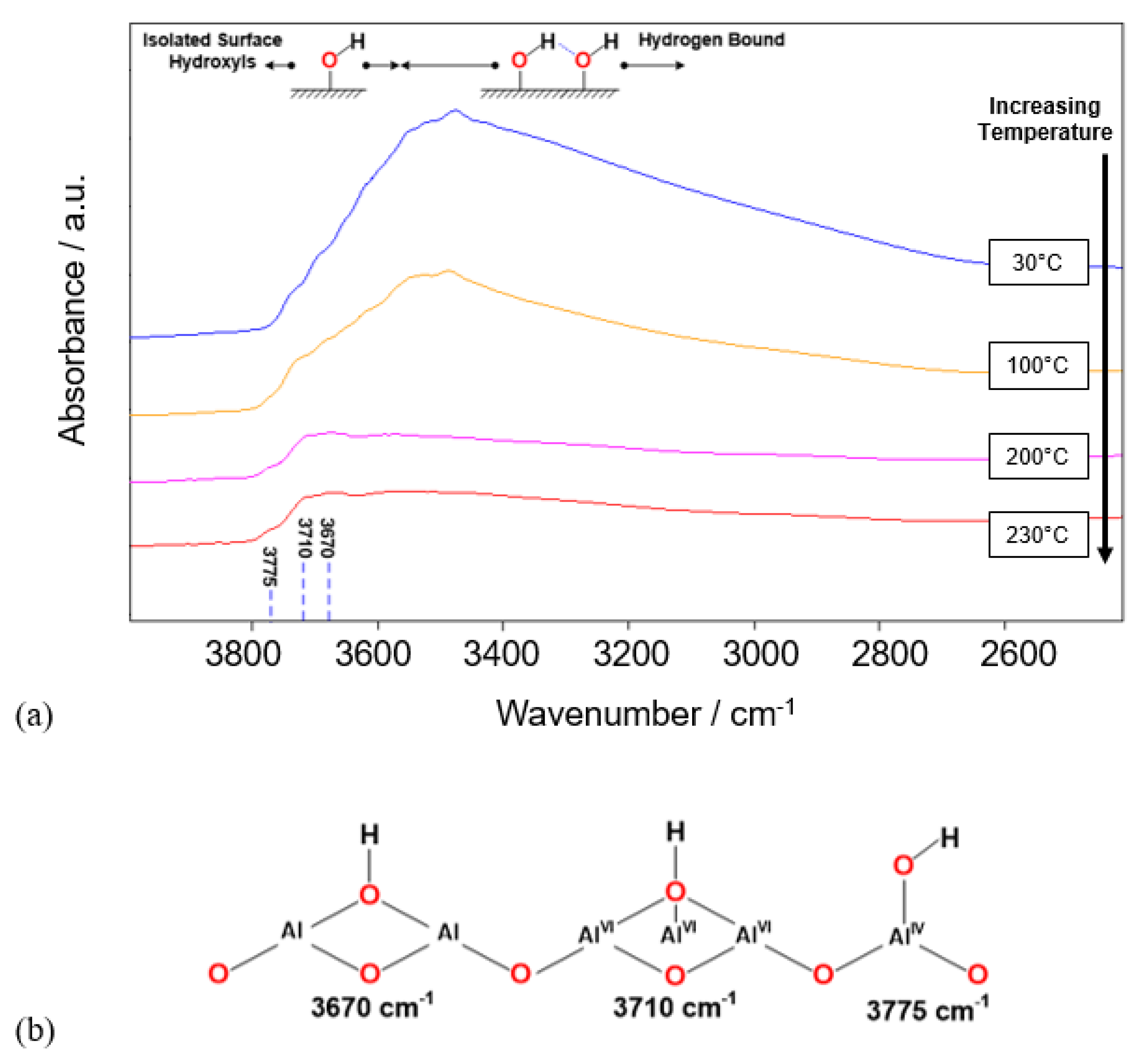
The dehydration behavior for -alumina is shown in Figure 3. There is a broad –OH absorbance which decreases upon heating from 30–100 °C. The strong absorbance centered around 3500 cm−1 is attributed to associated hydroxyl groups and the presence of molecular water. The surface hydroxyls appearing on -alumina are bridged (3670 cm−1), tri-bridged (3710 cm−1) and fully isolated octahedral (3775 cm−1). The and -alumina powders contained two similar groups, however, the 3720 cm−1 site was not present. The -alumina structure contains Al coordinated octahedrally throughout the bulk, but also shows absorbances for tetrahedrally coordinated Al surface hydroxyls. This is consistent with the idea that the surface should be considered an extended defect, where unique surface sites occur to surface reorganization and relaxation. The presence of such peaks has been reported in the literature [36,37] and is shown in Figure 3.
Alumina surfaces are “activated” through heating, which liberates physisorbed molecular water to render surface hydroxyls available for interactions with adsorbates. The drying regime used in this work was selected based on the low temperature of 230 °C to maintain the integrity of the boehmite structure. Under this protocol, the three aluminas produced surface hydroxyls, which were structurally similar according to literature models after dehydration. Boehmite produced a single, sharp absorbance at 3665 cm−1, distinguishing itself from the and phases. This absorbance is assigned as the bulk bridge hydroxyls present in the crystal structure as described in the introduction. We use the information gathered here to better understand the behavior of metallic aluminum powders, discussed in the next section.
3.2. Metallic Aluminum
One of the primary interests of this work is to study the behavior of naturally and artificially aged aluminum powders subjected to thermal treatments. The data presented in the previous section served as comparison behavior under the same conditions for the various phases. Herein, we used variability in aluminum powder surfaces as a function of age and water exposure to further understand and explore the surface phases.
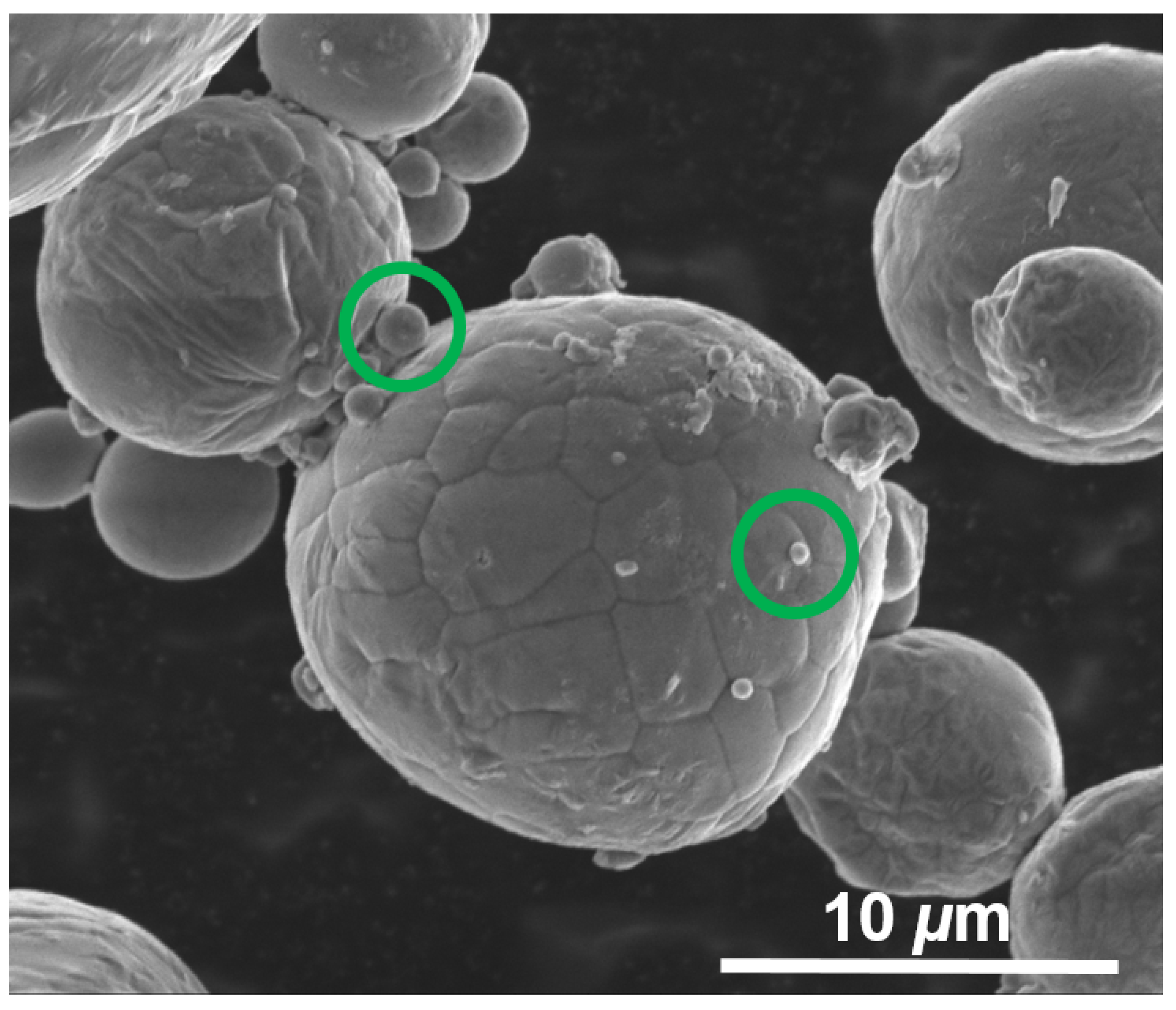
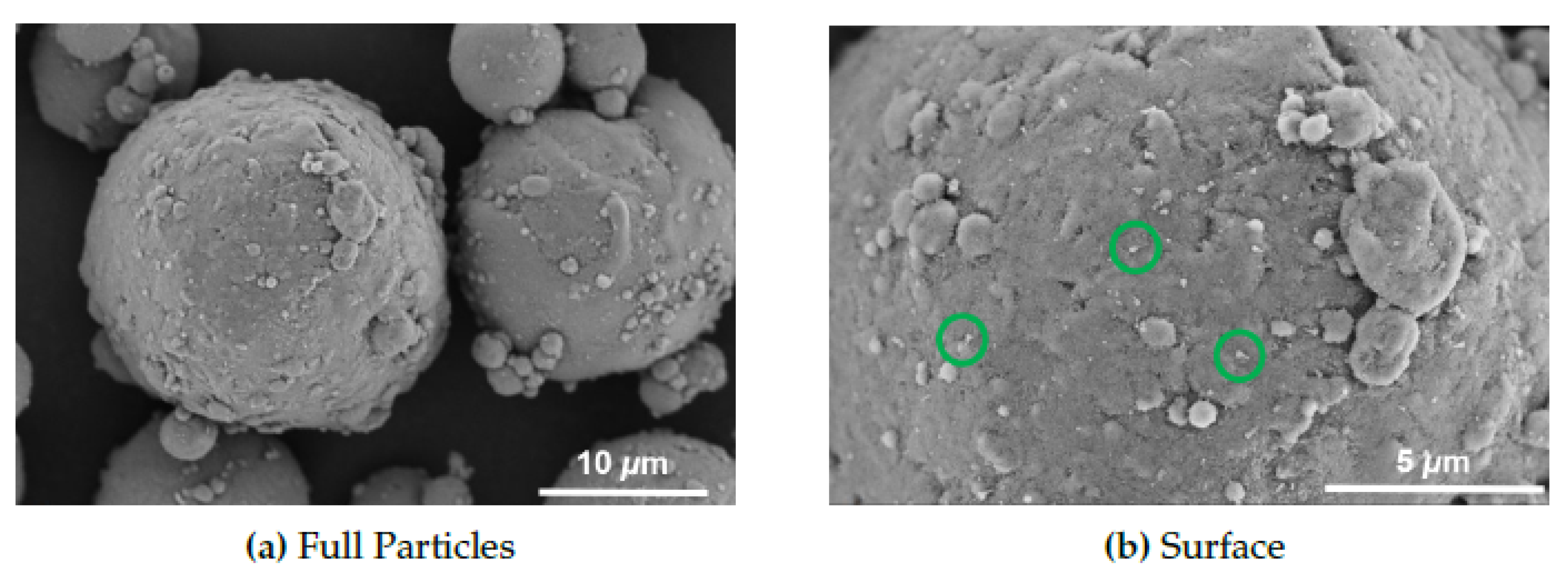
Basic image analysis using scanning electron microscopy (SEM) revealed surface features which we can corroborate with the IR data presented in this section. Figure 4 shows representative micrographs of AR-Al particles, where we see few morphological features. The particle surface has attached satellite particles, which is due to the gas atomization method used in their production [38]. The surface appears fairly smooth with a scale-like appearance or fracture lines that form during the oxidation process. These are discontinuities in the oxide structure, likely formed during the high temperature gas atomization process and formation of grain boundaries and micro-fractures.
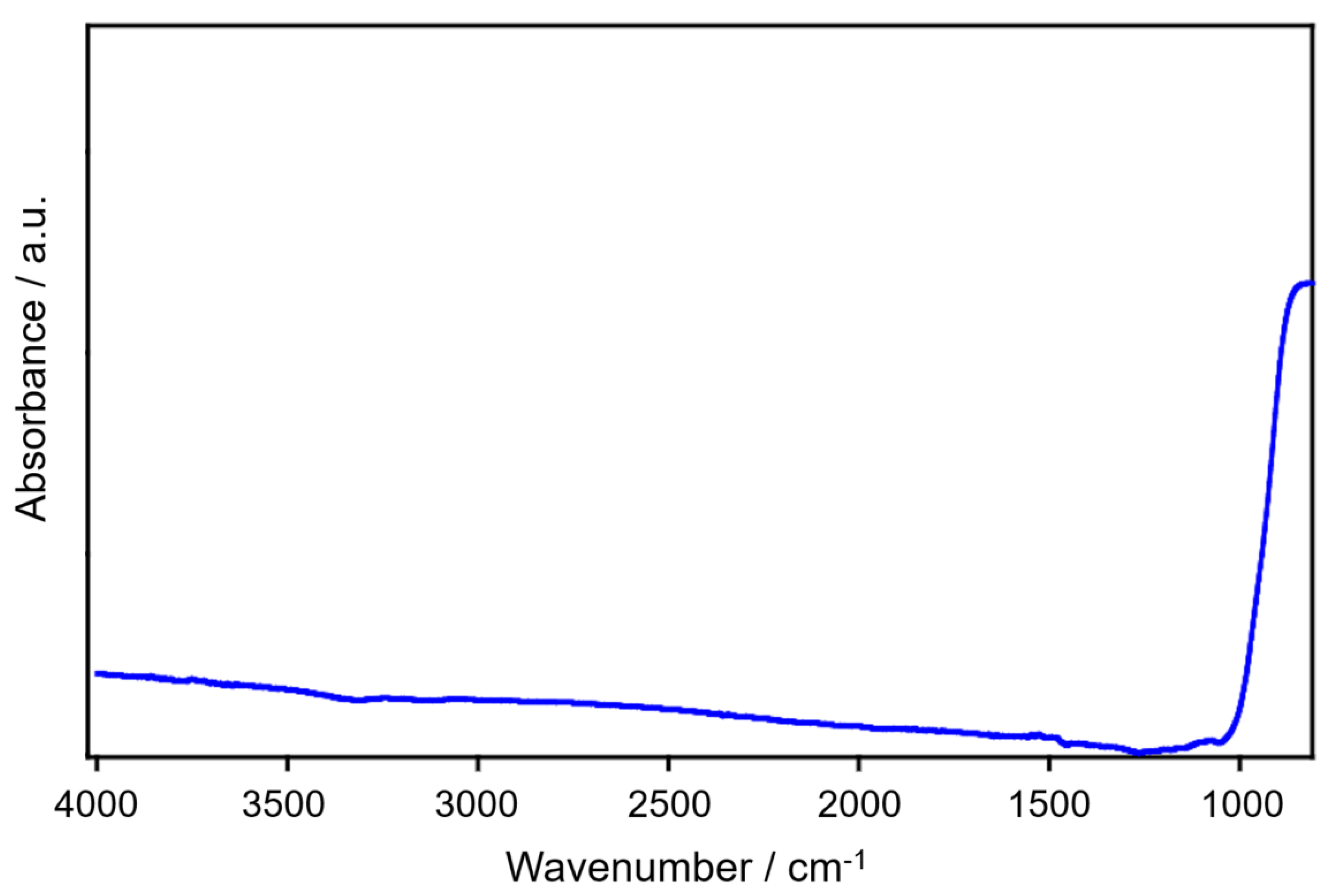
The DRIFT spectrum for AR-Al did not reveal not any detectable absorbance peaks in the surface hydroxyl region of the spectrum at any of the measured temperatures. The only detectable absorbance was at 950 cm−1, which is an Al-O vibration from the amorphous oxide as seen in Figure 5. This absorbance is close to the cutoff of the CaF2 windows, so a well-defined peak was not observed. The lack of a detectable molecular water peak made it difficult to ascertain that the physisorbed water was lost, however, this drying temperature was shown to be sufficient for water removal on the model oxide surfaces. The ambient temperature spectrum is shown in Figure 5 as there were no detectable changes observed as a function of drying the powder. Further investigations could be performed, such as thermogravimetric analysis to better understand the water loss on the as-received particle surfaces.
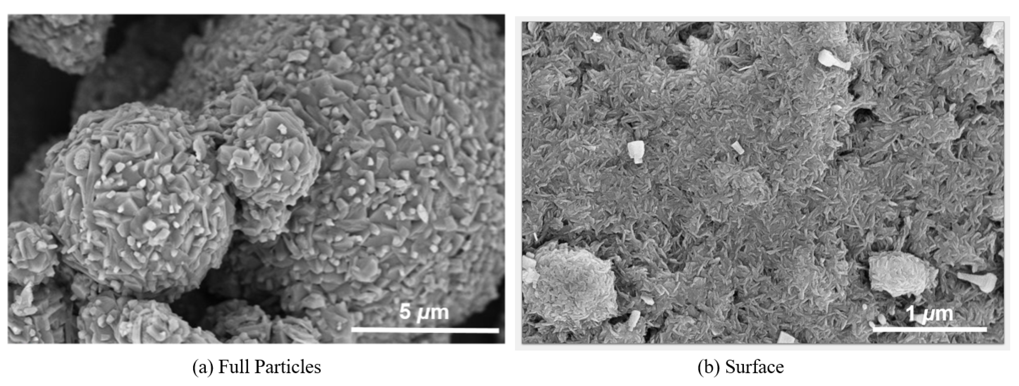
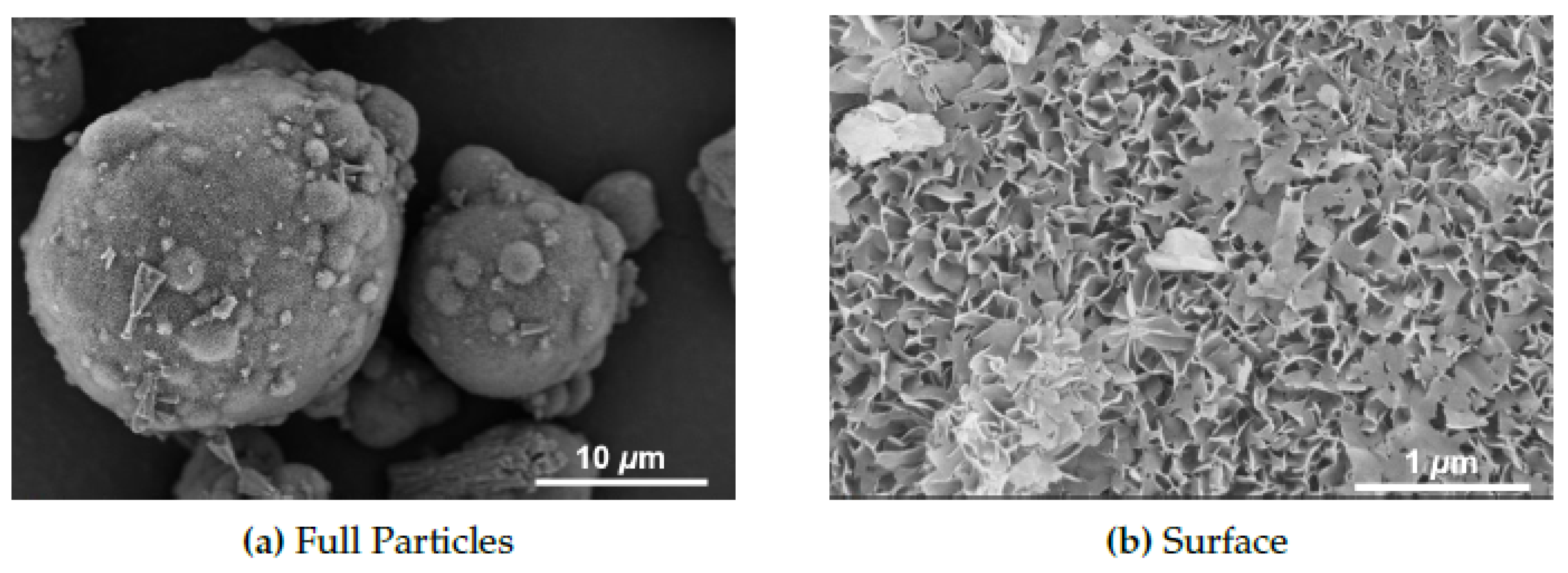
Representative SEM micrographs of the NA-Al particles are shown in Figure 6 and reveal signs of aging from the morphological changes manifested through pyramidal structure. The tri-hydroxide has been shown to grow preferentially in the Z direction, which is why we see outward growth on the surface of the particles [15]. These features are sub-micron sized and we also note the loss of the scale-like surface structure from aging. The satellite particles appear in a similar manner to the AR-Al and do not appear to have been dislodged during the aging process.
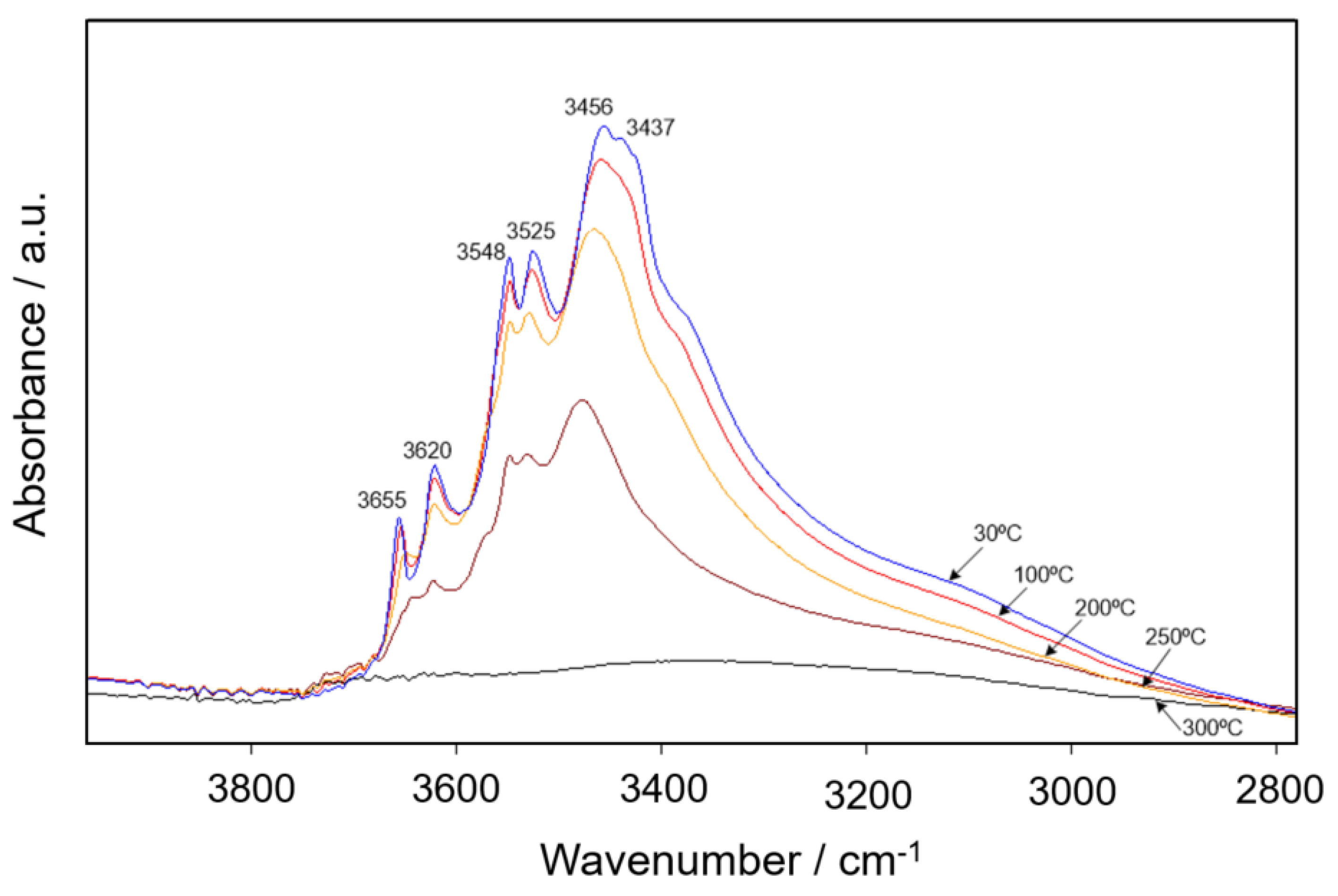
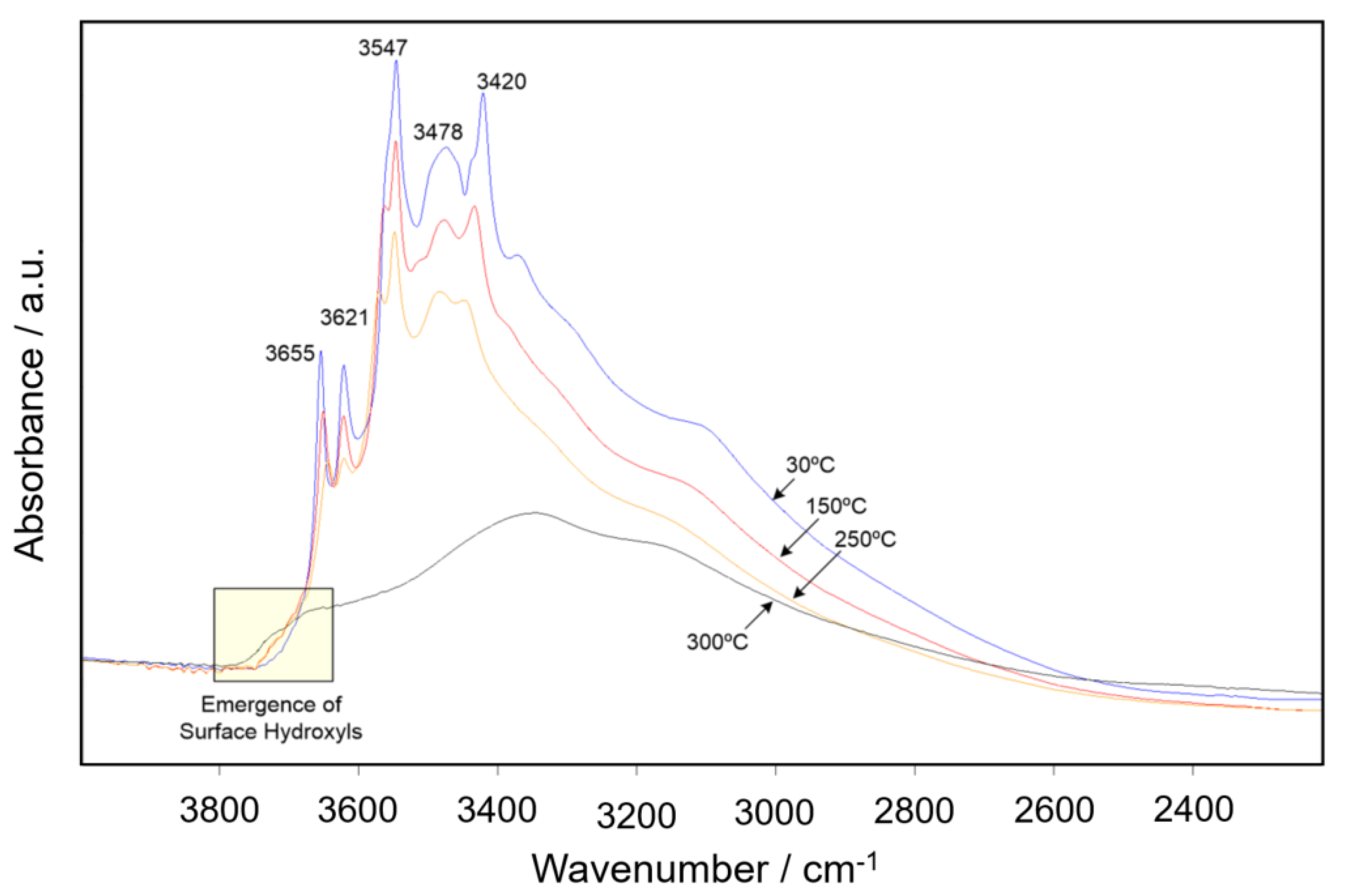
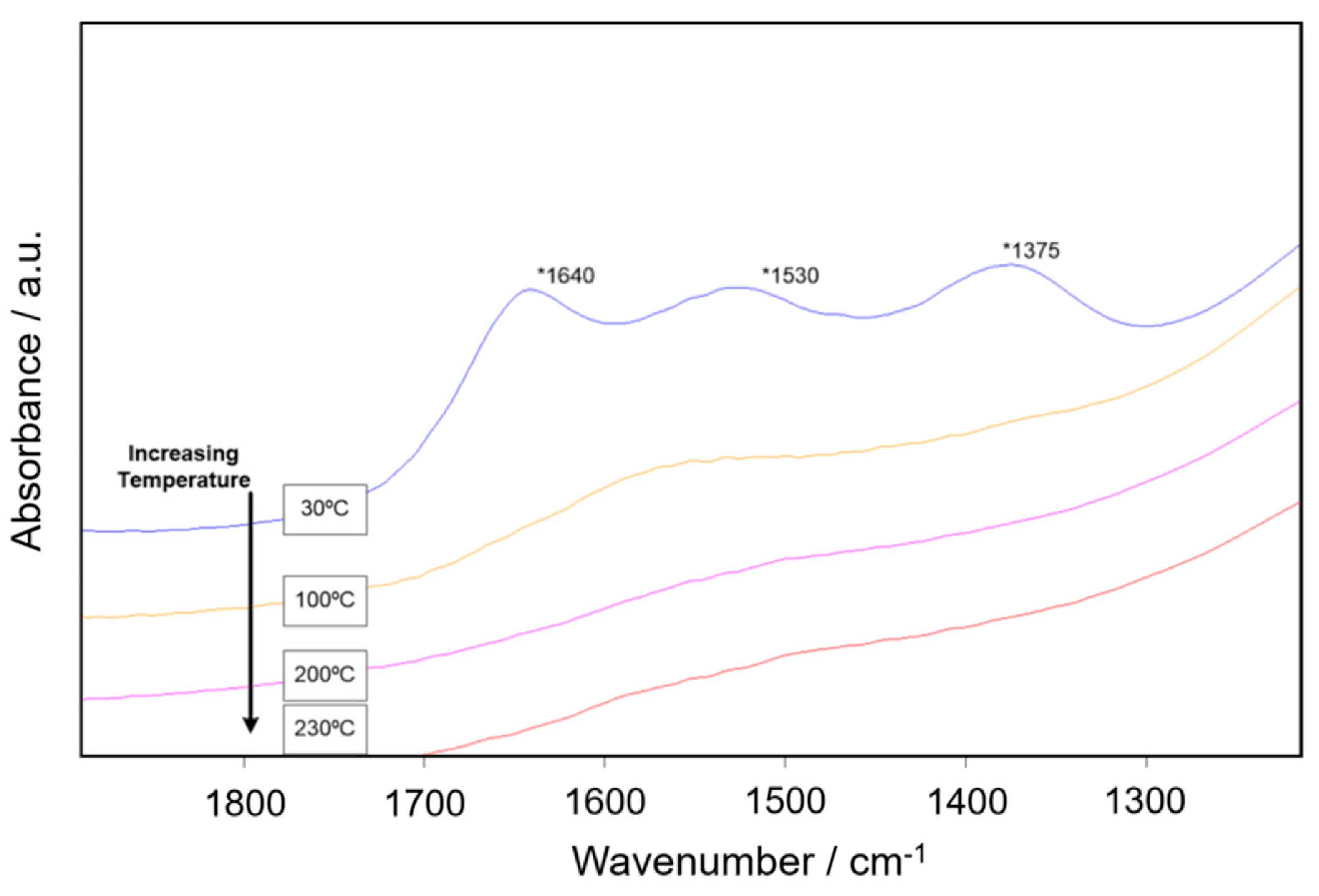
In order to observe the decomposition of the oxy- and tri-hydroxides, we raised the ceiling temperature to 300 °C, which yielded compelling results. The samples were dried to 300 °C for one hour to push the full decomposition of the tri-hydroxides as initial experiments equilibrated at 230 °C did not fully decompose. The naturally aged sample was dehydrated to 300 °C and its decomposition shown in Figure 7. Initially, there are several absorbance peaks attributed to the tri-hydroxides stretches as similar to those reported in the literature. Upon increasing temperature, these absorbance peaks decreased in intensity until there were no longer any detectable absorbance bands observed after reaching 300 °C. This behavior suggests that the tri-hydroxides complexes were decomposed and no detectable hydroxyl content was left on the surface after drying. The absorbance peaks observed at 3655, 3620, 3548, and 3525 cm−1 are typical of the tri-hydroxide, bayerite polymorph of Al(OH)3 [39]. The additional absorbance peaks, 3464 and 3437, have been identified as the reactive surface groups on the tri-hydroxide complexes on Gibbsite, another polymorph of the tri-hydroxide [40]. It is likely that there is a combination of tri-hydroxide phases on the particle surface. This particular sample did not show any isolated or surface hydroxyls for comparison.
Figure 8 reveals the micrographs associated with the AA-Al sample. We see a large assortment of plate and needle-like structures that fully cover the particle surfaces (Figure 8a). There is also possible fusion occurring at the different particle interfaces. The higher magnification image shown also reveals a fine needle-like structure on the surface, with additional geometric shapes on top of the layer (Figure 8b). These structures are quite remarkable and show that the scale-like features on the as-received powder are no longer visible after this type of aging in high RH with temperature (85 °C).
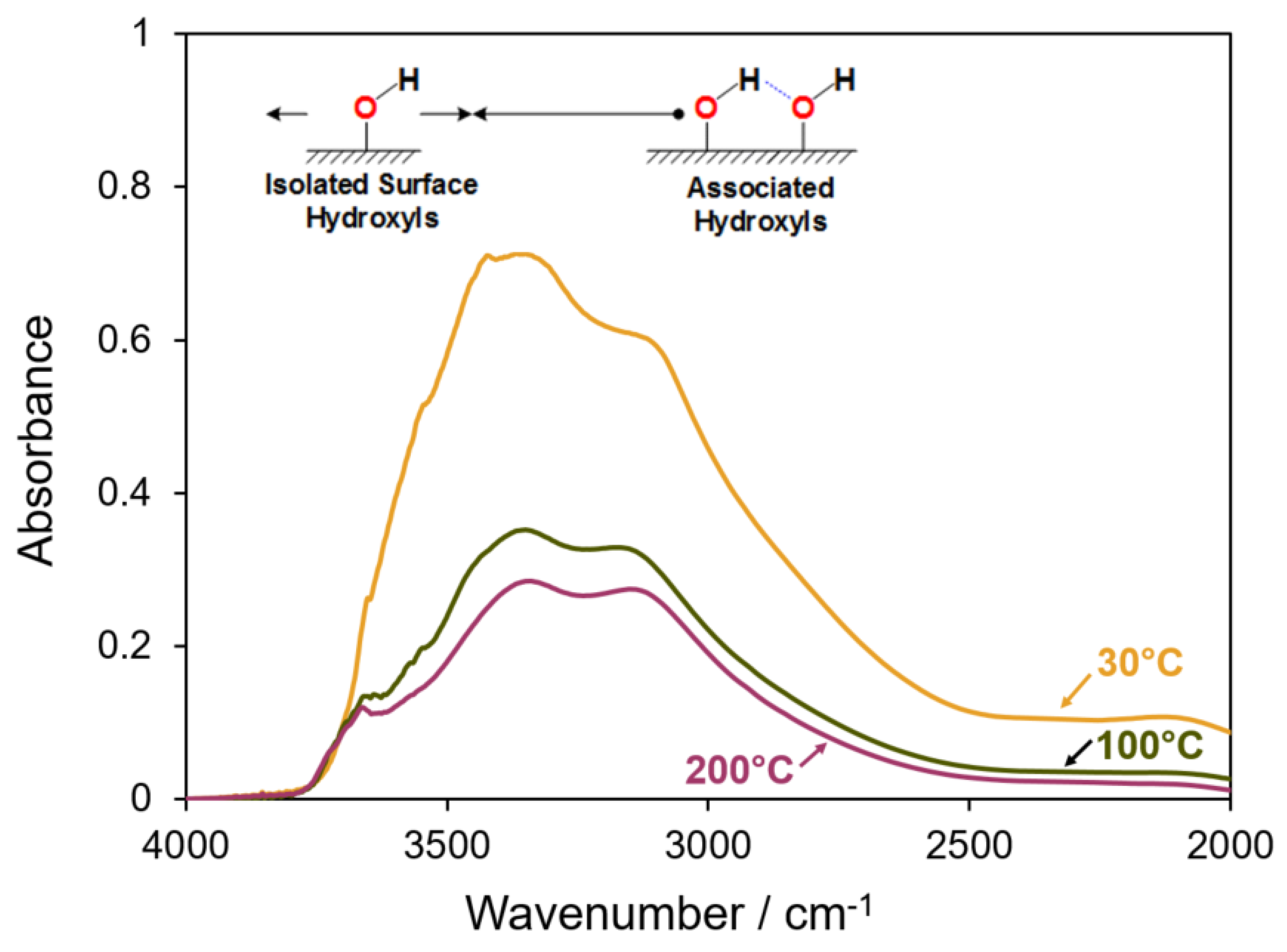
Shown in Figure 9 is the dehydration of AA-Al aluminum surface. The spectrum at room temperature appears differently than the NA-Al sample with additional absorbance peaks in the hydroxide region. The different shapes of bayerite clusters, such as the needles, rods, and pyramids observed in the SEM images, present different IR absorbances. Upon increasing the temperature to 300 °C, there is a loss of tri-hydroxide absorbance peaks, however, the boehmite absorbance peaks observed at 3100 and 3300 cm−1 are maintained. Surface hydroxyls emerge at 3670 and 3730 cm−1. The surface hydroxyl peaks observed on this sample are consistent with those observed on the boehmite model oxide presented earlier, without the presence of the tri-bridged hydroxyl at 3700 cm−1. It is possible that, due to the short-range order associated with the amorphous oxide layer, that this hydroxyl structure may not be present in high enough concentration and fell below the detection limit of the instrumentation.
SEM images of the WI-Al particles are shown in Figure 10. Here, we can see some larger tri-hydroxide structures at the particle surface, similarly to the naturally aged powder (a) and the boehmite surface at higher magnification (b). These findings are in agreement with others reports showing a needle and platelet-like structure consistent with boehmite [41,42,43]. The formation of bayerite clusters is favorable in solution preparation of boehmite surfaces at the temperatures used in this work. Additionally, the satellite particles were not dislodged as a result of stirring in solution.
The IR spectra for drying WI-Al are shown in Figure 11; After dehydration above 100 °C, the boehmite absorbances for the associated symmetric and asymmetric modes appear distinctly with some blue-shifting, consistent with the model oxide, boehmite that we presented in the aluminum oxides section. As the sample continues to dry, the tri-hydroxide content clearly falls below the detection limit by 200 °C. It is likely that these isolated structures were likely decomposed into boehmite structures as described in the literature [44]. It is also possible that they were overshadowed by the high absorbance of the associated hydroxyl groups for boehmite. With continued heating and equilibration, the surface hydroxyl peaks begin to appear. Similarly to the AA-Al, we find peaks at 3730 and 3665, in addition to the mode found at 3700 cm−1. These hydroxyls are bridged and tri-bridged structures based on the Peri and Tsyganenko models [17,18,19]. In this sample, there is likely a much higher concentration of the tri-bridged hydroxyl, due to the intentional growth of a boehmite layer. The AA-Al sample was exposed to high temperature and high humidity, which resulted in higher tri-hydroxide content. Although it decomposed into a boehmite-like structure, the SEM images show that the surface structures on the AA-Al sample were primary tri-hydroxide geometric shapes (Figure 8).
Due to the amorphous nature and short-range order of the surface oxides, oxy-hydroxides, and tri-hydroxides, the aged surfaces are complex. It has been reported that the amorphous oxide layer on aluminum is a meta-stable form of alumina, which we were unable to unambiguously identify on the AR-Al. Interestingly, both the AA and WI samples contained surface hydroxyls similar in absorbance location to alumina, with the 3730 and 3670 being the predominant type on both powders. This could potentially mean that some of the boehmite was decomposed into alumina at the particle surface. In the temperature regime that we studied, it is unlikely that the surface structure was crystalline -alumina. As the density of the crystalline phase is higher than the amorphous state, the detection limits of the instrumentation may limit the ability to quantify these groups. The most notable difference is the lack of the 3720 associated water type “hydroxyl”. In this instance, it is likely that there were not any available sites to support this structure, as both surfaces were fully hydrated.
4. Conclusions
In this work, we reveal the dehydration and decomposition behavior of alumina and metallic aluminum powders using in situ DRIFTS. The key insight we gleaned from this data is that the structure of the oxide and hydration dictates whether surface hydroxyls are accessible through drying or decomposition of the heavily oxidized surfaces of the metallic aluminum. The aluminas and artificially aged aluminum powder revealed, upon raising the temperature beyond the desorption temperature of physically absorbed water (100 °C), accessible hydroxyl groups of several different types. The most notable difference was the lack of detectable surface hydroxyls on the naturally aged sample. The increased temperature resulted in the decomposition from trihydroxide. We observed boehmite-like surfaces on the powders we artificially aged by exposure to high temperature and RH. The IR spectra of these powders showed decomposition of the trihydroxide into a boehmite-like surface.
An important follow on experiment to this work would be to investigate the SEM images resulting from heating using a temperature controlled stage in the SEM. We found interesting, three-dimensional surface structures resulting from the aging methods. How they change, physically, as a function of temperature is an important implication got their flow behavior. Decompositon of these features may change the inter-locking potential for particles to flow freely against eachother.
Author Contributions
Conceptualization, B.L.; Data curation, B.L.; Formal analysis, B.L.; Funding acquisition, B.L.; Investigation, B.L., T.T.B.; Project administration, B.L.; Writing—review, editing, B.L., T.T.B. All authors have read and agreed to the published version of the manuscript.
Funding
This material is based upon work supported by the Office of Naval Research under contract No. N00024-12-D-6402, Delivery Order No. 0037.
Institutional Review Board Statement
Not applicable.
Informed Consent Statement
Not applicable.
Data Availability Statement
Not applicable.
Acknowledgments
The authors wish to thank Joshua J. Stapleton, Carlo G. Pantano, and Benjamin J. Lear, of the Materials Research Institute and Department of Chemistry at PSU for their insightful conversations, assistance in the development, and collection of the data presented in this article. Julie M. Anderson collected the SEM images for this work and for that we are most grateful.
Conflicts of Interest
The authors declare no conflict of interest. The funders had no role in the design of the study; in the collection, analyses, or interpretation of data; in the writing of the manuscript, or in the decision to publish the results.
Abbreviations
The following abbreviations are used in this manuscript:
| AR-Al | As-received aluminum |
| AA-Al | Artificially aged aluminum |
| BET | Bruneauer-Emmett-Teller |
| DTGS | Deuterated Triglycine Sulfate |
| DRIFTS | Diffuse Reflectance Infrared Fourier Transform spectroscopy |
| MCT | Mercury Cadmium Telluride |
| NA-Al | Naturally aged aluminum |
| RH | Relative Humidity |
| WI-Al | Water-immersed aluminum |
| XPS | X-ray Photoelectron Spectroscopy |
Appendix A
Table A1 gives a summary of the bulk and surface area properties of the as received aluminum oxide and aluminum powders. Surface area was measured using a standard N2 adsorption isotherm and the BET method. Boehmite and gamma alumina were found to have comparable surface areas, however alpha alumina was significantly lower which is attributed to the larger particle size (300 nm vs. 50 nm).

Table A1.
A summary of the bulk and surface area properties of the as received aluminum oxide and aluminum powders.
Table A1.
A summary of the bulk and surface area properties of the as received aluminum oxide and aluminum powders.
| Sample | Surface Area (m2/g) | Average Particle Size (d50, nm) |
|---|---|---|
| Boehmite | 153.7007 | 50 |
| Alpha | 17.0227 | 300 |
| Gamma | 139.4133 | 50 |
| Aluminum | 0.2277 | 20 (µm) |
XPS analysis of the as-received aluminas and aluminum were completed to measure the surface composition. Binding energies for the Al 2p peak for the oxidized component for boehmite, alpha, and gamma alumina are reported in the literature as varying by 0.1–0.5 eV. The values measured in this study were 73.6, 74.2 and 74.1 eV respectively and are in agreement with the published literature. The C 1s peak was shown to have two binding energies of 285 eV and 288 eV maxima which are expected to be due to adventitious carbon and carbonate on the surface. Metallic aluminum presented similar values including the metallic Al peak at 71.3 eV.
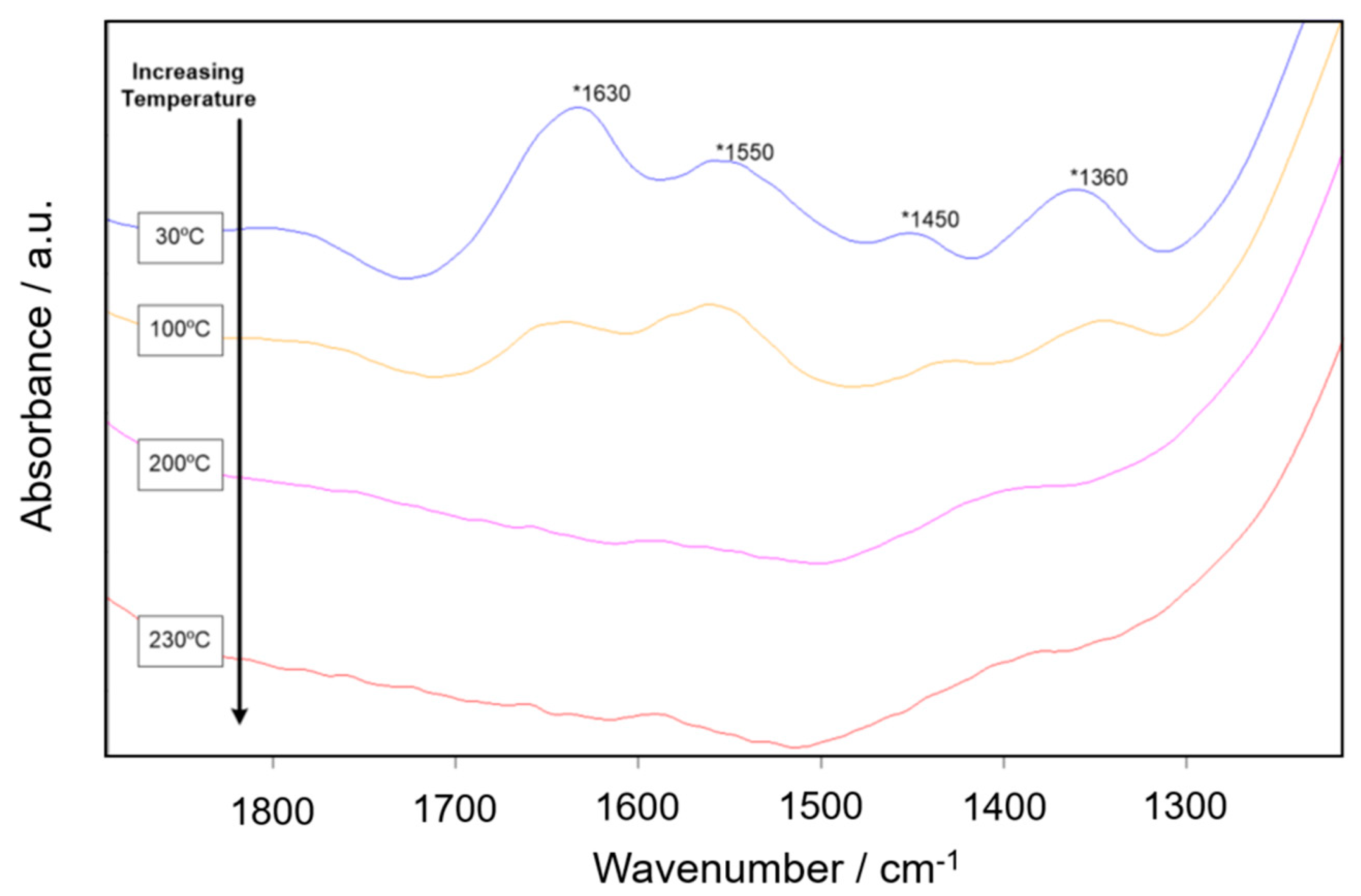
The dehydration spectra also contain carbonates, which is corroborated with the XPS findings. Carbon dioxide from the atmosphere has been shown to form carbonate like complexes at the aluminum oxide surface. Boehmite and gamma alumina both contained carbonates detectable using DRIFTS. The spectrum of alpha alumina was complicated with high absorbance in this area, however, carbonates were found in the XPS data. With increasing temperature, the absorbance peaks for the carbonates were shown to gradually reduce in intensity for boehmite and gamma alumina. Heating to 230 °C removed carbonate material from the surface of the aluminum oxide powders. The loss of carbonate for the surface of boehmite is shown in Figure A1 and for gamma alumina in Figure A2.
Figure A1.
The loss of carbonate for the surface of boehmite.

Figure A2.
The loss of carbonate for gamma alumina.

References
- Pines, H.; Haag, W.O. Alumina: Catalyst and support I. Alumina, its intrinsic acidity and catalytic activity. J. Am. Chem. Soc. 1960, 82, 2471–2483. [Google Scholar] [CrossRef]
- Morterra, C.; Magnacca, G. A case study: Surface chemistry and surface structure of catalytic aluminas as studied by vibrational spectroscopy of adsorbed species. Catal. Today 1996, 27, 497–532. [Google Scholar] [CrossRef]
- Busca, G. Catalytic materials based on silica and aluminum: Structural features and generation of surface acidity. Prog. Mater. Sci. 2019, 104, 215–219. [Google Scholar] [CrossRef]
- Pang, W.; Li, Y.; DeLuca, L.T.; Liang, D.; Qin, Z.; Liu, X.; Xu, H.; Fan, X. Effect of metal nanopowders on the performace of solid rocket propellants: A review. Nanomaterials 2021, 11, 2749. [Google Scholar] [CrossRef]
- Babuk, V.; Dolotkazin, I.; Gamsov, A.; Glebov, A.; DeLuca, L.T.; Galfetti, L. Nanoaluminum as a solid propellant fuel. J. Propul. Power 2009, 25, 482–489. [Google Scholar] [CrossRef]
- Ingenito, A.; Bruno, C. Using aluminum for space propulsion. J. Propul. Power 2004, 20, 1056–1063. [Google Scholar] [CrossRef]
- Maile, F.J.; Pfaff, G.; Reynders, P. Effect pigments—Past, present and future. Prog. Org. Coat 2005, 54, 150–163. [Google Scholar] [CrossRef]
- Morterra, C.; Cerrato, G.; Visca, M.; Lenti, D.M. Surface characterization of some TiO2-based pigments. Part 3. Coating of the pigments. J. Mater. Chem. 1992, 2, 341–355. [Google Scholar] [CrossRef]
- Ariffin, N.; Abdullah, M.M.A.B.; Postawa, P.; Zamree AbdRahim, S.; Mohd Arif Zainol, M.R.R.; Jaya, R.P.; Śliwa, A.; Omar, M.F.; Wysłocki, J.J.; Błoch, K.; et al. Effect of aluminium powder on kaolin-based geopolymer characteristic and removal of Cu2+. Materials 2021, 14, 814. [Google Scholar] [CrossRef]
- Köhler, M.; Fiebig, S.; Hensel, J.; Dilger, K. Wire and arc additive manufacturing of aluminum components. Metals 2019, 9, 608. [Google Scholar] [CrossRef] [Green Version]
- Shinkaryov, A.S.; Cherkasova, M.V.; Pelevin, I.A.; Ozherelkov, D.Y.; Chernyshikhin, S.V.; Kharitonova, N.A.; Gromov, A.A.; Nalivaiko, A.Y. Aluminum Powder Preparation for Additive Manufacturing Using Electrostatic Classification. Coatings 2021, 11, 629. [Google Scholar] [CrossRef]
- Popov, V.V.; Grilli, M.L.; Koptyug, A.; Jaworska, L.; Katz-Demyanetz, A.; Klobčar, D.; Balos, S.; Postolnyi, B.O.; Goel, S. Powder bed fusion additive manufacturing using critical raw materials: A review. Materials 2021, 14, 909. [Google Scholar] [CrossRef] [PubMed]
- Grilli, M.L.; Valerini, D.; Rizzo, A.; Yilmaz, M.; Song, C.; Hu, G.; Mikhaylov, A.; Chierchia, R.; Rinaldi, A. A comparative study of the mechanical and tribological Properties of thin Al2O3 coatings fabricated by atomic layer deposition and radio frequency sputtering. Phys. Status Solidi (A) 2021, 219, 2100398. [Google Scholar] [CrossRef]
- Rutkowska, I.; Marchewka, J.; Jeleń, P.; Odziomek, M.; Korpyś, M.; Paczkowska, J.; Sitarz, M. Chemical and Structural Characterization of Amorphous and Crystalline Alumina Obtained by Alternative Sol–Gel Preparation Routes. Materials 2021, 14, 1761. [Google Scholar] [CrossRef]
- Bauer, D.M.; Schwarzenbock, E.; Ludwig, I.; Schupp, N.; Palm, F.; Witt, G. Investigations on aging behaviour of aluminum powders during a lifetime simulation for the LBM process. In Proceedings of the European Congress and Exhibition on Powder Metallurgy, Hamburg, Germany, 9–13 October 2016. [Google Scholar]
- Peng, X.; Kong, L.; Fuh, J.Y.H.; Wang, H. A review of post-processing technologies in additive manufacturing. J. Manuf. Mater. Process 2021, 2, 38. [Google Scholar] [CrossRef]
- Peri, J.B.; Hannan, J. Surface hydroxyl groups on gamma-alumina. J. Phys. Chem. 1960, 64, 1526–1530. [Google Scholar] [CrossRef]
- Peri, J.B. A model for the surface of gamma-alumina. J. Phys. Chem. 1969, 69, 220–230. [Google Scholar] [CrossRef]
- Tsyganenko, A.A.; Filimonov, V.N. Infrared spectra of surface hydroxyls groups and crystalline structure of oxides. Spectrosc. Lett. 1972, 5, 477–487. [Google Scholar] [CrossRef]
- Knozinger, H.; Ratnasamy, P. Catalytic aluminas: Surface models and characterization of surface sites. Catal. Rev. Sci. Eng. 1978, 17, 31–70. [Google Scholar] [CrossRef]
- Busca, G.; Lorenzelli, V.; Ramis, G.; Willey, R.J. Surface Sites on spinel-type and corundum type metal oxide powders. Langmuir 1993, 9, 1492–1499. [Google Scholar] [CrossRef]
- Morterra, C.; Coluccia, S.; Ghiotti, G.; Zecchina, A. An IR spectroscopic characterization of alpha aluminum surface properties, carbon dioxide adsorption. J. Phys. Chem. 1977, 104, 275–290. [Google Scholar]
- Morterra, C.; Emanual, E.; Cerrato, G.; Magnacca, G. Infrared Study of some surface properties of boehmite. J. Chem. Soc. Faraday Trans. 1992, 104, 339–348. [Google Scholar] [CrossRef]
- Morterra, C.; Bolis, V.; Magnacca, G. IR spectroscopic and microcalorimetric characterization of lewis acid sites on Al2O3 using adsorbed CO. Langmuir 1994, 10, 1812–1824. [Google Scholar] [CrossRef]
- Kubaschewski, O.; Hopkins, B.E. Oxidation of Aluminum. In Oxidation of Metals and Alloys; Butterworths: Oxford, UK, 2007; pp. 32–58. [Google Scholar]
- Hunter, M.S.; Fowle, P. Natural and Thermally Formed Oxide Films on Aluminum. J. Electrochem. Soc. 1956, 103, 482–485. [Google Scholar] [CrossRef]
- Diggle, J.W.; Vijh, A.K. The Aluminum-Water System. In Oxides and Oxide Films; Dekker: New York, NY, USA, 1976; pp. 32–58. [Google Scholar]
- Anderson, I.E.; Foley, J.C. Determining the role of surfaces and interfaces in the powder metallurgy processing of aluminum alloy powders. Surf. Interface. Anal. 2001, 31, 599–608. [Google Scholar] [CrossRef]
- Ludwig, B.; Miller, T.F. Rheological and surface chemical characterization of alkoxysilane treated, fine aluminum powders showing enhanced flowability and fluidization behavior for delivery applications. Powder Technol. 2015, 283, 380–388. [Google Scholar] [CrossRef]
- Ludwig, B.; Gray, J.L. The effect of gas phase polydimethylsiloxane surface treatment of metallic aluminum particles: Surface characterization and flow behavior. Particuology 2017, 30, 92–101. [Google Scholar] [CrossRef] [Green Version]
- Fripiat, J.J.; Bosmans, H.J.; Rouxhet, P.G. Proton mobility in solids. I. Hydrogenic vibration modes and proton delocalization in boehmite. J. Phys. Chem. 1967, 71, 1097–1111. [Google Scholar] [CrossRef]
- Ballinger, T.H.; Yates, J.T., Jr. IR spectroscopic detection of lewis acid sites on alumina using adsorbed carbon monoxide. Correlation with aluminum-hydroxyl group removal. Langmuir 1991, 7, 3014–3045. [Google Scholar] [CrossRef]
- Digne, M.; Sautet, P.; Raybaud, P.; Euzen, P.; Toulhoat, H. Use of DFT to achieve a rational understanding of acid–basic properties of γ-alumina surfaces. Langmuir 1991, 7, 3014–3045. [Google Scholar] [CrossRef]
- Hart, R.K. The oxidation of aluminium in dry and humid oxygen atmospheres. Proc. R. Soc. A Math. Phys. Eng. Sci. 1956, 236, 66–68. [Google Scholar]
- Liu, X. DRIFTS study of surface of γ-alumina and its dehydroxylation. J. Phys. Chem. C 2000, 112, 5066–5073. [Google Scholar] [CrossRef]
- Mao, C.F.; Vannice, M.A. High surface area alpha-aluminas III. Oxidation of ethylene, ethylene oxide, and acetaldehyde over silver dispersed on high surface area alpha-alumina. Appl. Catal. A Gen. 1995, 122, 61–76. [Google Scholar] [CrossRef]
- Shirai, T.; Ishizaki, C.; Ishizaki, K. Tetrahedral aluminum ions on high purity sub-micron. alpha-alumina powder surfaces. J. Ceram. Soc. 2006, 114, 415–417. [Google Scholar] [CrossRef] [Green Version]
- Obzbilen, S. Satellite formation mechanism in gas atomized powders. Powder Metall. 1999, 42, 70–78. [Google Scholar] [CrossRef]
- Rothbauer, R.; Zigan, F. Refinement of the structure of bayerite including a proposal for the H positions. Z. Kristallogr. Krist. 1967, 125, 317–331. [Google Scholar]
- Phambu, N.; Humbert, B.; Burneau, A. Relation between the infrared spectra and the lateral specific surface areas of gibbsite samples. Langmuir 2000, 16, 6200–6207. [Google Scholar] [CrossRef]
- Hart, R.K. The formation of films on aluminum immersed in water. J. Chem. Soc. Faraday Trans. 1957, 53, 1020–1027. [Google Scholar] [CrossRef]
- Kimura, F.; Yamaguchi, E.; Horie, N.; Suzuki, G.; Kajihara, Y. Formation of boehmite crystals on microblasted aluminum surface to enhance performance of metal–polymer direct joining. Matt. Lett. 2020, 260, 126963. [Google Scholar] [CrossRef]
- Ma, R.; Jiang, Q.; Chen, J. The superhydrophobic surface constructed with boehmite micro-nanostructure. J. Mater. Sci. 2020, 55, 5795–5807. [Google Scholar] [CrossRef]
- Sato, T. The dehydration of aluminum tri-hydrate. J. Appl. Chem. 1959, 9, 331–340. [Google Scholar] [CrossRef]
Figure 1.
Dehydration of boehmite from 30–230 °C: (a) spectra labeled by temperature and surface hydroxyl peaks labeled by wavenumber: contains absorbance peaks at 3730, 3700, and 3665 cm−1, which can be attributed to bridged and tri-bridged surface hydroxyls (b).
Figure 1.
Dehydration of boehmite from 30–230 °C: (a) spectra labeled by temperature and surface hydroxyl peaks labeled by wavenumber: contains absorbance peaks at 3730, 3700, and 3665 cm−1, which can be attributed to bridged and tri-bridged surface hydroxyls (b).

Figure 2.
Dehydration of -alumina from 30–230 °C: (a) spectra labeled by temperature and surface hydroxyl peaks labeled by wavenumber: contains absorbance peaks at 3760, 3720, and 3670 cm−1, which can be attributed to bridged, physically adsorbed water, and a fully surface hydroxyl group (b).
Figure 2.
Dehydration of -alumina from 30–230 °C: (a) spectra labeled by temperature and surface hydroxyl peaks labeled by wavenumber: contains absorbance peaks at 3760, 3720, and 3670 cm−1, which can be attributed to bridged, physically adsorbed water, and a fully surface hydroxyl group (b).

Figure 3.
Dehydration of -alumina from 30–230 °C: (a) spectra labeled by temperature and surface hydroxyl peaks labeled by wavenumber: contains absorbance peaks at 3775, 3710, and 3670 cm−1, which can be attributed to bridged, tri-bridged, and fully isolated hydroxyl groups (b).
Figure 3.
Dehydration of -alumina from 30–230 °C: (a) spectra labeled by temperature and surface hydroxyl peaks labeled by wavenumber: contains absorbance peaks at 3775, 3710, and 3670 cm−1, which can be attributed to bridged, tri-bridged, and fully isolated hydroxyl groups (b).

Figure 4.
Scanning electron micrograph of as-received aluminum (AR-Al) sample showing few surface structural features other than satellite particles, which are an artifact of their production method (labeled by green circles).
Figure 4.
Scanning electron micrograph of as-received aluminum (AR-Al) sample showing few surface structural features other than satellite particles, which are an artifact of their production method (labeled by green circles).

Figure 5.
AR-Al sample DRIFTS spectrum, showing no detectable absorbance bands above the CaF2 window cutoff at 900 cm−1. Initial high absorbance band attributed to Al-O at around 950 cm−1.
Figure 5.
AR-Al sample DRIFTS spectrum, showing no detectable absorbance bands above the CaF2 window cutoff at 900 cm−1. Initial high absorbance band attributed to Al-O at around 950 cm−1.

Figure 6.
SEM micrographs detailing the particle surface of the naturally aged (NA-Al) sample: (a) full particles show outward growths of tri-hydroxide phases; (b) surface images reveal finer structure of complexes as indicated by green circles.
Figure 6.
SEM micrographs detailing the particle surface of the naturally aged (NA-Al) sample: (a) full particles show outward growths of tri-hydroxide phases; (b) surface images reveal finer structure of complexes as indicated by green circles.

Figure 7.
NA-Al sample, showing the dehydration behavior from ambient to 300 °C. Ambient temperature absorbance peaks found at 3655, 3620, 3548, and 3525 are consistent with tri-hydroxides and also observed in the SEM images (Figure 6).
Figure 7.
NA-Al sample, showing the dehydration behavior from ambient to 300 °C. Ambient temperature absorbance peaks found at 3655, 3620, 3548, and 3525 are consistent with tri-hydroxides and also observed in the SEM images (Figure 6).

Figure 8.
Scanning electron micrographs of AA-Al sample, showing full coverage of particle surface with platelet-like structures which are a form of the tri-hydroxide (a), High magnification image shows surfaces of complexes (b); this finding can be corroborated with the IR spectra observed for this sample.

Figure 9.
AA-Al sample, showing the dehydration behavior from ambient to 300 °C. Individual spectra are labeled by temperature and hydroxyl peaks identified wavenumber.
Figure 9.
AA-Al sample, showing the dehydration behavior from ambient to 300 °C. Individual spectra are labeled by temperature and hydroxyl peaks identified wavenumber.

Figure 10.
SEM micrographs detailing the particle surface of the water-immersed (WI-Al) sample: (a) particle shows some outward extrusions, consistent with some presence of the tri-hydroxide; (b) higher magnification surface image showing the boehmite surface features, considered nano-platelets or needles.

Figure 11.
WI-Al sample, showing the dehydration behavior from ambient to 200 °C. Individual spectra are labeled by temperature and hydroxyl peaks identified by wavenumber.
Figure 11.
WI-Al sample, showing the dehydration behavior from ambient to 200 °C. Individual spectra are labeled by temperature and hydroxyl peaks identified by wavenumber.

Publisher’s Note: MDPI stays neutral with regard to jurisdictional claims in published maps and institutional affiliations. |
© 2022 by the authors. Licensee MDPI, Basel, Switzerland. This article is an open access article distributed under the terms and conditions of the Creative Commons Attribution (CC BY) license (https://creativecommons.org/licenses/by/4.0/).
Share and Cite
MDPI and ACS Style
Ludwig, B.; Burke, T.T. Infrared Spectroscopy Studies of Aluminum Oxide and Metallic Aluminum Powders, Part I: Thermal Dehydration and Decomposition. Powders 2022, 1, 47-61. https://0-doi-org.brum.beds.ac.uk/10.3390/powders1010005
AMA Style
Ludwig B, Burke TT. Infrared Spectroscopy Studies of Aluminum Oxide and Metallic Aluminum Powders, Part I: Thermal Dehydration and Decomposition. Powders. 2022; 1(1):47-61. https://0-doi-org.brum.beds.ac.uk/10.3390/powders1010005
Chicago/Turabian StyleLudwig, Bellamarie, and Taryn T. Burke. 2022. "Infrared Spectroscopy Studies of Aluminum Oxide and Metallic Aluminum Powders, Part I: Thermal Dehydration and Decomposition" Powders 1, no. 1: 47-61. https://0-doi-org.brum.beds.ac.uk/10.3390/powders1010005