
Infrared Spectroscopy Studies of Aluminum Oxide and Metallic Aluminum Powders, Part II: Adsorption Reactions of Organofunctional Silanes


The Applied Research Laboratory, The Pennsylvania State University, University Park, PA 16804, USA
Powders 2022, 1(2), 75-87; https://0-doi-org.brum.beds.ac.uk/10.3390/powders1020007
Submission received: 9 February 2022
/
Revised: 7 March 2022
/
Accepted: 24 March 2022
/
Published: 1 April 2022
(This article belongs to the Special Issue Feature Papers in Powders)
Abstract
:A gas phase, probe molecule doser was fabricated and connected to a diffuse reflectance infrared Fourier transform spectroscopy (DRIFTS) reaction chamber to study the reactions and stability of two organosilanes with the surfaces of metallic aluminum and boehmite powders in situ. Two metallic aluminum powder surfaces were studied, including an as-received, native oxide layer surface, and a laboratory prepared, boehmite-like surface. Neat boehmite powder was also used for reference and comparison to the laboratory prepared surface. We found that the metalloxane bond (Al-O-Si) was observed in the 1100–950 cm−1 region for all surfaces, which indicates chemisorption between the adsorbate and available surface hydroxyls. We were also able to draw correlations between the loss of surface –OH and the subsequent growth of –CH for additional confirmation of adsorbate retention. Hydrothermal stability was probed through intentional exposure to water after chlorotrimethyl silane dosing, which showed adsorbate loss through fractional decreases in intensity of the –CH stretches. These results provide clear evidence of metalloxane bonds formed on aluminum powder and insight into their stability, supporting the identification of these bonds on bulk scale silane treated powders.
1. Introduction
The modification of metal-oxide powders with organosilanes is a common surface functionalization method used for a wide variety of scientific and engineering applications [1,2,3,4,5,6,7,8]. Organosilane-modified, aluminum-oxide particles have been studied extensively due to their importance as heterogeneous catalysts, fillers, and polymer/inorganic composites [5,9]. By altering the surface properties, the chemical compatibility with polymeric and other matrices improves substantially.
Metallic aluminum powder can also be processed in a similar manner to aluminum oxides due to its native oxide shell that encompasses the particle surface [10]. The surface analysis of aluminum powders and their treatment with organosilanes receives less attention than aluminum-oxides, leaving a knowledge gap in the direct identification of the metalloxane bonds on aluminum vs. aluminum-oxide powders. One possible reason for this is the tendency to evaluate the bulk properties of metal powders over their molecular, surface-level properties, as the bulk properties likely qualify the powder for use. To gain deeper insights, we extend the understanding of organosilane-modified, aluminum oxide powders to metallic aluminum powders using in situ Fourier transform infrared spectroscopy (FTIR) reaction studies. By using IR, we can observe distinct IR absorbance peaks to identify unique surface structures.
In an ambient environment, metallic aluminum reacts with both oxygen and water to form a stable, amorphous oxide surface. Auxiliary reactions with atmospheric water further hydrate the oxide to form surface hydroxyls and various hydrated forms such as bayerite (Al(OH)3) and boehmite (-AlO(OH)). The hydroxyl groups form with varying structures depending on the local Al/O environment, which can include both isolated and associated groups. The different molecular surface configurations of the aluminum oxide hydroxyls lead to subtle differences in their vibrational properties, resulting in unique IR absorption peaks [11,12,13,14,15,16,17,18]. In part I of this work, we used the same setup to study the dehydration and decomposition behavior, and thus extended it to adsorption studies. This fortuitous property allows for surface level studies of the dehydration, reactivity, and rearrangement behavior of both aluminum oxide and metallic aluminum powders. Herein, we study the reaction of the organosilane, chlorotrimethylsilane (CTMS) with various aluminum oxide surfaces, utilizing this property to monitor the reaction in situ through the loss of hydroxyls as the reaction proceeds.
The two most common types of organosilanes are alkoxysilanes and chlorosilanes, which take the form of R4-nSi(OR)n and R4-n SiXn, respectively, where the R group can vary depending on the required compatibility of the coated material. These types of silanes can be functional or non-functional, allowing for use as coupling agents or adhesion promoters, or simply as surface modifiers. Chlorosilanes can be hydrolyzed with trace amounts of water and at high temperature in the gas phase [1]. In this case, the chlorosilane reacts directly with the surface hydroxyl and does not require an initial hydrolyzing step. As a result, metalloxane bonds (M-O-Si) form without the water producing, condensation reaction step required for hydrolyzed organosilanes.
For our study, we selected a simple monochlorosilane, CTMS, to use in the gas phase to remove any complicating factors such as incomplete hydrolysis and self-condensation reactions. Additionally, the gas phase reaction enables a simplistic in situ monitoring capability using a DRIFTS reaction chamber. Recent work has shown similar methods using FTIR to characterize the surface of aluminum with silanes and planar substrates based on unique absorbance peaks [19,20,21]. In these works, the characteristic signature peaks enabled conclusive identification of the silane or siloxane components. Beyond the ambient characterization, additional studies have been using in situ IR to study adsorption and reactivity [22,23]. These studies also show how various components change with respect to thermal cycling.
In this work, we demonstrate the ability to follow surface reactions with powders in situ using a gas-phase doser connected to a DRIFTS reaction chamber. The samples selected for this study were boehmite, as-received aluminum powder (AR-Al), and an artificially grown boehmite surface on metallic aluminum particles (B-Al). We selected boehmite as a reference surface because naturally oxidized aluminum surfaces can have a pseudoboehmite-like structure [24,25]. Boehmite is also the hydrated form of -alumina, making it an ideal surface for our study related to aluminum powders and its relevance in the production of catalysts. Due to the low surface area of the aluminum particles used in this study, we used methods developed by others to grow a thicker boehmite layer on the aluminum particles to increase signal intensity for clearer identification of the surface behavior [26]. By using differential spectral analysis, we show the growth of metalloxane absorbance peaks on metallic aluminum and boehmite powders when dosed with CTMS. Due to the possibility of a self-dimerization reaction between CTMS molecules, we also show the interaction of hexamethyldisiloxane (HMDSO) with boehmite and B-Al as an additional reference. To probe stability, we dosed the surface treated powders with water and monitored for any changes as a result. In summation, this work shows the formation of metalloxane bonds on aluminum/aluminum oxide powders through a comprehensive study of reactivity, saturation, and stability.
2. Materials and Methods
2.1. Materials
Ethanol (Pharmco, Decon Labs, King of Prussia, PA, USA), boehmite (50 nm, Sasol, Johannesburg, South Africa), hexadimethylsiloxane (HMDSO, Sigma Aldrich, St. Louis, MO, USA) and aluminum powder (20 μm, AR-Al, Valimet H15, Stockton, CA, USA) were all used as received. CTMS (Sigma Aldrich, triple distilled, high purity >99.9%) was purified just prior to use by distillation under N2. The purity of CTMS was verified by gas phase FTIR.
2.2. Powder Preparation (Boehmite-Al)
Powdered aluminum (100 g) was dispersed in distilled H2O (50 mL, 70 °C) and allowed to stir for 1 h. The resulting product was recovered via Hirsch funnel, rinsed with ethanol three times and dried under N2 prior to analysis.
2.3. Diffuse Reflectance Infrared Spectroscopy, Ambient and Heating Experiments
All DRIFTS measurements were collected on a Fourier transform infrared spectrometer (Bruker, Vertex V70, Billerica, MA, USA) using a Praying Mantis® accessory (Harrick Scientific, Pleasantville, NY, USA) equipped with an environmental reaction chamber. Spectra were collected using a liquid nitrogen cooled Mercury-Cadmium-Telluride (MCT) detector and CaF2 windows (900 cm−1 cutoff) at 6 cm−1 resolution and 400 scans. CTMS silane dosing onto KBr used a deuterated triglycine sulfate (DTGS) detector at 16 cm−1 resolution and 100 scans. Samples were prepared dilute in KBr (25% w/w) to provide access to the low frequency region or neat for maximum sensitivity where noted. Surface temperature calibrations were conducted on all powders’ surfaces to verify the dosing temperature.
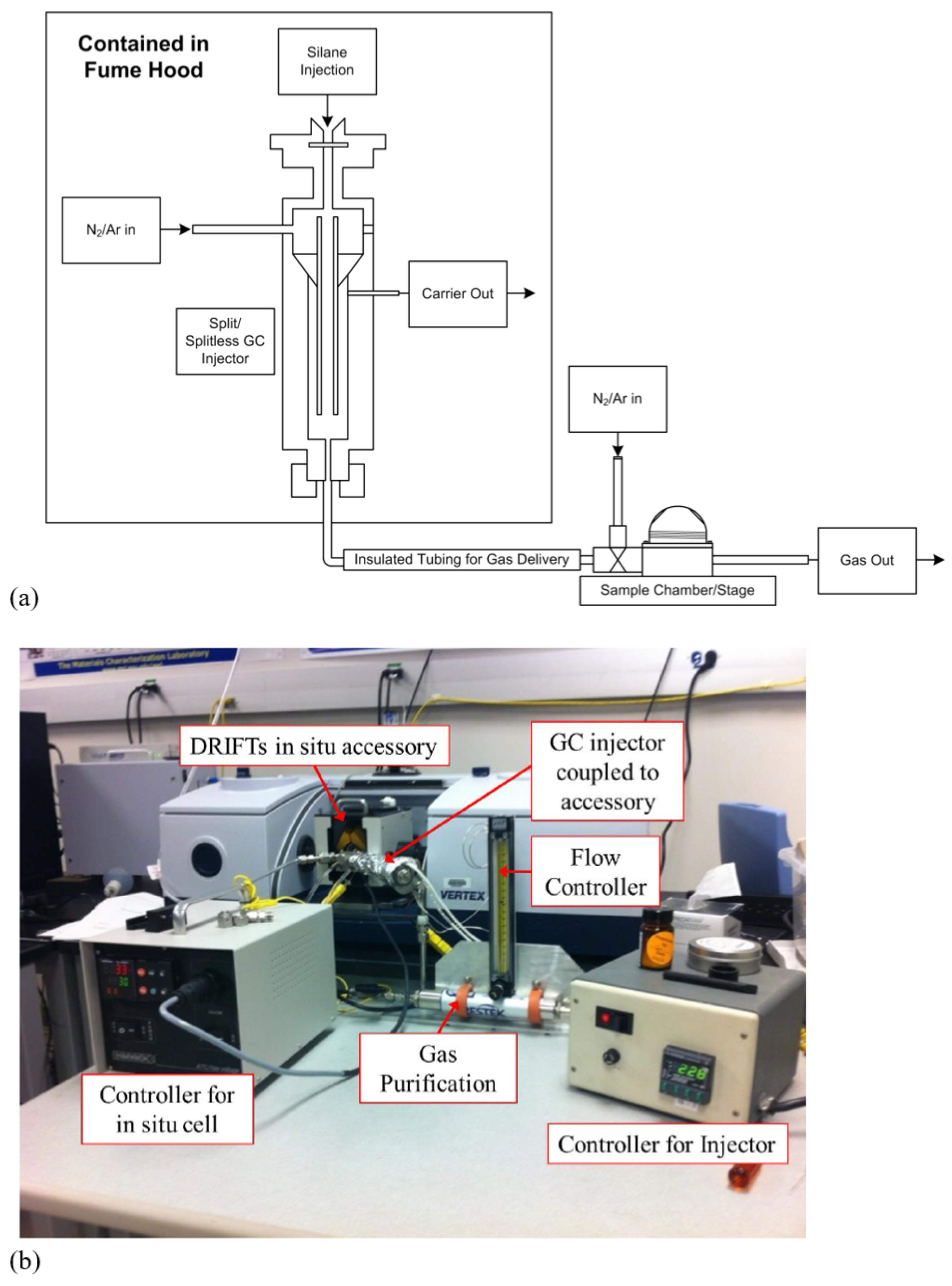
The custom dosing apparatus was fabricated using a standard split/splitless GC injector (Restek, Bellefonte, PA, USA) with a heating jacket (Cole–Parmer, Vernon Hills, IL, USA) and purge gas was purified using a super clean triple-gas (moisture, oxygen, hydrocarbon, Restek) filter. The doser was equipped with a flow controller (Cole–Parmer) calibrated to Ar and a spot-welded thermocouple on the injector to measure doser temperature. A constant doser temperature of 110 °C was used for all experiments with an Ar purge (80 cc/min). A small copper insert was designed to provide better thermal conduction from the bottom of the sample holder into powdered samples. The insert and in situ cell were dried in air for 30 min at 350 °C prior to use. Wet KBr (15 mg) was loaded into the insert and dried for 30 min at 350 °C. Appropriate temperature reference spectra were collected after outgassing of KBr. Powder samples ( 15 mg) were loaded into the copper insert and wet reference spectra collected. The sample was raised to the appropriate surface temperature and outgassed for 1 h prior to injection. If needed, the sample temperature was lowered to ambient to collect a dried spectrum for referencing; the sample was allowed to equilibrate at dosing temperature again for ten minutes. Injections of 0.5 μL CTMS were typically used. In saturation experiments, 0.5 μL and 1.0 μL incremental injections were used and the time elapsed between doses was 10 min. After all dosing was completed, the sample was allowed to equilibrate for 30 min and spectra collected. After equilibration, the sample was cooled to ambient temperature and additional spectra collected. Water dosing experiments were completed at ambient temperature and with 0.5 μL injection of H2O. After ten minutes of equilibration, the sample was raised to the decomposition surface temperature and allowed to dry for ten minutes. The sample was cooled to ambient and the resulting product spectra were collected. A simplified schematic of the doser setup is shown in Figure 1a in addition to a photograph of the instrumental setup Figure 1b.
2.4. Surface Temperature Calibration Experiments
The Praying Mantis® (Harrick Scientific) DRIFTS cell comes with a temperature thermocouple which is placed directly above the heater, however, at the bottom of the sample cup. To determine the actual surface temperature as related to the set point temperature of the controller, a separate thermocouple was placed directly on the surface of the powder. The thermocouple was inserted through a rubber septum, which was subsequently placed where a window would normally be on the hood of the in situ cell. The cell temperature was incremented using a 110 V proportional-interval-derivative (PID) temperature controller (Harrick Scientific) in steps of 10 °C with the corresponding surface temperature recorded as a function of set point. Due to significant differences between the set point and measured surface temperature, a small, copper sample cup insert was fabricated to increase heat transfer to the sample surface. The temperature differential was decreased significantly and all experiments were completed using this setup.
3. Results and Discussion
3.1. As-Received and Prepared DRIFTS Spectra
The DRIFTS spectra of boehmite, aluminum with a boehmite-surafce (B-Al), and as-received aluminum (AR-Al) powders are shown in Figure 2. As a reference, boehmite was prepared dilute in KBr due to its high surface area and resulting in over absorbance in the –OH region. The boehmite powder sample showed characteristic absorbance peaks, observed from the hydrogen bond layers at 3290 and 3090 cm−1 for the asymmetric and symmetric modes respectively; boehmite has a strong peak at 3670 cm−1, which is attributed to a bridged surface hydroxyl [18]. The absorbance intensity for the surface hydroxyls is damped due to the presence of physisorbed molecular water, which necessitates a drying step for the surface reaction to occur. Broad shoulder peaks beyond 3670 cm−1 were also observed and are attributed to additional surface hydroxyl peaks. The corresponding B-Al sample showed broad absorbance bands for –OH stretches centered around 3300 and 3100 cm−1 which are characteristic of the bulk –OH modes within the structure of boehmite. Additional peaks were observed at 3548 and 3424 cm−1, which were attributed to bayerite complexes [27]. The bulk –OH deformation of boehmite was observed at 1070 cm−1 under ambient conditions with physisorbed water and carbonate peaks at 1645, 1505, and 1400 cm−1, all consistent with values in the literature [28]. Low intensity surface hydroxyl peaks at 3743 and 3700 cm−1 were present on the surface prior to drying. The only observable absorbance peak for AR-Al was at 900 cm−1 (Al-O), which was cutoff due to the use of CaF2 windows for the in situ cell. No other absorption bands were expected for the AR–Al powder.
All samples were dehydrated to provide reproducible reference surfaces, induce activation, and remove physisorbed molecular water from interring with the surface reaction. The drying behavior of the powders was presented in Part I of this two part series.
3.2. CTMS Treatment of Boehmite and Aluminum Powders
We confirmed the purity of CTMS by showing the lack of dimer present prior to dosing through gas phase IR. Additional control experiments consisted of CTMS/KBr dosing to verify that CTMS did not absorb on KBr to prevent misinterpretation of the data. KBr windows were used for these experiments to allow access to the low frequency –CH3 rocking and Si-Cl stretching absorbance modes. Stretching modes for –CH were observed at 2970 and 2910 cm−1 and deformations for –CH3 were observed at 1255, 852 and 638 cm−1. The Si-Cl stretching mode was observed at 483 cm−1. All of these values were consistent with experimental data reported in the literature [29].
Figure 3 reveals the CTMS dosing spectra of the dilute-boehmite, B-Al, and AR-Al surfaces using a single dose (0.5 μL) and the spectra shown in differential form. The CTMS exposure to the dilute boehmite surface resulted in negative peaks at 3730, 3700, and 3670 cm−1. Similarly, the B-Al surface exposure to CTMS resulted in negative peaks observed at 3740, 3705, and 3665 cm−1, indicating the depletion or consumption of multiple types of surface hydroxyls. There was an increase in intensity of the associated hydroxyl portion of the spectrum, which would suggest a potential hydrogen bonding type surface interaction. Due to the low absorbance associated with the AR-Al surface, there were not any detectable absorbance peaks in the –OH region, which is consistent with the as-prepared powder surface. Evidence for the retention of the trimethyl silyl group is shown through positive absorbance peaks at 2956 for the –CH stretching mode, and at 1255 cm−1 for the –CH3 deformation, which were observed on all three samples. It should be noted that the small, negative peaks at 3735 and 1567 cm−1 for AR-Al are artifacts of molecular water present in the beam path outside of the in situ cell and from the low intensity absorbance peaks of the surface.
All three samples showed positive peaks near the anticipated Al-O-Si absorbance peak region. We observed a complex feature on the boehmite sample at 1000 cm−1, with a low energy shoulder at 950 cm−1. On the B-Al surface, we detected another complex feature at 1070 with a shoulder at 1040 cm−1. Similarly to the B-Al surface, the AR-Al sample presented a stretching mode at 1075 cm−1, however without a strongly absorbing shoulder peak. Both the AR-Al and B-Al features are in close proximity to the 1075 cm−1 assignment given by Paul et al. and accounts for the bonding interaction [30]. A corresponding broad absorbance appearance was not observed in the 3500–3000 cm−1 region on the AR-Al sample would suggest a hydrogen bonding type interaction and provides additional support for the assignment of the Al-O-Si absorbance.
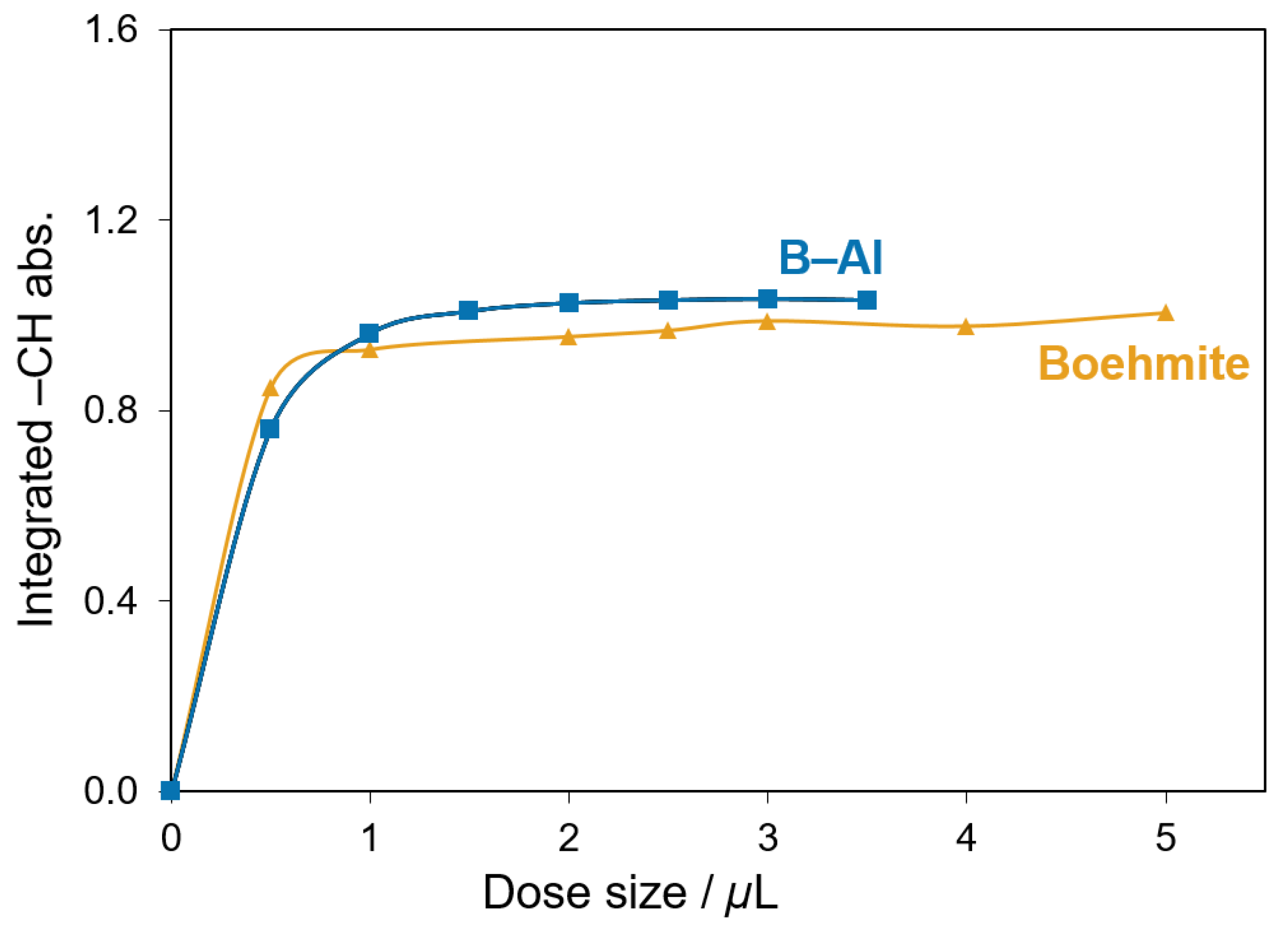
Additional evidence for the chemisorption of the trimethyl silyl group can be drawn from the from the saturation experiments shown in Figure 4. The dilute boehmite and B-Al samples showed high intensity peaks in the –CH stretching region (3000–2800 cm−1) and their growth was used to monitor the adsorption of CTMS. The total integrated area indicated the saturation point when the –CH area value remained stable despite additional doses. As shown in Figure 4, the saturation point for boehmite and B-Al occurred after 2 μL. The intensity of the –CH stretch was low on the AR-Al sample, however the –CH3 presented an additional opportunity for determining a correlation. Figure 5 reveals that the AR-Al sample required fewer doses to reach saturation, which occurred at 1 μL.
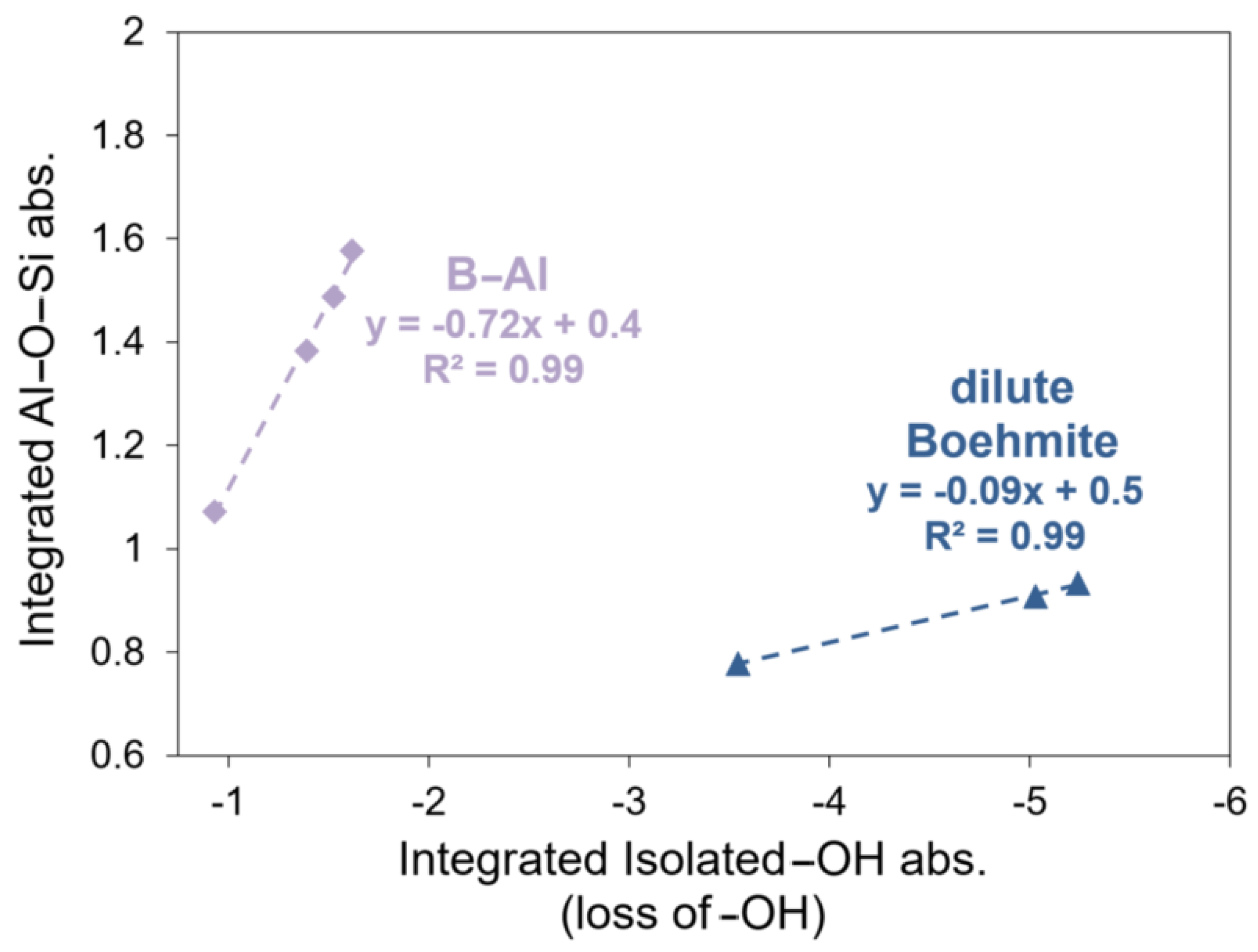
Figure 6 shows the linear correlations extracted from the integrated areas produced from the boehmite and B-Al saturation experiments, which have R2 values of 0.99. The negative slopes of the best fit lines agree with the loss of –OH groups with the growth of the Al-O-Si feature. These experiments indicated a semi-quantitative relationship between the reaction of surface hydroxyls and the formation of alkyl components for boehmite and B-Al. This relationship can be drawn from the integrated area of the Al-O-Si and surface retention of –CH3 on the AR-Al sample, which is shown in Figure 7. The R2 value for this regression was 0.94.
The potential for dimerization of the CTMS into hexadimethylsiloxane (HMDSO) and subsequent hydrogen bonding was addressed through intentional dosing with HMDSO. This served as a control experiment to compare the peaks observed in the surface hydroxyl and ether-type bond regions. Gas phase analysis of HMDSO was completed in an analogous manner to CTMS on both boehmite and B-Al. The low absorbance of the AR-Al sample precluded conclusive analysis and warrants future investigation. The gas phase control spectrum for HDMSO showed absorbance peaks at 2965 and 2907 cm−1 for the –CH stretches, 1255 cm−1 for the -CH3 deformation, and at 1074 cm−1 for the Si-O-Si. All values were consistent with previous reference values in the literature [31].
The absorbance peaks for Si-O-Si and Al-O-Si are difficult to resolve, although there should be a discernible shift due to the differing masses of Al vs. Si and their ionic radii of 118 pm and 110 pm respectively. Their reported signature values are within a few wave numbers of each other (1075 vs. 1070 cm−1). For the CTMS dosing experiments, the presence of dimer in the CTMS dosing liquid was ruled out by the lack of a Si-O-Si absorbance peak found in the gas phase spectrum completed just prior to the dosing experiments. Spectra for the dosing experiments using HMDSO are shown in Figure 8. The Si-O-Si absorbance from HMDSO appeared at 1070 cm−1, which is near the Al-O-Si feature observed on B-Al and AR-Al using CTMS. We also observed no further increase in the associated –OH group bonding as a result of dimer dosing when compared to CTMS adsorption. The Cl group can participate in H-bonding, as reported by Paul, however he reported this phenomenon at lower temperatures than those used in this study [30].
When dosed on dilute boehmite, the Si-O-Si absorbance was red shifted 60 cm−1 and appeared as a complex absorbance with a high-energy component at 1050 cm−1 and a low energy shoulder at 980 cm−1. However, it was still was not equivalent to the CTMS dosing absorbance appearing at 1000 and 950 cm−1. When comparing the CTMS and HMDSO differential spectra, there are strong broad bands observed in the associated hydroxyl region because of CTMS exposure. This is not the case in HMDSO, where there is only a small increase in the associated hydroxyl absorbance. The interaction of Cl with a surface hydroxyl could possibly explain this phenomenon, which presents as an H-bonding type interaction. The surface hydroxyls are also perturbed in dimer dosing; however, this is likely due to a hydrogen bonding type interaction of the O in the HMDSO with the Al-OH surface groups. Hydrogen bonding between surface –OH groups on alumina and ether type compounds has been presented in the literature [32].
We believe that there are a combination of surface reactions occurring through reviewing these results. The lack of a broad –OH absorbance increase in the differential spectrum for the HDMSO dosing suggests that there may be hydrogen bonding between the surface –OH and Cl of the CTMS. This is apparent on the B-Al sample and less so on the boehmite sample. The reasoning is likely attributable to the surface structure differences, where boehmite has a more ordered structure than the amorphous aluminum powder surface. An interesting area of study would be the quantification of surface hydroxyls on these samples, to further understand the surface adsorption method.
3.3. Hydrothermal Stability of CTMS/Aluminum Oxide Interfacial Interaction
To produce an environment where humidity could influence the surface reactions, water was introduced to the CTMS dosed samples to probe the stability of the Al-O-Si bond. A small amount of water (0.5 μL) was injected after the saturated surface had been allowed to equilibrate for 30 min. Figure 9 shows the differential absorbance spectra for water dosing on dilute boehmite and B-Al surfaces. The growth of a peak at 1630 cm−1 was observed on both surfaces and was attributed to molecular water; this likely accounts for the –OH perturbation via physisorption of molecular water. An explanation for this result could be the steric hindrance of the trimethylsilyl moiety, that is, additional hydroxyl groups were not accessible as a result of this group. The water would act as an adsorbate on the surface –OH groups, which would present as negative absorbance in this process. There was not a significant peak in the area of the Al-O-Si, indicating that the bond was not significantly hydrolyzed due to induced water exposure.
Water exposure to the B-Al surface produced a slight loss of –CH absorbance, loss of surface hydroxyls, and a large increase in the associated hydroxyl region, which was not observed in the dilute boehmite model surface. The loss of surface hydroxyls is attributed to the physisorption of molecular water to isolated hydroxyls groups that did not react with CTMS. The loss of –CH suggests fractional loss of the adsorbate from water exposure. The water could displace the weakly hydrogen bound CTMS or react with it to form HCl and a silanol.
4. Conclusions
For this work, we used an in situ DRIFTS reaction chamber to monitor the adsorption of CTMS on boehmite and metallic aluminum powders. With this tool, we have demonstrated the ability to follow dehydration, surface chemical, and stability reactions with temperature and environmental controls of powder relevant to multiple research areas. Specifically, we found that CTMS reacts with all of the presented surfaces, boehmite, boehmite-like aluminum, and as-received aluminum powders, to form Al-O-Si bonds identifiable using differential IR spectroscopy. Using integrated areas as qualitative representations of surface character, we were able to show correlations between the consumption of hydroxyl groups and the growth of metalloxane bonds with the presence of –CH and –CH3 absorbance peaks, indicating retention of the trimethylsilyl unit.
We utilized two additional probe molecules to support our findings from the CTMS dosing experiments. Firstly, to investigate to influence of potential dimerization reactions, HDMSO dosing was tested to compare with the CTMS results. When comparing CTMS results with HMDSO experiments, a significant difference was observed between the two: there was an increase in associated –OH bonding on the B-Al sample, which was not observed on boehmite. We believe this suggests a hydrogen bonding type interaction is also occurring on the B-Al surface. Secondly, to probe stability of the Al-O-Si bond, water exposure was used and showed adsorbate loss on the B-Al and boehmite surfaces. We found negative peaks in the Al-O-Si region, which are likely due to the partial hydrolysis of the Al-O-Si bond.
Our results provide compelling assignments of the Al-O-Si bond formation on the surface of metallic aluminum powders, both heavily oxidized and as-received. Additionally, these results were collected in situ in a highly controlled environment, where the complications from possible dimerization of the silane and atmospheric challenges do not apply. To this end, we believe our work herein provides additional evidence of metalloxane bond formation on aluminum powders and will support the surface characterization of bulk aluminum powders at the surface molecular level. Additional probe molecules, such as carbon monoxide (CO) and pyridine, could also be used to corroborate these data. Pyridine, specifically, is a common probe molecule used to identify surface hydroxyl groups. This property could be compelling in future research to support the understand of boehmite, B-Al, and AR-Al surfaces.
Funding
This material is based upon work supported by the Office of Naval Research under contract No. N00024-12-D-6402, Delivery Order No. 0037.
Institutional Review Board Statement
Not applicable.
Informed Consent Statement
Not applicable.
Data Availability Statement
Not applicable.
Acknowledgments
The author wishes to thank Maria Medeiros for funding this effort. I also wish to thank Joshua J. Stapleton, Carlo G. Pantano, Benjamin J. Lear, and Julie M. Anderson of the Pennsylvania State University Materials Research Institute at PSU for their insightful conversations and assistance in collecting the data presented in this article. Taryn T. Burke was also helpful in the preparation of this document.
Conflicts of Interest
The author declares no conflict of interest. The funders had no role in the design of the study; in the collection, analyses, or interpretation of data; in the writing of the manuscript, or in the decision to publish the results.
Abbreviations
The following abbreviations are used in this manuscript:
| AR-Al | As-received aluminum |
| B-Al | Boehmite-like surface aluminum |
| CTMS | Chlorotrimethyil silane |
| DTGS | Deuterated Triglycine Sulfate |
| DRIFTS | Diffuse Reflectance Infrared Fourier Transform spectroscopy |
| HMDSO | Hexamethyldisiloxane |
| KBr | Potassium Bromide |
| MCT | Mercury Cadmium Telluride |
| PID | Proportional-interval-derivative |
References
- Plueddemann, E.P. Silane Coupling Agents; Springer: New York, NY, USA, 2013. [Google Scholar]
- Van Ooij, W.J.; Zhu, D.; Stacy, M.; Seth, A.; Mugada, T.G.; Hi, J.; Puomi, P. Corrosion protection properties of organofunctional silanes—An overview. Catal. Tsinghu Sci. Technol. 2005, 10, 639–664. [Google Scholar] [CrossRef]
- Subramanian, V.; Van Ooij, W.J. Silane based metal pretreatments as alternatives to chromating: Shortlisted. Surf. Eng. 2005, 15, 168–172. [Google Scholar] [CrossRef]
- Arkles, B.; Steinmetz, J.; Zazyczny, J.; Zolotnitsky, M. Stable, water-borne silane coupling agents. In Proceedings of the 46th Annual Reinforced Plastics/Composites Institute, Society of Plastic Industry (SPI), Washington, DC, USA, 18–21 February 1991. [Google Scholar]
- Mittal, K. Silanes and Other Coupling Agents, 1st ed.; Taylor and Francis: New York, NY, USA, 2009. [Google Scholar]
- Ludwig, B.; Miller, T.F. Rheological and surface chemical characterization of alkoxysilane treated, fine aluminum powders showing enhanced flowability and fluidization behavior for delivery applications. Powder Technol. 2015, 283, 380–388. [Google Scholar] [CrossRef]
- Ludwig, B.; Gray, J.L. The effect of gas phase polydimethylsiloxane surface treatment of metallic aluminum particles: Surface characterization and flow behavior. Particuology 2017, 30, 92–101. [Google Scholar] [CrossRef] [Green Version]
- Ludwig, B.; Millington-Smith, D.; Dattani, R.; Adair, J.H.; Posatko, E.P.; Mawby, L.M.; Ward, S.K.; Sills, C.A. Evaluation of the hydrodynamic behavior of powders of varying cohesivity in a fluidized bed using the FT4 Powder Rheometer®. Powder Technol. 2020, 371, 106–114. [Google Scholar] [CrossRef]
- Slavov, S.V.; Chuang, K.T.; Sanger, A.R. Modification of gamma-alumina with chlorotrimethylsilane. J. Phys. Chem. 1995, 9, 17019–17027. [Google Scholar] [CrossRef]
- Hunter, M.S.; Fowle, P. Natural and thermally formed oxide films on aluminum. J. Electrochem. Soc. 1956, 103, 482–485. [Google Scholar] [CrossRef]
- Peri, J.B.; Hannan, J. Surface hydroxyl groups on gamma-alumina. J. Phys. Chem. 1960, 64, 1526–1530. [Google Scholar] [CrossRef]
- Peri, J.B. A model for the surface of gamma-alumina. J. Phys. Chem. 1969, 69, 220–230. [Google Scholar] [CrossRef]
- Misra, C.; Wefers, K. Oxides and Hydroxides of Aluminum: Alcoa Technical Paper; No. 19, Revised; Aluminum Company of America: Pittsburgh, PA, USA, 1987. [Google Scholar]
- Tsyganenko, A.A.; Filimonov, V.N. Infrared spectra of surface hydroxyls groups and crystalline structure of oxides. Spectrosc. Lett. 1972, 5, 477–487. [Google Scholar] [CrossRef]
- Knozinger, H.; Ratnasamy, P. Catalytic aluminas: Surface models and characterization of surface sites. Catal. Rev. Sci. Eng. 1978, 17, 31–70. [Google Scholar] [CrossRef]
- Busca, G.; Lorenzelli, V.; Ramis, G.; Willey, R.J. Surface sites on spinel-type and corundum type metal oxide powders. Langmuir 1993, 9, 1492–1499. [Google Scholar] [CrossRef]
- Morterra, C.; Coluccia, S.; Ghiotti, G.; Zecchina, A. An IR spectroscopic characterization of alpha aluminum surface properties, carbon dioxide adsorption. J. Phys. Chem. 1977, 104, 275–290. [Google Scholar]
- Morterra, C.; Emanual, E.; Cerrato, G.; Magnacca, G. Infrared study of some surface properties of boehmite. J. Chem. Soc. Faraday Trans. 1992, 104, 339–348. [Google Scholar] [CrossRef]
- Zeng, D.; Liu, Z.; Bai, S.; Zhao, J. Preparation and Characterization of a Silane Sealed PEO Coating on Aluminum Alloy. Coatings 2021, 11, 549. [Google Scholar] [CrossRef]
- Du, X.Q.; Liu, Y.W.; Chen, Y. Enhancing the corrosion resistance of aluminum by superhydrophobic silane/graphene oxide coating. Appl. Phys. A 2021, 127, 580. [Google Scholar] [CrossRef]
- Wu, S.; Wu, R.; Jiang, H.; Yuan, Z.; Chen, Q. Preparation and Characterization of Superhydrophobic Silane-Based Multilayer Surface Coatings on Aluminum Surface. J. Mater. Eng. Perform. 2022. [Google Scholar] [CrossRef]
- Cao, Y.; Zheng, X.; Du, Z.; Shen, L.; Zheng, Y.; Au, C.; Jiang, L. Low-temperature H2S removal from gas streams over γ-FeOOH, γ-Fe2O3, and α-Fe2O3: Effects of the hydroxyl group, defect, and specific surface area. Ind. Eng. Chem. Res. 2019, 58, 19353–19360. [Google Scholar] [CrossRef]
- Melchers, S.; Schneider, J.; Emeline, A.V.; Bahnemann, D.W. Effect of H2O and O2 on the adsorption and degradation of acetaldehyde on anatase surfaces—An in situ ATR-FTIR study. Catalysts 2018, 8, 417. [Google Scholar] [CrossRef] [Green Version]
- Altenpohl, D.G. Use of boehmite films for corrosion protection of aluminum. Corrosion 1962, 18, 143t–153t. [Google Scholar] [CrossRef]
- Altenpohl, D.; Post, W. Hydrated oxide films on aluminum their growth and their importance in electrolytic capacitors. J. Electrochem. Soc. 1962, 108, 628. [Google Scholar] [CrossRef]
- Hart, R.K. The formation of films on aluminum immersed in water. J. Chem. Soc. Faraday Trans. 1957, 53, 1020–1027. [Google Scholar] [CrossRef]
- Rothbauer, R.; Zigan, F. Refinement of the structure of bayerite including a proposal for the H positions. Z. Kristallogr. Krist. 1967, 125, 317–331. [Google Scholar]
- Morterra, C.; Magnacca, G. A case study: Surface chemistry and surface structure of catalytic aluminas, as studied by vibrational spectroscopy of adsorbed species. Catal. Today 1996, 27, 497–532. [Google Scholar] [CrossRef]
- Montejo, M.; Ureña, F.P.; Márquez, F.; Ignatyev, I.S.; González, J.L. Vibrational spectrum of chlorotrimethylsilane. Spectrochim. Acta A 2005, 62, 293–301. [Google Scholar] [CrossRef]
- Paul, D.K.; Ballinger, T.H.; Yates, J.T. Rhodium surface chemistry on a chemically modified alumina support. J. Phys. Chem. 1990, 94, 4617–4622. [Google Scholar] [CrossRef]
- Carteret, C.; Labrosse, A. Vibrational properties of polysiloxanes: From dimer to oligomers and polymers. 1. Structural and vibrational properties of hexamethyldisiloxane (CH3)3SiOSi (CH3)23. J. Raman Spectrosc. 2010, 41, 996–1004. [Google Scholar] [CrossRef]
- Chen, J.G.; Basu, P.; Ballinger, T.H.; Yates, J.T., Jr. A transmission infrared spectroscopic investigation of the reaction of dimethyl ether with alumina surfaces. Langmuir 1989, 5, 352–356. [Google Scholar] [CrossRef]
Figure 1.
Simplified pictorial of the gas phase doser connected to an inert reaction chamber with purge gas capability (a) and photograph of the instrument setup connected to the doser and various auxillary equipment (b).
Figure 1.
Simplified pictorial of the gas phase doser connected to an inert reaction chamber with purge gas capability (a) and photograph of the instrument setup connected to the doser and various auxillary equipment (b).

Figure 2.
Comparison of dilute boehmite (blue), boehmite-surface (B-Al, purple), and as-received (AR-Al, magenta) DRIFTS spectra. Major absorbance peaks in the -OH region (3800–3000 cm−1) were observed for boehmite and B-Al, which are attributed to surface (>3500 cm−1) and bulk –OH stretching modes (<3500 cm−1); there were no detectable absorbance peaks in this region for AR-Al. Absorbance peaks were observed in the 1700–1400 cm−1 for carbonates and <1200 cm−1 for Al-OH and Al-O.

Figure 3.
Results from CTMS dosing experiments for dilute Boehmite, B-Al, and AR-Al, showing positive growth in the Al-O-Si (1100–900 cm−1), methyl group deformation (1255 cm−1), and –CH stretch (3000–2800 cm−1) and negative peaks in the surface hydroxyl region (3800–3400 cm−1) for boehmite and B-Al.

Figure 4.
Saturation curves for boehmite (gold) and B-Al (blue) CTMS dosing experiments with dosing increments of 0.5 μL, indicating saturation points for all three surfaces based on dose size and stable –CH or integrated peak areas. The B-Al and boehmite samples remained relatively stable past 2 μL.

Figure 5.
Saturation curve for AR-Al, indicating saturation using the –CH3 integrated peak areas; saturation occurred after 2 μL total CTMS. The sample remained relatively stable past the 1 μL dose.
Figure 5.
Saturation curve for AR-Al, indicating saturation using the –CH3 integrated peak areas; saturation occurred after 2 μL total CTMS. The sample remained relatively stable past the 1 μL dose.

Figure 6.
Integrated –OH and Al-O-Si, showing correlation between the disappearances of surface hydroxyl groups associated with the growth of Al-O-Si (first points included as the sample had reached saturation).
Figure 6.
Integrated –OH and Al-O-Si, showing correlation between the disappearances of surface hydroxyl groups associated with the growth of Al-O-Si (first points included as the sample had reached saturation).

Figure 7.
Integrated –CH3 vs. Al-O-Si area data for AR-Al, showing a positive correlation with the growth of Al-O-Si with the increase in –CH3.
Figure 7.
Integrated –CH3 vs. Al-O-Si area data for AR-Al, showing a positive correlation with the growth of Al-O-Si with the increase in –CH3.

Figure 8.
Comparison of the CTMS (blue, solid) and HDMSO (magenta, dashed) in situ dosing experiments for dilute boehmite and B-Al showing the growth of the methyl deformation at 1255 cm−1, the -CH stretches (3000–2800 cm−1), and subsequent loss of surface –OH (3800–3400 cm−1).
Figure 8.
Comparison of the CTMS (blue, solid) and HDMSO (magenta, dashed) in situ dosing experiments for dilute boehmite and B-Al showing the growth of the methyl deformation at 1255 cm−1, the -CH stretches (3000–2800 cm−1), and subsequent loss of surface –OH (3800–3400 cm−1).

Figure 9.
Differential absorbance spectra for B-Al and boehmite after water dosing; Little loss observed in the Al-O-Si region (1080–1000 cm−1), with growth of the molecular water peak (1630 cm−1), and perturbation of the surface hydroxyl region (3800–3400 cm−1).
Figure 9.
Differential absorbance spectra for B-Al and boehmite after water dosing; Little loss observed in the Al-O-Si region (1080–1000 cm−1), with growth of the molecular water peak (1630 cm−1), and perturbation of the surface hydroxyl region (3800–3400 cm−1).

Publisher’s Note: MDPI stays neutral with regard to jurisdictional claims in published maps and institutional affiliations. |
© 2022 by the author. Licensee MDPI, Basel, Switzerland. This article is an open access article distributed under the terms and conditions of the Creative Commons Attribution (CC BY) license (https://creativecommons.org/licenses/by/4.0/).
Share and Cite
MDPI and ACS Style
Ludwig, B. Infrared Spectroscopy Studies of Aluminum Oxide and Metallic Aluminum Powders, Part II: Adsorption Reactions of Organofunctional Silanes. Powders 2022, 1, 75-87. https://0-doi-org.brum.beds.ac.uk/10.3390/powders1020007
AMA Style
Ludwig B. Infrared Spectroscopy Studies of Aluminum Oxide and Metallic Aluminum Powders, Part II: Adsorption Reactions of Organofunctional Silanes. Powders. 2022; 1(2):75-87. https://0-doi-org.brum.beds.ac.uk/10.3390/powders1020007
Chicago/Turabian StyleLudwig, Bellamarie. 2022. "Infrared Spectroscopy Studies of Aluminum Oxide and Metallic Aluminum Powders, Part II: Adsorption Reactions of Organofunctional Silanes" Powders 1, no. 2: 75-87. https://0-doi-org.brum.beds.ac.uk/10.3390/powders1020007