Article

Biodegradation of Chlorantraniliprole and Flubendiamide by Some Bacterial Strains Isolated from Different Polluted Sources

by
Mohamed A. Fahmy
1,*,
Samir H. Salem
1,
Shaza Y. A. Qattan
2,
Mohammed A. S. Abourehab
3,
Mada F. Ashkan
4,
Diana A. Al-Quwaie
4,
Hassan I. Abd El-Fattah
1 and
Behairy A. Akl
1
1
Department of Agricultural Microbiology, Faculty of Agriculture, Zagazig University, Zagazig 44511, Egypt
2
Biological Sciences Department, Microbiology, Faculty of Science, King Abdulaziz University, Jeddah 80203, Saudi Arabia
3
Department of Pharmaceutics, College of Pharmacy, Umm Al-Qura University, Makkah 21955, Saudi Arabia
4
Biological Sciences Department, College of Science & Arts, King Abdulaziz University, Rabigh 21911, Saudi Arabia
*
Author to whom correspondence should be addressed.
Submission received: 5 October 2022 / Revised: 6 November 2022 / Accepted: 16 November 2022 / Published: 28 November 2022
(This article belongs to the Special Issue Microbial Bioremediation of Environmental Pollution)

Abstract

:
This study aimed to isolate, purify, and identify some bacteria from different sources known to be contaminated with pesticides and evaluate their ability to degrade two important pesticides, chlorantraniliprole (CAP), and flubendiamide (FBD). In our study, six isolates showed maximum growth in the presence of CAP and FBD in the growth media as a sole carbon source. The isolates were purified and then identified by biochemical and morphological tests, MALD-TOF-MS, and 16S rRNA techniques, as Bacillus subtilis subsp. subtilis AZFS3, Bacillus pumilus AZFS5, Bacillus mojavensis AZFS15, Bacillus paramycoides AZFS18, Pseudomonas aeruginosa KZFS4, and Alcaligenes aquatilis KZFS11. The degradation ability of studied bacterial strains against pesticides was estimated under different conditions (temperatures, pH, salt, and incubation time). The results reveal that the optimal conditions for all bacterial strains’ growth were 30–35 °C, pH 7.0, 0.0–0.5% NaCl, and an incubation period of 11 days at 150 rpm in the presence of diamide insecticides at 50 mg/L. The capacity of six bacterial strains of CO2 production and degradation ability against various diamide pesticides and other pesticide groups (Profenofos, Cypermethrin, Carbofuran, and Malathion) were evaluated. The results show that the Pseudomonas aeruginosa KZFS4 (LC599404.1) strain produced the highest CO2 content, about 1.226 mg CO2/16 day, with efficacy in the biodegradation of FBD-CAP (78.6%), while the absorbance of bacterial growth (OD 600) on various pesticides ranged from 1.542 to 1.701. Additionally, Consortium-(No. 3)-mix-6-strains gave 1.553 mg CO2/16 days with efficacy (99.6%) and turbidity of 2.122 to 2.365 (OD 600) on various pesticides. In conclusion, the six bacterial strains could play an important role in the biodegradation process of pollutants in soils.

1. Introduction

Pesticides are chemicals used to get rid of pests, protect crops, and improve the overall productivity and quality of agricultural production [1]. The amount of pesticides used worldwide has increased as evidenced by the increase in active ingredients from 1990 (2285 Gg/year) to 2016 (4088 Gg/year). Pesticide consumption has recently surpassed four million tonnes per year [2,3]. The overuse of these chemicals has led to major problems, including contamination of the water, soil, and, to a lesser extent, the air. Frequently, the residual pesticide content in soil contamination exceeds the legal limits. In these cases, the difficulty is obtaining agricultural soil suitable for producing environmentally friendly crops while reducing the number of toxic chemicals [1,4]. Pesticide poisoning and the contamination of food, water, and the environment are global public health issues [5]. Furthermore, each country’s legislation on pesticide use, pesticide residues allowed, and pesticide registrations, if any, varies greatly [1,6].
Pesticides cause behavioral, physiological, and morphological changes in aquatic and terrestrial organisms [7,8]. However, these xenobiotics infiltrate the food chain and reach people through biomagnification [9]. Pesticide exposure has been shown to have neurological and physiological effects, such as tremors, fasciculations, convulsions, coma, pulmonary edema, respiratory failure, and irregularities in cardiac conduction [10,11].
Several different classes of pesticides are organized according to the application in which they are used in controlling weeds and insect pests [12]. Most pesticides are used as insecticides to control a wide variety of insects. Cholinesterase inhibitors (organophosphates and carbamates), pyrethroids, neonicotinoids, and ryanoids [13] include several chemical compounds such as Chlorantraniliprole (C18H14BrCl2N5O2) (CAP) and flubendiamide (C23H22F7IN2O4S) (FBD), known as diamide insecticides, characterized with a favorable toxicity profile, diverse activity, and a sharing of the same target site, the ryanodine receptor. Therefore, it is grouped under the ryanodine receptor modulator, Group 28 of the Insecticide Resistance Action Committee (IRAC) Behavior Classification [14,15].
Metabolic processes used by indigenous bacteria can be exploited for degradation, as bioremediation is an environmentally friendly, cost-effective, and efficient method compared to physical and chemical ones. Several methods are available for the degradation of pollutants, depending on the type of bacteria or enzyme used. The removal efficiency of these processes depends on the type of pollutant and the environmental and chemical conditions of the soil [1,4,16].
Bioremediation is a process that reduces pesticide leakage in agricultural soils through biodegradation processes, especially by using microorganisms that can efficiently eliminate pesticides from the soil and water. The efficiency of this process depends on the characteristics of pesticides, such as how long they remain in the soil, how well they are distributed, and how well they are absorbed by plants [16]. The toxic pesticides could be removed from the soil via efficient and expensive methods such as excavation, physical removal, and in situ fixation. Bioremediation returns an area to its natural state by breaking down and removing harmful substances through natural processes [4].
Microorganisms were found to be effective agents in controlling or reducing the hazard of toxic pollution, along with other processes. Each soil’s characteristics and environmental conditions can affect how effectively pesticides are removed from the environment. Bhatt et al. [17] showed that soil microorganisms could help degrade pesticides and be a source of energy, carbon, and other nutrients.
The key factor in bioremediation is reducing pesticide toxicity to harmless levels. Regulatory authorities issue various guidelines [18]. For successful bioremediation, a strong and capable bacterial strain is required to degrade the toxic substance to a harmless one. The final step in the bioremediation of pesticides involves oxidizing the parent compound to CO2 and H2O. During the oxidation of the pesticide, there is a transfer of electrons from the pesticide to any electron acceptor, whether in aerobic conditions (oxygen) or anaerobic conditions (nitrate, iron, and sulfate) [18,19].
Therefore, this study aimed at isolating, purifying, and identifying some bacteria from different sources known to be contaminated with pesticides and evaluating their ability to degrade two important pesticides, CAP and FBD and FBD-CAP-mixture plus different pesticides, through lab-scale experiments.

2. Material and Methods

2.1. Pesticides and Media

CAP and FBD (purity, 98.1%) were procured from Sigma, (Giza, Egypt). The other pesticides, profenofos (PFS), cypermethrin (CYP), carbofuran (CFN), and malathion (MLN), were obtained from the Department of Pesticides, Faculty of Agriculture, Zagazig University, Zagazig, Egypt. The stock solution of pesticides was prepared at a concentration of 1000 mg/L as recommended by Gao et al. [20]. The concentration of pesticides added to the media was adjusted for the requirements of each experiment. The mineral salt medium (MSM) consisted of (mg/L): K2HPO4, 500; KH2PO4, 250; NaCl, 500; (NH4)2SO4, 230; CaCl2.2H2O, 7.5; MgSO4.7H2O, 100; MnSO4.7H2O, 100; FeCl3, 1 mg; Distilled water: 1000 mL at pH = 7.0, as described by Atlas and Synder [21], while the Trypticase soy broth (TSB) medium was obtained from Sigma, Egypt.

2.2. Sampling, Enrichment, and Bacteria Isolation

Soil, leaves, and water samples were collected from two areas with a known history of contamination with pesticides, adjacent to two factories producing pesticides and chemicals in Egypt (Abu-Zabal chemical plant, fertilizer factory AZF, with coordinates “30.2415° N 31.3522° E”; and Kafr El-Zayat pesticides and chemicals factory KZF, with coordinates “30.8285° N 31.8138° E”. The samples were collected in plastic bags or bottles containing 500 g or mL and stored at 4 °C until enrichment [21]. MSM was used for culture enrichment. A total of 10 g or mL of each sample was added to 100 mL of a 250 mL Erlenmeyer flask containing 40 mg/L of FBD-CAP mixture and incubated at 30 °C for a week with shaking at 150 rpm according to the method of Setlhare et al. [22]. Fresh media were sub-cultured until a stable culture was obtained.
Portions of each culture were spread on MSM plates supplemented with 40 mg/L FBD-CAP and incubated at 30 °C until growth was visible. Pure cultures were obtained by streaking individual morphologically different colonies on trypticase soy medium plates. The purified colonies were selected and stored as 20% (v/v) glycerol stocks at 70 °C. The highest six active bacterial isolates were coded as AZFS3, AZFS5, AZFS15, AZFS18, KZFS4, and KZFS11 and used for further experiments.

2.3. Characterization of Bacterial Isolates

Based on their morphological, biochemical, and physiological characteristics, a series of biochemical and physiological tests were performed to predict the identities of the six isolates [23,24,25], Academic Park, Faculty of Medicine, Alexandria University, Egypt, and Matrix-assisted laser desorption ionization-time of flight mass spectrometry (MALDI-TOF-MS) was used to confirm the isolates. In brief, one large colony or multiple small colonies (enough to fill about one-half of a 10-μL inoculating loop) of a bacterial isolate to be tested was suspended in 70% ethanol in a 1.5-mL microcentrifuge tube. Extraction of bacteria, matrix preparation, spotting of the steel target plate, and calibration of the instrument were performed as previously described [26,27,28,29]. Extracts of bacteria were run through a Bruker MALDI-TOF MicroFlex LT mass spectrometer to generate spectra, and the Biotyper Software (Version 2.0.4, Bruker Daltonik GmbH, Bremen, Germany) was used to analyze the results.

2.4. Molecular Identification of Isolates

The six selected bacterial isolates were molecularly identified based on the determination of the nucleotide sequence of their 16S rRNA gene followed by multi-sequence alignment compared to the most similar overseas strains documented in GenBank. Total DNA extracts were prepared and purified as described by Sambrook et al. [30], and PCR amplification was conducted using two universal primers (27F: 5′-AGA GTT TGA TCC TGG CTC AG-3′ and 1492R: 5′-GGY TAC CTT GTT ACG ACT T-3′) according to the protocol of Srivastava et al. [31] in a volume of 25 µL using Veriti 96 well thermal cycler (Applied Biosystems®, San Francisco, CA, USA) (Baek et al., 2010).
PCR products were purified using the QIA quick PCR purification for determining their nucleotide sequences using the Applied Biosystems 3500 Genetic Analyzer (ABI 3500 sequencer, item: 30,496) at a capillary length of 50 cm and 36 cm, using a peak block ramp rate of 3.9 °C/s, reaction volume ranges of 10–100 mL, a sample ramp rate of ±3.35 °C/s, and a temperature range (metric) of 4.0–99.9 °C. The phylogenetic tree was constructed using the neighbor-joining method [32]. The trees replicate (%), associated with taxa clustered, was calculated with the bootstrap test [33]. The evolutionary distances were computed using the maximum composite as described by Tamura et al. [34]. The evolutionary distances were computed using the maximum composite described by the evolutionary analyses using the Molecular Evolutionary Genetics Analysis Version X (MEGAX) software [35].

2.5. Optimization of Growth Culture Conditions and Pesticide Biodegradation

Fresh bacterial inoculum was prepared by cultivating in TSB tubes to an optical density of 600 nm and adding 1 mL of inoculum equivalent to (107 CFU/mL) from the six bacterial strains in 250 mL of MS broth containing 50 mg/L of CAP and FBD mixture according to the method of Gao et al. [20] under different growth conditions (temperatures: 25, 30, 35, and 40 °C; pH degrees: 6.0, 7.0, 8.0, and 9.0; NaCl concentrations: 0, 0.5, 2.5, and 5.0%; 0, control means that no salt is added to the MS broth during its preparation; and interval incubation periods: 3, 7, 11, and 16 days at the agitation of 150 rpm using a shaking incubator). The optical density (OD) at 600 nm (OD 600) was determined by using a spectrophotometer (UV-2101/3101 PC; Shimadzu Corporation, Analytical Instruments Division, Kyoto, Japan) following the modifications described by Ouided and Abderrahmane [36] and John et al. [37]. Referring to the one variable at a time (OVAT) technique, all factors were held constant in these experiments, with only the targeted variable being changed for the optimization of process variables [22,38]. In all of the previous experiments, three additional flasks without CAP and FBD mixture were used as control.

2.6. Laboratory-Scale Bioremediation by Determine Microbial CO2 Production

A respirometric biodegradation test was conducted in a special unit consisting of a one-liter wide-mouth jar, Pyrex, with a special lid [39,40]. With some modifications, 100 g/d. wt. Soil and sandy loam were brought to 55% of water-holding capacity (WHC) by adding the defined amount of distilled water containing the desired nutrient concentration. The nitrogen and phosphorus corrections were performed using (NH4)2SO4: 0.60 mg/100 g of soil and phosphorus KH2PO4: 0.1 mg/100 g of soil. Afterward, 2.0 mL of each bacterial strain (107 CFU/mL) was added individually and from the prepared consortium from the 6-strain inoculant adjusted in the same volume with sterilized water to the equipped soil, thoroughly mixed, and put carefully in an open glass vessel specific to the used laboratory unit and then treated with 10 mg/kg of FBD-CAP-mixture [41]. At the same time, three controls were used as follows: Control (1): Three additional units without the FBD-CAP mixture and without bacterial strains were used as a control for soil respiration; control (2): three units with FBD-CAP mixture and without inoculum as soil bacteria respiration control; and control (3): six bacterial strains and their consortium with soil without the FBD-CAP mixture.
The prepared soil jars were incubated at 28 °C, and the titration was carried out after 3, 7, 11, and 16 days of incubation. The output of CO2 was trapped in 100 mL of KOH 0.1 N and determined by titrating the residual KOH with a standard solution of HCl (0.1 N), (1.0 mL of 0.1 N HCl equivalent to 2.20 mg CO2). The obtained results were calculated after taking the amount of liberated CO2 from control jars into account and expressed as mg CO2/100 g.d.wt. Sandy loam soil samples were collected from an organic farm in Sharkia Governorate, Egypt, at coordinates “30.35697° N, 31.61435° E”. The soil was not previously contaminated with pesticide compounds and was characterized as fertile and healthy. The physicochemical analyses of the soil were described and presented in Table 1 [42,43],

2.7. Biodegradation of Different Pesticide Groups

Inoculum was prepared by growing bacteria in 50 mL of TSB overnight at 30 °C on a rotary shaker at 150 rpm. Cultures were pelleted by centrifugation at 6000× g for 10 min. Cells were washed three times with 25 mL of sterile 0.0125 M phosphate buffer (pH 7.2), and 107 CFU/mL were used. The six bacterial pesticide degraders and their consortia were tested for their ability to grow on some diamide pesticides CAP, FBD, and CAP-FBD mixture as well as some different pesticides (Profenofos, Cypermethrin, Carbofuran, and Malathion) as a sole source of carbon for energy [37,44]. This experiment was conducted in duplicates using MSM in 250 mL Erlenmeyer flasks and the presence of 100 mg/L of each pesticide [45,46]. The experimental conditions were a pH of 7.0, an incubation temperature of 35 °C, a salinity of NaCl of 0.5 g, an incubation period of 11 days, and shaking at 150 rpm. At the same time, three additional flasks without the FBD-CAP mixture were used as controls. In the last part of the experiment, the absorbance of bacterial growth at 600 nm was measured.

2.8. Statistical Examinations

CoStat statistical program (6.311) was used to conduct statistical data analyses. A one-way ANOVA was conducted to compare various treatments. We cannot conclude that a significant difference exists if the p-value is <0.05. The one-way ANOVA test was conducted by a post hoc test using the Duncan test to make multiple comparisons between the means of various treatments. Duncan’s multiple range test fails at the 5% probability level for the means labeled with the same letter in more than one column.

3. Results

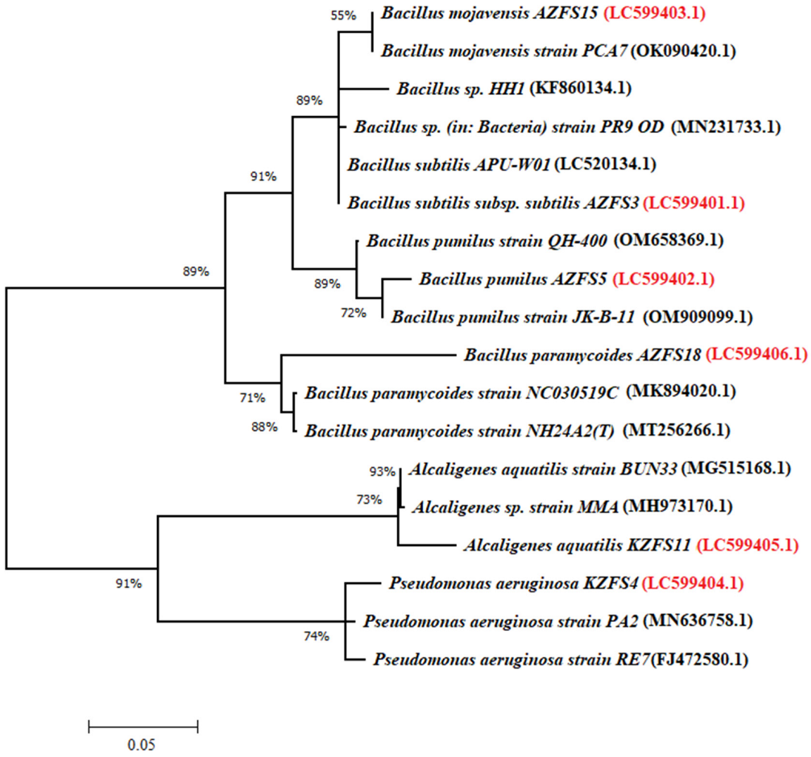
3.1. Identification of Bacterial Strains

In this study, six bacterial isolates named AZFS3, AZFS5, AZFS15, AZFS18, KZFS4, and KZFS11 were isolated, purified, and selected based on their ability to grow in a culture medium containing CAP and FBD as sole sources of carbon, and were used as bio-degrading agents for pesticide biodegradation.
Recent developments in mass spectrometry have made it possible to rapidly and accurately identify bacteria and fungi [47,48] by comparing the sample spectra to those of the reference database in this study, the keys of Bergey’s Manual of Systematic Bacteriology (2012). William et al. [49] identified the selected bacterial isolates up to the genus and species levels. In addition, as a more advanced method, the MALDI-TOF-MS strategy was also used to confirm the identification of the selected bacterial isolates.
Biotyper Software compares the spectrum of tested organisms to peak, list-based entries of bacterial strains in the database and provides similarity scores and the closest matches to the organisms. A score below 1.699 is unreliable. This advanced strategy employed in this study for directly identifying pesticide-degrading bacteria was much faster and more specific than other proposed methods. The same conclusion was reached by Stevenson et al. [29] and Moussaoui et al. [50]. Additionally, Bille et al. [47] mentioned that, in 99.2% of cases, this technique allowed for the correct identification of bacteria grown on solid media (2609 out of 2630 organisms).
The bacterial isolate AZFS3 was identified based on the nucleotide sequence of the 16S rRNA gene and documented in Genbank as the strain of Bacillus subtilis subsp. subtilis under the accession number LC599401.1. Results in Figure 1 representing the phylogenetic relationship of this isolate compared to the most similar overseas strains showed 96% similarity between AZFS3 isolate and the 16S rRNA gene of Bacillus subtilis strain (MT111013.1). This result was completely in harmony with MALDI-TOF, morphological, and biochemical properties illustrated in Table 2, Table 3 and Table 4. The MALDI-TOF scores for isolates AZFS5, AZFS15, AZFS18, KZFS4, and KZFS11 were 2.332, 2.141, 2.323, 2.129, and 2.120, close to Bacillus pumilus, Bacillus mojavensis, Bacillus paramycoides, Pseudomonas aeruginosa, and Alcaligenes aquatilis, respectively, and these results were confirmed among analyses of the 16S rRNA gene of the other five isolates.
The sequence similarities of AZFS5 (LC599402.1), AZFS15 (LC599403.1), AZFS18 (LC599406.1), KZFS4 (LC599404.1), and KZFS11 (LC599405.1) were 97, 99, 91, 98, and 96% with Bacillus pumilus strain (KJ574403.1), Bacillus mojavensis (OK090420.1), Bacillus paramycoides strain (MN093405.1), Pseudomonas aeruginosa strain (JN412064.1), and Alcaligenes aquatilis strain (CP032153.1), respectively.
The results in Table 3 prove that conventional identification using morphological and biochemical methods and identification obtained from cultures using the Bruker Daltonies MALDI-TOF-MS instrument produced identical results, but the latter was a simple and quick identification. Data from 16S rRNA sequencing and MALDI-TOF-MS for identification were used to confirm [51,52], because ribosomal proteins and ribosomal nucleic acids have evolved together [27,53].

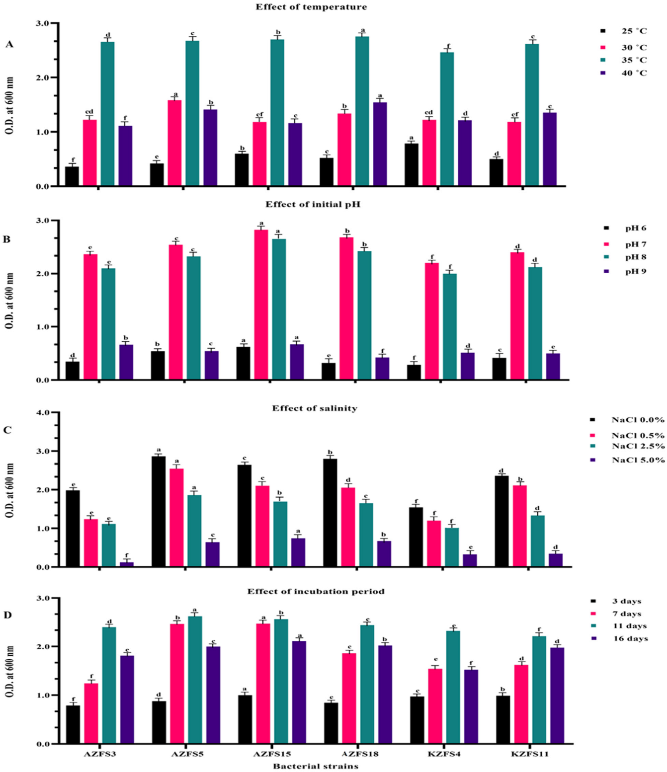
3.2. Factors Affecting Bacterial Growth through Biodegradation of Diamide Pesticides

In this study, only four factors were changed to optimize cultural growth conditions: temperature, salt, pH, and incubation time; the bacteria were left to grow. The experiments were done on a laboratory scale with six active bacterial strains in the presence of 50 mg/L of CAP-FBD mixture as a pesticide indicator.

3.2.1. Effect of Temperature

Temperature, for example, affects pesticide bioavailability and biodegradation [54,55]. The changes in the bacterial growth of the six tested bacterial strains in MSM supplemented with CAP-FBD mixture as affected by different incubation temperatures are given in Figure 2A. The growth dynamics of the tested bacteria were determined by measuring the optical densities during the experiment, which lasted for 11 days of incubation (pH 7.0, salinity 0.0% NaCl). The tested bacterial strains could grow at various temperatures of 25, 30, 35, and 40 °C. There was an increase in turbidity (bacterial growth) at O.D. 600 nm, a 2–3-fold increase compared to the control.
Additionally, the incubation temperature was increased from 25 to 40 °C, and the best range of growth was from 30 to 35 °C. The results also revealed that the highest bacterial biomass of the tested strains was observed at 35 °C and showed a positive correlation between the temperature degree and the bacterial biomass production. These results confirmed those obtained by Lin et al. [56], who reported that temperature significantly influenced cypermethrin biodegradation by Streptomyces sp., strain HU-S-01, and also showed that cypermethrin biodegradation occurred between 30 and 35 °C.
Moreover, Siddique et al. [57] found that the optimal temperature for the degradation of HCH isomers (a and c) was similar; increasing the temperature to 30 °C accelerated endosulfan biodegradation [58,59]. The biodegradation of pesticides was observed most accelerated at 35 °C [54,60]. Meantime, [61] confirmed that the degradation of flouroxypar was accelerated at 35 °C. Additionally, Farhan et al. [62] isolated Bacillus sp. Ct3 from chlorpyrifos-contaminated cotton soils, which biodegraded 88% of chlorpyrifos (125 mgL−1) in 8 days. The optimal temperature for profiling Bacillus sp. Ct3 biodegradation of chlorpyrifos was 35 °C.

3.2.2. Effect of Initial pH

The pH level of the medium is the primary factor in regulating microbial growth. The changes in the optical density (O.D. 600 nm) of the six tested bacterial strains were monitored for 11 days of incubation at 35 °C, and 0% NaCl is given in Figure 2B. The results show that when the initial pH of the tested media was between 7.0 and 8.0, all tested strains showed a significant rise in bacterial growth. This alteration in pH value caused considerable bacterial growth rates to increase by five and six-fold in pH 7.0 and 8.0 and one-fold in the case of pH 9.0 compared to the control. The data also reveal that two bacterial strains, Bacillus mojavensis AZFS15 and Bacillus paramycoides AZFS18, had the highest bacterial biomass at pH 7.0, while the rest of the bacterial strains showed the highest bacterial biomass at pH 7.0 but with slightly less growth. At pH 6.0 and 9.0, bacteria achieved the lowest growth values. The differences depend on thebacteria type and its ability to use the CAP-FBD mixture as a source of carbon and energy in MSM.
Accordingly, results were obtained by Vidali [63], Karpouzas, and Walker [64]. When they tested ethoprofos, pH 7.0 was optimal for growth, followed by pH 8.0. Additionally, Mohan et al. [65] found that when the environment was optimized at pH 8.0, the biodegradation of endosulfan was faster. During the biodegradation of endosulfan in soil, a pH range of 7.0 ± 0.1 was observed [59]. Moreover, Sidal and Yilmaz [66] found that an initial pH of 7.0 and 30 °C was optimal for Pseudomonas sp. to produce rhamnolipid and the highest biosurfactant concentration. The surface active properties of biosurfactant formed by Lactobacillus pentosus were also impacted by decreases in pH and temperature, with the highest stability and surface tension reduction at pH 8.0 [67]. The optimal biosurfactant production by P. aeruginosa and B. subtilis 181 was observed at pH 7.0 and 37 °C [68,69].
In this study, at pH 6.0 and 9.0, there were obvious decreases in the bacterial biomass for all tested bacterial strains used. Similar findings were reported by Cycoń et al. [70], who revealed that low pH may inhibit the activity of bacteria and enzymes associated with pesticide transformation. Furthermore, Abo-Amer [71] proposed that acidic and alkaline pH inhibited diazinon degradation.

3.2.3. Salinity Effect

Salinity’s impact on pesticide biodegradation was also reported to be rarely evaluated. Due to the potential for cell membrane disruption, protein denaturing, such as enzymes, and osmotic force changes caused by high NaCl concentrations in the medium, bacterial biodegradation of diamide pesticides was weak in these conditions [72]. In this experiment, the ability of six studied strains to grow at four different NaCl concentrations (0.0, 0.5, 2.5, and 5.0%), was determined at 35 °C, pH 7.0, and incubation for 11 days with 50 mg/L of CAP-FBD mixture.
The experimental results in Figure 2C showed that all bacterial strains were very sensitive to the NaCl concentrations, as indicated by the obvious decline in their growth rates. Salinity has a big effect on all biological processes and is likely to slow down bacterial growth and, as a result, make it harder for the bioremediation of pesticides. On the other hand, these bacterial strains were mainly isolated from soil and freshwater contaminated with pesticides, not from marine environments. Data in Figure 2C show that the control treatment (0% NaCl) was the most suitable concentration for most tested bacterial strains. The conclusion could be summarized that the most suitable concentrations for the most tested bacterial strains were within 0.0 and 0.5%.
The findings that were obtained in this study are consistent with those that Yun et al. [73] found; as soil salinity increased, metabolism and the rate of chlorpyrifos degradation were reduced. As salinity increased, it took longer for desethyl lactofen and acifluorfen to biodegrade [74]. Furthermore, salinity may impact microorganisms and thus the biodegradation of lactofen and metabolites. Lactofen residues may be more severe in higher salinity soils and require attention [72]. Minai-Tehrani et al. [75] examined the effects of various NaCl concentrations (0.0–5.0%) on contaminated soil. They found that biodegradation was higher in 0.0% NaCl (41%), while the reduction was higher in 1.0% NaCl (35%), and 5.0% NaCl showed the lowest reduction (12% and 8%, respectively).
On the other hand, salts may also accelerate the degradation of pesticides in soils due to other factors. It was found that salts enhance the degradation of parathion in soil because salts catalyzed the surface hydrolysis of organophosphorus esters [76]. Moreover, the effects of salinity on solubility and extracellular enzymes may transform pesticide degradation [73]. The effect of salt content on pesticide bioremediation in the saline–alkaline soil condition has received little attention, and little is known about the Effect of salinity on the bioremediation process of contaminated soils [55]. Furthermore, limited information is available on the degradation of pesticides in saline soils.

3.2.4. Effect of Incubation Period

The influence of the incubation period (3, 7, 11, and 16 days) on the growth of the selected six bacterial strains at different time intervals was studied using an MSM, pH 7.0, and incubation at 35 °C. The bacterial growth in terms of bacterial biomass as a function of time was determined spectrophotometrically at 600 nm Figure 2D.
The results presented in Figure 2D provided that as the time of incubation proceeded, most bacterial strains showed an obvious increase in bacterial growth (biomass). The highest bacterial biomass formation was observed at 11 days of incubation. After the experiment, at 16 days of incubation, there was a progressive decrease in bacterial biomass formation in all the tested bacteria. This reduction could be attributed to the absence of available carbon and energy sources in the medium or the presence of toxic metabolite compounds in the medium.
The obtained results are in the same line as Hussain et al. [77]. During the first three days of incubation, very little degradation was reported. After 7 days of incubation, the biodegradation of both isomers of endosulfan was between 22 and 60% (a-endosulfan) and between 18 and 58% (b-endosulfan). The removal of both isomers of endosulfan by bacterial strains ranged from 43 to 93% after 14 days of incubation. Based on Sharma et al. [78], Bacillus sp. and Micrococcus sp. demonstrated 71.6% degradation after 10 days of incubation at 0.1% v/v chlorpyrifos. Bacillus species showed 40% and 44% at the same concentration levels. Incubating the isolate Staphylococcus aureus for two weeks made it more effective at breaking down 80% of the total compound in the media. Additionally, Doolotkeldieva et al. [79] reported that active Bacillus polymyxa and Pseudomonas fluorescens bacterial strains were used in consortia and individual cultures. They have demonstrated high rates of degrading activity on the pesticide Aldrin in 12 days.

3.3. Changes in the Amount of CO2 Evolution

The main concept behind using microbes to clean up pesticide pollution in the environment is to use organic pesticides as carbon and nitrogen sources. Complex pesticide compounds are broken down into simple compounds or completely decomposed into CO2, H2O, and NH3, reducing pesticide residues and toxicity [80]. Through the degradation process, bacteria get energy from these waste products. The degradation process occurs best when the temperature, pH of the soil, and amount of water in the soil are all optimum. In this connection, a respirometric biodegradation experiment was carried out in a special biometric flask (1.0 L) using sandy loam soil, which was spiked with CAP-FBD mixture at (10 mg/kg) and inoculated with the highest active diamide-pesticide degraders used in this study.
The six bacterial strains were used individually and also used in consortia as follows: consortium (no.1), used two short-rods Pseudomonas aeruginosa KZFS4 and Alcaligenes aquatilis KZFS11; consortium (no.2), used four bacilli (Bacillus subtilis subsp. subtilis AZFS3, Bacillus pumilus AZFS5, Bacillus mojavensis AZFS15, and Bacillus paramycoides AZFS18; and consortium (no.3) mixed all the aforementioned six strains (two short-rods and four bacilli) to estimate the true and effective metabolic activity of the tested bacteria, individually and in association, during the biodegradation process by measurement of CO2 production. Table 5 shows how CO2 was measured when the tested bacterial strains were put in an incubator at 28 °C for 16 days.
The obtained results indicate that there were large differences between the tested active pesticide degraders in their capacities to biodegrade or mineralize the CAP-FBD mixture at 10 mg/kg, as indicated by the rate of CO2 output from the soil in question and the calculation of the average rate of CO2 evolution. Additionally, the rate of CO2 production was calculated for each treatment during the 16 days. Pseudomonas aeruginosa KZFS4 showed the highest individual average rate of CO2 production from biodegradation of FBD-CAP, at 1.226 mg CO2/16 day (78.6%) and total CO2 production at 46.352 mg CO2/16 days, followed by the B. subtilis subsp. subtilis AZFS3 strain recorded 0.901 mg CO2/16 day (57.7%) and total CO2 production at 33.668 mg CO2/16 day. Additionally, the lowest average rate of CO2 production was registered at 0.810 mg CO2/16 day (51.9%) in the case of Bacillus mojavensis AZFS15. Our findings are consistent with that of Gilani et al. [81] who reported that Pseudomonas is a diverse genus with numerous enzymes and catabolic processes involved in the biodegradation of pesticides. According to reports, Pseudomonas putida MAS-1 is 90% more effective in degrading chlorpyrifos. In aerobic conditions, Pseudomonas ATCC 700113 utilizes 3,5,6-trichloro-2-pyridinyl as its only carbon and energy source, degrading into CO2, water, chloride, ammonium, and other unknown polar metabolites [81,82].
Some studies have evaluated the relationship between microbial respiration, microbial biomass, and pesticide biodegradation in various soils [83,84]. Their results show that the breakdown of pesticides (alachlor, fluometuron, chlorsulfuron, dicamba, metsulfuron, and 2,4-D) was linked to the respiration of bacteria and biomass.
Table 5 reveals that the CO2 rate of tested bacteria reached its peak after 3 days of incubation, in the case of bacteria and consortium, except for Bacillus subtilis subsp. subtilis AZFS3 and Bacillus pumilus AZFS5 reaching their peak in 7 days. The amounts of CO2 decreased gradually until the experiment’s end in both individual bacterial strains and consortia. This decline in CO2 production might be attributed to the decline in the density of the microbial population; which was previously attributed to the exhaustion of the available degradable organic fraction, and [85] mentioned that this decline could be attributed to toxic compounds in soil or the lack of assimilable carbon and energy sources.
Significant amounts of CO2 uptake were recorded during the experiment when the active diamide-pesticide-degrading bacteria were mixed and added to the soil as a consortium (no. 1, no. 2, and no. 3). The rate of CO2 production from FBD-CAP reached its peak after 3 days of incubation, being 0.665, 0.688, and 0.712 mg CO2/day/100 g.d.wt.soil. The average rate of CO2 output from FBD-CAP degradation was 1.397, 1.446, and 1.553 mg CO2/16 day, with biodegradation efficiencies of 89.6, 92.8, and 99.6%. Total CO2 production was 51.162, 57.195, and 70.021 mg CO2/16 day, in the cases of consortia no. 1, 2, and 3, respectively. The mixed bacterial strains as a specific consortium showed more growth and degradation of the CAP-FBD mixture of 10 mg/kg used in this study, as indicated by the values of CO2 production and the average rate of biodegradation efficiency.
Additionally, it is interesting to notice from the data in Table 5 that the amount of CO2 evolved at 16 days and, in the case of consortium no.3, reached a zero value. The six bacterial strains used, in association forms, exhausted or mineralized the available degradable CAP-FBD mixture completely, and the density of the microbial population declined due to the exhaustion of available nutrients and energy sources. In cases where a pollutant is toxic, or there are not enough suitable microorganisms, using a bacterial consortium as an inoculum has some benefits over bio stimulation by native microorganisms (quality or quantity). Due to their synergistic effects, the microorganisms’ consortia can degrade diamide pesticides more quickly than single isolates.
Xu et al. [86] enriched a bacterial consortium to test their ability to biodegrade neonicotinoid insecticides under different conditions (pH values, temperatures, and various pollutant concentrations). They could see that pesticide contaminants were broken down quickly and that the consortium had synergistic effects, indicating that the consortium was better at biodegrading pollutants in an unstable environment.
The findings demonstrate that the consortium could improve the degradation rate of the CAP-FBD mixture, and adding the number of carbon sources could increase the biodegradation rate of the CAP-FBD mixture. These results are in line with Pino and Peñuela [87].

3.4. Biodegradation of Diamide Pesticides

Based on the findings of the performed experiments in this study, it could be inferred that the six tested bacterial strains have the ability for diamide pesticide degradation. In this experiment, the ability of these bacteria to break down a variety of pesticides (Profenofos, Cypermethrin, Carbofuran, and Malathion), and use diamide pesticides in the forms of (CAP-FBD mixture, FBD, and CAP) 100 mg/L as sole sources of carbon and energy in MSM under optimal conditions, was tested. The registered bacterial growth at O.D. 600 nm of the six tested bacterial strains as well as the consortium of six bacteria, given the best result in the previous experiment, were used in this study as an efficient bacterium in pesticide degradation as affected by different carbon sources as shown in Table 6.
As for the first group of carbon sources, CAP-FBD mixture, FBD, and CAP, with 100 mg/L, the tested bacteria could grow and utilize them as indicated by the values of their optical densities, which ranged as follows: from 1.233 to 2.365, from 1.216 to 2.119, and from 1.300 to 2.234, respectively, as shown in Table 6. Similar results regarding the efficient degradation of pesticides by the tested bacteria were ascertained. Around the world, many soil bacteria with the potential for bioremediation have been identified [88]. Microorganisms usepesticides that contaminate the soil for their energy or as a source of nutrients, which is the primary mechanism behind bioremediation.
Several factors affect bioremediation, such as bioavailability, substrate, and environmental factors. Additionally, Góngora-Echeverría et al. [89] proposed that microbial bioremediation can be used to successfully detoxify toxic pesticide residues that have accumulated in the environment. A microbial consortium can degrade multiple types of pesticides in natural habitats. These consortia are more effective at degrading pesticides than single strains. Additionally, some studies have tested the bioremediation capabilities of some natural soil microorganism consortia, while others have isolated microorganisms and then tested the individual capacity of microbes to break down pesticides [90,91,92].
From the obtained results, the most superior pesticide degraders were as follows: Pseudomonas aeruginosa KZFS4 individually gave the highest growth in all pesticides from 1.542 to 1.701 O.D. (600 nm), and the consortium (no.3) showed the highest capacities to utilize these carbon sources used in this study and mineralize them to CO2, H2O, and biomass from 2.119 to 2.365 O.D. (600 nm), and these bacteria strains provided themselves as pesticide degraders. These findings are consistent with the results reported by Randika et al. [88] and Naphade et al. [93]. It has been reported that various soil bacterial strains with pesticide detoxification abilities have been identified and successfully used in the bioremediation of pesticide-contaminated sites. Several studies have been conducted in this field all over the world. Some soil bacteria with bioremediation potential, such as Pseudomonas aeruginosa, can degrade chlorpyrifos, cypermethrin, endosulfan, and other pesticides. Pseudomonas species can degrade neonicotinoids [19,94].
The bacterial growth of the tested strains, six bacterial strains as well as the consortium (no. 3), used in this study, as affected by the type of other different pesticide groups 2 (Profenofos, Cypermethrin, Carbofuran, and Malathion) 100 mg/L as a carbon source in MSM, was also representing in Table 6. The data show that all the tested bacterial strains could metabolize all the tested pesticides but at different degradation rates, as indicated by the values of their optical densities owing to their growth. The turbidity values of bacteria on Profenofos and Cypermethrin were (1.133–2.122) and (1.244–2.222), respectively, while on Carbofuran and Malathion, they were (1.219–2.324) and (1.131–2.311), respectively.
Similar results were asserted regarding the efficient degradation of pesticides by the tested bacteria. The most well-known natural isolates capable of degrading organophosphates are Pseudomonas diminuta MG and Flavobacterium ATCC 27551 [95,96]. Profenofos is a common organophosphate pesticide [97]. Since there is intense rivalry among microorganisms for carbon sources in the environment and this bacterium uses profenofos as an energy source, this gives it a significant advantage in the biodegradation of pesticides by adding them to contaminated soil [96]. Furthermore, Ghani et al. [98] stated that using profenofos-degrading bacterial strains with growth-promoting properties on tomato plants can significantly boost growth, break down the profenofos, and reduce the toxic effects of profenofos in pesticide stress conditions.
As previously stated, only the Pseudomonas aeruginosa KZFS4 (LC599404.1) strain had the greatest ability to metabolize and utilize this group 2 compound. Based on these results, it could be deduced that these six bacterial strains have a high ability to utilize a diverse range of pesticide residues. These findings are in line with that of Kadhim et al. [99], who demonstrated that the efficiency of malathion biodegradation is higher in consortia than in individual bacterial isolates. Consortia degraded Malathion at a rate of 98.32%. However, used individual bacterial cultures by Kadhim et al. [99] reported that the ability of bacterial isolates to biodegrade malathion was examined using 2.0 mL of each isolate’s active culture per 100 mL of MSM containing 500 mg/L of malathion. For 10 days, the test system was incubated. Malathion monoculture was broken down by Pseudomonas putida and Staphylococcus vitulinus at a rate of 47.18% and 44.24%, respectively.
Future studies must clarify other factors affecting the capability and efficiency of pesticide biodegradation, such as nutrients, oxygen content, and the physical state of pesticides in the field under realistic conditions, to improve these bacteria’s viability as potential commercials. On the other hand, more research is also needed from an economic point of view to find ways to reuse biopesticides in this work.

4. Conclusions

The findings of this study indicate that consortia can bio-degrade and metabolize some diamide insecticides and other pesticides. Six different bacterial strains with the potential for degradation of diamide pesticides were identified, and strain P. aeruginosa KZFS4 has the maximum potential to degrade the diamide pesticides, which was identified as Pseudomonas aeruginosa KZFS4 by 16S rRNA sequence analysis. The optimum conditions for biodegradation of CAP-FBD mixture using the six bacterial strains of this study at 50 mg/L concentration were pH (7.0), temperature (30–35 °C), salinity (0.0–0.5% NaCl), and incubation time (11 days) were suitable for the majority of tested bacterial strains, and these parameters with these bacterial strains would be applicable in the bioremediation process as a final goal on an industrial scale and with pesticide spills. Pseudomonas aeruginosa KZFS4 could be the strong isolate that could help break down diamides found in different environments. An active consortium (no.3) was selected, which had shown strong degrading activity rates on the diamide insecticides and other pesticides and thus could be a promising inoculum candidate.

Author Contributions

Conceptualization, M.A.F. and H.I.A.E.-F.; methodology, M.A.F., S.H.S., H.I.A.E.-F., M.F.A., D.A.A.-Q., S.Y.A.Q., M.A.S.A. and B.A.A.; software, M.A.F.; validation, M.A.F., S.H.S., H.I.A.E.-F. and B.A.A.; formal analysis, M.F.A. and D.A.A.-Q.; investigation, H.I.A.E.-F.; resources, M.A.F.; data curation, S.Y.A.Q. and M.A.S.A.; writing—original draft preparation, S.H.S. and H.I.A.E.-F.; writing—review and editing, M.A.F. and H.I.A.E.-F.; visualization, S.H.S.; funding acquisition, M.A.S.A. All authors have read and agreed to the published version of the manuscript.

Data Availability Statement

The raw reads have been submitted to the NCBI nucleotide database, with accession numbers LC599401.1, LC599402.1, LC599403.1, LC599404.1, LC599405.1, and LC599406.1. These datasets were derived from the public domain resources listed below: https://0-www-ncbi-nlm-nih-gov.brum.beds.ac.uk/nuccore, accessed on 15 November 2022.

Acknowledgments

The authors would like to thank the Deanship of scientific research at Umm Al-Qura University for supporting this work by grant code (23UQU4290565DSR102).

Conflicts of Interest

The authors declare that they have no conflicts of interest.

References

  1. Raffa, C.M.; Chiampo, F. Bioremediation of agricultural soils polluted with pesticides: A review. Bioengineering 2021, 8, 92. [Google Scholar] [CrossRef] [PubMed]
  2. Food and Agriculture Organization of the United Nations. Rome. 2018. Available online: http://faostat.fao.org (accessed on 12 March 2018).
  3. Faostat, F. Food and Agriculture Organization of the United Nations-Statistic Division. 2019. Available online: https://www.fao.org/faostat/en/#data (accessed on 15 November 2022).
  4. Gonçalves, C.R.; da Silva Delabona, P. Bioremediation of pesticides in Brazil: A brief overview. Environ. Adv. 2022, 8, 100220. [Google Scholar] [CrossRef]
  5. Boedeker, W.; Watts, M.; Clausing, P.; Marquez, E. The global distribution of acute unintentional pesticide poisoning: Estimations based on a systematic review. BMC Public Health 2020, 20, 1875. [Google Scholar] [CrossRef] [PubMed]
  6. Calvert, G.M.; Mehler, L.N.; Alsop, J.; De Vries, A.L.; Besbelli, N. Surveillance of pesticide-related illness and injury in humans. In Hayes’ Handbook of Pesticide Toxicology; Elsevier: Amsterdam, The Netherlands, 2010; pp. 1313–1369. [Google Scholar]
  7. Xing, H.; Li, S.; Wang, Z.; Gao, X.; Xu, S.; Wang, X. Histopathological changes and antioxidant response in brain and kidney of common carp exposed to atrazine and chlorpyrifos. Chemosphere 2012, 88, 377–383. [Google Scholar] [CrossRef] [PubMed]
  8. David, M.; Marigoudar, S.R.; Patil, V.K.; Halappa, R. Behavioral, morphological deformities and biomarkers of oxidative damage as indicators of sublethal cypermethrin intoxication on the tadpoles of D. melanostictus (Schneider, 1799). Pestic. Biochem. Phys. 2012, 103, 127–134. [Google Scholar] [CrossRef]
  9. Ellgehausen, H.; Guth, J.A.; Esser, H.O. Factors determining the bioaccumulation potential of pesticides in the individual compartments of aquatic food chains. Ecotoxicol. Environ. Saf. 1980, 4, 134–157. [Google Scholar] [CrossRef]
  10. David, M.; Sangeetha, J.; Shrinivas, J.; Harish, E.; Naik, V. Effects of deltamethrin on haematological indices of indian major carp, Cirrhinus mrigala (Hamilton). Int. J. Pure Appl. Zool. 2015, 3, 37–43. [Google Scholar]
  11. Majumdar, B.B.; Guha, G.; Ray, A.N.; Bala, B. Toxic intracerebral demyelination in a case of suicidal Cypermethrin poisoning. Ann. Trop. Med. Public Health 2012, 5, 615. [Google Scholar] [CrossRef]
  12. Özkara, A.; Akyıl, D.; Konuk, M. Pesticides, environmental pollution, and health. In Environmental Health Risk-Hazardous Factors to Living Species; IntechOpen: London, UK, 2016. [Google Scholar]
  13. Ishaaya, I. Biochemical Sites of Insecticide Action and Resistance; Springer Science & Business Media: Berlin, Germany, 2012. [Google Scholar]
  14. Sharma, A.; Kumar, V.; Shahzad, B.; Tanveer, M.; Sidhu, G.P.S.; Handa, N.; Kohli, S.K.; Yadav, P.; Bali, A.S.; Parihar, R.D. Worldwide pesticide usage and its impacts on ecosystem. SN Appl. Sci. 2019, 1, 1446. [Google Scholar] [CrossRef] [Green Version]
  15. Aghris, S.; Alaoui, O.T.; Laghrib, F.; Farahi, A.; Bakasse, M.; Saqrane, S.; Lahrich, S.; Mhammedi, E. Extraction and determination of flubendiamide insecticide in food samples: A review. Curr. Res. Food Sci. 2022, 5, 401–413. [Google Scholar] [CrossRef]
  16. Ortiz-Hernández, M.L.; Rodríguez, A.; Sánchez-Salinas, E.; Castrejón-Godínez, M.L. Bioremediation of soils contaminated with pesticides: Experiences in Mexico. In Bioremediation in Latin America; Springer: Berlin/Heidelberg, Germany, 2014; pp. 69–99. [Google Scholar]
  17. Bhatt, P.; Bhatt, K.; Sharma, A.; Zhang, W.; Mishra, S.; Chen, S. Biotechnological basis of microbial consortia for the removal of pesticides from the environment. Crit. Rev. Biotechnol. 2021, 41, 317–338. [Google Scholar] [CrossRef]
  18. Hussain, S.; Siddique, T.; Arshad, M.; Saleem, M. Bioremediation and phytoremediation of pesticides: Recent advances. Crit. Rev. Environ. Sci. Technol. 2009, 39, 843–907. [Google Scholar] [CrossRef]
  19. Obaid, R.J. Synthesis and biological evaluation of some new imidazo[1,2-c]pyrimido [5,4-e]pyrimidin-5-amine derivatives. J. Umm Al-Qura Univ. Appl. Sci. 2021, 7, 16–22. [Google Scholar]
  20. Gao, W.; Li, D.; You, H. Functional characterization and genomic analysis of the chlorantraniliprole-degrading strain Pseudomonas sp. GW13. Bioengineering 2019, 6, 106. [Google Scholar] [CrossRef] [Green Version]
  21. Atlas, R.M.; Synder, J.W. Reagents, stains, and media: Bacteriology. In Manual of Clinical Microbiology, 11th ed.; John Wiley & Sons Inc.: Hoboken, NJ, USA, 2011; pp. 272–303. [Google Scholar]
  22. Setlhare, B.; Kumar, A.; Aregbesola, O.; Mokoena, M.; Olaniran, A. 2, 4-dichlorophenol degradation by indigenous Pseudomonas sp. PKZNSA and Klebsiella pneumoniae KpKZNSA: Kinetics, enzyme activity and catabolic gene detection. Appl. Biochem. Microbiol. 2021, 57, 656–665. [Google Scholar] [CrossRef]
  23. Logan, N.A.; De Vos, P. Genus I. bacillus. Bergey’s Man. Syst. 2009, 3, 21–128. [Google Scholar]
  24. Kersters, K.; De Ley, J. Genus Alcaligenes Castellani and Chalmers 1919, 936AL. In Bergey’s Manual of Systematic Bacteriology, 9th ed.; LWW Publication: Philadelphia, PA, USA, 1992; Volume 1, pp. 361–373. [Google Scholar]
  25. Garrity, G.M.; Brenner, D.J.; Krieg, N.; Staley, J. Bergey’s Manual of Systematic Bacteriology. The Proteobacteria, Part C: The Alpha-, Beta-, Delta-, and Epsilonproteobacteria, Springer: New York, NY, USA, 2005.
  26. Nacef, M.; Chevalier, M.; Chollet, S.; Drider, D.; Flahaut, C. MALDI-TOF mass spectrometry for the identification of lactic acid bacteria isolated from a French cheese: The maroilles. Int. J. Food Microbiol. 2017, 247, 2–8. [Google Scholar] [CrossRef]
  27. Sauer, S.; Kliem, M. Mass spectrometry tools for the classification and identification of bacteria. Nat. Rev. Microbiol. 2010, 8, 74–82. [Google Scholar] [CrossRef]
  28. Biswas, S.; Rolain, J.-M. Use of MALDI-TOF mass spectrometry for identification of bacteria that are difficult to culture. J. Microbiol. Methods 2013, 92, 14–24. [Google Scholar] [CrossRef]
  29. Stevenson, L.G.; Drake, S.K.; Murray, P.R. Rapid identification of bacteria in positive blood culture broths by matrix-assisted laser desorption ionization-time of flight mass spectrometry. J. Clin. Microbiol. 2010, 48, 444–447. [Google Scholar] [CrossRef] [Green Version]
  30. Sambrook, J.; Fritsch, E.; Maniatis, T. Molecular Cloning. A Laboratory Manual, 2nd ed.; Cold Spring Harbor, Laboratory Press: New York, NY, USA, 1989. [Google Scholar]
  31. Srivastava, S.; Singh, V.; Kumar, V.; Verma, P.C.; Srivastava, R.; Basu, V.; Gupta, V.; Rawat, A.K. Identification of regulatory elements in 16S rRNA gene of Acinetobacter species isolated from water sample. Bioinformation 2008, 3, 173. [Google Scholar] [CrossRef] [PubMed] [Green Version]
  32. Saitou, N.; Nei, M. The neighbor-joining method: A new method for reconstructing phylogenetic trees. Mol. Biol. Evol. 1987, 4, 406–425. [Google Scholar] [PubMed]
  33. Felsenstein, J. Confidence limits on phylogenies: A justification. Evolution 1985, 39, 783–791. [Google Scholar] [CrossRef] [PubMed]
  34. Tamura, K.; Nei, M.; Kumar, S. Prospects for inferring very large phylogenies by using the neighbor-joining method. Proc. Natl. Acad. Sci. USA 2004, 101, 11030–11035. [Google Scholar] [CrossRef] [PubMed] [Green Version]
  35. Kumar, S.; Stecher, G.; Tamura, K. MEGA7: Molecular Evolutionary Genetics Analysis Version 7.0 for Bigger Datasets. Mol. Biol. Evol. 2016, 33, 1870–1874. [Google Scholar] [CrossRef] [Green Version]
  36. Ouided, B.; Abderrahmane, B. Isolation and characterization of glyphosate-degrading bacteria from different soils of Algeria. Afr. J. Microbiol. Res. 2013, 7, 5587–5595. [Google Scholar] [CrossRef] [Green Version]
  37. John, E.M.; Sreekumar, J.; Jisha, M. Optimization of chlorpyrifos degradation by assembled bacterial consortium using response surface methodology. Soil Sediment Contam Int. J. 2016, 25, 668–682. [Google Scholar] [CrossRef]
  38. Dubey, S.; Upadhyay, S.N.; Sharma, Y.C. Optimization of removal of Cr by γ-alumina nano-adsorbent using response surface methodology. Ecol. Eng. 2016, 97, 272–283. [Google Scholar] [CrossRef]
  39. Isermeyer, H. Eine einfache Methode zur bestimmung der bodenatmung und der karbonate im boden. Z. Für. Pflanz. Düngung Bodenkd. 1952, 56, 26–38. [Google Scholar] [CrossRef]
  40. Mukherjee, I.; Das, S.K.; Kumar, A. Degradation of flubendiamide as affected by elevated CO2, temperature, and carbon mineralization rate in soil. Environ. Sci. Pollut. Res. 2016, 23, 19931–19939. [Google Scholar] [CrossRef]
  41. Wu, M.; Li, G.; Chen, X.; Liu, J.; Liu, M.; Jiang, C.; Li, Z. Rational dose of insecticide chlorantraniliprole displays a transient impact on the microbial metabolic functions and bacterial community in a silty-loam paddy soil. Sci. Total Environ. 2018, 616, 236–244. [Google Scholar] [CrossRef]
  42. Sparks, D.L.; Page, A.L.; Helmke, P.A.; Loeppert, R.H. Methods of Soil Analysis, Part 3: Chemical Methods; John Wiley & Sons: Hoboken, NJ, USA, 2020; Volume 14. [Google Scholar]
  43. Page, A.; Miller, R.; Keeney, D. Methods of Soil Analysis, part 2. Chem. Microbiol. Prop. 1982, 2, 643–698. [Google Scholar]
  44. Malatova, K. Isolation and Characterization of Hydrocarbon Degrading Bacteria from Environmental Habitats in Western New York State. Master’s Thesis, Rochester Institute of Technology, Rochester, NY, USA, November 2005. [Google Scholar]
  45. Jadhav, S.S.; David, M. Biodegradation of flubendiamide by a newly isolated Chryseobacterium sp. strain SSJ1. 3 Biotech 2016, 6, 31. [Google Scholar] [CrossRef] [Green Version]
  46. Geed, S.; Kureel, M.; Shukla, A.; Singh, R.; Rai, B. Biodegradation of malathion and evaluation of kinetic parameters using three bacterial species. Resour.-Effic. Technol. 2016, 2, S3–S11. [Google Scholar] [CrossRef]
  47. Bille, E.; Dauphin, B.; Leto, J.; Bougnoux, M.-E.; Beretti, J.-L.; Lotz, A.; Suarez, S.; Meyer, J.; Join-Lambert, O.; Descamps, P. MALDI-TOF MS Andromas strategy for the routine identification of bacteria, mycobacteria, yeasts, Aspergillus spp. and positive blood cultures. Clin. Microbiol. Infect 2012, 18, 1117–1125. [Google Scholar] [CrossRef] [Green Version]
  48. Dubois, D.; Leyssene, D.; Chacornac, J.P.; Kostrzewa, M.; Schmit, P.O.; Talon, R.; Bonnet, R.; Delmas, J. Identification of a variety of Staphylococcus species by matrix-assisted laser desorption ionization-time of flight mass spectrometry. J. Clin. Microbiol. 2010, 48, 941–945. [Google Scholar] [CrossRef] [Green Version]
  49. William, B.W.; Paul, D.V.; George, M.G.; Dorothy, J.; Noel, R.K.; Wolfgang, L.; Fred, A.R.; Karl, S. Bergeys Manual of Systematic Bacteriology; LWW Publication: Philadelphia, PA, USA, 2012; Volume 171, p. 190. [Google Scholar]
  50. Moussaoui, W.; Jaulhac, B.; Hoffmann, A.-M.; Ludes, B.; Kostrzewa, M.; Riegel, P.; Prévost, G. Matrix-assisted laser desorption ionization time-of-flight mass spectrometry identifies 90% of bacteria directly from blood culture vials. Clin. Microbiol. Infect. 2010, 16, 1631–1638. [Google Scholar] [CrossRef] [Green Version]
  51. Bizzini, A.; Durussel, C.; Bille, J.; Greub, G.; Prod’Hom, G. Performance of matrix-assisted laser desorption ionization-time of flight mass spectrometry for identification of bacterial strains routinely isolated in a clinical microbiology laboratory. J. Clin. Microbiol. 2010, 48, 1549–1554. [Google Scholar] [CrossRef] [Green Version]
  52. Wang, Y.R.; Qian, C.; Cui, S.H.; Li, F.Q. Characterization of Staphylococcus aureus isolated from clinical specimens by matrix assisted laser desorption/ionization time-of-flight mass spectrometry. Biomed. Environ. Sci. 2013, 26, 430–436. [Google Scholar]
  53. Teramoto, K.; Kitagawa, W.; Sato, H.; Torimura, M.; Tamura, T.; Tao, H. Phylogenetic analysis of Rhodococcus erythropolis based on the variation of ribosomal proteins as observed by matrix-assisted laser desorption ionization-mass spectrometry without using genome information. J. Biosci. Bioeng. 2009, 108, 348–353. [Google Scholar] [CrossRef]
  54. Odukkathil, G.; Vasudevan, N. Toxicity and bioremediation of pesticides in agricultural soil. Rev. Environ. Sci. Biotechnol. 2013, 12, 421–444. [Google Scholar] [CrossRef]
  55. Mali, H.; Shah, C.; Patel, D.H.; Trivedi, U.; Subramanian, R. Degradation insight of organophosphate pesticide chlorpyrifos through novel intermediate 2, 6-dihydroxypyridine by Arthrobacter sp. HM01. Bioresour. Bioprocess. 2022, 9, 31. [Google Scholar] [CrossRef]
  56. Lin, Q.; Chen, S.; Hu, M.; Haq, M.; Yang, L.; Li, H. Biodegradation of cypermethrin by a newly isolated actinomycetes HU-S-01 from wastewater sludge. Int. J. Environ. Sci. Technol. 2011, 8, 45–56. [Google Scholar] [CrossRef] [Green Version]
  57. Siddique, T.; Okeke, B.C.; Arshad, M.; Frankenberger, W.T. Biodegradation kinetics of endosulfan by Fusarium ventricosum and a Pandoraea species. J. Agric. Food Chem. 2003, 51, 8015–8019. [Google Scholar] [CrossRef] [PubMed]
  58. Philip, L. Bioremediation of endosulfan contaminated soil and water—Optimization of operating conditions in laboratory scale reactors. J. Hazard. Mater. 2006, 136, 354–364. [Google Scholar]
  59. Arshad, M.; Hussain, S.; Saleem, M. Optimization of environmental parameters for biodegradation of alpha and beta endosulfan in soil slurry by Pseudomonas aeruginosa. J. Appl. Microbiol. 2008, 104, 364–370. [Google Scholar] [CrossRef]
  60. Singh, B.K.; Walker, A.; Wright, D.J. Bioremedial potential of fenamiphos and chlorpyrifos degrading isolates: Influence of different environmental conditions. Soil Biol. Biochem. 2006, 38, 2682–2693. [Google Scholar] [CrossRef]
  61. Tao, L.; Yang, H. Fluroxypyr biodegradation in soils by multiple factors. Environ. Monit. Assess. 2011, 175, 227–238. [Google Scholar] [CrossRef]
  62. Farhan, M.; Ahmad, M.; Kanwal, A.; Butt, Z.A.; Khan, Q.F.; Raza, S.A.; Qayyum, H.; Wahid, A. Biodegradation of chlorpyrifos using isolates from contaminated agricultural soil, its kinetic studies. Sci. Rep. 2021, 11, 10320. [Google Scholar] [CrossRef]
  63. Vidali, M. Bioremediation. An overview. Pure Appl. Chem. 2001, 73, 1163–1172. [Google Scholar] [CrossRef]
  64. Karpouzas, D.G.; Walker, A. Factors influencing the ability of Pseudomonas putida epI to degrade ethoprophos in soil. Soil Biol. Biochem. 2000, 32, 1753–1762. [Google Scholar] [CrossRef]
  65. Mohan, S.V.; Sirisha, K.; Rao, R.S.; Sarma, P. Bioslurry phase remediation of chlorpyrifos contaminated soil: Process evaluation and optimization by Taguchi design of experimental (DOE) methodology. Ecotoxicol. Environ. Saf. 2007, 68, 252–262. [Google Scholar] [CrossRef]
  66. Sidal, U.; Yilmaz, E.S. Production of rhamnolipid (A Biosurfactant) using free and immobilized cells of Pseudomonas sp. Kafkas Univ. Vet. Fak. Derg. 2012, 18, 285–289. [Google Scholar]
  67. Vecino Bello, X.; Devesa-Rey, R.; Cruz, J.M.; Moldes, A.B.n. Study of the synergistic effects of salinity, pH, and temperature on the surface-active properties of biosurfactants produced by Lactobacillus pentosus. J. Agric. Food Chem. 2012, 60, 1258–1265. [Google Scholar] [CrossRef]
  68. Saikia, R.R.; Deka, S.; Deka, M.; Sarma, H. Optimization of environmental factors for improved production of rhamnolipid biosurfactant by Pseudomonas aeruginosa RS29 on glycerol. J. Basic Microbiol. 2012, 52, 446–457. [Google Scholar] [CrossRef]
  69. Al-Araji, L.I.Y.; Issa, L. Biosurfactant Production by Pseudomonas aeruginosa 181. Ph.D. Thesis, Universiti Putra Malaysia, Seri Kembangan, Malaysia, 2004. [Google Scholar]
  70. Cycoń, M.; Wójcik, M.; Piotrowska-Seget, Z. Biodegradation of the organophosphorus insecticide diazinon by Serratia sp. and Pseudomonas sp. and their use in bioremediation of contaminated soil. Chemosphere 2009, 76, 494–501. [Google Scholar] [CrossRef]
  71. Abo-Amer, A.E. Biodegradation of diazinon by Serratia marcescens DI101 and its use in bioremediation of contaminated environment. J. Microbiol. Biotechnol. 2011, 21, 71–80. [Google Scholar] [CrossRef]
  72. Jing, X.; Yang, J.; Wang, T. Effects of salinity on herbicide lactofen residues in soil. Water Air Soil Pollut. 2018, 229, 3. [Google Scholar] [CrossRef]
  73. Yun, E.-Y.; Ro, H.-M.; Lee, G.-T.; Choi, W.-J. Salinity effects on chlorpyrifos degradation and phosphorus fractionation in reclaimed coastal tideland soils. Geosci. J. 2010, 14, 371–378. [Google Scholar] [CrossRef]
  74. de Souza Silva, C.M.M.; Fay, E.F. Effect of salinity on soil microorganisms. Soil Health Land Use Manag. 2012, 10, 177–198. [Google Scholar]
  75. Minai-Tehrani, D.; Herfatmanesh, A.; Azari-Dehkordi, F.; Minuoi, S. Effect of salinity on biodegradation of aliphatic fractions of crude oil in soil. Pak. J. Biol. Sci. 2006, 9, 1531–1535. [Google Scholar] [CrossRef] [Green Version]
  76. Mingelgrin, U.; Yaron, B. The effect of calcium salts on the degradation of parathion in sand and soil. Soil Sci. Soc. Am. J. 1974, 38, 914–917. [Google Scholar] [CrossRef]
  77. Hussain, S.; Arshad, M.; Saleem, M.; Khalid, A. Biodegradation of α-and β-endosulfan by soil bacteria. Biodegradation 2007, 18, 731–740. [Google Scholar] [CrossRef] [PubMed]
  78. Sharma, B.; Saxena, S.; Datta, A.; Arora, S. Spectrophotometric analysis of degradation of chlorpyrifos pesticide by indigenous microorganisms isolated from affected soil. Int. J. Curr. Microbiol. App. Sci. 2016, 5, 742–749. [Google Scholar] [CrossRef]
  79. Doolotkeldieva, T.; Konurbaeva, M.; Bobusheva, S. Microbial communities in pesticide-contaminated soils in Kyrgyzstan and bioremediation possibilities. Environ. Sci. Pollut. Res. 2018, 25, 31848–31862. [Google Scholar] [CrossRef] [Green Version]
  80. Ye, X.; Dong, F.; Lei, X. Microbial resources and ecology-microbial degradation of pesticides. Nat. Res. Conserv. Res. 2018, 1, 242. [Google Scholar] [CrossRef]
  81. Gilani, R.A.; Rafique, M.; Rehman, A.; Munis, M.F.H.; ur Rehman, S.; Chaudhary, H.J. Biodegradation of chlorpyrifos by bacterial. J. Basic Microbiol. 2015, 55, 1–15. [Google Scholar]
  82. Feng, Y.; Minard, R.D.; Bollag, J.M. Photolytic and microbial degradation of 3,5,6-trichloro-2-pyridinol. Environ. Toxicol. Chem. Int. J. 1998, 17, 814–819. [Google Scholar] [CrossRef]
  83. Voos, G.; Groffman, P. Relationships between microbial biomass and dissipation of 2,4-D and dicamba in soil. Biol. Fertil. Soils 1997, 24, 106–110. [Google Scholar] [CrossRef]
  84. Anderson, J. Herbicide degradation in soil: Influence of microbial biomass. Soil Biol. Biochem. 1984, 16, 483–489. [Google Scholar] [CrossRef]
  85. Sabaté, J.; Vinas, M.; Solanas, A. Laboratory-scale bioremediation experiments on hydrocarbon-contaminated soils. Int. Biodeterior. Biodegrad. 2004, 54, 19–25. [Google Scholar] [CrossRef]
  86. Xu, B.; Xue, R.; Zhou, J.; Wen, X.; Shi, Z.; Chen, M.; Xin, F.; Zhang, W.; Dong, W.; Jiang, M. Characterization of acetamiprid biodegradation by the microbial consortium ACE-3 enriched from contaminated soil. Front. Microbiol. 2020, 11, 1429. [Google Scholar] [CrossRef]
  87. Pino, N.; Peñuela, G. Simultaneous degradation of the pesticides methyl parathion and chlorpyrifos by an isolated bacterial consortium from a contaminated site. Int. Biodeterior. Biodegrad. 2011, 65, 827–831. [Google Scholar] [CrossRef]
  88. Randika, J.; Bandara, P.; Soysa, H.; Ruwandeepika, H.; Gunatilake, S. Bioremediation of pesticide-contaminated soil: A review on indispensable role of soil bacteria. J. Agric. Sci.-Sri Lanka 2022, 17, 19–43. [Google Scholar] [CrossRef]
  89. Góngora-Echeverría, V.R.; García-Escalante, R.; Rojas-Herrera, R.; Giácoman-Vallejos, G.; Ponce-Caballero, C. Pesticide bioremediation in liquid media using a microbial consortium and bacteria-pure strains isolated from a biomixture used in agricultural areas. Ecotoxicol. Environ. Saf. 2020, 200, 110734. [Google Scholar] [CrossRef]
  90. McAllister, K.A.; Lee, H.; Trevors, J.T. Microbial degradation of pentachlorophenol. Biodegradation 1996, 7, 1–40. [Google Scholar] [CrossRef]
  91. Geetha, M.; Fulekar, M. Bioremediation of pesticides in surface soil treatment unit using microbial consortia. Afr. J. Environ. Sci. Technol. 2008, 2, 036–045. [Google Scholar]
  92. Werren, J.H. Symbionts provide pesticide detoxification. Proc. Natl. Acad. Sci. USA 2012, 109, 8364–8365. [Google Scholar] [CrossRef] [Green Version]
  93. Naphade, S.R.; Durve, A.A.; Bhot, M.; Varghese, J.; Chandra, N. Isolation, characterization and identification of pesticide tolerating bacteria from garden soil. Eur. J. Exp. Biol. 2012, 2, 1943–1951. [Google Scholar]
  94. Javaid, M.K.; Ashiq, M.; Tahir, M. Potential of biological agents in decontamination of agricultural soil. Scientifica 2016, 2016, 1598325. [Google Scholar] [CrossRef] [Green Version]
  95. Mulbry, W.W.; Karns, J.S.; Kearney, P.; Nelson, J.; McDaniel, C.; Wild, J. Identification of a plasmid-borne parathion hydrolase gene from Flavobacterium sp. by southern hybridization with opd from Pseudomonas diminuta. Appl. Environ. Microbiol. 1986, 51, 926–930. [Google Scholar] [CrossRef] [PubMed]
  96. Malghani, S.; Chatterjee, N.; Yu, H.X.; Luo, Z. Isolation and identification of profenofos degrading bacteria. Braz. J. Microbiol. 2009, 40, 893–900. [Google Scholar] [CrossRef] [PubMed] [Green Version]
  97. WHO. The WHO Recommended Classification of Pesticides by Hazard and Guidelines to Classification 2009; World Health Organization: Geneva, Switzerland, 2010. [Google Scholar]
  98. Ghani, M.U.; Asghar, H.N.; Niaz, A.; Ahmad Zahir, Z.; Nawaz, M.F.; Häggblom, M.M. Efficacy of rhizobacteria for degradation of profenofos and improvement in tomato growth. Int. J. Phytoremediation 2022, 24, 463–473. [Google Scholar] [CrossRef] [PubMed]
  99. Kadhim, F.; Rabee, A.M.; Abdalraheem, E. Biodegradation of malathion by selected bacterial isolates. J. Int. Environ. Appl. Sci. 2015, 10, 361–366. [Google Scholar]
Figure 1. Neighbor-joining tree showing the phylogenetic relationships between four Bacillus strains, pseudomonas, and Alcaligenes, and the type strains based on 16S rRNA gene sequences. The numbers at the branch nodes are bootstrap percentages (100 replicates), and the bar indicates a 0.5% sequence.
Processes 10 02527 g001
Figure 2. Bacterial growth of the tested six bacterial strains on MS broth with FBD-CAP. Mixture 50 mg/L, (A) effect of temperature, (B) effect of initial pH, (C) effect of salinity, and (D) effect of incubation period. Means and standard deviations of three replicates. Different letters on top of the bar indicate significant differences (p < 0.05).
Processes 10 02527 g002
Table 1. Physico-chemical analyses of soils used for CO2 production experiments.
CharactersValues
Physical analysis
Clay%12.1
Silt%10.9
Sand%77.0
Textural classSandy loam
CaCO3 (g kg−1)11.5
Chemical analysis
pH (1:2.5 at 25 °C)8.09
EC dSm−1 (1:5 at 25 °C) *0.52
Na+0.30
K+1.30
Ca2+1.60
Mg2+2.22
CO32−Nil
HCO32.30
Cl0.66
SO42−2.44
Organic matter (g kg−1)9.40
Available nutrients (mg kg−1 soil)
N60.50
P18.6
K90.5
* EC, Electrical conductivity.
Table 2. Morphological, physiological, and biochemical characteristics of the selected six active pesticide-degrading bacterial isolates.
Isolates CodeAZFS3AZFS5AZFS15AZFS18KZFS4KZFS11
1-Gram reaction++++
2-Cell shapeL-RodL-RodL-RodL-RodS-rodS-rod
3-Motility+++++
4-Spore formation+++NO-
5-Oxygen requirement
Aerobic growth
Anaerobic growth

+

+

+

+
+/−

+

+
6-Voges-proskauer++++
7-Catalase reaction+++++
8-Oxidase reaction++++
9-Urease
10-Nitrate reduction+++++
11-Uitlization of
• Citrate
• Propionate

+

+

+

+
+

+
ND


12-Hydrolysis of
-Casein
-Gelatin
-Starch

+
+
+

+
+

ND
+

+
+
+

ND
+
ND


13-Growth in media with
• 0% NaCl
• 2% NaCl
• 5% NaCl
• −7% NaCl
• −10% NaCl

+
+
+
+
ND

+
+
+
+
+

+
+
+
+
+

+
+
+
+
+

+
+
+


+
+
+
+
+
14-Growth at pH
• 5.0
• 6.0
• 7.0
• 8.0
• 9.0
• 10.0

+
+
+
+
+

+
+
+
+
+

+
+
+
+
+

+
+
+
+
+

+
+
+
+
+


+
+
+
15-Growth at
• 20 °C
• 30 °C
• 40 °C
• 50 °C
• 60 °C

+
+
+
−−

+
+
+
−−

+
+
+
+

+
+
+
−−

+
+
+
−−

+
+
+
−−
16-Acid production from
(1) L-Arabinose
(2) D-Glucose
(3) Glycogen
(4) D-Mannitol
(5) D-Mannose
(6) L-Rhamnose
(7) Starch
(8) D-Xylose
(9) Trehalose
(10) Inulin

+
+
+
+
+
−+
+
+

+
+
+
+
+

+
+
ND
+
+
+
ND
+
+
ND

+
+
+
+
d
+
+

−+
ND
+

+
+
+
−ND
+
ND
ND
ND
ND
(+): Positive results, (−): Negative results, NO: Not Observed, ND: Not Detected, d: 11–89% of strains are positive.
Table 3. Classification results are rated to Bruker Daltonik MALDI Biotyper.
No.Isolate CodeAnalyte NameOrganism (Best Match)Score Value
1AZFS3C1 (+++)Bacillus subtilis subsp. subtilis DSM102.121
2AZFS5C2 (+++)Bacillus pumilus BB08-12.332
3AZFS15C3 (++)Bacillus mojavensis DSM 92052.141
4KZFS18C6 (+++)Bacillus paramycoides BY92.323
5KZFS4C4 (++)Pseudomonas aeruginosa F12.129
6KZFS11C5 (++)Alcaligenes aquatilis QD1682.120
A (+++), B (++).
Table 4. Results of identification processes due to analysis of 16S rRNA gene sequencing of four Bacillus strains, Alcaligenes, and Pseudomonas strains isolated from soil contaminated with pesticides.
Source of SoilID SampleName of StrainAccessions NumbersIdentity (%)
AZFAZFS3Bacillus subtilis subsp. subtilis AZFS3LC599401.1100
AZFS5Bacillus pumilus AZFS5LC599402.1100
AZFS15Bacillus mojavensis AZFS15LC599403.1100
AZFS18Bacillus paramycoides AZFS18LC599406.1100
KZFKZFS4Pseudomonas aeruginosa KZFS4LC599404.1100
KZFS11Alcaligenes aquatilis KZFS11LC599405.1100
Table 5. Carbon dioxide evaluation (mg CO2/100 g.d. wt. soil.) from sandy loam soil treated with FBD-CAP mixture at (10 mg/kg) as affected by inoculation with the highest microbial active pesticide degraders.
Bacterial StrainIncubation at 28 °C for 16 Days
3 Days7 Days11 Days16 Daysmg CO2/16 Day% ***
T. *FBD-CAP **T.FBD-CAPT.FBD-CAPT.FBD-CAPT.FBD-CAPFBD-CAP
AZFS3 110.103 i0.202 i14.443 e0.388 e8.021 e0.210 e1.101 a0.101 f33.668 e0.901 e57.7
AZFS5 211.231 h0.244 h13.571 f0.368 f6.401 f0.129 f0.202 e0.122 c31.405 f0.863 g55.4
AZFS15 315.525 f0.301 f8.122 i0.265 i5.111 i0.115 i0.233 b0.129 a28.991 i0.810 i51.9
AZFS18 414.362 g0.298 g10.703 g0.299 g5.959 h0.121 h0.229 d0.121 d31.253 h0.839 h53.8
KZFS4 519.125 d0.481 d17.021 d0.404 d10.005 c0.230 g0.201 f0.111 e46.352 d1.226 d78.6
KZFS11 616.212 e0.322 e8.613 h0.277 h6.213 g0.122 f0.231 c0.123 b31.269 g0.844 f54
Consortia (No.1) 722.125 c0.665 c20.021 c0.499 c9.005 d0.225 c0.011 h0.008 h51.162 c1.397 c89.6
Consortia (No.2) 825.125 b0.688 b22.021 b0.515 b10.006 b0.231 b0.044 g0.012 g57.195 b1.446 b92.8
Consortia (No.3) 929.891 a0.712 a23.912 a0.535 a16.218 a0.306 a0.00.070.021 a1.553 a99.6
* T. Total CO2 production during 3, 7, 11, and 16 days, ** CO2 production from FBD-CAP degradation, ***, percent of degradation FBD-CAP mixture after 16 days (Total CO2 production from FBD-CAP at 10 mg/L (1 mg/100 g) was 1.559). (1–6) six bacterial strains, 7 consortia (No.1) 2 short rods, 8 consortia (No.2) 4 Bacillus, 9 consortia (No.6) Mix 6 strains. Different letters represent significant differences (Duncan’s test significant difference test at p < 0.05) among all treatments.
Table 6. Growth and ability of the selected six bacterial strains and consortium (no.3), to use some diamide pesticides and other different pesticides (100 mg/L) in Mineral Salt Medium (MSM) at 35 °C.
Bacterial StrainsVarious Pesticides Types
Bacterial Growth (O.D. 600 nm)
Diamide Pesticide Group (1)Other Different Pesticides Group (2)
FBD-CAP *FBDCAPPFSCYPCFNMLN
Bacillus subtilis subsp. subtilis AZFS31.300 e1.320 e1.31 e1.33 c1.251 f1.219 g1.321 e
Bacillus pumilus AZFS51.233 f1.306 f1.356 d1.133 g1.244 g1.333 e1.245 f
Bacillus mojavensis AZFS151.366 d1.359 d1.300 f1.301 e1.356 e1.367 d1.131 g
Bacillus paramycoides AZFS181.300 e1.216 g1.311 e1.260 f1.359 d1.300 f1.329 d
Pseudomonas aeruginosa KZFS41.610 b1.701 b1.601 b1.599 b1.542 b1.621 b1.701 b
Alcaligenes aquatilis KZFS111.521 c1.455 c1.483 c1.322 d1.479 c1.433 c1.494 c
Consortium (No.3)2.365 a2.119 a2.234 a2.122 a2.222 a2.324 a2.311 a
* Different letters represent significant differences (Duncan’s test significant difference test at p < 0.05) among all treatments. * FBD-CAP chlorantraniliprole and flubendiamide mixture, CAP = chlorantraniliprole, PFS = Profenofos, CYP = Cypermethrin, CFN = Carbofuran, and MLN = Malathion.
Publisher’s Note: MDPI stays neutral with regard to jurisdictional claims in published maps and institutional affiliations.

