Review

Advancements in Canadian Biomaterials Research in Neurotraumatic Diagnosis and Therapies

1
Neuroscience Program, Ottawa Hospital Research Institute, Ottawa, ON K1Y 4E9, Canada
2
Neurosciences, Faculty of Medicine, University of Ottawa, Ottawa, ON K1Y 4E9, Canada
3
Department of Chemical and Biological Engineering, University of Ottawa, Ottawa, ON K1N 6N5, Canada
4
Department of Chemistry and Chemical Biology, McMaster University, Hamilton, ON L8S 4M1, Canada
5
Department of Mechanical Engineering, Division of Biomedical Engineering, University of Saskatchewan, Saskatoon, SK S7N 5A9, Canada
6
Department of Mechanical Engineering and Medical Sciences, University of Victoria, Victoria, BC V8W 2Y2, Canada
7
Department of Chemistry, Carleton University, Ottawa, ON K1S 5B6, Canada
8
Biomedical Engineering Department and Faculty of Dentistry, McGill University, Montreal, QC H3A 2B4, Canada
9
Division of Neurosurgery, Department of Surgery, The Ottawa Hospital, Ottawa, ON K1Y 4E9, Canada
*
Author to whom correspondence should be addressed.
Submission received: 3 May 2019 / Revised: 27 May 2019 / Accepted: 30 May 2019 / Published: 3 June 2019
(This article belongs to the Special Issue Biomaterials and Tissue Engineering)

Abstract

:
Development of biomaterials for the diagnosis and treatment of neurotraumatic ailments has been significantly advanced with our deepened knowledge of the pathophysiology of neurotrauma. Canadian research in the fields of biomaterial-based contrast agents, non-invasive axonal tracing, non-invasive scaffold imaging, scaffold patterning, 3D printed scaffolds, and drug delivery are conquering barriers to patient diagnosis and treatment for traumatic injuries to the nervous system. This review highlights some of the highly interdisciplinary Canadian research in biomaterials with a focus on neurotrauma applications.

1. Introduction

Neurotrauma can affect structures of the brain or spinal cord and often results in loss of motor, sensory, and cognitive functions. While significant advances have taken place to help us understand the pathophysiology of neurotrauma, our diagnosis and treatment strategies remain limited. Although we have made significant strides in neuroprotection and neural regeneration, many of the therapeutic agents cannot be simply administered systemically, i.e., oral and intravenous, as they can be either readily inactivated, cannot cross the blood–brain/spinal cord barrier (BBB/BSCB) [1,2], are rapidly degraded in the circulatory system [3], or cause significant systemic side effects when systemically administered [4]. Outcomes of attempts to stimulate the central nervous system’s axonal regeneration have been limited due to poor control of the extent and direction of the growth processes. Strategies to stimulate regeneration involving cell implantations, including stem and progenitor cell therapies have also been met with challenges such as cell survival, integration and restoration of the damaged neural circuitry [5]. As a result, biomaterials have been developed to overcome these issues in order to improve the diagnosis and treatment of neurotrauma patients.
Biomaterials are used for medical applications and are intended to interact with biological systems [6]. The successful creation of biomaterials is highly interdisciplinary, involving collaborations between experts with different backgrounds, including engineers, chemists, physicists, biologists, neuroscientists, and clinicians. In this review, we will focus on the current advances made by Canadian biomaterial researchers with respect to neurotrauma diagnosis and repair with potential clinical and preclinical applications.

2. Diagnostic Biomaterials

Traditional uses of biomaterials for diagnosis have been limited to polymeric biomaterial probes for magnetic resonance imaging (MRI) and positron emission tomography (PET) modalities [7]. Emerging diagnostic biomaterials can both improve the accuracy of patient diagnosis and expedite the development of neural regenerative treatments.

2.1. Contrast Agents

Aptamers are short, single stranded synthetic nucleic acids (DNA or RNA) selected from combinatorial libraries for their ability to bind specifically to molecular targets [8]. Aptamers have excellent molecular recognition abilities that make them uniquely suited for drug delivery, diagnostic, and therapeutic applications [9,10]. Using systematic evolution of ligands by exponential enrichment (SELEX), DeRosa et al. developed and studied different aptamers for a variety of molecular targets, including neurotransmitters, cancer biomarkers, and viruses [11,12], and further developed aptamer-based MRI and computed tomography (CT) contrast agents by conjugating the aptamers and the contrast agents (as seen in Figure 1) for detection of intercranial thrombus in neurotraumatic patients [13,14]. Current research efforts are focused on developing new aptamer-based contrast agents to image fibrin in attempts to localize intercranial blood clots for early detection, as clinical treatment options for intercranial blood clots and prevention of their complications are highly dependent on early detections [15].
Development of a non-invasive imaging modality for scaffold monitoring can improve biomaterial design to gain a better understanding of the implanted scaffold, such as in vivo degradation process during preclinical development. Specifically, non-invasive imaging would eliminate the need for open surgical exploration and histological examination, thereby reducing animal sacrifice and decreasing the cost associated with animal studies [16]. For example, synchrotron-based imaging methods have been used to visualize and characterize scaffolds once the scaffolds are implanted [17,18]. This interesting approach to monitoring scaffolds implanted in animal models may eventually find applications in human patients.

2.2. Non-Invasive Axonal Tracing

Anterograde and retrograde axonal tracing techniques are well established tracing techniques in the field of neuroscience and allow for assessment of neuronal connections between neurons and neural projections [19,20]. Axon tracing techniques are commonly used in neural regenerative therapies. Traditional axon tracers, such as biotinylated dextran amine (BDA), Fluoro-Gold (FG) and 1,1′-dioctadecyl-3,3,3,3-tetramethyl-indocarbocyanine perchlorate (DiI) are used to enable detailed microscopy observation of axons but are limited by their inability to provide non-invasive observations of axon regeneration [21,22,23,24]. Using superparamagnetic iron oxide (SPIO), Du et al. have designed a novel, tri-functional bio-nanomaterial that incorporates a superparamagnetic core for MRI contrast, a fluorescent shell for optical detection, and a BDA-functionalized shell for potential axonal tracing, as seen in Figure 2 [25]. The results show that the SPIOs are biocompatible and that they can be readily taken up by live cells. The research also demonstrates that the SPIOs can generate sufficient MRI signals for MR imaging. This interesting nanoparticle-based fluorescent anterograde tracer can serve as a diagnostic tool that could potentially enable axonal tracing using MRI thus significantly reducing the numbers of animals used in scientific studies.

3. Therapeutic Biomaterials

Recently, there has been extensive interest in the development of three-dimensional (3D) polymer scaffolds for neural regeneration and repair after central nervous system (CNS) injuries. Although basic neuroscience continues to be the predominant focus in the field of axon regeneration, research focuses have also been placed on the development of bioengineered strategies that are clinically relevant to help patients who sustain neural injuries. For example, tissue engineering scaffolds made from biomaterials have combined both neuroscience and engineering approaches [26,27,28,29,30]. To this end, scaffolds are designed to act as structural supports for axonal growth and/or as vehicles for cell transplantation and drug delivery of therapeutic agents to promote nerve regeneration. Unlike tissues from other organs that typically form fibrous scars after injuries, damaged CNS tissues undergo liquefactive necrosis and cavitation [31]. Therefore, biomaterials for CNS applications are required to anchor and protect cells in addition to supporting cell growth and functions. These materials commonly include naturally occurring biomaterials such as Matrigel and collagen, and synthetic biomaterials, such as poly(lactic-co-glycolic acid) (PLGA) and poly(ethylene glycol) (PEG) [32,33,34,35,36]. Naturally occurring biomaterials have excellent biocompatibility due to their intrinsic properties being similar to the extracellular matrix (ECM) in the body [37]. Unfortunately, due to their biological origins, natural polymers are also vulnerable to degradation which may significantly compromise the mechanical stability over time. In comparison, synthetic biomaterials are relatively biologically inert and consequently lack cell-material interactions; however, these synthetic biomaterials have well-defined compositions [38], are generally mechanically strong, and can be readily tailored for neurotrauma applications. In addition, since both cell adhesion motifs and material architectures have been shown to be important in promoting nerve regenerations, researchers have attempted to modify synthetic materials to achieve better cell-material interactions. For example, poly(L-lactide) (PLLA) has been surface modified with laminin to improve neural cell adhesion and stem cell survival [39,40,41]. Specifically, He et al. synthesized PLLA nanofibers by electrospinning and modified the PLLA surface by adding multilayers of laminin-chitosan. The researchers showed that the resulting scaffold mimicked the structures and functionalities of ECM. In fact, PLLA-laminin-chitosan scaffold was shown to promote greater neurite outgrowth from dorsal root ganglion (DRG) neurons and neonatal mouse cerebellum C17.2 stem cells in comparison with the native PLLA scaffolds [41]. In addition, scaffold architectures, both microarchitecture — which encompasses topographical features of cells—and macroarchitecture—which encompasses spatial organization in a tissue level—have been demonstrated to provide frameworks for successful neural regeneration and tissue integration. For example, architectures of microfibers prepared from PLGA, poly(ɛ-caprolactone) (PCL), and poly(ether sulfone) (PES) have been investigated for their effects on neural stem cell proliferation and differentiation. It was found that fiber diameters in the range of 500–800 nm promoted optimal cell proliferation and neuronal differentiation [38,42,43].

3.1. Cell Patterning Using Biomaterials

Novel patterning techniques have been used to direct neuronal growth directionalities and promote nerve regenerations. Hyaluronic acid (HA) is a transparent biomaterial that is naturally found in the extracellular matrix and present in connective tissues in the body. Since the material is slightly negatively charged, it does not allow cell adhesion [14].However, when modified with an arginine-glycine-aspartic acid-serine (RGDS) peptide sequence, a cell permissive fibronectin recognition site, the originally cell non-permissive HA can be rendered cell permissive after the RGDS modification. To this end, RGDS peptide sequence has been covalently linked to 2-nitrobenzyl, a photolabile functional group that can completely mask the cell adhesive property of the RGDS sequence when it is chemically attached to the peptide, i.e., photocaged, and resume the cell adhesive property of the peptide upon exposure to lights, i.e., photo uncaged, rendering the peptide cell permissive. As a result, these photocaged peptides are initially bound to the HA matrix to form a cell non-permissive HA matrix, which is subsequently patterned with cell permissive regions by selectively uncaging the HA surface by light illumination. Using this method, Goubko et al. have successfully prepared alternating cell permissive and non-permissive patterns on two-dimensional hydrogel surfaces and have discovered that by caging RGDS at different amino acid residue locations, the photolysis and the stability of the caged peptides are significantly different [44,45]. This new observation has significant impact on these caged peptides and scaffold patterning used in various applications in neural regeneration.

3.2. 3D Bioprinted Bioactive Biomaterials

Scaffolds with living cells and/or bioactive molecules can be fabricated using 3D bioprinting techniques, where a scaffold is fabricated by laying down threads or fibers of bioink, i.e., the mixture of hydrogel, living cells, and/or bioactive molecules, in a controllable layer-by-layer manner [46]. Specifically, Chen et al. have illustrated the feasibility of bioprinting scaffolds from alginate hydrogel or a composite hydrogel mixture of alginate and hyaluronic acid, with both controlled microstructure and controlled distribution of living Schwann cells for use in peripheral nerve repair applications [26,47,48,49,50,51]. In addition, the researchers also developed methods to address key issues involved in the 3D bioprinting process, including printability, cell viability, and neurite outgrowth [26,47,48,52]. To mimic biological features that are important in promoting axon regeneration, scaffolds are also micro-patterned with laminin [53]. In this study, the researchers combine laminin and chitosan to promote axon guidance in cultured adult dorsal root ganglion (DRG) neurons. Using a dispensing-based rapid prototyping (DBRP) technique, the researchers create two-dimensional grid patterns by dispensing the two compositions, i.e., chitosan or laminin-blended chitosan substrates, in orthogonal directions. Results from this experiment demonstrates that DRG neurites on these patterns preferentially follow the laminin-blended chitosan pathways, suggesting that patterned scaffold can enhance and direct axonal growth in vitro. In addition, further study demonstrates that 3D printed scaffolds loaded with living Schwann cells are effective in promoting axon growth in vitro and shows exciting potentials in nerve regeneration in vivo [29].
Bioactive scaffolds, including 3D printed devices, multifunctional electrospun scaffolds, and drug releasing microspheres have been used to promote differentiation of pluripotent stem cells into neural tissues [54,55,56,57,58,59,60,61,62]. In particular, novel 3D printed fibrin formulations produced by the Willerth Lab have been shown to promote mouse and human induced pluripotent stem cell differentiations to form neural tissues that consist of different neuronal phenotypes [54,55,60,61]. As shown in Figure 3, the researchers use genipin to crosslink fibrin matrix in order to increase the resulting fibrin scaffold stability while decreasing the degradation rate of fibrin. The results show that genipin crosslinking alters the physical characteristics of the fibrin scaffolds and influences the behaviour of the differentiating cells seeded inside. Furthermore, this provides additional insight into how 3D fibrin scaffolds can be optimized to promote neural differentiation from pluripotent stem cells, which has real potential in neuronal tissue engineering to repair damaged nervous systems [56,58,59]. Three-dimensional cell culture and bioprinting hydrogel technologies can also be used to simulate in vivo biochemical environments to improve drug screening, understand complex mechanisms of disease, and create implantable cell-hydrogel constructs for tissue repairs [63]. For example, Wylie et al. have developed a temporal hydrogel patterning system to mimic the dynamic biochemical environment of the extracellular matrix [64] and have developed 3D patterning using two-photon chemistry inside transparent 3D agarose hydrogels [65,66]. In the future, it can be expected that these technologies can be used to control cellular activities and design artificial tissues or organs for both in vitro drug screening and in vivo implantations.

3.3. 3D Biomaterials for Drug Delivery Systems

In addition to providing support for tissue structures, emerging neural therapeutic biomaterials have been used to combine tissue engineering scaffolds with drug delivery. While drug related research has long been focused on the discovery and synthesis of new and more potent drugs to treat diseases, recently there has been a shift in research focus towards the delivery of different drug formulations within the body [67]. The demand for drug delivering systems in the nervous system ranges from the need for specific, local, and sustained release of drugs to targeted regenerative processes, and this is particularly true for the CNS. While many therapies have shown promises to enhance regeneration in preclinical studies, translating these therapies to human patients remains a significant challenge. The BBB/BSCB is a protective barrier of the CNS, protecting the brain and the spinal cord against harmful agents; as a result, this barrier also significantly reduces efficacies of systemic drug delivery methods, i.e., oral and intravenous [68]. Given the special structure of the BBB/BSCB, new therapeutic drug designs or elaborations of efficient delivery methods are required for CNS drug deliveries [69]. For example, in the case of spinal cord injury, serial injections or continuous osmotic mini-pump infusions have been used to deliver high concentrations of therapeutic drugs to the spinal cord intrathecal space via infusion pumps [70]. Intrathecal delivery is commonly used in patients with cancer-related pains to allow for specific and focused dosing of opioids for pain control while circumventing systematic side effects [71,72]. Direct intrathecal drug delivery has also been shown to be a promising delivery method in spinal cord injury (SCI) animal models [73,74,75]. However, these methods present several key limitations, such as uneven distribution of the released drugs with serial injections, increased risk of infection, and obstructed catheters [68]. To solve these issues, injectable hydrogels have been used to facilitate intrathecal drug delivery. For example, Hamann et al. have studied an injectable hydrogel formulation composed of a high concentration of collagen, which can be quickly solidified after intrathecal injection into the subarachnoid space [76]. This collagen based hydrogel has been shown to persist in the injection site for at least 8 weeks without eliciting an inflammatory reaction [76]. Another interesting example of localized drug delivery for CNS regeneration is an injectable guanosine 5′-diphosphate (GDP)-crosslinked chitosan sponge [77,78]. This injectable chitosan sponge has been shown to enhance remyelination post SCI due to its unique chemical composition and its localized delivery of neurotrophic factors that promote oligodendrocyte progenitor differentiation. Similarly, the same chitosan sponges are also shown to be excellent candidates to encapsulate and deliver growth factors such as bone morphogenetic proteins (BMPs), with over 85% encapsulation efficiency and can differentiate calvarial pre-osteoblasts into functional mature osteoblasts [79]. A schematic illustration of preparations of the GDP cross-linked chitosan sponge is shown in Figure 4. Interestingly, Wylie et al. have recently reported a hydrogel-based delivery system with the ability to tune the release of bioactive proteins for up to 100 days using a competitive affinity mechanism [80]. The competitive affinity release is a result of the disruption of protein–hydrogel interactions by a competitive binder. More specifically, streptavidin (SA)–antibody conjugates are incorporated in an agarose–desthiobiotin hydrogel through desthiobiotin–SA complexation, which is designed to be disrupted by dissolution of sparingly soluble biotin derivative pellets to competitively bind SA. As a result of the competitive binding pairs, the presence of biotin derivatives in the releasing solution continually compete for binding sites made available by the streptavidin(SA)–antibody conjugate inside of the hydrogel, thereby achieving release of the attached antibody from the hydrogel. While the model drug used in this study is an antibody, it can be expected that other protein based drugs or molecules, such as growth factors can also be released in a similar fashion to achieve drug release for an extended period of time. This is significant because in comparison with the invasive nature of traditional methods to deliver drugs to the CNS, this long-term drug delivery system is expected to minimize injection frequencies for potential therapies.
It is well established that the presence of myelin-associated growth inhibitory proteins such as Nogo-A is one of the main reasons why the adult CNS cannot successfully regenerate after SCI. These inhibitory proteins, i.e., Nogo-A, bind and activate the Nogo-66 receptor that is known to facilitate axonal growth inhibition, leading to profound inhibition of growth cone motilities. To overcome this inhibitory environment, alginate microspheres have been used as drug delivery systems to deliver Nogo receptor-blocking peptides to antagonize the inhibitory effect of Nogo-A on axon growth [81,82]. These microspheres, used either alone or incorporated into tissue engineering scaffolds, can be used to overcome—at least partially—the inhibitory environments in the CNS to improve nerve regeneration.
Axonal guidance channels that act as physical guidance and create a permissive environment for regenerating neurons have been used as part of entubulation strategies in the SCI [83] and peripheral nervous system (PNS) [84] nerve regeneration repair applications. For example, Tsai et al. have prepared hollow fiber guidance channels that have been shown to promote axonal regeneration in a complete rat spinal cord transection model [85]. In addition, when these channels are further filled with matrices such as collagen, fibrin, and methylcellulose supplemented with different growth factors, total axon densities within these matrix-filled channels have been showed to increase significantly in comparison with the unfilled channels, suggesting that matrix filled nerve guides in combination with growth factors can improve axonal regeneration after the SCI [83].

4. Conclusions

Canadians researchers have been at the forefront at developing biomaterials-based strategies to overcome the complex challenges associated with neurotrauma, particularly with respect to biomaterials for diagnosis, imaging and therapeutic scaffolds for drug delivery and axon guidance. Although the effects of traumatic brain and spinal cord injuries are devastating for patients and their families, it is expected that the application of biomaterials in the field of neurotrauma will expedite the development and implementation of promising therapies and ultimately enhance the successful translation of these therapies to humans.

Author Contributions

Conceptualization, S.C., S.M.W. and E.C.T.; writing—original draft preparation, S.C., A.M.A., T.L., T.R.S., R.W., X.C. (Xiongbiao Chen), S.M.W., M.D., M.T., X.C. (Xudong Cao), and E.C.T.; writing—review and editing, X.X. S.C., A.M.A., T.L., T.R.S., R.W., X.C. (Xiongbiao Chen), S.M.W., M.D., M.T., X.C. (Xudong Cao) and E.C.T.; supervision, X.C. (Xudong Cao) and E.C.T.

Funding

NSERC: CIHR, and Canadian Stem Cell Network.

Acknowledgments

The authors would like to acknowledge Mrs. Nella Bianconi, the University of Ottawa and the Ottawa Hospital Research Institute for administrative support and helpful discussions.

Conflicts of Interest

The authors declare no conflict of interest.

References

  1. Pardridge, W.M. The blood-brain barrier: Bottleneck in brain drug development. J. Neuro. Rx 2005, 2, 3–14. [Google Scholar] [CrossRef]
  2. Pardridge, W.M. Blood–brain barrier delivery. J. Drug Discov. Today 2007, 12, 54–61. [Google Scholar] [CrossRef]
  3. Popovic, N.; Brundin, P. Therapeutic potential of controlled drug delivery systems in neurodegenerative diseases. J. Int. J. Pharm. 2006, 314, 120–126. [Google Scholar] [CrossRef] [PubMed]
  4. Aloe, L.; Luisa Rocco, M.; Omar Balzamino, B.; Micera, A. Nerve growth factor: A focus on neuroscience and therapy. J. Curr. Neuropharmacol. 2015, 13, 294–303. [Google Scholar] [CrossRef]
  5. Trounson, A.; McDonald, C. Stem cell therapies in clinical trials: Progress and challenges. J. Cell Stem Cell 2015, 17, 11–22. [Google Scholar] [CrossRef]
  6. Orive, G.; Anitua, E.; Pedraz, J.L.; Emerich, D. Biomaterials for promoting brain protection, repair and regeneration. J. Nat. Rev. Neurosci. 2009, 10, 682. [Google Scholar] [CrossRef] [PubMed]
  7. Herth, M.M.; Barz, M.; Moderegger, D.; Allmeroth, M.; Jahn, M.; Thews, O.; Zentel, R.; Rösch, F. Radioactive labeling of defined HPMA-based polymeric structures using [18F] FETos for in vivo imaging by positron emission tomography. Biomacromolecules 2009, 10, 1697–1703. [Google Scholar] [CrossRef] [PubMed]
  8. Bernard, E.D.; Beking, M.A.; Rajamanickam, K.; Tsai, E.C.; Derosa, M.C. Target binding improves relaxivity in aptamer-gadolinium conjugates. JBIC 2012, 17, 1159–1175. [Google Scholar] [CrossRef]
  9. Zhou, W.; Huang, P.J.; Ding, J.; Liu, J. Aptamer-based biosensors for biomedical diagnostics. Analyst 2014, 139, 2627–2640. [Google Scholar] [CrossRef] [Green Version]
  10. Mattice, C.M.; DeRosa, M.C. Status and Prospects of Aptamers as Drug Components. BioDrugs 2015, 29, 151–165. [Google Scholar] [CrossRef]
  11. Ruscito, A.; McConnell, E.M.; Koudrina, A.; Velu, R.; Mattice, C.; Hunt, V.; McKeague, M.; DeRosa, M.C. In vitro selection and characterization of DNA aptamers to a small molecule target. Curr. Prot. Chem. Biol. 2017, 9, 233–268. [Google Scholar] [CrossRef] [PubMed]
  12. McConnell, E.M.; Ventura, K.; Dwyer, Z.; Hunt, V.; Koudrina, A.; Holahan, M.R.; DeRosa, M.C. In vivo use of a multi-DNA aptamer-based payload/targeting system to study dopamine dysregulation in the central nervous system. ACS Chem. Neurosci. 2018, 10, 371–383. [Google Scholar] [CrossRef]
  13. Smith, M.; McKeague, M.; DeRosa, M.C. Synthesis, transfer, and characterization of core-shell gold-coated magnetic nanoparticles. MethodsX 2019, 6, 333–354. [Google Scholar] [CrossRef]
  14. McConnell, E.M.; Holahan, M.R.; DeRosa, M.C. Aptamers as promising molecular recognition elements for diagnostics and therapeutics in the central nervous system. Nucleic Acid Ther. 2014, 24, 388–404. [Google Scholar] [CrossRef] [PubMed]
  15. Longmire, M.; Choyke, P.L.; Kobayashi, H. Dendrimer-based contrast agents for molecular imaging. Curr. Top. Med. Chem. 2008, 8, 1180–1186. [Google Scholar] [CrossRef] [PubMed]
  16. Harrington, J.K.; Chahboune, H.; Criscione, J.M.; Li, A.Y.; Hibino, N.; Yi, T.; Villalona, G.A.; Kobsa, S.; Meijas, D.; Duncan, D.R.; et al. Determining the fate of seeded cells in venous tissue-engineered vascular grafts using serial MRI. FASEB J. 2011, 25, 4150–4161. [Google Scholar] [CrossRef]
  17. Izadifar, Z.; Honaramooz, A.; Wiebe, S.; Belev, G.; Chen, X.; Chapman, D. Low-dose phase-based X-ray imaging techniques for in situ soft tissue engineering assessments. Biomaterials 2016, 82, 151–167. [Google Scholar] [CrossRef]
  18. Zhu, N.; Chapman, D.; Cooper, D.; Schreyer, D.J.; Chen, X. X-ray diffraction enhanced imaging as a novel method to visualize low-density scaffolds in soft tissue engineering. Tiss. Eng. Part C Methods 2011, 17, 1071–1080. [Google Scholar] [CrossRef]
  19. Schofield, B.R.; Schofield, R.M.; Sorensen, K.A.; Motts, S.D. On the use of retrograde tracers for identification of axon collaterals with multiple fluorescent retrograde tracers. J. Neurosci. 2007, 146, 773–783. [Google Scholar] [CrossRef] [Green Version]
  20. Li, Z.; Dergham, A.; McCulloch, H.; Qin, Y.; Yang, X.; Zhang, J.; Cao, X. Facile synthesis of Gd-doped CdTe quantum dots with optimized properties for optical/MR multimodal imaging. JBIC J. Biol. Inorg. Chem. 2017, 22, 1151–1163. [Google Scholar] [CrossRef]
  21. Glover, J.C.; Petursdottir, G.; Jansen, J.K. Fluorescent dextran-amines used as axonal tracers in the nervous system of the chicken embryo. J. Neurosci. Methods 1986, 18, 243–254. [Google Scholar] [CrossRef]
  22. Fritzsch, B.; Wilm, C. Dextran amines in neuronal tracing. J. Trends Neurosci. 1990, 13, 14. [Google Scholar] [CrossRef]
  23. Lue, T.F. Erectile dysfunction. N. Engl. J. Med. 2000, 342, 1802–1813. [Google Scholar] [CrossRef] [PubMed]
  24. Puigdellívol-Sánchez, A.; Valero-Cabré, A.; Prats-Galino, A.; Navarro, X.; Molander, C. On the use of fast blue, fluoro-gold and diamidino yellow for retrograde tracing after peripheral nerve injury: Uptake, fading, dye interactions, and toxicity. J. Neurosci. Methods 2002, 115, 115–127. [Google Scholar] [CrossRef]
  25. Du, Y.; Qin, Y.; Li, Z.; Yang, X.; Zhang, J.; Westwick, H.; Tsai, E.; Cao, X. Development of multifunctional nanoparticles towards applications in non-invasive magnetic resonance imaging and axonal tracing. J. Biol. Inorg. Chem. 2017, 22, 1305–1316. [Google Scholar] [CrossRef]
  26. Rajaram, A.; Chen, X.B.; Schreyer, D.J. Strategic design and recent fabrication techniques for bioengineered tissue scaffolds to improve peripheral nerve regeneration. Tiss. Eng. Part B Rev. 2012, 18, 454–467. [Google Scholar] [CrossRef]
  27. Sarker, M.; Naghieh, S.; McInnes, A.D.; Schreyer, D.J.; Chen, X. Strategic Design and Fabrication of Nerve Guidance Conduits for Peripheral Nerve Regeneration. Biotechnol. J. 2018, 13, e1700635. [Google Scholar] [CrossRef]
  28. Wang, M.; Zhai, P.; Chen, X.; Schreyer, D.J.; Sun, X.; Cui, F. Bioengineered scaffolds for spinal cord repair. Tiss. Eng. Part B Rev. 2011, 17, 177–194. [Google Scholar] [CrossRef]
  29. Ning, L.; Sun, H.; Lelong, T.; Guilloteau, R.; Zhu, N.; Schreyer, D.J.; Chen, X. 3D bioprinting of scaffolds with living Schwann cells for potential nerve tissue engineering applications. Biofabrication 2018, 10, 035014. [Google Scholar] [CrossRef]
  30. Sarker, M.; Saman, N.; McInnes, A.D.; Schreyer, D.J.; Xiongbiao, C. Regeneration of peripheral nerves by nerve guidance conduits: Influence of design, biopolymers, cells, growth factors, and physical stimuli. Prog. Neurobiol. 2018, 171, 125–150. [Google Scholar] [CrossRef]
  31. Adigun, O.O.; Al-Dhahir, M.A. Anatomy, Head and Neck, Cerebrospinal Fluid; StatPearls Publishing LLC: Treasure Island, FL, USA, 2019. [Google Scholar]
  32. Bible, E.; Chau, D.Y.; Alexander, M.R.; Price, J.; Shakesheff, K.M.; Modo, M. The support of neural stem cells transplanted into stroke-induced brain cavities by PLGA particles. Biomaterials 2009, 30, 2985–2994. [Google Scholar] [CrossRef] [PubMed]
  33. Tate, C.C.; Shear, D.A.; Tate, M.C.; Archer, D.R.; Stein, D.G.; LaPlaca, M.C. Laminin and fibronectin scaffolds enhance neural stem cell transplantation into the injured brain. J. Tiss. Eng. Regen. Med. 2009, 3, 208–217. [Google Scholar] [CrossRef] [PubMed]
  34. Jin, K.; Wang, X.; Xie, L.; Mao, X.O.; Greenberg, D.A. Transgenic ablation of doublecortin-expressing cells suppresses adult neurogenesis and worsens stroke outcome in mice. Proc. Natl. Acad. Sci. USA 2010, 107, 7993–7998. [Google Scholar] [CrossRef] [PubMed] [Green Version]
  35. Zhang, J.; Tokatlian, T.; Zhong, J.; Ng, Q.K.; Patterson, M.; Lowry, W.E.; Carmichael, S.T.; Segura, T. Physically associated synthetic hydrogels with long-term covalent stabilization for cell culture and stem cell transplantation. Adv. Mater. 2011, 23, 5098–5103. [Google Scholar] [CrossRef] [PubMed]
  36. Guan, J.; Zhu, Z.; Zhao, R.C.; Xiao, Z.; Wu, C.; Han, Q.; Chen, L.; Tong, W.; Zhang, J.; Han, Q.; et al. Transplantation of human mesenchymal stem cells loaded on collagen scaffolds for the treatment of traumatic brain injury in rats. Biomaterials 2013, 34, 5937–5946. [Google Scholar] [CrossRef] [PubMed]
  37. Bachman, H.; Nicosia, J.; Dysart, M.; Barker, T.H. Utilizing Fibronectin Integrin-Binding Specificity to Control Cellular Responses. Adv. Wound Care 2015, 4, 501–511. [Google Scholar] [CrossRef] [Green Version]
  38. Lim, S.H.; Liu, X.Y.; Song, H.; Yarema, K.J.; Mao, H.Q. The effect of nanofiber-guided cell alignment on the preferential differentiation of neural stem cells. Biomaterials 2010, 31, 9031–9039. [Google Scholar] [CrossRef] [PubMed] [Green Version]
  39. Mahairaki, V.; Lim, S.H.; Christopherson, G.T.; Xu, L.; Nasonkin, I.; Yu, C.; Mao, H.Q.; Koliatsos, V.E. Nanofiber matrices promote the neuronal differentiation of human embryonic stem cell-derived neural precursors in vitro. Tiss. Eng. Part A 2011, 17, 855–863. [Google Scholar] [CrossRef]
  40. Nakaji-Hirabayashi, T.; Kato, K.; Iwata, H. Improvement of neural stem cell survival in collagen hydrogels by incorporating laminin-derived cell adhesive polypeptides. Bioconjug. Chem. 2012, 23, 212–221. [Google Scholar] [CrossRef]
  41. He, L.; Tang, S.; Prabhakaran, M.P.; Liao, S.; Tian, L.; Zhang, Y.; Xue, W.; Ramakrishna, S. Surface modification of PLLA nano-scaffolds with laminin multilayer by LbL assembly for enhancing neurite outgrowth. Macromol. Biosci. 2013, 13, 1601–1609. [Google Scholar] [CrossRef] [PubMed]
  42. Christopherson, G.T.; Song, H.; Mao, H.Q. The influence of fiber diameter of electrospun substrates on neural stem cell differentiation and proliferation. Biomaterials 2009, 30, 556–564. [Google Scholar] [CrossRef]
  43. Liao, J.; Guo, X.; Grande-Allen, K.J.; Kasper, F.K.; Mikos, A.G. Bioactive polymer/extracellular matrix scaffolds fabricated with a flow perfusion bioreactor for cartilage tissue engineering. Biomaterials 2010, 31, 8911–8920. [Google Scholar] [CrossRef] [PubMed] [Green Version]
  44. Goubko, C.A.; Majumdar, S.; Basak, A.; Cao, X. Hydrogel cell patterning incorporating photocaged RGDS peptides. Biomed. Microdev. 2010, 12, 555–568. [Google Scholar] [CrossRef]
  45. Goubko, C.A.; Basak, A.; Majumdar, S.; Jarrell, H.; Khieu, N.H.; Cao, X. Comparative analysis of photocaged RGDS peptides for cell patterning. J. Biomed. Mater. Res. A 2013, 101, 787–796. [Google Scholar] [CrossRef]
  46. Chen, D.X.; Glaser, B. Extrusion Bioprinting of Scaffolds for Tissue Engineering Applications; Springer: Cham, Switzerland, 2019. [Google Scholar]
  47. Rajaram, A.; Schreyer, D.J.; Chen, D.X. Use of the polycation polyethyleneimine to improve the physical properties of alginate-hyaluronic acid hydrogel during fabrication of tissue repair scaffolds. J. Biomater. Sci. Polym. Ed. 2015, 26, 433–445. [Google Scholar] [CrossRef]
  48. England, S.; Rajaram, A.; Schreyer, D.J.; Chen, X. Bioprinted fibrin-factor XIII-hyaluronate hydrogel scaffolds with encapsulated Schwann cells and their in vitro characterization for use in nerve regeneration. Bioprinting 2017, 5, 1–9. [Google Scholar] [CrossRef]
  49. Wang, M.-D.; Zhai, P.; Schreyer, D.J.; Zheng, R.-S.; Sun, X.-D.; Cui, F.-Z.; Chen, X.-B. Novel crosslinked alginate/hyaluronic acid hydrogels for nerve tissue engineering. J. Front. Mater. Sci. 2013, 7, 269–284. [Google Scholar] [CrossRef]
  50. Sarker, M.; Naghieh, S.; McInnes, A.D.; Ning, L.; Schreyer, D.; Chen, X. Bio-fabrication of peptide-modified alginate scaffolds: Printability, mechanical stability and neurite outgrowth assessments. Bioprinting 2019, e00045. [Google Scholar] [CrossRef]
  51. Naghieh, S.; Sarker, M.; Abelseth, E.; Chen, X. Indirect 3D bioprinting and characterization of alginate scaffolds for potential nerve tissue engineering applications. J. Mech. Behav. Biomed. Mater. 2019, 93, 183–193. [Google Scholar] [CrossRef] [Green Version]
  52. Ning, L.; Betancourt, N.; Schreyer, D.J.; Chen, X. Characterization of cell damage and proliferative ability during and after bioprinting. ACS Biomater. Sci. Eng. 2018, 4, 3906–3918. [Google Scholar] [CrossRef]
  53. Zhu, N.; Li, M.; Guan, Y.; Schreyer, D.; Chen, X. Effects of laminin blended with chitosan on axon guidance on patterned substrates. Biofabrication 2010, 2, 045002. [Google Scholar] [CrossRef]
  54. Kolehmainen, K.; Willerth, S.M. Preparation of 3D fibrin scaffolds for stem cell culture applications. J. JoVE 2012, 61, e3641. [Google Scholar] [CrossRef] [PubMed]
  55. Edgar, J.M.; Robinson, M.; Willerth, S.M. Fibrin hydrogels induce mixed dorsal/ventral spinal neuron identities during differentiation of human induced pluripotent stem cells. J. Acta Biomater. 2017, 51, 237–245. [Google Scholar] [CrossRef] [PubMed]
  56. Mohtaram, N.K.; Ko, J.; Montgomery, A.; Carlson, M.; Sun, L.; Wong, A.; Robinson, M.; Jun, M.B.-G.; Willerth, S.M. Multifunctional electrospun scaffolds for promoting neuronal differentiation of induced pluripotent stem cells. J. Biomater. Tiss. Eng. 2014, 4, 906–914. [Google Scholar] [CrossRef]
  57. Gomez, J.C.; Edgar, J.M.; Agbay, A.M.; Bibault, E.; Montgomery, A.; Mohtaram, N.K.; Willerth, S.M. Incorporation of retinoic acid releasing microspheres into pluripotent stem cell aggregates for inducing neuronal differentiation. J. Cell. Mol. Bioeng. 2015, 8, 307–319. [Google Scholar] [CrossRef]
  58. Mohtaram, N.K.; Ko, J.; King, C.; Sun, L.; Muller, N.; Jun, M.B.G.; Willerth, S.M. Electrospun biomaterial scaffolds with varied topographies for neuronal differentiation of human-induced pluripotent stem cells. J. Biomed. Mater. Res. Part A 2015, 103, 2591–2601. [Google Scholar] [CrossRef]
  59. Mohtaram, N.; Ko, J.; Agbay, A.; Rattray, D.; Neill, P.; Rajwani, A.; Vasandani, R.; Thu, H.; Jun, M.; Willerth, S. Development of a glial cell-derived neurotrophic factor-releasing artificial dura for neural tissue engineering applications. J. Mater. Chem. B 2015, 3, 7974–7985. [Google Scholar] [CrossRef] [Green Version]
  60. Montgomery, A.; Wong, A.; Gabers, N.; Willerth, S.M. Engineering personalized neural tissue by combining induced pluripotent stem cells with fibrin scaffolds. J. Biomater. Sci. 2015, 3, 401–413. [Google Scholar] [CrossRef]
  61. Robinson, M.; Douglas, S.; Willerth, S.M. Mechanically stable fibrin scaffolds promote viability and induce neurite outgrowth in neural aggregates derived from human induced pluripotent stem cells. J. Sci. Rep. 2017, 7, 6250. [Google Scholar] [CrossRef]
  62. Agbay, A.; De La Vega, L.; Nixon, G.; Willerth, S. Guggulsterone-releasing microspheres direct the differentiation of human induced pluripotent stem cells into neural phenotypes. J. Biomed. Mater. 2018, 13, 034104. [Google Scholar] [CrossRef]
  63. Lim, D.-K.; Wylie, R.G.; Langer, R.; Kohane, D.S. Selective binding of C-6 OH sulfated hyaluronic acid to the angiogenic isoform of VEGF165. J. Biomater. 2016, 77, 130–138. [Google Scholar] [CrossRef] [PubMed]
  64. Lambert, C.R.; Nijsure, D.; Huynh, V.; Wylie, R.G. Hydrogels with reversible chemical environments for in vitro cell culture. J. Biomed. Mater. 2018, 13, 045002. [Google Scholar] [CrossRef]
  65. Wylie, R.G.; Ahsan, S.; Aizawa, Y.; Maxwell, K.L.; Morshead, C.M.; Shoichet, M.S. Spatially controlled simultaneous patterning of multiple growth factors in three-dimensional hydrogels. J. Nat. Mater. 2011, 10, 799. [Google Scholar] [CrossRef]
  66. Wylie, R.G.; Shoichet, M.S. Three-dimensional spatial patterning of proteins in hydrogels. J. Biomacromol. 2011, 12, 3789–3796. [Google Scholar] [CrossRef]
  67. Fenton, O.S.; Olafson, K.N.; Pillai, P.S.; Mitchell, M.J.; Langer, R. Advances in biomaterials for drug delivery. J. Adv. Mater. 2018, 30, 1705328. [Google Scholar] [CrossRef] [PubMed]
  68. Lu, C.-T.; Zhao, Y.-Z.; Wong, H.L.; Cai, J.; Peng, L.; Tian, X.-Q. Current approaches to enhance CNS delivery of drugs across the brain barriers. J. Int. J. Nanomed. 2014, 9, 2241. [Google Scholar] [CrossRef]
  69. Pardridge, W.M. Drug transport across the blood–brain barrier. J. Cereb. Blood Flow Metab. 2012, 32, 1959–1972. [Google Scholar] [CrossRef]
  70. Xing, F.; Yong, R.J.; Kaye, A.D.; Urman, R.D. Intrathecal drug delivery and spinal cord stimulation for the treatment of cancer pain. J. Curr. Pain Headache Rep. 2018, 22, 11. [Google Scholar] [CrossRef] [PubMed]
  71. Burchiel, K.J.; Hsu, F.P. Pain and spasticity after spinal cord injury: Mechanisms and treatment. J. Spine 2001, 26, S146–S160. [Google Scholar] [CrossRef]
  72. Nagel, S.J.; Wilson, S.; Johnson, M.D.; Machado, A.; Frizon, L.; Chardon, M.K.; Reddy, C.G.; Gillies, G.T.; Howard, M.A., III. Spinal cord stimulation for spasticity: Historical approaches, current status, and future directions. J. Neuromodul. Technol. Neur. Interf. 2017, 20, 307–321. [Google Scholar] [CrossRef]
  73. Kojima, A.; Tator, C.H.; Neurology, E. Epidermal growth factor and fibroblast growth factor 2 cause proliferation of ependymal precursor cells in the adult rat spinal cord in vivo. J. Neuropathol. Exp. Neurol. 2000, 59, 687–697. [Google Scholar] [CrossRef]
  74. Namiki, J.; Kojima, A.; Tator, C.H. Effect of brain-derived neurotrophic factor, nerve growth factor, and neurotrophin-3 on functional recovery and regeneration after spinal cord injury in adult rats. J. Neurotrauma 2000, 17, 1219–1231. [Google Scholar] [CrossRef]
  75. Kojima, A.; Tator, C.H. Intrathecal administration of epidermal growth factor and fibroblast growth factor 2 promotes ependymal proliferation and functional recovery after spinal cord injury in adult rats. J. Neurotrauma 2002, 19, 223–238. [Google Scholar] [CrossRef]
  76. Hamann, M.C.J.; Tsai, E.C.; Tator, C.H.; Shoichet, M.S. Novel intrathecal delivery system for treatment of spinal cord injury. J. Exp. Neurol. 2003, 182, 300–309. [Google Scholar] [CrossRef]
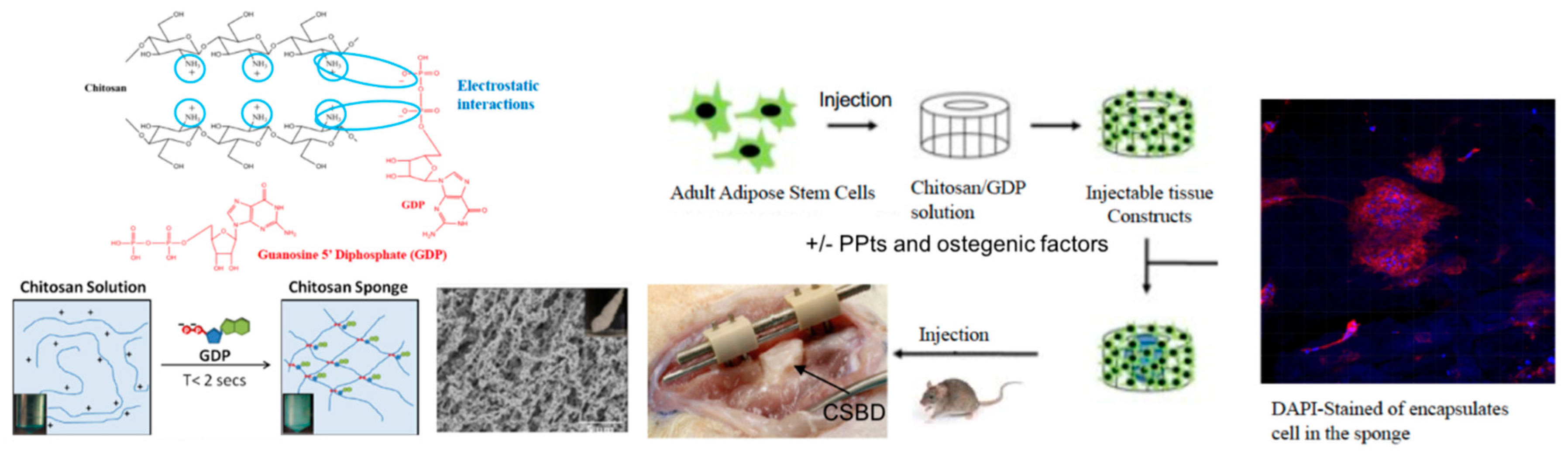
  77. Mekhail, M.; Daoud, J.; Almazan, G.; Tabrizian, M. Rapid, Guanosine 5′-Diphosphate-Induced, Gelation of Chitosan Sponges as Novel Injectable Scaffolds for Soft Tissue Engineering and Drug Delivery Applications. J. Adv. Healthc. Mater. 2013, 2, 1126–1130. [Google Scholar] [CrossRef]
  78. Mekhail, M.; Tabrizian, M. Injectable Chitosan-Based Scaffolds in Regenerative Medicine and their Clinical Translatability. J. Adv. Healthc. Mater. 2014, 3, 1529–1545. [Google Scholar] [CrossRef] [PubMed]
  79. Nayef, L.; Mekhail, M.; Benameur, L.; Rendon, J.S.; Hamdy, R.; Tabrizian, M. A combinatorial approach towards achieving an injectable, self-contained, phosphate-releasing scaffold for promoting biomineralization in critical size bone defects. J. Acta Biomater. 2016, 29, 389–397. [Google Scholar] [CrossRef] [PubMed]
  80. Huynh, V.; Wylie, R.G. Competitive Affinity Release for Long-Term Delivery of Antibodies from Hydrogels. Angewandte Chem. Int. Ed. 2018, 57, 3406–3410. [Google Scholar] [CrossRef] [PubMed]
  81. Zhai, P.; Chen, X.; Schreyer, D.J. An in vitro study of peptide-loaded alginate nanospheres for antagonizing the inhibitory effect of Nogo-A protein on axonal growth. J. Biomed. Mater. 2015, 10, 045016. [Google Scholar] [CrossRef]
  82. Zhai, P.; Chen, X.; Schreyer, D.J. PLGA/alginate composite microspheres for hydrophilic protein delivery. J. Mater. Sci. Eng. C 2015, 56, 251–259. [Google Scholar] [CrossRef]
  83. Tsai, E.C.; Dalton, P.D.; Shoichet, M.S.; Tator, C.H. Matrix inclusion within synthetic hydrogel guidance channels improves specific supraspinal and local axonal regeneration after complete spinal cord transection. J. Biomater. 2006, 27, 519–533. [Google Scholar] [CrossRef] [PubMed]
  84. Chiono, V.; Tonda-Turo, C. Trends in the design of nerve guidance channels in peripheral nerve tissue engineering. J. Prog. Neurobiol. 2015, 131, 87–104. [Google Scholar] [CrossRef] [PubMed]
  85. Tsai, E.C.; Dalton, P.D.; Shoichet, M.S.; Tator, C.H. Synthetic hydrogel guidance channels facilitate regeneration of adult rat brainstem motor axons after complete spinal cord transection. J. Neurotrauma 2004, 21, 789–804. [Google Scholar] [CrossRef] [PubMed]
Figure 1. High resolution transmission electron microscopy (HRTEM) images of small, monodispersed Fe3O4 nanoparticles (NPs) as contrast agents in diphenyl ether observed under different magnifications. Reprinted under a Creative Commons Attribution 4.0 International License from Reference [13] from journal MethodsX.
Processes 07 00336 g001
Figure 2. Trifunctional nanomaterial SPIO@SiO2(FITC)-BDA and its application as an axonal tracer. (a) Schematic representation of the axon tracer design showing a superparamagnetic iron oxide (SPIO) core, a middle layer of Fluorescein isothiocyanate (FITC)-doped silica, and the external layer of biotinylated dextran amine (BDA). Fluorescent confocal micrographs of (b) undifferentiated and (c) differentiated N2a cells that have taken up SPIO@SiO2(FITC)-BDA nanoparticles. Reprinted with permission from Reference [25].
Processes 07 00336 g002
Figure 3. Scanning electron microscopy images of three-dimensional printed fibrin scaffolds crosslinked with different concentrations of genipin: (a) 0 μM, (b) 1 μM, (c) 2.5 μM, (d) 5 μM, and (e) 10 μM. Reprinted under a Creative Commons Attribution 4.0 International License from Reference [61] from the journal Scientific Reports.
Processes 07 00336 g003
Figure 4. Electrostatic interaction between anionic guanosine 5′-diphosphate (GDP) and cationic chitosan leads to the formation of highly porous scaffold. The encapsulation of adult adipose stem cells (ASCs) and pyrophosphatase (PPtase) can break down GDP and release phosphate ions to promote the biomineralization in a critical size bone defect (CSBD). The image shows the chitosan sponge in CBSD to illustrate the localization of the sponge. There is no need for open surgery in clinical use since the sponge can be directly injected into the CSBD.
Processes 07 00336 g004

