Article

In Vitro Antidiabetic Activity Affecting Glucose Uptake in HepG2 Cells Following Their Exposure to Extracts of Lauridia tetragona (L.f.) R.H. Archer

Department of Life and Consumer Sciences, College of Agriculture and Environmental Sciences, University of South Africa, Johannesburg 1709, South Africa
*
Author to whom correspondence should be addressed.
Submission received: 29 November 2019 / Revised: 23 December 2019 / Accepted: 24 December 2019 / Published: 30 December 2019
(This article belongs to the Special Issue Extraction, Characterization and Pharmacology of Natural Products)

Abstract

:
The incidence of diabetes is on the rise and one of the medically active plants used for the treatment of diabetes in South Africa is Lauridia tetragona. The aim of this study is to investigate the antidiabetic property of the polyphenolics (PP) compounds isolated from the methanolic extract of Lauridia tetragona. The α-amylase, α-glucosidase, dipeptidyl peptidase IV (DPPIV), lipase inhibitory activities, and glucose uptake in HepG2 were investigated. The methanolic extract fractions of L. tetragona yielded six fractions (PP1–PP6) all of which showed weak inhibition against DPPIV and lipase compared to the standards. However, PP4 and PP6 showed the best inhibition against α-amylase (IC50 of 359.3 ± 2.11 and 416.82 ± 2.58 μg/mL, respectively) and α-glucosidase (IC50 of 95.93 ± 2.34 and 104.49 ± 2.21 μg/mL, respectively) and only PP4 (173.6%) resulted in enhanced glucose uptake in HepG2 cells compared to berberine (129.89%) and metformin (187.16%) used as positive controls. The previous investigation on PP4 and PP6 showed the presence of polyphenolics such as ferulic acid, coumaric acid, and caffeic acid. The results of this study suggest that L. tetragona could be suitable as an antidiabetic agent and justifies the folkloric use of the plant to treat diabetes.

1. Introduction

Diabetes mellitus (DM) is a chronic and serious metabolic disease that is characterized by the imbalance in glucose homeostasis leading to raised blood glucose. In the two major categories of diabetes, type 2 is the most prevalent as it accounts for around 90% of all cases of diabetes [1]. The combination of sundry lifestyle, genetic susceptibility, viral infections, and toxins has been implicated to play important roles in type 1 diabetes while type 2 diabetes has been strongly linked with overweight or obesity, increasing age, ethnicity, and family history [2]. The current global prevalence of diabetes of 9.3% could rise to 10.2% by 2030 and 10.9% by 2045 [3]. Approximately 50% of diabetes cases in the world are undiagnosed because those living with the disease are unaware of their condition and the majority of these cases are from low-income and middle-income countries [4]. The incidence of undiagnosed diabetes in Africa is even higher at 69.2%, indicating that diabetes is emerging as a serious health concern in countries such as South Africa where high rates of maternal and child mortality, injury-related disorders, non-communicable diseases, and infectious diseases make available resources limited to meet the high cost of managing diabetes [4]. The high cost of available antidiabetic therapies together with the myriad of adverse side effects prompted the exploration of cheaper and more effective antidiabetic compounds from medicinal plants. Studies using extracts from plants have shown potential in improving diabetes following a significant improvement in glucose uptake [5,6,7,8,9]. Most of these plants have been reported to contain polyphenols as one of their active principles and recent reports also suggest the antidiabetic property of phenolics compounds [10,11]. α-glucosidase is the enzyme that catalyzes the key step of carbohydrate digestion and glucose release. Therefore, glucosidase inhibitors are therapeutic agents that can reduce the level of postprandial blood glucose by preventing the hydrolysis of glucose by carbohydrate metabolizing enzymes viz. α glucosidase and α-amylase. The inhibitory activities of phenolic compounds against α-amylase and α-glucosidase have been evaluated to show their potential as good sources of new antidiabetic drugs to improve insulin resistance and increase glucose uptake. Therefore, the inhibition of starch digestive enzymes is an effective approach towards the control of postprandial glycemia. Another effective approach is the use of protease dipeptidyl peptidase (DPP)-IV inhibitors and incretin-based treatments. Incretin is a gut-derived hormone that stimulates a decrease in blood glucose levels by stimulating glucose-induced insulin secretion [12,13]. One of the routinely used plants for the management of diabetes in South Africa is L. tetragona, which belongs to the Celastraceae family [14]. L. tetragona contains high levels of polyphenols with anti-oxidant and hepatoprotective activities that make an important subject for health-related studies [15]. The present study is aimed at investigating some in vitro antidiabetic activities of the polyphenolic-rich fraction of L. tetragona based on ethnomedicinal claims regarding its use in the treatment of diabetes. The in vitro assays involved monitoring glucose uptake in HepG2 cells as well as monitoring the inhibition of carbohydrate and lipid metabolizing enzymes.

2. Materials and Methods

The chemicals used such as the 4-Nitrophenyl β-d-glucopyranoside (PNPG), Dulbecco’s Modified Eagle Medium (DMEM), α-amylase type VI-B from porcine pancreas (A3176-1MU), α-glucosidase type I from Baker’s yeast (G5003-100UN), dipeptidyl peptidase IV (DPPIV), human (D3446-10UG) and FBS (fetal bovine serum) were purchased from Sigma (South Africa). Unless otherwise specified, all reagents were purchased from Sigma (Johannesburg, South Africa).

2.1. Plant Material

The plant material was collected and authenticated by botanists at the South African Biodiversity Institute (SANBI), Pretoria, South Africa, before the leaves were washed and pulverized using an electric blender (Hamilton Beach, Prince Edward, Canada) and then extracted in methanol (Sigma, Johannesburg, South Africa) for 24 h before being filtered (Whatman No.1, Sigma, Johannesburg, South Africa). The filtrate was concentrated under reduced pressure at 40 °C and stored at −4 °C.

2.2. Isolation of Polyphenolics from Lauridia tetragona

The polyphenolic compounds were separated, isolated, and identified as previously described [15] with modifications. The recovered phenolic fractions from the thin-layer chromatography (TLC) were dissolved in distilled water and re-extracted with ethyl acetate (EtOAc) in a separating funnel. The resulting EtOAc layer was collected and concentrated using a rotary evaporator and designated as the EtOAc fraction. The aqueous layer was then extracted with n-butanol as described above and collected as the n-butanol and aqueous fractions, respectively. The EtOAc fraction was first eluted with 100% MeOH through a flash chromatography silica gel column (40 μm) containing grade 62 silica gel (pore size 150 Å, 60–200 mesh) and then separated using a differential gradient of MeOH in chloroform. Six fractions (PP1–PP6) were collected and concentrated using a Genevac EZ-2 series evaporator. All the fractions were investigated for anti-enzyme activity (DPPIV, α-glucosidase, and α-amylase). The fractions that showed the best enzyme inhibition were then introduced to HepG2 cells to determine glucose uptake.

2.3. Determination of Enzyme Inhibitory Activity

2.3.1. α-Amylase

The α-amylase inhibitory activity was determined as previously described [5]. Briefly, duplicate 5 µL aliquots of 10 mg α-amylase in 100 mL phosphate buffer and 15 µL of the extract samples (15.625–250 µg/mL) that were previously diluted in a phosphate buffer were reacted in separate wells of a 96 well plate and incubated at 37 °C for 10 min. Duplicate control wells containing buffer instead of the enzyme were included. Then, 2% starch solution (20 µL) was added to each reaction well before incubating at 37 °C for exactly 30 min before 75 µL of iodine reagent was added to each well. Separate wells containing acarbose (Sigma A8980, South Africa) instead of the extracts were also prepared and used as standard. The absorbance at 580 nm was determined using a Varioskan Flash spectrophotometer (Thermo Scientific, Waltham, MA USA) for each well and the percentage inhibition was calculated using the equation:
% inhibition = ([A − B]/A) × 100,
where A = the absorbance of the control reaction mixture and B = the absorbance of the reaction mixture containing enzyme.

2.3.2. α-Glucosidase

The inhibition of the α-glucosidase enzyme was carried out as previously described [16]. Briefly, duplicate aliquots of extract fractions (5 µL), α-glucosidase enzyme (20 µL, 50 µg/mL), and potassium phosphate buffer (60 µL, 67 mM, pH 6.8) were reacted inside wells of a 96 well plate and incubated at 37 °C for 5 min. The controls containing buffer instead of enzyme and lacking substrate were included. Thereafter, a 10 µL aliquot of 10 mM PNPG was added to each well before incubation at 37 °C for 20 min. This was followed with the addition (100 µL) of 100 mM Na2CO3 solution (Sigma, Johannesburg, South Africa) to each well before the absorbance was read at 405 nm using a Varioskan Flash spectrophotometer (Thermo Scientific, Waltham, MA USA). Separate wells containing epigallocatechin gallate (EGCG) (Sigma, Johannesburg, South Africa) instead of the extracts were also prepared and used as standard. The percent of inhibition was calculated using the equation:
% inhibition = ([A − C]/A) × 100,
where A = the absorbance of the control solution and C = the absorbance of the experimental wells.

2.3.3. Dipeptidyl Peptidase-IV

The inhibitory effect against DPPIV activity was based on a previously described method [17]. Briefly, about 30 μg/mL of each fraction was incubated with 0.1 mg/mL of DPPIV enzyme (D3446, Sigma Aldrich, South Africa) at 37 °C for 30 min in an incubator containing 5% CO2. Thereafter, 50 μL of 10 μM Gly-Pro p-nitroanilide hydrochloride (Gly-Pro-ρNA, Sigma G0513) substrate was added followed by incubation at 37 °C for 30 min. Diprotin A (Sigma Aldrich, South Africa, D3822) (50 μg/mL) was used as a standard inhibitor instead of the investigated samples. The hydrolysis of substrate Gly-Pro- ρNA by DPPIV enzyme to release p-nitroanilide (ρNA) was measured by a change in absorbance at 405 nm using a Varioskan Flash spectrophotometer (Thermo Scientific, Waltham, MA, USA). The inhibitory effects were expressed as the percentage inhibition of DPPIV activity.

2.3.4. Pancreatic Lipase

The in vitro inhibition of the lipase from the porcine pancreas (L3126-25G, Sigma, South Africa) enzyme was performed using a 96 well microplate as previously described [18]. Duplicate 10 μL aliquots of each of the extract fractions diluted from 50–200 μg/mL together with tetrahydrolipstatin (100 μM) were incubated at 37 °C for 15 min with 40 μL of 50 mg/mL pancreatic lipase enzyme. Thereafter, 170 μL of the 10 mM p-nitrophenyl butyrate (PNPB) substrate was added to each well before incubating at 37 °C for 15 min before the absorbance of each well was read at 405 nm using a Varioskan Flash spectrophotometer. Control with the standard (Orlistat) was prepared as well as the control without the enzyme inhibitor (phosphate buffer only) and without the substrate (PNPB) were also prepared.
Inhibition activity (%) = [1 − (abs of test sample/abs of enzyme control)] × 100.

2.4. Cell Culture

The human hepatoma cell line (HepG2) was a donation from Professor Monde Ntwasa (University of South Africa). All experiments were approved by the College of Agriculture and Environmental Sciences (CAES) Health Research Ethics Committee of the University of South Africa (REC Reference No: 2018/CAES/093; date of approval: 6 September 2018). The HepG2 cells were cultured in DMEM supplemented with 10% FBS and 1% penicillin–streptomycin in a humidified incubator at 37 °C with 5% CO2. The cells at 80–90% confluence were split and then seeded 5 × 10⁶ cells/mL in 96 well plates.

2.5. Cell Viability Assay

To evaluate the changes in cell viability in the presence of different plant extract fractions, the different concentrations (10–100 μg/mL) of the investigated samples were added and incubated for a further 24 h. The cell viability was carried out using CellTiter 96® non-radioactive cell proliferation assay kit (G4000 and G4100, Promega, South Africa) according to the manufacturer’s instructions. The absorbance was measured using a Varioskan Flash spectrophotometer (Thermo Scientific, Waltham, MA, USA) set at 570 nm.

2.6. Glucose Uptake Assay

HepG2 cells that were seeded in culture plates at 1 × 10⁵ cells/mL were allowed to adhere and grow at 37 °C for 24 h in a humidified incubator supplemented with 5% CO2. Before assaying, cells were preincubated at 37 °C for 48 h with different concentrations (10 μL, 25–100 μg/mL) of the PP4 and PP6. Thereafter, the spent culture medium was aspirated and replaced with 25 μL of the incubation medium (DMEM that had been diluted with 8 mM glucose, 0.1% Bovine serum albumin (BSA), and Phosphate-Buffered Saline (PBS) before it was incubated at 37 °C for 3 h. Thereafter, 10 μl of the incubation medium was removed from all the wells and transferred into a new 96 well plate. The concentration of glucose in the medium was determined using the glucose assay kit (Sigma GAGO20, Johannesburg, South Africa) according to the manufacturer’s instructions. The absorbance was then measured at 540 nm using a Varioskan Flash spectrophotometer. The glucose used by the cells was calculated by subtracting the cell-containing wells from the cell-free wells. The cell viability was also determined using the cell viability assay as previously described in Section 2.5. The positive controls used were 0.1 μg/mL metformin and 10 μg/mL berberine while the untreated cells served as the negative control containing only the incubation buffer without the test sample.

2.7. Statistical Analyses

Where applicable, all the data were analyzed statistically by one-way ANOVA analysis of variance and Dunnett’s post-test and were expressed as mean ± SD. The level of statistical significance was set at p < 0.05. Graphs were generated using GraphPad Prism software (Version 4, GraphPad Software, San Diego, CA, Canada, 2005).

3. Results

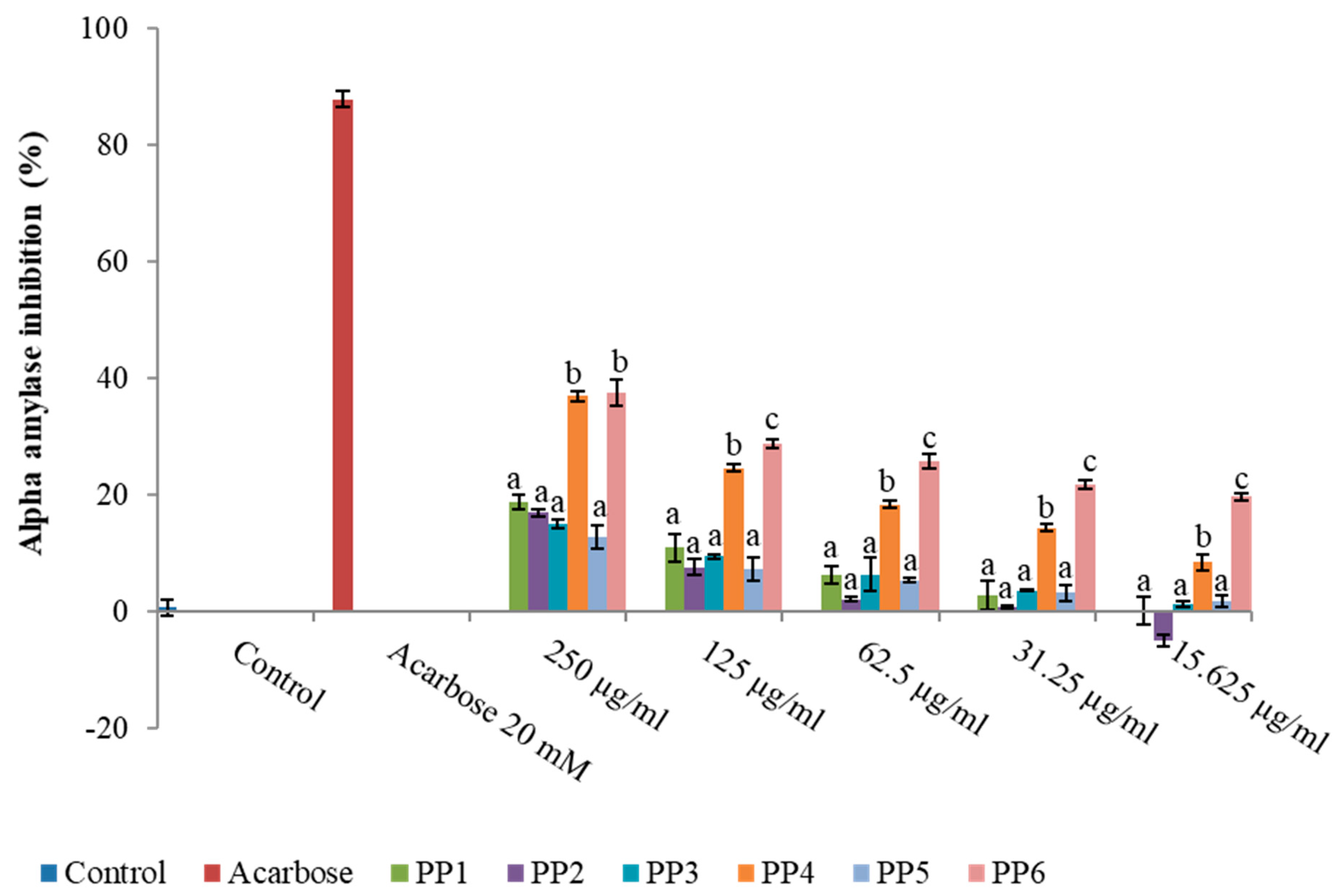
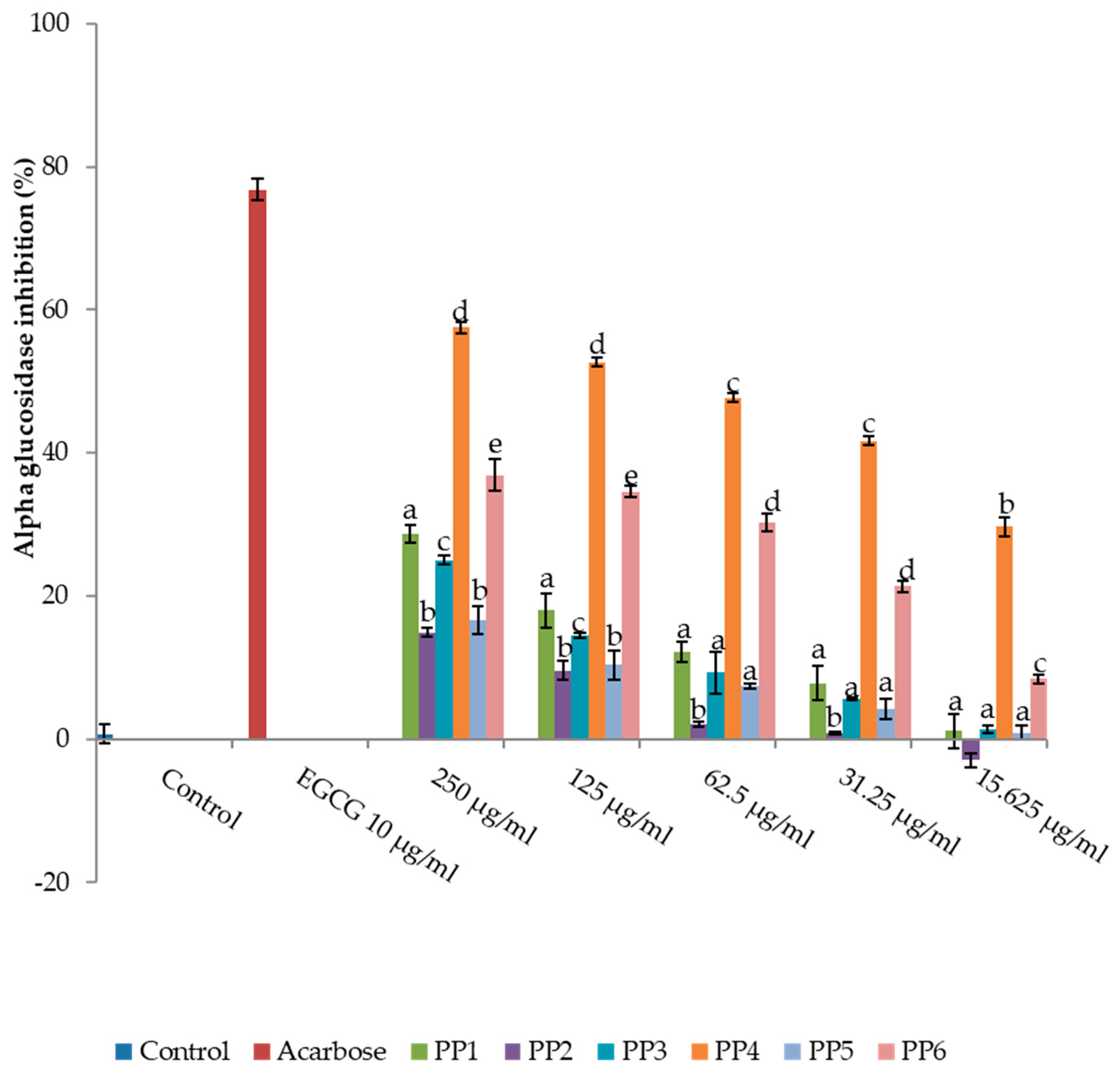
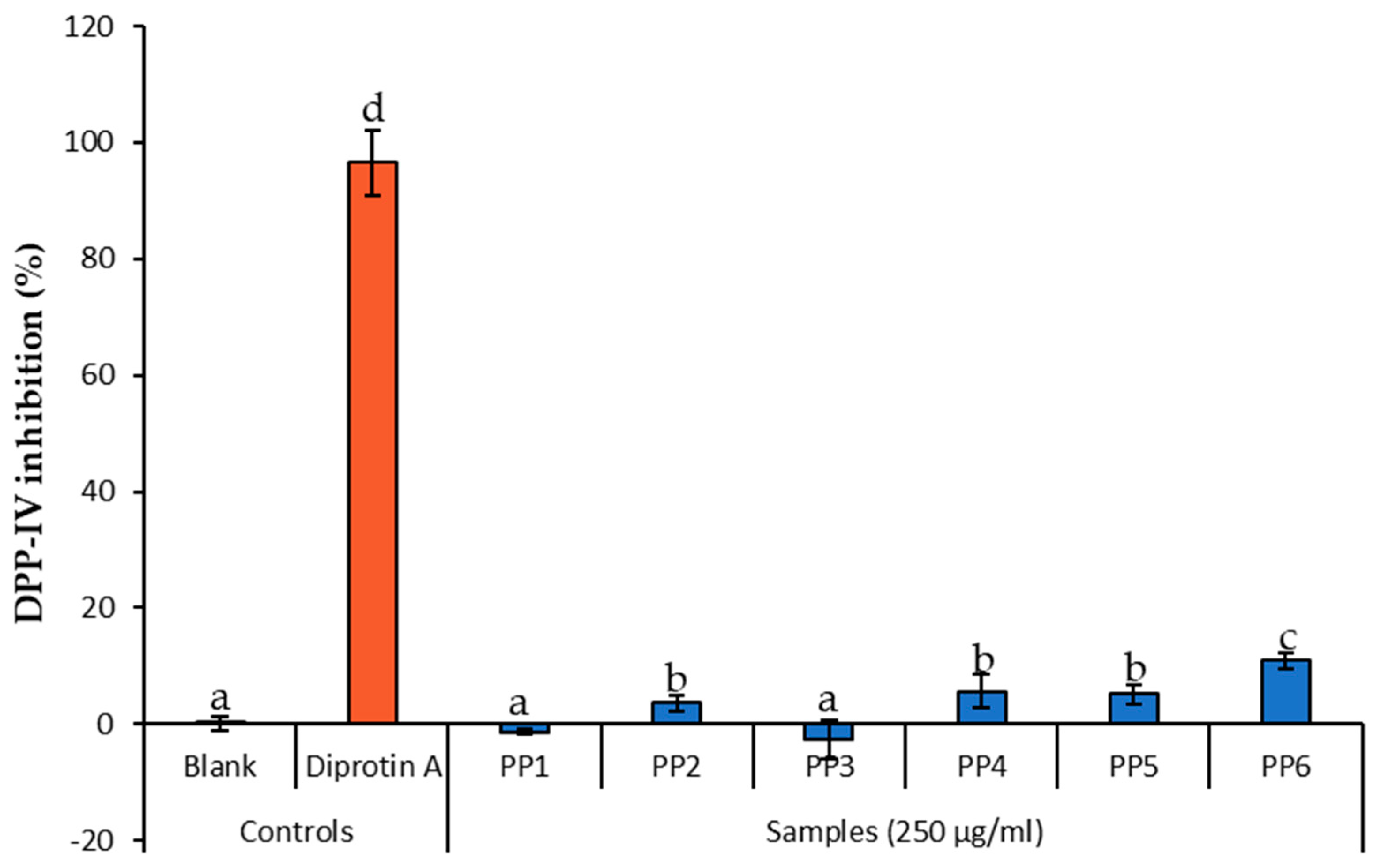
3.1. Inhibitory Activity of L. tetragona on Amylase, Glucosidase, and DPPIV

The methanolic extract of L. tetragona leaves was successively fractionated before each fraction was investigated for α-amylase, α-glucosidase, and DPPIV inhibitory activities. The inhibitory effects of the different fractions from the methanolic extract of L. tetragona on enzymes linked with type 2 diabetes are shown in Figure 1, Figure 2 and Figure 3. All the tested fractions showed a concentration dependent inhibition on both the amylase and glucosidase enzymes. The highest percentage inhibition observed on amylase was at 250 μg/mL by both PP4 and PP6 and was significantly different (p < 0.05) from acarbose. Conversely, all the PPs showed mild inhibition against DPPIV that were significantly lower than diprotin A. However, PP4 showed the highest inhibition percentage against α-glucosidase, but it was still lower than epigallocatechin gallate which was used as the control. Overall, PP4 had a better inhibitory capacity in all the fractions. This is shown by the IC50 values 359.3 ± 2.11 and 95.93 ± 2.34 μg/mL for α-amylase and α-glucosidase, respectively, followed by PP6 with 416.82 ± 2.58 and 104.49 ± 2.21 μg/mL for α-amylase and α-glucosidase, respectively (Table 1).

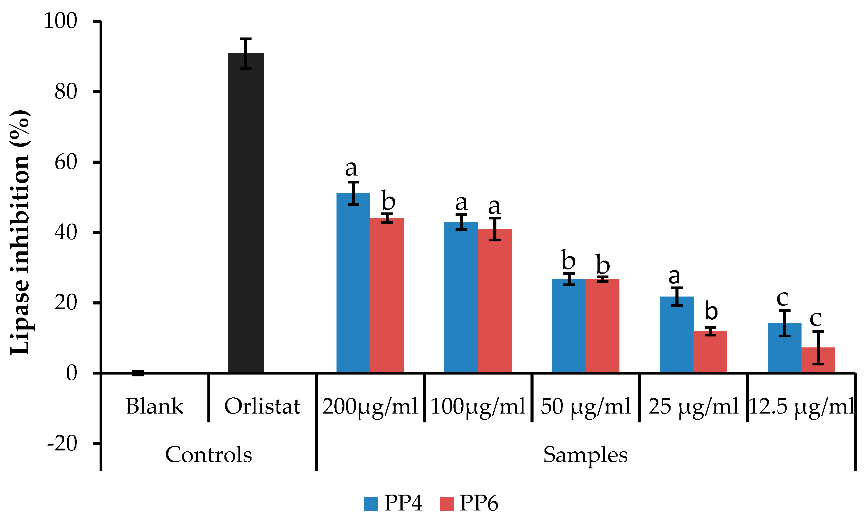
3.2. Inhibitory Activity of L. tetragona on Pancreatic Lipase

The previous results suggest the antidiabetic potential of PP4 and PP6, hence these samples were tested for their inhibitory activity on pancreatic lipase (Figure 4). Both samples showed lower pancreatic lipase inhibition compared to the standard (Orlistat). PP4 showed significantly higher lipase inhibition at 200 μg/mL but not significantly different (p < 0.05) when compared with PP6 at lower concentrations. However, PP6 showed higher activity with lower IC50 compared to PP4 (Table 1).

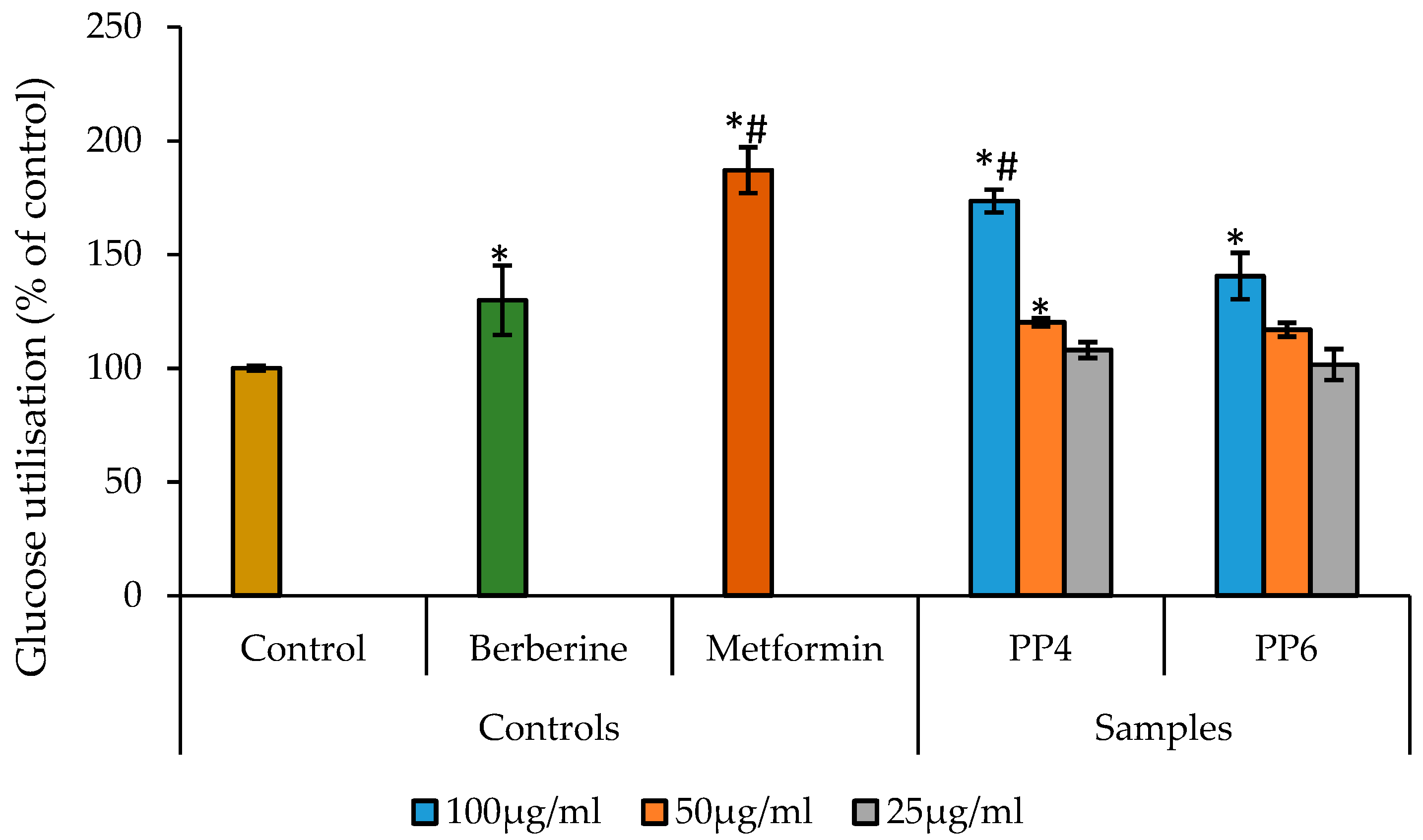
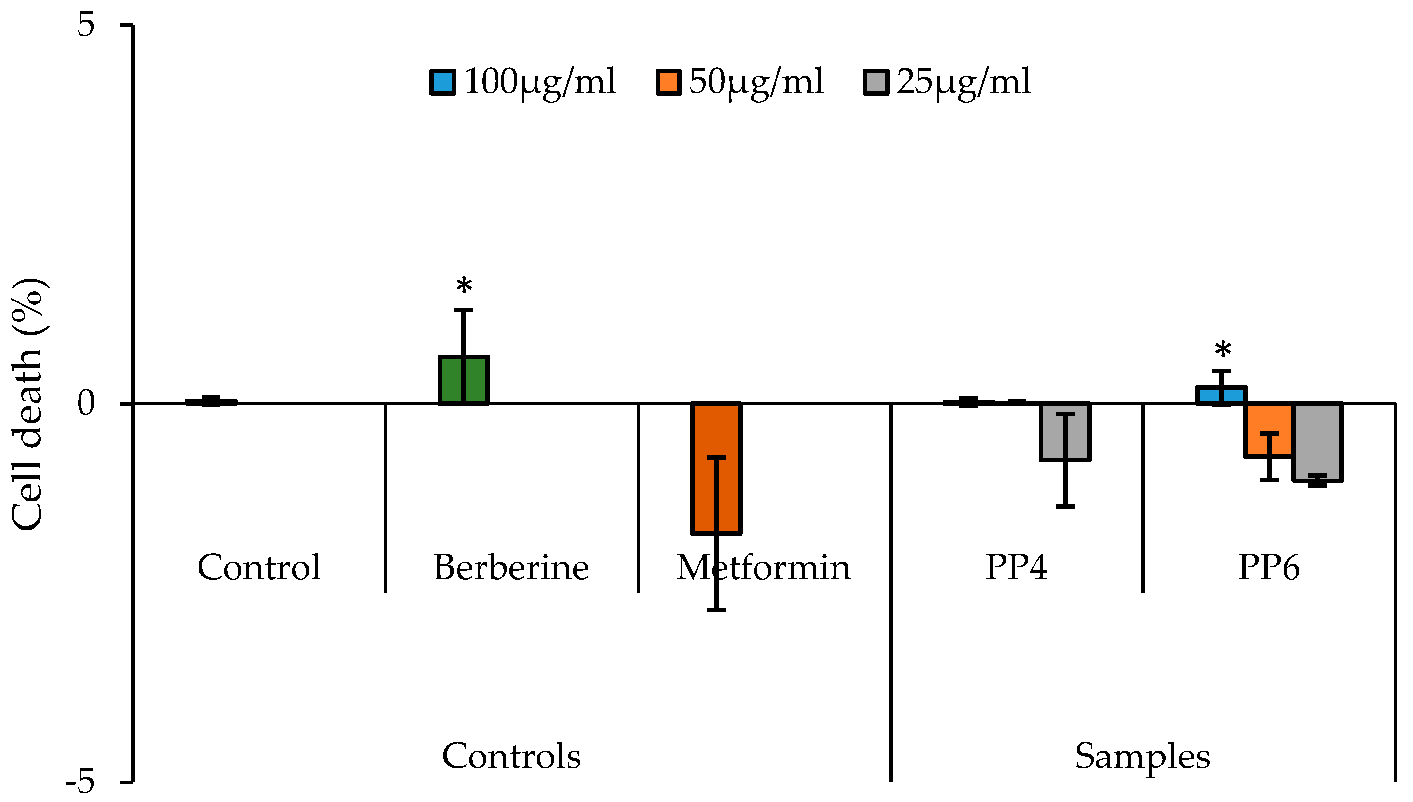
3.3. The Effect of PP4 and PP6 on Glucose Uptake Using HepG2 Cells

The effects of the different concentrations of the samples (10–100 μg/mL) on the cell viability were evaluated. The cell viability was initially evaluated to check for the cytotoxicity of PP4 and PP6. This was then used to confirm the actual dosage required for the glucose utilization assay in the HepG2 cells. The CellTiter 96® based assays showed no cytotoxic effects. The glucose uptake in HepG2 cells treated with both PP4 and PP6 at different concentrations (25, 50, and 100 μg/mL) is shown in Figure 5. Both PP4 and PP6 caused significant (p < 0.05) increase in glucose utilization in HepG2 cells at 100 μg/mL, and only PP4 showed a significant increase at 50 μg/mL. However, both PP4 and PP6 showed a concentration-dependent increase in glucose uptake and a significant increase compared to the untreated control. PP4 (173.6%) exhibited higher glucose utilization at 100 μg/mL compared to berberine (129.89%) and was not significantly different from metformin (187.16%), which was used as the positive control.
The toxicity assay revealed that both PP4 and PP6 were not toxic to HepG2 cells at all the concentrations investigated, except PP6 at 100 μg/mL but was not significantly different from the berberine (Figure 6). The percentage of cell death observed in berberine and PP6 (100 μg/mL) was lower than 5%. The low percentage of cell death observed in the PP4, PP6, metformin, and berberine treatments are an indication of the significant reduction in glucose utilization in the cells.

4. Discussion

Diabetes has emerged as one of the most serious metabolic disorders in the world because of the seriousness of related complications and its rapid onset worldwide [19]. The crucial role of natural products in drug discovery is evident as the use of medicinal plants in the prevention and treatment of diabetes continues to grow because of their beneficial effects. Natural products are also being used as alternative sources for safer and cheaper diabetes treatment with fewer side effects [20]. In this study, the antidiabetic activities of polyphenols isolated from L. tetragona and their inhibitory potential against enzymes associated with diabetes and their glucose uptake in HepG2 cells were evaluated. Polyphenols are molecules naturally found in plants that are structurally characterized by the presence of one or more phenol groups [21]. Generally, polyphenols are known to possess antioxidant activities thereby protecting cells against oxidative stress in both plants and humans [22]. In addition to this, polyphenols also possess antidiabetic activities, particularly as good α-glucosidase and α-amylase inhibitors. The inhibitory activities of the compounds isolated against α-amylase and α-glucosidase were investigated in comparison with acarbose. The inhibitory activities observed in this study are consistent with recent reports on the inhibitory activities of polyphenolic compounds against α-amylase and α-glucosidase [5,17,21,23,24]. Although all the fractions showed mild inhibition against these enzymes, only PP4 and PP6 showed better activities which was evident by their IC50 values. The preliminary investigations of active compounds in PP4 and PP6 revealed the presence of polyphenols such as ferulic acid, coumarin, 3-Isopropylcatechol, and caffeine [15]. Previous studies have reported the α-amylase and α-glucosidase inhibitory activities of ferulic, coumarin, and caffeic acid [25,26,27,28,29,30]. There is paucity of data on the inhibitory properties of ferulic acids, coumarin, 3-Isopropylcatechol, and caffeic acid on the DPPIV. However, reports suggest that polyphenols possess inhibitory properties against different molecular targets that are involved in the metabolism of glucose, such as the stimulation of the L-cells and β-cells to secrete GLP1 and insulin, respectively. The inhibition of DPPIV to increase the half-life of GLP1 and stimulation of the peripheral response to insulin—thereby increasing the overall effects of the GLP1-insulin signaling—has also been reported [31]. Different reports have also described the inhibitory potentials of polyphenols as potent inhibitors of specific targets related to type 2 diabetes physiopathology, including protein tyrosine phosphatase 1B (PTP1B), DPPIV, and sodium-glucose cotransporter 2, among many other targets [32,33,34]. The oral administration of ferulic acid (50 mg/kg) has been reported to significantly decrease blood glucose levels and increase plasma insulin levels in type 2 diabetic mice by elevating hepatic glycogen synthesis and glucokinase activity [31]. Therefore, it could be suggested that the ferulic acid content in PP4 and PP6 were responsible for the activities observed. Pancreatic lipase is involved predominantly with the breakdown of dietary lipids and their assimilation in the intestine; hence, pancreatic lipase inhibitors are used to prevent the absorption of fats in the diet, making them ideal for the management of obesity or overweight [35,36,37]. Previous reports have suggested that polyphenols such as caffeic acid, p-coumaric acid, quercetin, and capsaicin from hot peppers are good pancreatic lipase inhibitors [38]. The inhibitory activities of PP4 and PP6 indicate the importance of L. tetragona in the control of lipid deposition in the body and ultimately in the management of diabetes. The glucose uptake result using HepG2 showed that both PP4 and PP6 possess glucose uptake comparable to berberine. However, only PP4 showed significant glucose uptake that was comparable to metformin. This indicates that PP4 could be similar to metformin by increasing the glucose utilized in the liver. Metformin is a biguanide that activates the AMP-activated protein kinase (AMPK) in the liver, thereby initiating a cascade of events that lead to the inhibition of lipids and glucose synthesis which improves the sensitivity of the liver to insulin [39,40,41]. There have been reports on phenols—not only on the suppression of liver glucose release but also on the enhancement of liver glucose uptake—which intensifies the importance of phenols in the regulation of intracellular signaling pathways [42]. Therefore, in this study, it could be speculated that the glucose utilization observed in the HepG2 cells for both PP4 and PP6 may be due to the polyphenols present in these samples as previously reported [15]. It is worth noting that toxicity has been one of the major concerns of herbal treatments. This study clearly shows that samples were not cytotoxic to the hepatocytes at the tested concentrations and that the glucose uptake observed was not due to the cytotoxic effects of the tested samples.

5. Conclusions

In summary, the findings of this study suggest that polyphenols isolated from L. tetragona extract showed good hypoglycemic activity that may be linked to the inhibition of crucial enzymes associated with diabetes. Further investigations are required to characterize these compounds and determine their potential antidiabetic pharmacological activity.

Author Contributions

Conceptualization, S.O.; investigation, S.O.; resources, J.D.; data curation, S.O.; writing—original draft preparation, S.O.; writing—review and editing, J.D.; supervision, J.D.; funding acquisition, J.D. All authors have read and agreed to the published version of the manuscript.

Funding

This research received no external funding. The APC was funded by the University of South Africa, South Africa.

Acknowledgments

University of South Africa, South Africa.

Conflicts of Interest

The authors declare no conflict of interest.

References

  1. Holman, N.; Young, B.; Gadsby, R. Current prevalence of Type 1 and Type 2 diabetes in adults and children in the UK. Diabet. Med. 2015, 32, 1119–1120. [Google Scholar] [CrossRef] [PubMed]
  2. International Diabetes Federation. IDF Diabetes Atlas, 9th ed.; International Diabetes Federation: Brussel, Belgium, 2019; Available online: https://diabetesatlas.org/upload/resources/2019/IDF_Atlas_9th_Edition_2019.pdf (accessed on 28 November 2019).
  3. Saeedi, P.; Petersohn, I.; Salpea, P.; Malanda, B.; Karuranga, S.; Unwin, N.; Colagiuri, S.; Guariguata, L.; Motala, A.A.; Ogurtsova, K.; et al. Global and regional diabetes prevalence estimates for 2019 and projections for 2030 and 2045: Results from the International Diabetes Federation Diabetes Atlas, 9th edition. Diabetes Res. Clin. Pract. 2019, 107843. [Google Scholar] [CrossRef] [PubMed] [Green Version]
  4. Pheiffer, C.; Pillay-van Wyk, V.; Joubert, J.D.; Levitt, N.; Nglazi, M.D.; Bradshaw, D. The prevalence of type 2 diabetes in South Africa: A systematic review protocol. BMJ Open 2018, 8, e021029. [Google Scholar] [CrossRef] [PubMed] [Green Version]
  5. Odeyemi, S.W.; Afolayan, A.J. Identification of Antidiabetic Compounds from Polyphenolic-rich Fractions of Bulbine abyssinica A. Rich Leaves. Pharmacognosy Res. 2018, 10, 72–80. [Google Scholar] [CrossRef] [PubMed]
  6. Semaan, D.G.; Igoli, J.O.; Young, L.; Marrero, E.; Gray, A.I.; Rowan, E.G. In vitro anti-diabetic activity of flavonoids and pheophytins from Allophylus cominia Sw. on PTP1B, DPPIV, alpha-glucosidase and alpha-amylase enzymes. J. Ethnopharmacol. 2017, 203, 39–46. [Google Scholar] [CrossRef] [Green Version]
  7. Choudhury, H.; Pandey, M.; Hua, C.K.; Mun, C.S.; Jing, J.K.; Kong, L.; Ern, L.Y.; Ashraf, N.A.; Kit, S.W.; Yee, T.S.; et al. An update on natural compounds in the remedy of diabetes mellitus: A systematic review. J. Tradit. Complement. Med. 2018, 8, 361–376. [Google Scholar] [CrossRef]
  8. Adinortey, M.B.; Agbeko, R.; Boison, D.; Ekloh, W.; Kuatsienu, L.E.; Biney, E.E.; Affum, O.O.; Kwarteng, J.; Nyarko, A.K. Phytomedicines Used for Diabetes Mellitus in Ghana: A Systematic Search and Review of Preclinical and Clinical Evidence. Evid. Based Complement. Altern. Med. 2019, 2019, 1–23. [Google Scholar] [CrossRef] [Green Version]
  9. Naimi, M.; Vlavcheski, F.; Shamshoum, H.; Tsiani, E. Rosemary Extract as a Potential Anti-Hyperglycemic Agent: Current Evidence and Future Perspectives. Nutrients 2017, 9, 968. [Google Scholar] [CrossRef] [Green Version]
  10. Yamashita, Y.; Wang, L.; Nanba, F.; Ito, C.; Toda, T.; Ashida, H. Procyanidin Promotes Translocation of Glucose Transporter 4 in Muscle of Mice through Activation of Insulin and AMPK Signaling Pathways. PLoS ONE 2016, 11, e0161704. [Google Scholar] [CrossRef]
  11. Silva, B.; Oliveira, P.; Casal, S.; Alves, M.; Dias, T. Promising Potential of Dietary (Poly)Phenolic Compounds in the Prevention and Treatment of Diabetes Mellitus. Curr. Med. Chem. 2017, 24, 334–354. [Google Scholar] [CrossRef]
  12. Ali Asgar, M. Anti-Diabetic Potential of Phenolic Compounds: A Review. Int. J. Food Prop. 2013, 16, 91–103. [Google Scholar] [CrossRef]
  13. Kim, B.R.; Kim, H.Y.; Choi, I.; Kim, J.B.; Jin, C.H.; Han, A.R. DPP-IV Inhibitory Potentials of Flavonol Glycosides Isolated from the Seeds of Lens culinaris: In Vitro and Molecular Docking Analyses. Molecules 2018, 23, 1998. [Google Scholar] [CrossRef] [PubMed] [Green Version]
  14. Oyedemi, S.; Bradley, G.; Afolayan, A.J. Ethnobotanical survey of medicinal plants used for the management of diabetes mellitus in the Nkonkobe municipality of South Africa. J. Med. Plants Res. 2009, 3, 1040–1044. [Google Scholar]
  15. Odeyemi, S.; Dewar, J. Repression of Acetaminophen-Induced Hepatotoxicity in HepG2 Cells by Polyphenolic Compounds from Lauridia tetragona (L.f.) R.H. Archer. Molecules 2019, 24, 2118. [Google Scholar] [CrossRef] [PubMed] [Green Version]
  16. Sun, J.; Zhang, F.; Yang, M.; Zhang, J.; Chen, L.; Zhan, R.; Li, L.; Chen, Y. Isolation of α-glucosidase inhibitors including a new flavonol glycoside from Dendrobium Devonianum. Nat. Prod. Res. 2014, 28, 1900–1905. [Google Scholar] [CrossRef]
  17. Proença, C.; Freitas, M.; Ribeiro, D.; Tomé, S.M.; Araújo, A.N.; Silva, A.M.S.; Fernandes, P.A.; Fernandes, E. The dipeptidyl peptidase-4 inhibitory effect of flavonoids is hindered in protein rich environments. Food Funct. 2019, 10, 5718–5731. [Google Scholar] [CrossRef]
  18. Kim, Y.S.; Lee, Y.M.; Kim, H.; Kim, J.; Jang, D.S.; Kim, J.H.; Kim, J.S. Anti-obesity effect of Morus bombycis root extract: Anti-lipase activity and lipolytic effect. J. Ethnopharmacol. 2010, 130, 621–624. [Google Scholar] [CrossRef]
  19. Kasturi, S.; Surarapu, S.; Uppalanchi, S.; Anireddy, J.S.; Dwivedi, S.; Anantaraju, H.S.; Perumal, Y.; Sigalapalli, D.K.; Babu, B.N.; Ethiraj, K.S. Synthesis and α-glucosidase inhibition activity of dihydroxy pyrrolidines. Bioorg. Med. Chem. Lett. 2017, 27, 2818–2823. [Google Scholar] [CrossRef]
  20. Somtimuang, C.; Olatunji, O.J.; Ovatlarnporn, C. Evaluation of In Vitro α -Amylase and α -Glucosidase Inhibitory Potentials of 14 Medicinal Plants Constituted in Thai Folk Antidiabetic Formularies. Chem. Biodivers. 2018, 15, e1800025. [Google Scholar] [CrossRef]
  21. Ivey, K.L.; Croft, K.; Prince, R.L.; Hodgson, J.M. Comparison of flavonoid intake assessment methods. Food Funct. 2016, 7, 3748–3759. [Google Scholar] [CrossRef]
  22. Odeyemi, S.; Afolayan, A.; Bradley, G. In vitro anti-inflammatory and free radical scavenging activities of crude saponins extracted from Albuca bracteata Jacq. Bulb. African J. Tradit. Complement. Altern. Med. 2015, 12, 34–40. [Google Scholar] [CrossRef] [Green Version]
  23. Rasouli, H.; Hosseini-Ghazvini, S.M.-B.; Adibi, H.; Khodarahmi, R. Differential α-amylase/α-glucosidase inhibitory activities of plant-derived phenolic compounds: A virtual screening perspective for the treatment of obesity and diabetes. Food Funct. 2017, 8, 1942–1954. [Google Scholar] [CrossRef] [PubMed]
  24. Karakaya, S.; Gözcü, S.; Güvenalp, Z.; Özbek, H.; Yuca, H.; Dursunoğlu, B.; Kazaz, C.; Kılıç, C.S. The α-Amylase and α-glucosidase inhibitory activities of the dichloromethane extracts and constituents of Ferulago bracteata roots. Pharm. Biol. 2018, 56, 18–24. [Google Scholar] [CrossRef] [PubMed] [Green Version]
  25. Zhao, D.G.; Zhou, A.Y.; Du, Z.; Zhang, Y.; Zhang, K.; Ma, Y.Y. Coumarins with α-glucosidase and α-amylase inhibitory activities from the flower of Edgeworthia gardneri. Fitoterapia 2015, 107, 122–127. [Google Scholar] [CrossRef]
  26. Jeong, E.Y.; Cho, K.S.; Lee, H.S. α-amylase and α-glucosidase inhibitors isolated from Triticum aestivum L. sprouts. J. Korean Soc. Appl. Biol. Chem. 2012, 55, 47–51. [Google Scholar] [CrossRef]
  27. Alongi, M.; Anese, M. Effect of coffee roasting on in vitro α-glucosidase activity: Inhibition and mechanism of action. Food Res. Int. 2018, 111, 480–487. [Google Scholar] [CrossRef]
  28. Nyambe-Silavwe, H.; Williamson, G. Chlorogenic and phenolic acids are only very weak inhibitors of human salivary α-amylase and rat intestinal maltase activities. Food Res. Int. 2018, 113, 452–455. [Google Scholar] [CrossRef]
  29. Yang, C.Y.; Yen, Y.Y.; Hung, K.C.; Hsu, S.W.; Lan, S.J.; Lin, H.C. Inhibitory effects of pu-erh tea on alpha glucosidase and alpha amylase: A systemic review. Nutr. Diabetes 2019, 9, 23. [Google Scholar] [CrossRef] [Green Version]
  30. Liu, S.; Yu, Z.; Zhu, H.; Zhang, W.; Chen, Y. In vitro α-glucosidase inhibitory activity of isolated fractions from water extract of Qingzhuan dark tea. BMC Complement. Altern. Med. 2016, 16, 378. [Google Scholar] [CrossRef] [Green Version]
  31. Domínguez Avila, J.; Rodrigo García, J.; González Aguilar, G.; de la Rosa, L. The Antidiabetic Mechanisms of Polyphenols Related to Increased Glucagon-Like Peptide-1 (GLP1) and Insulin Signaling. Molecules 2017, 22, 903. [Google Scholar] [CrossRef] [Green Version]
  32. Potipiranun, T.; Adisakwattana, S.; Worawalai, W.; Ramadhan, R.; Phuwapraisirisan, P.; Potipiranun, T.; Adisakwattana, S.; Worawalai, W.; Ramadhan, R.; Phuwapraisirisan, P. Identification of Pinocembrin as an Anti-Glycation Agent and α-Glucosidase Inhibitor from Fingerroot (Boesenbergia rotunda): The Tentative Structure–Activity Relationship towards MG-Trapping Activity. Molecules 2018, 23, 3365. [Google Scholar] [CrossRef] [Green Version]
  33. He, J.H.; Chen, L.X.; Li, H. Progress in the discovery of naturally occurring anti-diabetic drugs and in the identification of their molecular targets. Fitoterapia 2019, 134, 270–289. [Google Scholar] [CrossRef]
  34. Adisakwattana, S.; Chantarasinlapin, P.; Thammarat, H.; Yibchok-Anun, S. A series of cinnamic acid derivatives and their inhibitory activity on intestinal α-glucosidase. J. Enzyme Inhib. Med. Chem. 2009, 24, 1194–1200. [Google Scholar] [CrossRef] [PubMed]
  35. Anil, K.H.; Prashith, K.T.; Vinayaka, K.; Swathi, D.; Venugopal, T. Anti-obesity (Pancreatic lipase inhibitory) activity of Everniastrum cirrhatum (Fr.) Hale (Parmeliaceae). Pharmacogn. J. 2011, 3, 65–68. [Google Scholar] [CrossRef] [Green Version]
  36. Lunagariya, N.A.; Patel, N.K.; Jagtap, S.C.; Bhutani, K.K. Inhibitors of pancreatic lipase: State of the art and clinical perspectives. EXCLI J. 2014, 13, 897–921. [Google Scholar] [PubMed]
  37. Birari, R.B.; Bhutani, K.K. Pancreatic lipase inhibitors from natural sources: Unexplored potential. Drug Discov. Today 2007, 12, 879–889. [Google Scholar] [CrossRef] [PubMed]
  38. Martinez-Gonzalez, A.I.; Alvarez-Parrilla, E.; Díaz-Sánchez, Á.G.; de la Rosa, L.A.; Núñez-Gastélum, J.A.; Vazquez-Flores, A.A.; Gonzalez-Aguilar, G.A. In vitro Inhibition of Pancreatic Lipase by Polyphenols: A Kinetic, Fluorescence Spectroscopy and Molecular Docking Study. Food Technol. Biotechnol. 2017, 55, 519–530. [Google Scholar] [CrossRef] [PubMed]
  39. Rena, G.; Hardie, D.G.; Pearson, E.R. The mechanisms of action of metformin. Diabetologia 2017, 60, 1577–1585. [Google Scholar] [CrossRef] [PubMed] [Green Version]
  40. Foretz, M.; Viollet, B. Regulation of hepatic metabolism by AMPK. J. Hepatol. 2011, 54, 827–829. [Google Scholar] [CrossRef] [PubMed]
  41. Viollet, B.; Guigas, B.; Sanz Garcia, N.; Leclerc, J.; Foretz, M.; Andreelli, F. Cellular and molecular mechanisms of metformin: An overview. Clin. Sci. (Lond.) 2012, 122, 253–270. [Google Scholar] [CrossRef] [PubMed] [Green Version]
  42. Hanhineva, K.; Törrönen, R.; Bondia-Pons, I.; Pekkinen, J.; Kolehmainen, M.; Mykkänen, H.; Poutanen, K. Impact of dietary polyphenols on carbohydrate metabolism. Int. J. Mol. Sci. 2010, 11, 1365–1402. [Google Scholar] [CrossRef] [PubMed]
Figure 1. The inhibition of α-amylase by different fractions from the methanolic extract of Lauridia tetragona. The values are expressed as mean ± SD (n = 5). Bars with different letters at each concentration are significantly different (p < 0.05).
Processes 08 00033 g001
Figure 2. The inhibition of α-glucosidase by different fractions from the methanolic extract of L. tetragona. The values are expressed as mean ± SD (n = 5). Bars with different letters at each concentration are significantly different (p < 0.05). EGCG = epigallocatechin gallate.
Processes 08 00033 g002
Figure 3. The inhibition of dipeptidyl peptidase-IV enzyme by different fractions from the methanolic extract of L. tetragona. The values are expressed as mean ± SD (n = 5). Bars with different letters are significantly different (p < 0.05).
Processes 08 00033 g003
Figure 4. The inhibition of pancreatic lipase by the active fractions from the methanolic extract of L. tetragona. The values are expressed as mean ± SD (n = 5). Bars with different letters at each concentration are significantly different (p < 0.05).
Processes 08 00033 g004
Figure 5. Effect of PP4 and PP6 isolated from L. tetragona extract on glucose uptake in HepG2 hepatocytes. The cells were treated with or without different concentrations of the samples for 48 h. Data expressed as mean ± SD (n = 5). A significant increase compared to the control is indicated by “*” (p < 0.05) whereas “#” indicates a significant increase compared to berberine (10 μg/mL) (p < 0.05). No significant increase was observed in the samples compared to metformin (0.1 μg/mL).
Processes 08 00033 g005
Figure 6. Toxicity of PP4 and PP6 isolated from L. tetragona extract to HepG2 cells used for glucose utilization assay. The data represent the mean ± SD (n = 5). A significant increase compared to the control (p < 0.05) is indicated by “*”.
Processes 08 00033 g006
Table 1. The Inhibitory effects of the different fractions from the methanolic extract of L. tetragona against α-amylase, α-glucosidase, and lipase activities (mean ± SD).
Sample IC50 Values (μg/mL)
α-Amylase α-Glucosidase Lipase
PP1650.34 ± 5.21437.68 ± 4.24-
PP2633.06 ± 3.22709.88 ± 2.12-
PP3873.22 ± 0.68507.23 ± 4.18-
PP4359.3 ± 2.1195.93 ± 2.34122.46 ± 1.03
PP51090.36 ± 2.41777.23 ± 4.44-
PP6416.82 ± 2.58104.49 ± 2.21119.47 ± 1.42
Acarbose21.07 ± 1.5--
EGCG-62.54 ± 1.61-
Orlistat37.11 ± 0.06
The data are expressed as mean ± SD (n = 5). Samples were tested in a set of five experiments. Acarbose and EGCG (epigallocatechin gallate) were used as positive controls. (-): Not determined.

