Article

The Biofungicide Activity of Some Plant Essential Oils for the Cleaner Production of Model Linen Fibers Similar to Those Used in Ancient Egyptian Mummification

by
Maisa M. A. Mansour
1,
Mervat EL-Hefny
2,*,
Mohamed Z. M. Salem
3 and
Hayssam M. Ali
4,5
1
Conservation Department, Faculty of Archaeology, Cairo University, Giza 12613, Egypt
2
Department of Floriculture, Ornamental Horticulture and Garden Design, Faculty of Agriculture (El-Shatby), Alexandria University, Alexandria 21545, Egypt
3
Forestry and Wood Technology Department, Faculty of Agriculture (EL-Shatby), Alexandria University, Alexandria 21545, Egypt
4
Botany and Microbiology Department, College of Science, King Saud University, P.O. Box 2455, Riyadh 11451, Saudi Arabia
5
Timber Trees Research Department, Sabahia Horticulture Research Station, Horticulture Research Institute, Agriculture Research Center, Alexandria 21526, Egypt
*
Author to whom correspondence should be addressed.
Submission received: 11 December 2019 / Revised: 29 December 2019 / Accepted: 3 January 2020 / Published: 7 January 2020
(This article belongs to the Special Issue Green Separation and Extraction Processes)

Abstract

:
In this work, the essential oils (EOs) from Eriocephalus africanus leaf, Vitex agnus-castus leaf and fruit, Cymbopogon citratus leaf, and Rosmarinus officinalis leaf were used as antifungal agents against isolated Aspergillus flavus, Cladosporium cladosporioides, and Penicillium chrysogenum from an ancient Egyptian child’s mummy. The isolated fungi were used to colonize the samples of linen fibers. The best oil was used as a novel natural product for the cleaner production of model linen fibers similar to those used in ancient Egyptian mummification. Standard and original linen fibers were compared with the infected Linen samples using Fourier transform infrared (FTIR) and X-ray diffraction (XRD) analyses. The FTIR revealed the changes in the molecular structure of the cellulose, hemicellulose, and lignin of the infected linen fibers. The cellulose crystallinity indices decreased to 64.61%, 52.69%, and 54.63% in the linen inoculated with A. flavus, C. cladosporioides, and P. chrysogenum compared to the control sample (72.08%), thereby affecting the chemical properties of the cellulose. The mycelia inhibition percentages of the three fungi reached 100% after the leaf EO from V. agnus-castus was applied, followed by C. citratus. The V. agnus-castus leaf EO applied at contraptions of 250, 500, 50, 1000, and 2000 µL/mL showed 100% inhibition for A. flavus and P. chrysogenum and reached 100% against C. cladosporioides at concentrations of 500, 750, 1000, and 2000 µL/mL. C. citratus leaf essential oil applied at concentrations of 500, 750, 1000, and 2000 µL/mL showed 100% inhibition to the growth of A. flavus and C. cladosporioides and reached 100% inhibition against the growth of P. chrysogenum at concentrations of 750, 1000 and 2000 µL/mL. This inhibition could be related to the main compounds of caryophyllene (23.13%), eucalyptol (20.59%), sabinene (β-thujene) (12.2%), γ-elemene (9%), and β-farnesene (6.14%) identified in V. agnus-castus leaf EO or due to the main compounds of β-citral (43.63%) and geranial (41.51%), as identified in the leaf EO of C. citratus by GC/MS. The morphological changes in the hyphae of the fungi were observed via SEM examination, where V. agnus-castus leaf EO, the best active oil, showed potent inhibition to fungi grown on the model linen fiber. In this way, the morphology and the structure of the hyphae were effectively changed. Our findings prove that the designed model linen fiber treated with V. agnus-castus leaf EO is able to preserve wrapping fibres and represents a novel natural alternative for effective fungicidal treatment.

Graphical Abstract

1. Introduction

Plants are rich in phenols, oxides, esters, monoterpenoids, and other compounds that have a broad spectrum of use for their potential antimicrobial activities [1,2,3,4,5,6,7,8,9,10,11]. Indeed, essential oils (EOs) have some important uses as antifungal agents, with many applications [2,4,5,12]. The EO obtained from the fresh leaves of Cympobogon citratus (Lemongrass) contain geranial (α-citral) and neral (β-citral) [13,14]. While in the other study, citronellal, limonene, ß-myrcene, and geraniol were found to be the main compounds [15]. Plant EOs have bactericidal and fungicidal effects against Gram-positive and -negative bacteria, molds, and yeasts [16,17].
The major chemical constituents of the EO from Rosmarinus officinalis (Rosemary) are camphor, 1,8-cineol, camphene, verbenone, and pinene [18,19], which have antifungal activity against Penicillium digitatum [20] and also act as antibacterial and antifungal agents against some spoilage organisms (Rezzoug et al., 2005).
The abundant compounds of the EOs obtained from Vitex agnus-castus L. fruits and leaves were sabinene, 1,8-cineole, and α-pinene, which have potential antimicrobial activities [21]. Oil from the leaves of V. agenus-castus showed the presence of eucalyptol, (E)-β-caryophyllene, and β-sitosterol as the main compounds [9].
Artemisia ketone, intermedeol, and γ-eudesmol were the main compounds in the EO of Eriocephalus africanus L. [22], while Artemisia ketone and ledol were the main compounds in the essential oil from the leaves of E. africanus grown in Egypt [5], with observable antibacterial activity. The organic solvent extracts (ethanol, methanol, and chloroform) of E. africanus exhibited antifungal activities against Candida albicans [23].
The cultural heritage deterioration caused by the activity of fungi such as Aspergillus, Penicillium, Alternaria, Cladosporium, Aureobasidium, Geotrichum, Mucor, Pullularia, and Rhizopus has become a global problem [24,25,26]. These deteriorating fungi can colonize archaeological objects, such as mummies, faiences, paintings, clothing, books, and manuscripts, thereby changing their structures through the chemical and physical processes of enzymatic degradation, the production of acids, and mechanical attacks [7,27,28]. Additionally, the biodeterioration caused by fungi results in discoloration, crushing, spots, cracking, loss of strength, smell, and loss of material [28,29].
Historical textile materials, when exposed to high humidity, can be easily decayed by cellulolytic fungi, where their mycelia penetrate into cellulosic material resulting in a loss of fiber strength [30,31,32]. At low temperatures and relative humidity (RH), some fungal contaminations by P. chrysogenum and C. cladosporioides can be found as white mycelium spots on both leathers and textiles [28,33,34]. Saprophytic fungi, including Penicillium, Alternaria, A. fumigatus, A. nidulans, Rhizopus and Chrysosporium, have been isolated from a mummy from the archaeological museum in Zagreb [35]. Recently, A. flavus, C. cladosporioides, and P. chrysogenum were isolated from a child’s mummy discovered in Egypt [36].
Therefore, the present study aimed firstly to evaluate the antifungal activity of five natural EOs against the growth of three fungi isolated from an Egyptian child’s mummy and secondly to identify the best active oils to treat the model linen fibers similar to those used in Egyptian mummification.
Changes in the functional chemical groups and cellulose crystallinity of the linen fibers affected by inoculation with the three fungi were measured by FTIR and XRD, while SEM examination was used to show the deterioration in the linen fibers and the inhibition of fungal growth after treating the model fibers with the best active EO.

2. Materials and Methods

2.1. Isolated Fungi from the Ancient Egyptian Child Mummy and Colonization Test

We used the following three fungi, Aspergillus flavus, Cladosporium cladosporioides, and Penicillium chrysogenum (under the accession numbers LC325160, LC325159, and LC325162, respectively), previously isolated from the mummy of a child aged 6 to 8 years and identified using internal transcribed spacer (ITS) sequencing [36].
For the colonization test, standard linen samples cut into 2 × 2 cm squares using a scalpel and saturated with liquid media and maintained on a malt extract agar (MEA) (Sigma–Aldrich, Darmstadt, Germany) with 5% NaCl (Sigma–Aldrich, Darmstadt, Germany) were inoculated with a spore suspension from each of the three fungi, according to the recommendations of Miller et al. [37], Mansour [36], and Mohamed et al. [7].

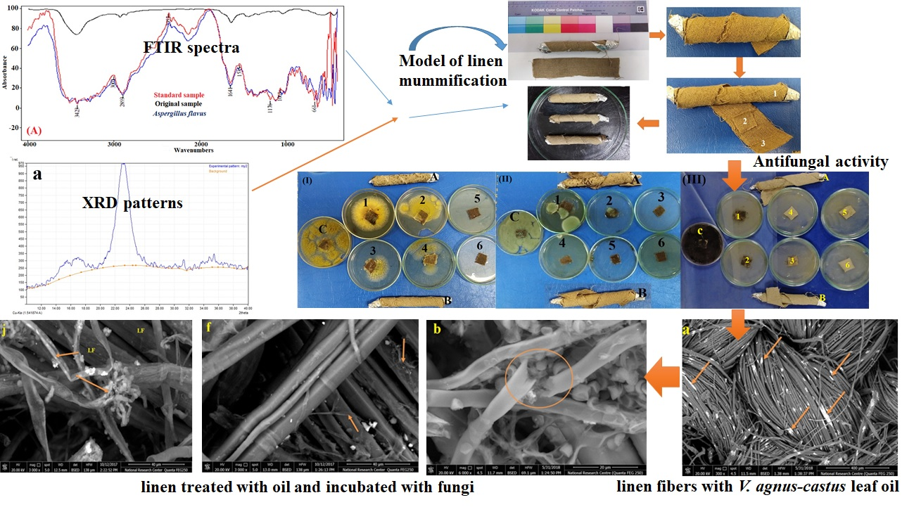
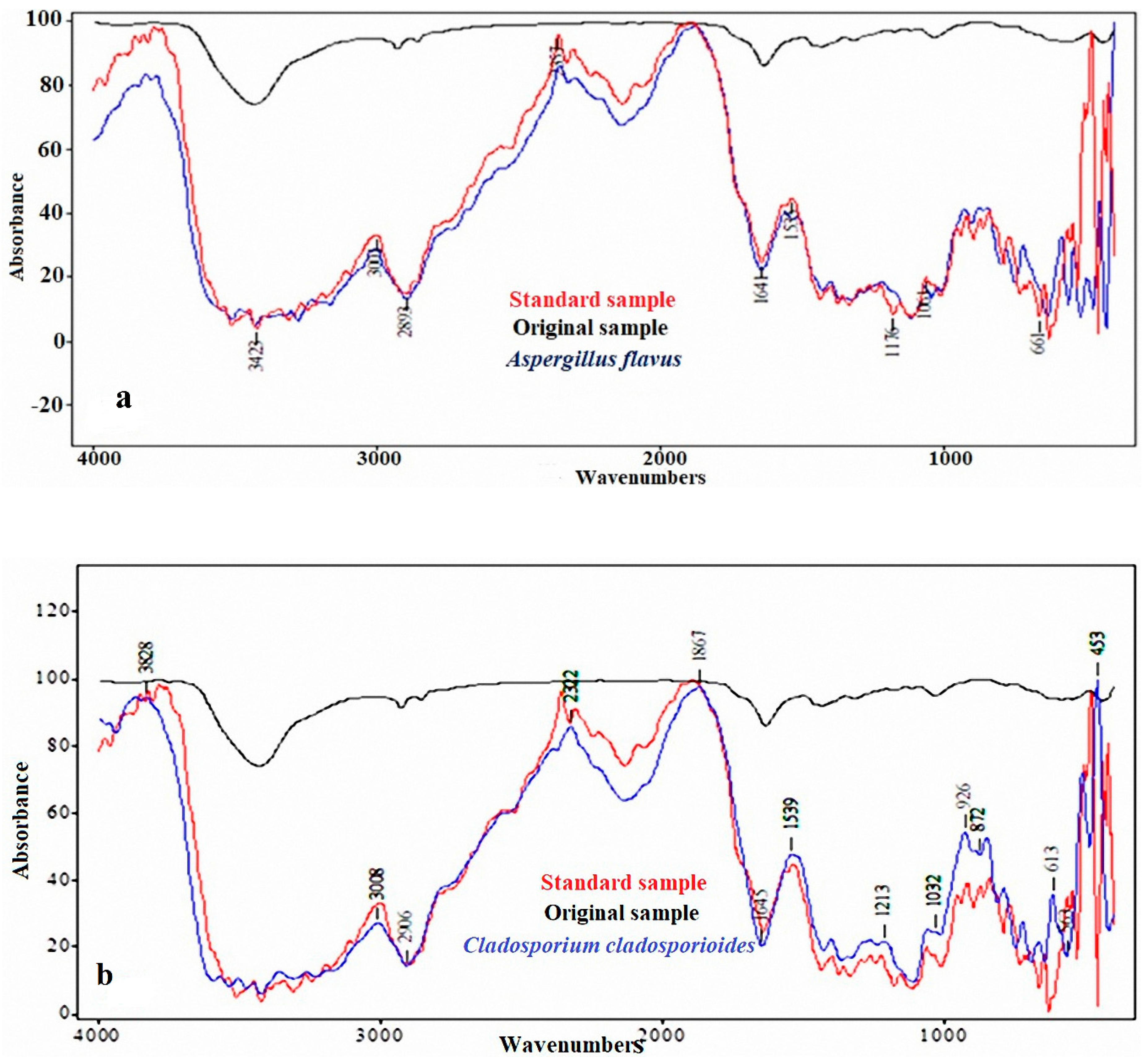
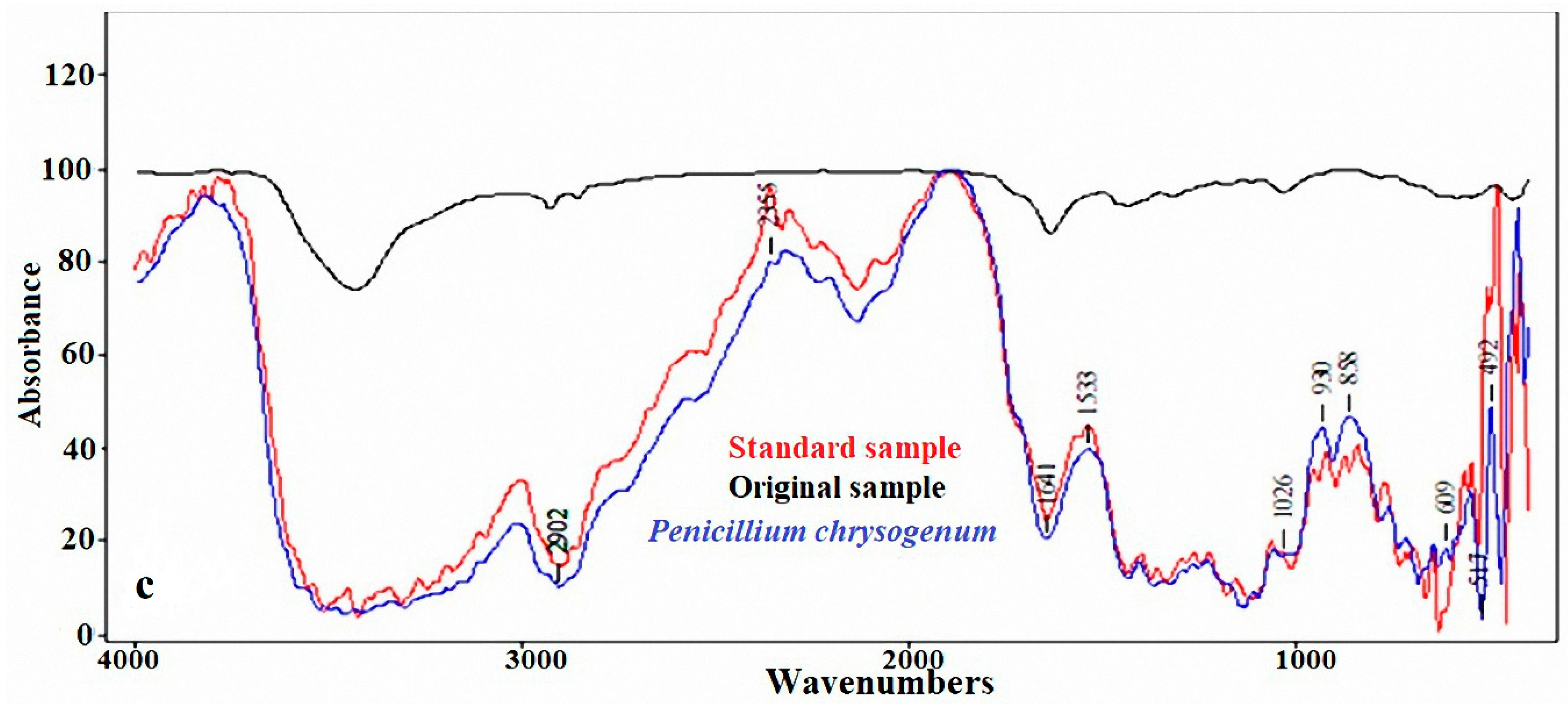
2.2. FTIR Analysis of the Standard, Original, and Infected Linen Samples

Three months after the inoculation of the linen fibers with each of the three fungi, an Fourier transform infrared (FTIR) spectrometer (Model 6100 Jasco, Tokyo, Japan) was used to analyze the samples. This analytical technique was performed on both the used linen fiber (2 cm × 2 cm) and on a linen sample from the original mummy (small fragments).

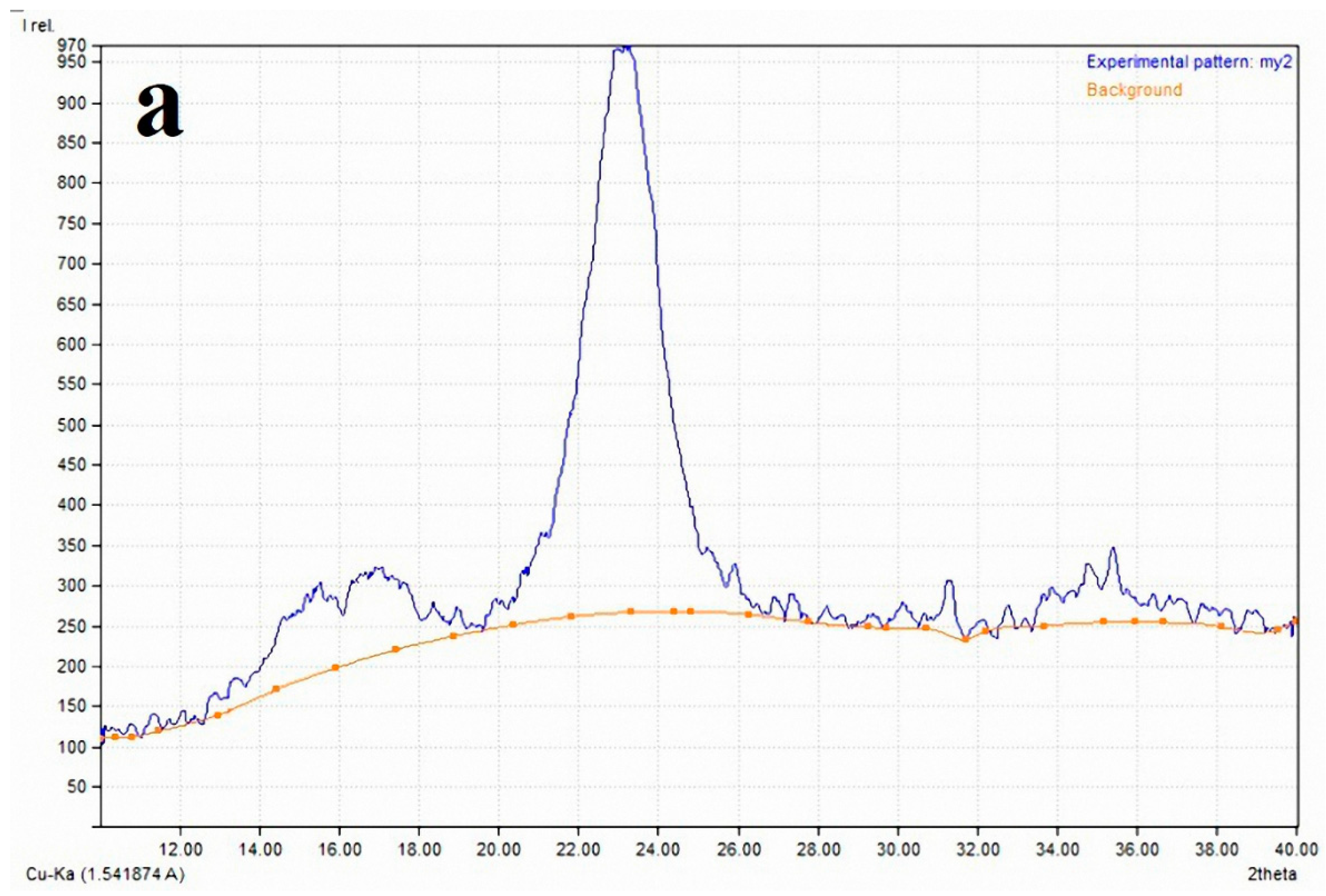
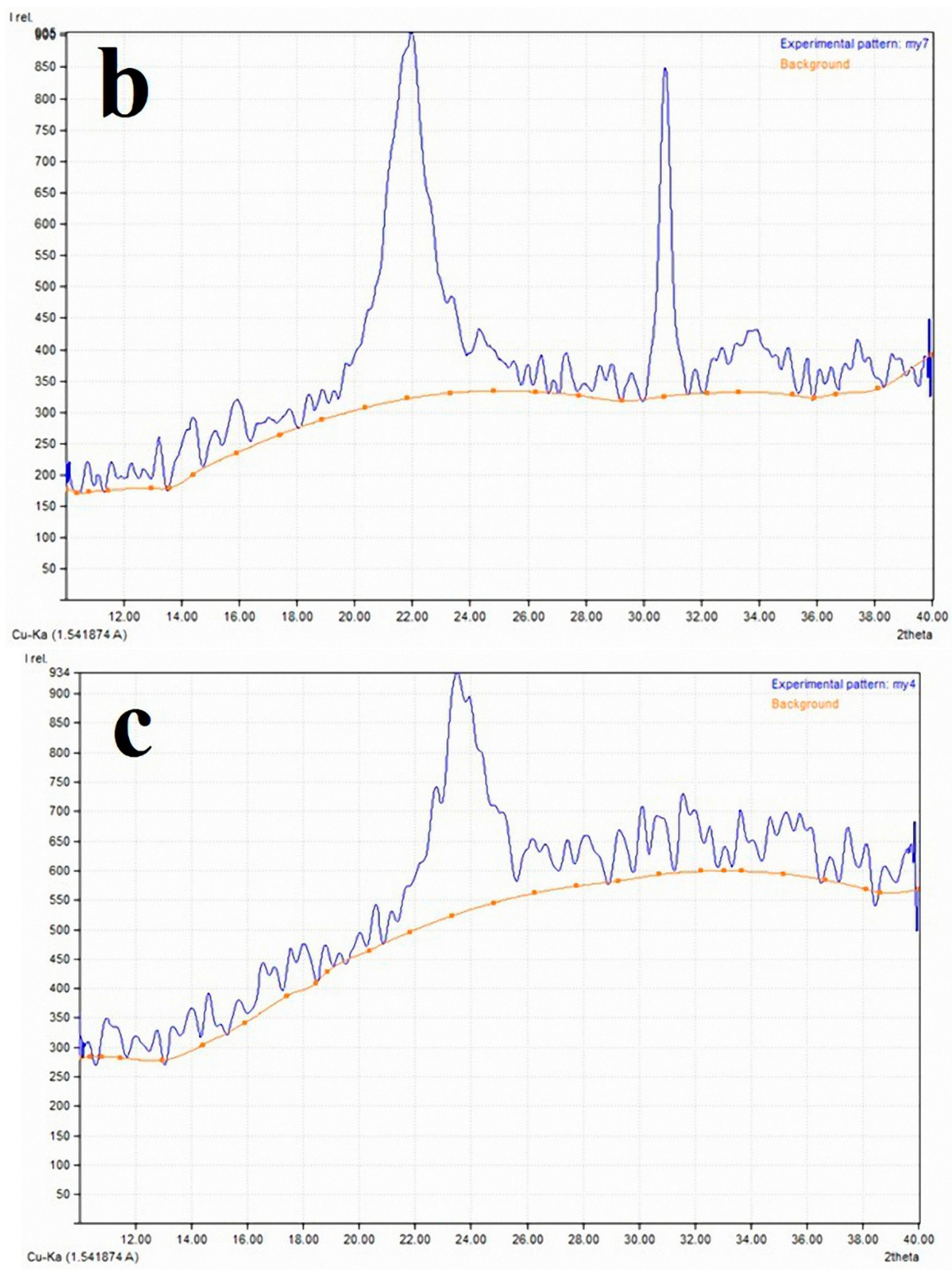
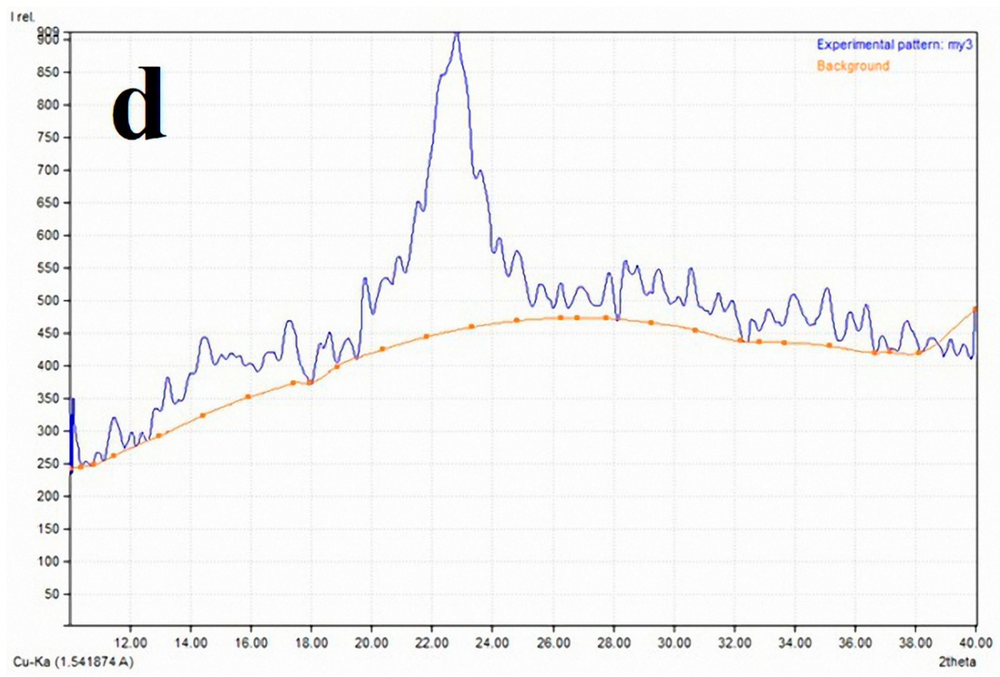
2.3. X-ray Diffraction Analysis (XRD) for the Crystallinity Index

The band positions of the crystalline and amorphous cellulose forms of the studied control and the samples colonized with A. flavus, C. cladosporioides, and P. chrysogenum were measured using a Philips Analytical X-ray (Philips–Eindhoven—The Netherlands) by means of Match 3 + PDF-4 2015 software program (Kreuzherrenstr, Bonn, Germany) diffractometer type: PW 1840, with a Cu tube anode, a generator tension of 40 kV, and a generator current of 25 mA). The crystallinity index (CI) was measured according to Segal et al. [38] with the following formula:
C I I 002 I a m I 002 × 100
where I002 is the diffraction intensity at the 002 peak position (2Ɵ ≈ 22.5°) and Iam at 2Ɵ = 18° (angle of amorphous cellulose).

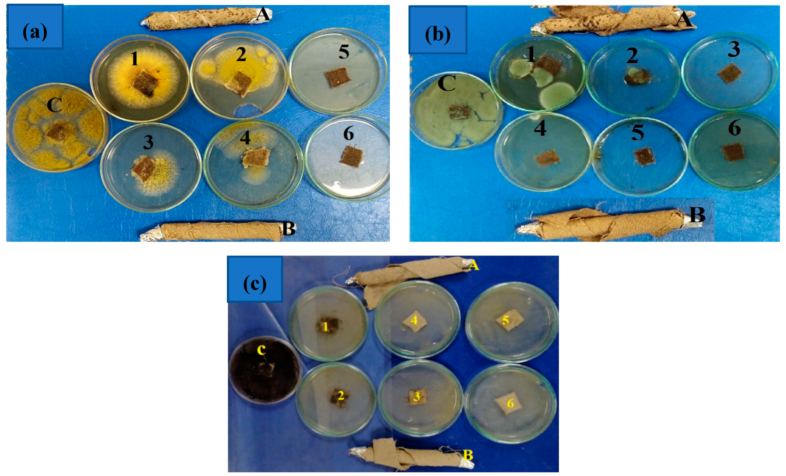
2.4. Antifungal Activity of Essential Oils

2.4.1. Plant Material and Essential Oil Extraction

The Eriocephalus africanus leaf, Vitex agnus-castus (leaf and fruit), Cymbopogon citratus leaf, and Rosmarinus officinalis leaf, were collected in 2017 from the nursery of the Department of Floriculture, Ornamental Plants and Garden Design, Faculty of Agriculture, Alexandria University, Alexandria, Egypt. The plants were identified and authenticated by the co-author Dr. Mervat El-Hefny. The essential oils (EOs) were extracted via the water distillation method using a proper essential oil trap (Clevenger’s apparatus) [39]. The EOs were kept dry in sealed Eppendorf tubes at 4 °C to wait for the chemical analysis.

2.4.2. GC/MS Analysis of the EOs

A Trace GC Ultra/Mass spectrophotometer ISQ (Thermo Scientific) instrument equipped with a flame ionization detector and a DB-5 narrow bore column (Agilent, Palo Alto, CA, USA) was used. The temperature program of the column oven and the other specifications of the operating system can be found in a previously published work [40]. Briefly, The GC–MS was equipped with a ZB-5MS Zebron capillary column (length 30 m × 0.25 mm ID, 0.25 µm film thickness; Agilent, Palo Alto, CA, USA). Helium, the carrier gas, with an average velocity of 39 cm/s, was used. Firstly, the temperature of the oven was held at 45 °C (2 min); then, the temperature was increased from 45 to 165 °C at 4 °C/min and 165–280 °C at 15 °C/min. the mass spectra were measured and collected under electron impact ionization at 70 electron volts and scanned from m/z 50–500 at five scans/second. The identification of the chemical compositions was performed based on an MS library search [41] and confirmed by measuring the Standard and Reverse Standard Indices [7,9,42,43].

2.4.3. Preparation of Oil Concentrations and Antifungal Activity

Firstly, the stock solution was 20 mL, wherein the oil amount was diluted in 10% dimethyl sulfoxide (2 mL Dimethyl sulfoxide (DMSO) diluted in 18 mL sterilized distilled water) (Sigma–Aldrich, Darmstadt, Germany). EOs were prepared at concentrations of 62, 125, 250, 500, 1000, and 2000 µL/mL by dissolving the EO in DMSO 10%, and a few drops of Tween-80 detergent were added to each EO concentration to emulsify the carrier oils in water. The inhibitory effects of the tested EOs on the linear growth of three fungi (Aspergillus flavus, Cladosporium cladosporioides, and Penicillium chrysogenum) were estimated. The measurement was performed in triplicate [1,44,45]. The mycelial growth inhibition (%) was calculated according to the following equation:
M G I % = A c A t A c × 100
where the MGI is mycelia growth inhibition, and AC and At are the average diameters of the fungal colonies of the control (10% DMSO + few drops of Tween-80) and the treatment, respectively.

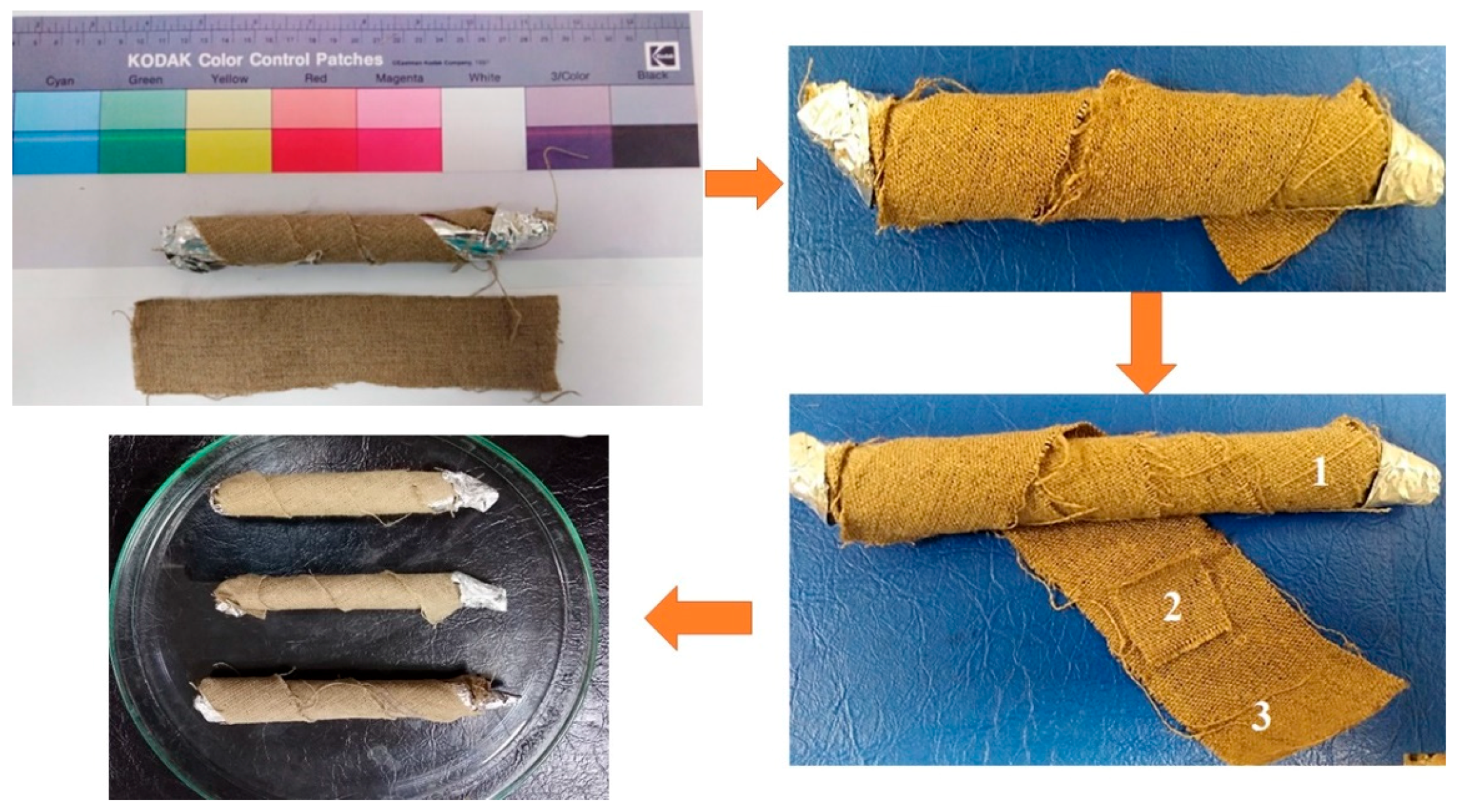
2.5. Model Linen of Mummification

Firstly, the linen samples were vapour treated with concentrated EO prepared at 0 (control), 125, 250, 500, 750, 1000, and 2000 µL/mL using the evaporation method [44,46,47] with minor modification. Linen samples were placed in petri dishes containing eight layers of Wattman No. 1 filter papers (Loba Chemie Pvt. Ltd., laboratory reagents & fine chemicals, Mumbai, India) overlaid by a polyethylene spacer mesh (Hebei Hightop Metal Mesh Co., Ltd., China). Dishes were autoclaved at 121 °C for 20 min and then left to cool. Each concentration of the EO was impregnated over the filter papers and kept for 48 h. This period allow the EO (fumigant) to evaporate and subsequently become absorbed by the linen samples.
A linen piece 15 cm long and 3 cm wide layer was fumigated with the best active concentrated EO resulting from the antifungal bioassay test. The sample model of the mummy was then produced and wrapped using three layers of linen pieces 15 cm long and 3 cm wide in the form of a roll and put in the Petri dish (15 cm) (Figure 1). The linen layers with infected each fungus were put between the treated linen layers with the best active EOs and the linen layers without any treatment in the form of a wide roll. Similarly, the fumigated linen pieces (2 × 2 cm) with different concentrations of the best EO were incubated in Petri dishes and inoculated with the spores of each fungus.

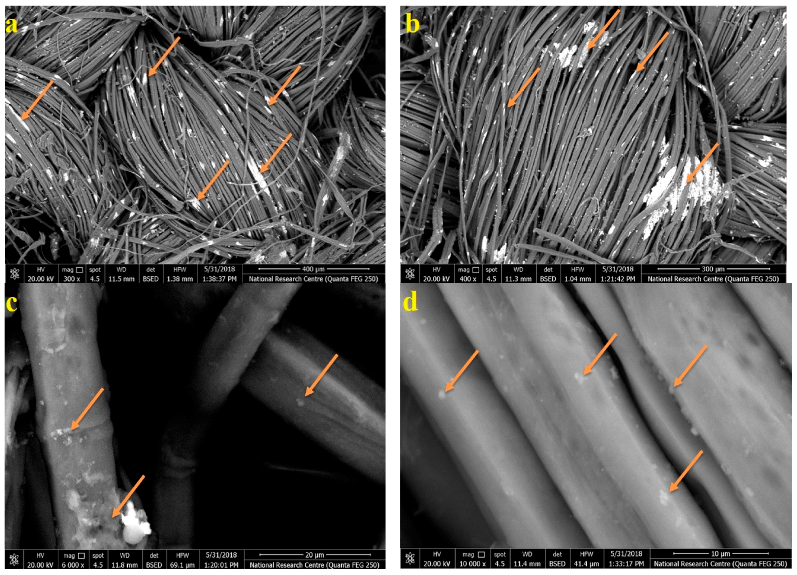
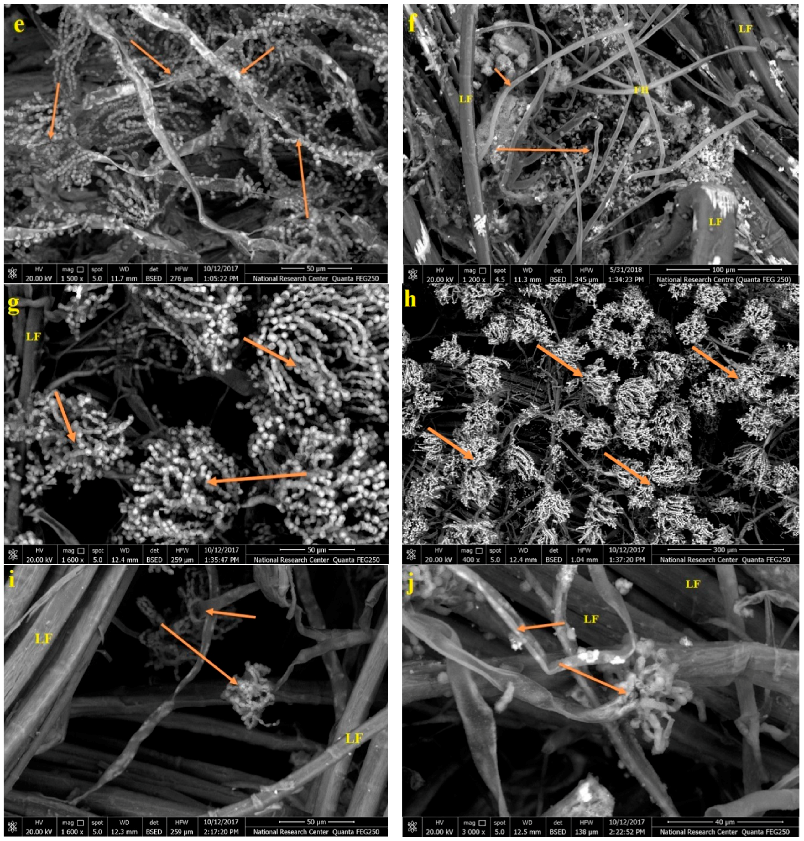
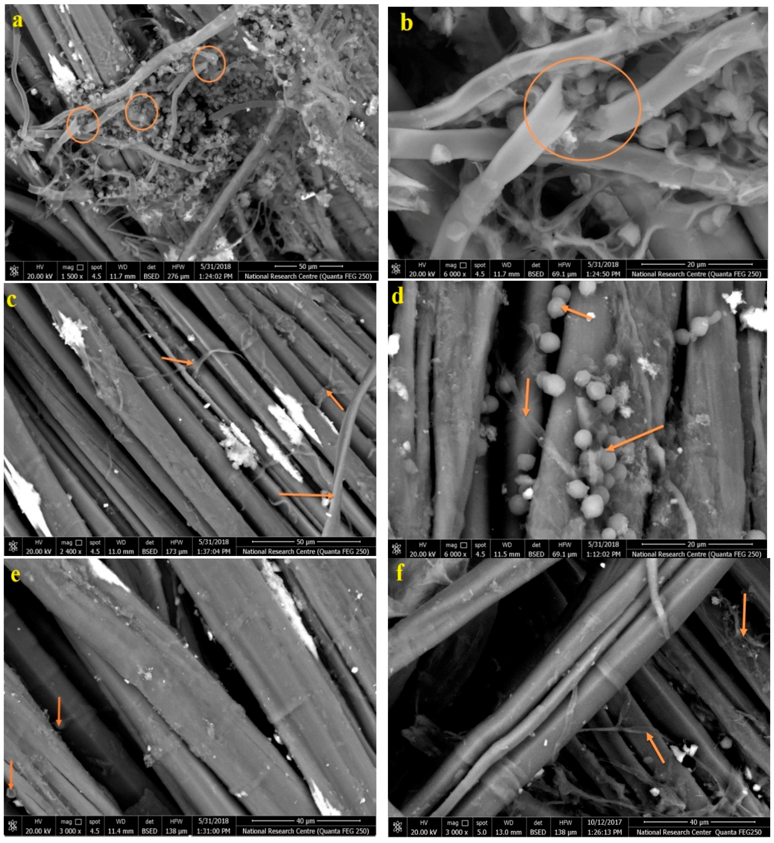
2.6. Scanning Electron Microscope (SEM) Examination of Inoculated Linen with Fungi

SEM analysis was used to examine the linen inoculated with the three fungi. The SEM model was a FEI Quanta 200 SEM FEG (FEI Company, Eindhoven, The Netherlands), which was used to study changes in the surface morphology and colonization of the fungi in both the inoculated and non-inoculated linen samples.

2.7. Statistical Analysis

The mycelial growth inhibition (%) of the fungi, as affected by the five EOs with 6 concentrations, was analyzed using a two-way ANOVA, while the mycelial growth inhibition (%) resulting from the best EO applied to the model mummification linen fiber was analyzed using a one-way ANOVA.

3. Results and Discussion

3.1. FTIR Analysis of the Linen

The changes in the wavenumbers of the functional groups of the main chemical structures of the linen fibers (standard, original, and infected with fungi) are shown in Figure 2a–c. The fungal spores possess a high absorption ability in the short-wavelength spectral region (1533 cm−1) due to chitin in the cell wall, as well as the high content of proteins [48]. Moreover, dramatic decreases of the band to a maximum at 1500 cm−1 were found, indicating a typical lignin [49]. Fungi have high absorption rate in the long-wavelength spectral region (1500–500 cm−1) due to the high content of carbohydrates [50].
Due to their chemical composition (especially cellulose), historical textile materials can easily decay via P. chrysogenum and C. cladosporioides when exposed to high humidity [28,32,34]. The mycelia of cellulolytic fungi lead to the loss of fibre strength as the fungi penetrate the cellulosic materials [30,31].
The assignment of OH stretching appeared at 3300–3400 cm−1, which is characteristic of OH stretching in alcohols and phenols of cellulose, lignin, or hemicellulose [11,44,51,52,53,54,55]. The band at 3423 cm−1 (–OH groups bending) in the spectrum of decayed and original samples became wider than the standard linen spectrum, and its peak shifted to a high wave, wherein hydroxyl groups on the backbone of the hemicelluloses were found.
Changes in the wavelengths at 1424 cm−1 and 1512 cm−1 confirmed the ability of fungi to decompose the lignin [55,56]. The bands’ relative intensities of absorption at 1510–1267 cm−1 increased significantly. The lower absorption peaks of the lignin [57] occurred under hydrolysis, oxidation, and the breaking of intermolecular bonds, which were caused by fungi. It was reported that the FTIR technique might be applied to detect mound fungi growth on materials [58]. This means that the primary functional groups that reoccurred in the infected linen and the original sample were attacked by microorganisms. The crystallinity degree of native cellulose is between 60% and 90% [59,60]. Furthermore, the cellulose fibers collapsed, which means that the samples decayed [61].

3.2. Cellulose Crystallinity of Linen Fibers

The results of the XRD (Figure 3) showed that there are noticeable differences in the amorphous areas in the linen affected by the three molds compared to the control (Cellulose Crystallinity = 72%), as shown in Figure 3a. This indicated that fungal degradation may change the d-spacing of the crystalline area of the cellulose and cause a slight shift to a higher theta degree of the band positions. Large decreases of 64.61%, 52.69%, and 54.63% in the crystallinity indices of the cellulose crystallinity of the linen inoculated with A. flavus (Figure 3b), C. cladosporioides (Figure 3c), and P. chrysogenum (Figure 3d) were found compared to the control sample (72.08%), which indicates that a change has occurred in the chemical properties of the cellulose.

3.3. Antifungal Activity of the EOs

The antifungal activities of the EOs of E. africanus leaf, V. agnus-castus leaf and fruit, C. citratus leaf, and R. officinalis leaf are presented in Table 1. The results of the mycelial inhibition percentages show that the leaf EO from V. agnus-castus possessed the best activities against the growth of A. flavus, C. cladosporioides, and P. chrysogenum, as most of the studied EO concentrations showed a 100% inhibition of fungal mycelial growth, followed by C. citratus EO.

3.4. Chemical Composition of the EOs

The phytochemical constituents of Eriocephalus africanus leaf EO can be found in our previously published work [5]. The phytochemical constituents of Cympobogon citratus EO presented in Table 2 show the main abundant compounds of β-citral (43.63%), geranial (α-citral or citral A) (41.51%), β-myrcene (12.37%), 1,3-diolein (1.57%), and 9-octadecenoic acid (0.92%). The EO of lemongrass contains mainly citral [15] and citronellal, limonene, and geraniol [12,15].
Table 3 shows the chemical constituents of the EO from Rosmarinus officinalis, where the main compounds were eucalyptol (30.75%), α-pinene (20.41%), camphor (18.15%), borneol (5.94%), ethyl iso-allocholate (6.77%), limonene (2.72%), linalool (2.74%), and camphene (2.59%). The main chemical compounds in the EO of R. officinalis grown in Pakistan were 1,8-cineol, camphor, α-pinene, limonene, camphene, and linalool [62], while cineole, camphor, and α-pinene were the main compounds in Brazilian plants [63]. In Tunisian R. officinalis, the EOs were 1.8-cineole and camphor [64]. Camphor (5–31%), 1,8-cineol (15–55%), camphene (2.5–12.0%), verbenone (2.2–11.1%), and pinene (9–26%) were found to be the major compounds in the EO of R. officinalis [18,19].
Table 4 shows the chemical constituents of EO from Vitex agnus-castus leaves, where the main compounds are caryophyllene (23.13%), eucalyptol (20.59%), sabinene (β-thujene) (12.2%), γ-elemene (9%), β-farnesene (6.14%), caryophyllene oxide (4.06%), 2-methylenecholestan-3-ol (3.35%), and 1-heptatriacotanol (2.77%). Table 5 presents the chemical constituents of V. agnus-castus fruit EO, where the main compounds are γ-elemene (41.59%), geranyllinalool (17.98%), eucalyptol (16.65%), nerolidyl acetate (11.63%), cis-caryophyllene (3.73%), α-pinene (2.58%), caryophyllene oxide (2.55%), and nerolidol (1.16%). The main chemical compounds identified in the EO of V. agnus-castus with strong antimicrobial activities were mostly 1,8-cineole or eucalyptol, sabinene, (E)-β-farnesene, (E)-caryophyllene, and α-terpinyl acetate [9,21,65,66,67]. Further, 1,8-cineole has been proven to have good antifungal activity against Aspergillus apple rot [21]. V. agnus-castus oil contains the main compounds of 1,8-cineole (eucalyptol) and 8-caryophyllene, which significantly inhibited the growth of F. oxysporum, R. solani, Sclerotinia sclerotiorum, and Verticillium dahlia [68]. Melia azedarach wood-treated with the n-hexane oil of V. agenus-castus leaves at a 1%, 2%, or 3% concentration showed good antifungal activity against F. culmorum, Rhizoctonia solani, and P. chrysogenum [9], where the main compounds were eucalyptol (22.1%), (E)-β-caryophyllene (18.3%), and β-sitosterol (12.4%). The leaf EO of V. agnus-castus showed potent antimicrobial activity against the growth of bacteria (Agrobacterium tumefaciens and Erwinia carotovora var. carotovora) and fungi (Alternaria alternata, Botrytis cinerea, F. oxysporum, and F. solani) [69].

3.5. Antifungal Activity of the Model Linen Treated with V. agnus-castus Leaf EO

The antifungal activity of the studied EOs illustrates that the leaf EO from V. agnus-castus has the highest and best activity against the three fungi. The samples of the linen fibers were incubated with each fungus in petri dishes containing concentrated oil (Figure 4).
The results of the antifungal activity after the application of V. agnus-castus leaf EO as a fumigant agent to the model linen fibers similar to those used for the mummy are shown in Table 6. The lowest concentrations of EO from the V. agnus-castus leaf when the linen pieces were fumigated caused a 100% growth inhibition of the three fungi (1000 µL/mL, 500 µL/mL, and 500 µL/mL for A. flavus, C. cladosporioides and P. chrysogenum, respectively).

3.6. SEM Examination

The model linen pieces treated with the lowest concentrations of the V. agnus-castus leaf EO caused 100% inhibition to the growth of fungi and, to compare them with the control treatment, were subjected to SEM examination. Figure 5a–d shows the SEM images with different magnifications of the linen fibers after treatment with EO without fungi. These photos show the distribution of the V. agnus-castus leaf EO spots over the fibers. As shown in Figure 5i,j, the linen fibers not treated with the EOs feature significant growth of fungal mycelium from A. flavus (e,f), C. cladosporioides (g,h), and P. chrysogenum (i,j).
Figure 6 shows the SEM images of the linen fibers treated with V. agnus-castus leaf EO and inoculated with fungi. SEM examination showed the morphological changes in the hyphae. The linen fibers treated with V. agnus-castus leaf EO showed clear damage in the fungus, which is evident in changing the form of the hyphae and the absorption and split in other cases (a,b). In the control group, the hyphae were round and full with a smooth surface (Figure 5e–j), while, like the colonized linen fibers treated with V. agnus-castus leaf EO, the structures of the hyphae of the fungi were damaged (Figure 6a–f), deformed, and had rough surfaces.
Previous studies have indicated the significant growth of fungal mycelium. Pulp paper from Whatman, cotton, or chemical pulp showed deterioration symptoms when inoculated with Trichoderma harzianum and Paecilomyces variotii [70]. Papyrus strips treated with some additives decreased the growth of A. flavus, A. niger, and C. gloeosporioides compared to the control treatment [10]. An erosion of fiber morphology occurred in Linen pulp fibers when inoculated with A. niger [11], but with the addition of some plant source additives, this growth decreased.
Finally, according to the SEM examination of linen fibres treated with EO and inoculated with the fungi, the V. agnus-castus leaf EO inhibited the growth of mycelium and was able to effectively destroy the morphology and the structure of the hyphae. This study suggests a novel eco-friendly treatment of model linen fibers similar to the method used in the ancient Egyptian mummification of the child mummy using the essential oil of V. agnus-castus leaf. This oil offered potent activity against the growth of A. flavus, C. cladosporioides, and P. chrysogenum, which were previously isolated from the child mummy and inoculated into the designed model linen fiber for mummification.

4. Conclusions

Isolated fungi A. flavus, C. cladosporioides, and P. chrysogenum from an ancient Egyptian mummy were examined for their roles as biodetergent agents in the linen fibres that were used. The analytical methods of FTIR and XRD showed that these three fungi affected the chemical composition of the linen samples through changes in the wavenumbers of the functional groups of the main chemical structures of the standard, original, and linen fibers infected with fungi. Five EOs extracted from E. africanus leaf, V. agnus-castus leaf and fruit, C. citratus leaf, and R. officinalis leaf were tested for their antifungal activity. V. agnus-castus leaf oil produced the best inhibition of fungal mycelial growth. The effectiveness of the innovated and treated model linen fibers with V. agnus-castus leaf EO was achieved, and SEM examination showed inhibited growth of the fungal mycelium, as well as changes in the morphology and structure of the hyphae.

Author Contributions

M.M.A.M., M.E.-H., M.Z.M.S. and H.M.A. designed the experiments, conducted the laboratory analyses, wrote parts of the manuscript, and interpreted the results; all co-authors contributed in writing and revising the article. All authors have read and agreed to the published version of the manuscript.

Funding

This research was funded by Researchers Supporting Project number (RSP-2019/123), King Saud University, Riyadh, Saudi Arabia.

Acknowledgments

Researchers Supporting Project number (RSP-2019/123) King Saud University, Riyadh, Saudi Arabia.

Conflicts of Interest

The authors declare no conflicts of interest.

References

  1. Salem, M.Z.M.; Zidan, Y.E.; Mansour, M.M.A.; El Hadidi, N.M.N.; Abo Elgat, W.A.A. Antifungal activities of two essential oils used in the treatment of three commercial woods deteriorated by five common mold fungi. Inter. Biodeterior. Biodegrad. 2016, 106, 88–96. [Google Scholar] [CrossRef]
  2. Ashmawy, N.A.; Al Farraj, D.A.; Salem, M.Z.M.; Elshikh, M.S.; Al-Kufaidy, R.; Alshammari, M.k.; Salem, A.Z.M. Potential impacts of Pinus halepensis Miller trees as a source of phytochemical compounds: Antibacterial activity of the cones essential oil and n-butanol extract. Agrofores. Syst. 2018. [Google Scholar] [CrossRef]
  3. Al-Huqail, A.A.; Behiry, S.I.; Salem, M.Z.M.; Ali, H.M.; Siddiqui, M.H.; Salem, A.Z.M. Antifungal, antibacterial, and antioxidant activities of Acacia saligna (Labill.) H. L. Wendl. flower extract: HPLC analysis of phenolic and flavonoid compounds. Molecules 2019, 24, 700. [Google Scholar] [CrossRef] [PubMed] [Green Version]
  4. Behiry, S.I.; Okla, M.K.; Alamri, S.A.; EL-Hefny, M.; Salem, M.Z.M.; Alaraidh, I.A.; Ali, H.M.; Al-Ghtani, S.M.; Monroy, J.C.; Salem, A.Z.M. Antifungal and antibacterial activities of Musa paradisiaca L. peel extract: HPLC analysis of phenolic and flavonoid contents. Processes 2019, 7, 215. [Google Scholar] [CrossRef] [Green Version]
  5. Behiry, S.I.; EL-Hefny, M.; Salem, M.Z.M. Toxicity effects of Eriocephalus africanus L. leaf essential oil against some molecularly identified phytopathogenic bacterial strains. Nat. Prod. Res. 2019. [Google Scholar] [CrossRef]
  6. Hamad, Y.K.; Abobakr, Y.; Salem, M.Z.M.; Ali, H.M.; Al-Sarar, A.S.; Al-Zabib, A.A. Activity of plant extracts/essential oils against some plant pathogenic fungi and mosquitoes: GC/MS analysis of bioactive compounds. BioResources 2019, 14, 4489–4511. [Google Scholar] [CrossRef]
  7. Mohamed, W.A.; Mansour, M.M.A.; Salem, M.Z.M. Lemna gibba and Eichhornia crassipes extracts: Clean alternatives for deacidification, antioxidation and fungicidal treatment of historical paper. J. Clean. Prod. 2019, 219, 846–855. [Google Scholar] [CrossRef]
  8. Okla, M.K.; Alamri, S.A.; Salem, M.Z.M.; Ali, H.M.; Behiry, S.I.; Nasser, R.A.; Alaraidh, I.A.; Al-Ghtani, S.M.; Soufan, W. Yield, phytochemical constituents, and antibacterial activity of essential oils from the leaves/twigs, branches, branch wood, and branch bark of Sour Orange (Citrus aurantium L.). Processes 2019, 7, 363. [Google Scholar] [CrossRef] [Green Version]
  9. Salem, M.Z.M.; Behiry, S.I.; EL-Hefny, M. Inhibition of Fusarium culmorum, Penicillium chrysogenum and Rhizoctonia solani by n-hexane extracts of three plant species as a wood-treated oil fungicide. J. Appl. Microbiol. 2019, 126, 1683–1699. [Google Scholar] [CrossRef]
  10. Taha, A.S.; Salem, M.Z.M.; Abo Elgat, W.A.A.; Ali, H.M.; Hatamleh, A.A.; Abdel-Salam, E.M. Assessment of the impact of different treatments on technological and antifungal properties of produced Papyrus (Cyperus papyrus L.) sheets. Materials 2019, 12, 620. [Google Scholar] [CrossRef] [Green Version]
  11. Taha, A.S.; Abo Elgat, W.A.A.; Salem, M.Z.M.; Ali, H.M.; Fares, Y.G.E.; Elshikh, M.S. Impact of some plant source additives on enhancing the properties and antifungal activities of pulp made from Linen fibers. BioResources 2019, 14, 6025–6046. [Google Scholar]
  12. Tajidin, N.E.; Ahmad, S.H.; Rosenani, A.B.; Azimah, H.; Munirah, M. Chemical composition and citral content in lemongrass (Cymbopogon citratus) essential oil at three maturity stages. Afri. J. Biotechnol. 2012, 11, 2685–2693. [Google Scholar] [CrossRef] [Green Version]
  13. Pengelly, A. The Constituents of Medicinal Plants (Eds): An Introduction to the Chemistry and Therapeutics of Herbal Medicine; CABI Publishing: England, UK, 2004; pp. 85–103. [Google Scholar]
  14. de Silva, B.C.; Guterres, S.S.; Weisheimer, V.; Schapoval, E.E.S. Antifungal activity of the lemongrass oil and citral against Candida species. Braz. J. Infect. Dis. 2008, 12, 63–66. [Google Scholar] [CrossRef] [PubMed] [Green Version]
  15. Schaneberg, B.T.; Khan, I.A. Comparison of extraction methods for marker compounds in the essential oil of lemongrass by GC. J. Agric. Food Chem. 2002, 50, 1345–1349. [Google Scholar] [CrossRef]
  16. Daferera, D.J.; Ziogas, B.N.; Polissiou, M.G. The effectiveness of plant essential oils on the growth of Botrytis cinerea, Fusarium sp. and Clavibacter michiganensis sub sp. michiganensis. Crop. Prot. 2003, 22, 39–44. [Google Scholar] [CrossRef]
  17. Naik, M.I.; Fomda, B.A.; Jaykumar, E.; Bhat, J.A. Antibacterial activity of lemongrass (Cymbopogon citratus) oil against some selected pathogenic bacteria. Asian Pac. J. Trop. Med. 2010, 3, 535–538. [Google Scholar] [CrossRef] [Green Version]
  18. Domokos, J.; Hethelyi, E.; Palinkas, J.; Szirmai, S.; Tulok, M.H. Essential oil of rosemary (Rosmarinus officinalis L.) of Hungarian origin. J. Essent. Oil Res. 1997, 9, 41–45. [Google Scholar] [CrossRef]
  19. Salido, S.; Altarejos, J.; Nogueras, M.; Sanchez, A.; Lugue, P. Chemical composition and seasonal variations of rosemary oil from southern Spain. J. Essen. Oil Res. 2003, 15, 10–14. [Google Scholar] [CrossRef]
  20. Hendel, N.; Larous, L.; Belbey, L. Antioxidant activity of rosemary (Rosmarinus officinalis L.) and its in vitro inhibitory effect on Penicillium digitatum. Int. Food Res. J. 2016, 23, 1725–1732. [Google Scholar]
  21. Stojković, D.; Soković, M.; Glamočlija, J.; Džamić, A.; Ćirić, A.; Ristić, M.; Grubišić, D. Chemical composition and antimicrobial activity of Vitex agnus-castus L. fruits and leaves essential oils. Food Chem. 2011, 128, 1017–1022. [Google Scholar] [CrossRef]
  22. Merle, H.; Verdeguer, M.; Blazquez, M.A.; Boira, H. Chemical composition of the essential oils from Eriocephalus africanus L. var. africanus populations growing in Spain. Flavour Fragr. J. 2007, 22, 461–464. [Google Scholar] [CrossRef]
  23. Salie, F.; Eagles, P.F.; Leng, H.M. Preliminary antimicrobial screening of four South African Asteraceae species. J. Ethnopharmacol. 1996, 52, 27–33. [Google Scholar] [CrossRef]
  24. Górny, R.L.; Reponen, T.; Willeke, K.; Schmechel, D.; Robine, E.; Boissier, M.; Grinshpun, S.A. Fungal fragments as indoor air biocontaminants. Appl. Environ. Microbiol. 2002, 68, 3522–3531. [Google Scholar] [CrossRef] [PubMed] [Green Version]
  25. Piñar, G.; Piombino-Mascali, D.; Maixner, F.; Zink, A.; Sterflinger, K. Microbial survey of the mummies from the Capuchin Catacombs of Palermo, Italy: Biodeterioration risk and contamination of the indoor air. FEMS Microbiol. Ecol. 2013, 86, 341–356. [Google Scholar] [CrossRef] [PubMed] [Green Version]
  26. Geweely, N.S.; Afifi, H.A.; Ibrahim, D.M.; Soliman, M.M. Efficacy of essential oils on fungi isolated from archaeological objects in Saqqara excavation, Egypt. Geomicrobiol. J. 2019, 36, 148–168. [Google Scholar] [CrossRef]
  27. Allsopp, D.E. Worldwide wastage: The economics of biodeterioration. Microbiol. Tod. 2011, 38, 150–153. [Google Scholar]
  28. Kavkler, K.; Gunde-Cimerman, N.; Zalar, P.; Demsar, A. Fungal contamination of textile objects preserved in Slovene museums and religious institutions. Int. Biodeterior. Biodegrad. 2015, 97, 51–59. [Google Scholar] [CrossRef]
  29. Sterflinger, K. Fungi: Their role in deterioration of cultural heritage. Fungal Biol. Rev. 2010, 24, 47–55. [Google Scholar] [CrossRef]
  30. Montegut, D.; Indictor, N.; Koestler, R. Fungal deterioration of cellulosic textiles: A review. Int. Biodeterior. 1991, 28, 209–226. [Google Scholar] [CrossRef]
  31. Sequeira, S.; Cabrita, E.J.; Macedo, M.F. Antifungals on paper conservation: An overview. Int. Biodeterior. Biodegrad. 2012, 74, 67–86. [Google Scholar] [CrossRef]
  32. Gutarowska, B.; Pietrzak, K.; Machnowski, W.; Milczarek, J.M. Historical textiles – a review of microbial deterioration analysis and disinfection methods. Tex. Res. J. 2017, 87, 2388–2406. [Google Scholar] [CrossRef]
  33. Montanari, M.; Melloni, V.; Pinzari, F.; Innocenti, G. Fungal biodeterioration of historical library materials stored in Compactus movable shelves. November 2012. Int. Biodeterior. Biodegrad. 2012, 75, 83–88. [Google Scholar] [CrossRef]
  34. Lech, T.; Ziembinska-Buczynska, A.; Krupa, N. Analysis of microflora present on historical textiles with the use of molecular techniques. Int. J. Conserv. Sci. 2015, 6, 137–144. [Google Scholar]
  35. Čavka, M.; Glasnović, A.; Janković, I.; Sikanjić, P.; Perić, B.; Brkljacić, B.; Mlinarić-Missoni, E.; Skrlin, J. Microbiological analysis of a mummy from the archeological museum in Zagreb. Coll. Antropol. 2010, 34, 803–805. [Google Scholar] [PubMed]
  36. Mansour, M. Impact of storage conditions on biodeterioration of ancient Egyptian child mummies by xerophilic fungi. Egy. J. Archaeol. Restorat. Stud. 2018, 8, 97–107. [Google Scholar]
  37. Miller, A.; Laiz, L.; Dionísio, A.; Macedo, M.; Saiz-Jimenez, C. Growth of phototrophic biofilms from limestone monuments under laboratory conditions. Int. Biodeterior. Biodegr. 2009, 63, 860–867. [Google Scholar] [CrossRef] [Green Version]
  38. Segal, L.; Creely, J.J.; Martin, A.E., Jr.; Conrad, C.M. An empirical method for estimating the degree of crystallinity of native cellulose using the X-ray diffraction. Tex. Res. J. 1959, 29, 786–794. [Google Scholar] [CrossRef]
  39. Salem, M.Z.M.; Ali, H.M.; El-Shanhorey, N.A.; Abdel-Megeed, A. Evaluation of extracts and essential oil from Callistemon viminalis leaves: Antibacterial and antioxidant activities, total phenolic and flavonoid contents. Asian Pac. J. Trop. Med. 2013, 6, 785–791. [Google Scholar] [CrossRef] [Green Version]
  40. Hussein, H.S.; Salem, M.Z.M.; Soliman, A.M. Repellent, attractive, and insecticidal effects of essential oils from Schinus terebinthifolius fruits and Corymbia citriodora leaves on two whitefly species, Bemisia tabaci and Trialeurodes ricini. Sci. Hortic. 2017, 216, 111–119. [Google Scholar] [CrossRef]
  41. Wiley/NIST. The Wiley/NBS Registry of Mass Spectral Data, 8th ed.; Wiley: New York, NY, USA, 2008. [Google Scholar]
  42. Salem, M.Z.M.; Mansour, M.M.A.; Elansary, H.O. Evaluation of the effect of inner and outer bark extracts of Sugar Maple (Acer saccharum var. saccharum) in combination with citric acid against the growth of three common molds. J. Wood Chem. Technol. 2019, 39, 136–147. [Google Scholar] [CrossRef]
  43. EL-Hefny, M.; Ashmawy, N.A.; Salem, M.Z.M.; Salem, A.Z.M. Antibacterial activity of the phytochemicals-characterized extracts of Callistemon viminalis, Eucalyptus camaldulensis and Conyza dioscoridis against the growth of some phytopathogenic bacteria. Microb. Pathog. 2017, 113, 348–356. [Google Scholar] [CrossRef] [PubMed]
  44. Salem, M.Z.M.; Zidan, Y.E.; Mansour, M.M.A.; El Hadidi, N.M.N.; Abo Elgat, W.A.A. Evaluation of usage three natural extracts applied to three commercial wood species against five common molds. Inter. Biodeterior. Biodegrad. 2016, 110, 206–226. [Google Scholar] [CrossRef]
  45. Salem, M.Z.M.; Mansour, M.M.A.; Mohamed, W.S.; Ali, H.M.; Hatamleh, A.A. Evaluation of the antifungal activity of treated Acacia saligna wood with Paraloid B-72/TiO2 nanocomposites against the growth of Alternaria tenuissima, Trichoderma harzianum, and Fusarium culmorum. Bio. Res. 2017, 12, 7615–7627. [Google Scholar]
  46. Lopez, P.; Sanchez, C.; Batlle, R.; Nerin, C. Solid-and vapor-phase antimicrobial activities of six essential oils: Susceptibility of selected foodborne bacterial and fungal strains. J. Agric. Food Chem. 2005, 53, 6939–6946. [Google Scholar] [CrossRef]
  47. Nedorostova, L.; Kloucek, P.; Kokoska, L.; Stolcova, M.; Pulkrabek, J. Antimicrobial proprieties of selected essential oils in vapour phase against foodborne bacteria. Food Control. 2009, 20, 157–160. [Google Scholar] [CrossRef]
  48. Zimmermann, B.; Tkalčec, Z.; Mešić, A.; Kohler, A. Characterizing aeroallergens by Infrared Spectroscopy of fungal spores and pollen. PLoS ONE 2015, 10, e0124240. [Google Scholar] [CrossRef] [Green Version]
  49. Gerd, W.; Margarete, P.; Dietrich, F. Hexafluoropropanol as Valuable Solvent for Lignin in UV and IR Spectroscopy. Holzforschung 1983, 37, 303–307. [Google Scholar]
  50. Kosa, G.; Kohler, A.; Tafintseva, V.; Zimmermann, B.; Forfang, K.; Afseth, N.K.; Tzimorotas, D.; Vuoristo, K.S.; Horn, S.J.; Mounier, J.; et al. Microtiter plate cultivation of oleaginous fungi and monitoring of lipogenesis by high-throughput FTIR spectroscopy. Microb. Cell Fact. 2017, 16, 101. [Google Scholar] [CrossRef] [Green Version]
  51. Zhang, Y.L.; Chen, J.; Lei, Y.; Zhou, Q.; Sun, S.; Noda, I. Discrimination of different red wine by Fourier transform infrared and two-dimensional infrared correlation spectroscopy. J. Mol. Struct. 2010, 974, 144–150. [Google Scholar] [CrossRef]
  52. Herrera, R.; Erdocia, X.; Llano-Ponte, R.; Labidi, J. Characterization of hydrothermally treated wood in relation to changes on its chemical composition and physical properties. J. Anal. Appl. Pyrol. 2014, 107, 256–266. [Google Scholar] [CrossRef]
  53. Zhou, C.; Jiang, W.; Cheng, Q.; Via, B.K. Multivariate calibration and model integrity for wood chemistry using Fourier transform infrared spectroscopy. J. Anal. Methods Chem. 2015, 1–9. [Google Scholar] [CrossRef] [PubMed] [Green Version]
  54. Gandolfo, D.S.; Mortimer, H.; Woodhall, J.W.; Boonham, N. Fourier transform infra-red spectroscopy using an attenuated total reflection probe to distinguish between Japanese larch, pine and citrus plants in healthy and diseased states. Spectrochim Acta A 2016, 163, 181–188. [Google Scholar] [CrossRef] [PubMed] [Green Version]
  55. Salem, M.Z.M.; Hamed, S.A.M.; Mansour, M.M.A. Assessment of efficacy and effectiveness of some extracted bio-chemicals as bio-fungicides on Wood. Drv. Ind. 2019, 70, 337–350. [Google Scholar] [CrossRef]
  56. Hong, Y.; Dashtban, M.; Chen, S.; Song, R.; Qin, W. Lignin in paper mill sludge is degraded by white-rot fungi in submerged fermentation. J. Microb. Biochem. Technol. 2015, 7, 177–181. [Google Scholar]
  57. Shi, J.; Xing, D.; Lia, J. FTIR Studies of the Changes in Wood Chemistry from Wood Forming Tissue under Inclined Treatment. Energy Procedia 2012, 16, 758–762. [Google Scholar] [CrossRef] [Green Version]
  58. Mohebby, B. Attenuated total reflection infrared spectroscopy of white-rot decayed beech wood. Int. Biodeterior. Biodegrad. 2005, 55, 247–251. [Google Scholar] [CrossRef]
  59. Silva-Carvalho, R.; Silva, J.P.; Ferreirinha, P.; Leitão, A.F.; Andrade, F.K.; Gil da Costa, R.M.; Cristelo, C.; Rosa, M.F.; Vilanova, M.; Gama, F.M. Inhalation of Bacterial Cellulose Nanofibrils Triggers an Inflammatory Response and Changes Lung Tissue Morphology of Mice. Toxicol. Res. 2019, 35, 45–63. [Google Scholar] [CrossRef]
  60. Duarte, E.B.; das Chagas, B.S.; Andrade, F.K.; Brígida, A.I.S.; Borges, M.F.; Muniz, C.R.; Souza Filho, M.D.S.M.; Morais, J.P.S.; Feitosa, J.P.A.; Rosa, M.F. Production of hydroxyapatite-bacterial cellulose nanocomposites from agroindustrial wastes. Cellulose 2015, 22, 3177–3187. [Google Scholar] [CrossRef]
  61. Warnock, M.; Davis, K.; Wolf, D.; Gbur, E. Soil Burial Effects on Biodegradation and Properties of Three Cellulosic Fabrics; University of Arkansas: Fayetteville, AR, USA, 2011. [Google Scholar]
  62. Hussain, A.I.; Anwar, F.; Chatha, S.A.S.; Jabbar, A.; Mahboob, S.; Nigam, P.S. Rosmarinus officinalis essential oil: Antiproliferative, antioxidant and antibacterial activities. Braz. J. Microbiol. 2010, 41, 1070–1078. [Google Scholar] [CrossRef] [Green Version]
  63. Takayama, C.; Meirade-Faria, F.; Almeida, A.C.A.; Dunder, R.J.; Manzo, L.P.; Socca, E.A.R.; Batista, L.M.; Salvador, M.J.; Souza-Brito, A.R.M.; Luiz-Ferreira, A. Chemical composition of Rosmarinus officinalis essential oil and antioxidant action against gastric damage induced by absolute ethanol in the rat. Asian Pac. J. Trop. Biomed. 2016, 6, 677–681. [Google Scholar] [CrossRef] [Green Version]
  64. Zaouali, Y.; Bouzaine, T.; Boussaid, M. Essential oils composition in two Rosmarinus officinalis L. varieties and incidence for antimicrobial and antioxidant activities. Food Chem Toxicol. 2010, 48, 3144–3152. [Google Scholar] [CrossRef] [PubMed]
  65. Zoghbi, M.G.B.; Andrade, E.H.; Maia, J.G.S. The essential oil of Vitex agnus-castus L. growing in the Amazon region. Flav. Frag. J. 1999, 14, 211–213. [Google Scholar] [CrossRef]
  66. Asdadi, A.; Hamdouch, A.; Oukacha, A.; Moutaj, R.; Gharby, S.; Harhar, H.; El Hadek, M.; Chebli, B.; Idrissi-Hassani, L.M. Study on chemical analysis, antioxidant and in vitro antifungal activities of essential oil from wild Vitex agnus-castus L. seeds growing in area of Argan tree of Morocco against clinical strains of Candida responsible for nosocomial infections. J. Mycol. Med. 2015, 25, e118–e127. [Google Scholar] [PubMed]
  67. Gonçalves, R.; Vanessa, F.; Ayres, S.; Carlos, E.; Carvalho, M.G.M.; Souza, A.C.; Guimarães, G.M.; Corrêa, C.H.G.; Martins, R.T.; Eliane, O.S.; et al. Chemical composition and antibacterial activity of the essential oil of Vitex agnus-castus L. (Lamiaceae). An. Da Acad. Bras. De Ciências 2017, 89, 2825–2832. [Google Scholar]
  68. Yilar, M.; Bayan, Y.; Onaran, A. Chemical composition and antifungal effects of Vitex agnus-castus L. and Myrtus communis L. Plants. Not. Bot. Horti. Agrobo. 2016, 44, 466–471. [Google Scholar] [CrossRef] [Green Version]
  69. Badawy, M.E.I.; Abdelgaleil, S.A.M. Composition and antimicrobial activity of essential oils isolated from Egyptian plants against plant pathogenic bacteria and fungi. Ind Crop. Prod. 2014, 52, 776–782. [Google Scholar] [CrossRef]
  70. Hassan, R.R.A.; Mansour, M.M.A. A microscopic study of paper decayed by Trichoderma harzianum and Paecilomyces variotii. J. Polym. Environ. 2018, 26, 2698–2707. [Google Scholar] [CrossRef]
Figure 1. Model of the linen mummification and the application of oil: 1—Linen without any treatment; 2—linen infected by fungi; 3—linen treated by oil.
Processes 08 00079 g001
Figure 2. Fourier transform infrared (FTIR) spectra of linen fibres as affected by inoculation with (a) Aspergillus flavus, (b) Cladosporium cladosporioides, and (c) Penicillium chrysogenum.
Processes 08 00079 g002aProcesses 08 00079 g002b
Figure 3. X-ray Diffraction Analysis (XRD) patterns for measuring the crystallinity index of the linen cellulose; (a) control; (b) linen inoculated with A. flavus; (c) linen inoculated with C. cladosporioides; (d) linen inoculated with P. chrysogenum.
Processes 08 00079 g003aProcesses 08 00079 g003bProcesses 08 00079 g003c
Figure 4. Photo of the antifungal activity of linen fumigated with different concentrations of V. agnus-castus leaf essential oil against (a) Aspergillus flavus, (b) Penicillium chrysogenum, and (c) C. cladosporioides. A: Linen infected with the fungus; B: linen treated with the oil; C: control treatment
Processes 08 00079 g004
Figure 5. SEM images of the linen fibers treated with V. agnus-castus leaf oil (ad); linen fibers colonized with A. flavus (e,f), C. cladosporioides (g,h), and P. chrysogenum (i,j). LF: Linen fiber; arrows refer to the growth of the fungi hayphae.
Processes 08 00079 g005aProcesses 08 00079 g005b
Figure 6. SEM images of the linen fibres treated with the oil and colonized with the fungi; (a,b) linen treated with oil and incubated with A. flavus; (c,d) linen treated with oil and incubated with C. cladosporioides; (e,f) linen treated with oil and incubated with P. chrysogenum. Arrows and circles refer to the growth and damage found in the fungal structures.
Processes 08 00079 g006
Table 1. The effects of essential oil (EO) concentrations on the inhibition of the fungal mycelia (%) of A. flavus, Cladosporium cladosporioides, and P. chrysogenum.
OilConcentration
µL/mL
Inhibition of Fungal Mycelial (%)
A. flavusC. cladosporioidesP. chrysogenum
E. africanus leaves0 (control)0.000.000.00
1250.007.04 ± 0.6418.14 ± 0.64
25037.41 ± 0.6462.59 ± 0.6446.29 ± 0.64
50061.85 ± 0.6465.18 ± 0.6458.52 ± 0.64
75068.88 ± 1.11100 ± 0.0075.92 ± 1.69
100080.37 ± 0.64100 ± 0.0080.74 ± 0.64
200087.77 ± 1.11100 ± 0.00100 ± 0.00
V. agnus-castus leaves0 (control)0.000.000.00
12576.29 ± 0.6440 ± 1.1151.85 ± 0.64
250100 ± 0.0071.48 ± 0.64100 ± 0.00
500 100 ± 0.00100 ± 0.00100 ± 0.00
750 100 ± 0.00100 ± 0.00100 ± 0.00
1000 100 ± 0.00100 ± 0.00100 ± 0.00
2000 100 ± 0.00100 ± 0.001000.00
C. citratus leaves0 (control)0.000.000.00
1258.15 ± 0.649.63 ± 6.412.59 ± 0.64
25049.26 ± 0.6474.07 ± 0.6419.63 ± 0.64
500100 ± 0.00100 ± 0.0054.81 ± 0.64
750100 ± 0.00100 ± 0.00100 ± 0.00
1000100 ± 0.00100 ± 0.00100 ± 0.00
2000100 ± 0.00100 ± 0.00100 ± 0.00
R. officinalis leaves0 (control)0.000.000.00
1253.71 ± 0.648.88 ± 1.115.18 ± 7.14
25042.59 ± 1.6915.92 ± 6.4118.14 ± 6.41
50058.88 ± 1.1118.88 ± 1.1151.48 ± 0.64
75074.44 ± 1.1120.74 ± 6.4169.63 ± 6.41
100088.51 ± 0.6441.48 ± 0.6488.51 ± 0.64
2000100 ± 0.00100 ± 0.00100 ± 0.00
V. agnus-cactus fruits0 (control)0.000.000.00
1256.66 ± 1.1113.33 ± 11.110.37 ± 0.64
25048.52 ± 0.6418.52 ± 0.6416.29 ± 0.64
50063.71 ± 0.6467.77 ± 1.1147.41 ± 0.64
75076.66 ± 1.11100 ± 0.0070.37 ± 0.64
100080.74 ± 0.64100 ± 0.0088.15 ± 1.28
2000100 ± 0.00100 ± 0.00100 ± 0.00
p-value<0.0001<0.0001<0.0001
Table 2. Chemical composition of the EO from lemongrass leaves.
Compound NameR.T.
(min)
Area %Molecular FormulaMolecular WeightStandard IndexReverse Standard Index
β-Myrcene8.3612.37C10H16136955951
β-Citral18.443.63C10H16O152934934
geranial (e-citral)19.4141.51C10H16O152926926
9-octadecenoic acid31.080.92C18H34O2282832795
1,3-Diolein 32.821.57C39H72O5620794758
R.T.: Retention time.
Table 3. Chemical composition of the EO from R. officinalis.
Compound NameR.T.
(min)
Area %Molecular FormulaMolecular WeightStandard IndexReverse Standard Index
α-Pinene6.9920.41C10H16136957956
Camphene7.72.59C10H16136961938
Limonene10.092.72C10H16136928924
Eucalyptol10.4630.75C10H18O154950947
Linalool12.862.74C10H18O154951938
Camphor15.918.15C10H16O152954952
Borneol16.175.94C10H18O154911907
Verbenone (2-Pinen-4-one)18.412.29C10H14O150929916
Ethyl iso-allocholate32.536.77C26H44O5436822801
R.T.: Retention time.
Table 4. Chemical composition of the essential oil from Vitex agnus-castus leaves.
Compound NameR.T.
(min)
Area %Molecular FormulaMolecular WeightStandard IndexReverse Standard Index
α-Thujene6.70.21C10H16136948912
α-Pinene70.47C10H16136958950
Sabinene (β-Thujene)8.3912.2C10H16136970962
β-Pinene8.630.56C10H17136949946
1-Octen-3-ol9.20.69C8H16O128945931
Terpinolene9.720.22C10H16136932881
Eucalyptol10.6820.59C10H18O154950950
γ-Terpinene11.190.46C10H16136136896
Linalool12.880.34C10H18O154946932
Terpinen-4-ol16.112C10H18O154930930
α-Terpineol16.841.67C10H18O154941938
Linalyl acetate17.240.31C12H20O2196896889
(−)-α-Gurjunene21.610.62C15H24204930915
Caryophyllene22.4223.13C15H24204960960
β-Farnesene22.736.14C15H24204943941
(+)-Aromadendrene23.663.26C15H24204930913
γ-Elemene24.919C15H25204927900
Viridiflorol27.020.33C15H26O222872812
(−)-Spathulenol27.581.79C15H24O220922891
Caryophyllene oxide27.654.06C15H24O220901869
Ledol27.880.97C15H26O222876846
β-Cadin-4-en-10-ol28.531.19C15H26O222843793
(Z)-9-octadecenoic acid30.010.55C18H34O2282777763
Arachidonic acid methyl ester30.941.98C21H34O2318844786
2-Methylenecholestan-3-ol31.683.35C28H48O400818765
1-Heptatriacotanol32.112.77C37H76O536826805
R.T.: Retention time.
Table 5. Chemical composition of the EOs from V. agnus-castus fruits.
Compound NameR.T.(min)Area %Molecular FormulaMolecular WeightStandard IndexReverse Standard Index
α-Pinene6.182.58C10H16136911913
β-Thujene7.431.32C10H16136899901
Eucalyptol9.5116.65C10H18O154928930
cis-Caryophyllene20.263.73C15H24204906907
γ-Elemene22.7441.59C15H24204876880
Caryophyllene oxide25.092.55C15H24O220773787
Preg-4-en-3-one,17α-hydroxy-17β-cyano-27.740.82C20H27NO2313744750
Geranyllinalool29.7917.98C20H34O290831835
(Nerolidyl acetate)30.3611.63C17H28O2264742743
Nerolidol31.531.16C15H26O222784813
R.T.: Retention time.
Table 6. Antifungal activity of the fumigated model linen fibers with V. agnus-castus leaf essential oil.
Oil Concentration (µL/mL)Growth Inhibition (%)
A. flavusC. cladosporioidesP. chrysogenum
0 (control)0.00 e0.00 d0.00 d
1259.25 ± 3.2 d75.92 ± 3.21 c29.63 ± 3.2 c
25038.88 ± 5.55 c78.14 ± 0.64 b92.59 ± 3.21 b
50072.22 ± 5.55 b100 a100 a
75075.92 ± 3.21 b100 a100 a
1000100 a100 a100 a
2000100 a100 a100 a
Means with the same letter within the same column are significantly difference according to the Least Significant Difference (LSD 0.05).

