Facile Synthesis of Bio-Template Tubular MCo2O4 (M = Cr, Mn, Ni) Microstructure and Its Electrochemical Performance in Aqueous Electrolyte


Abstract
:1. Introduction
2. Experimental
2.1. Synthesis
2.2. Characterization
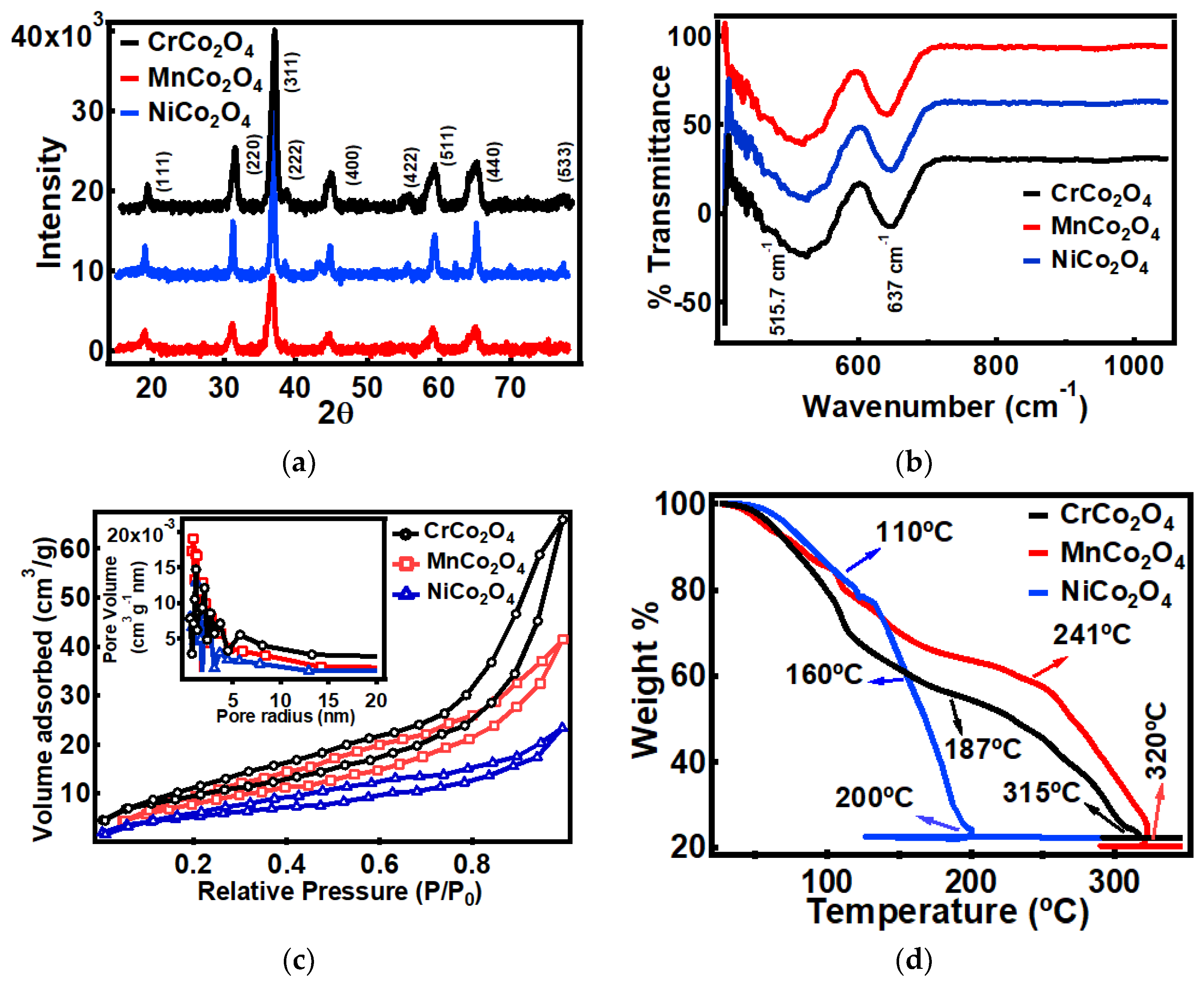
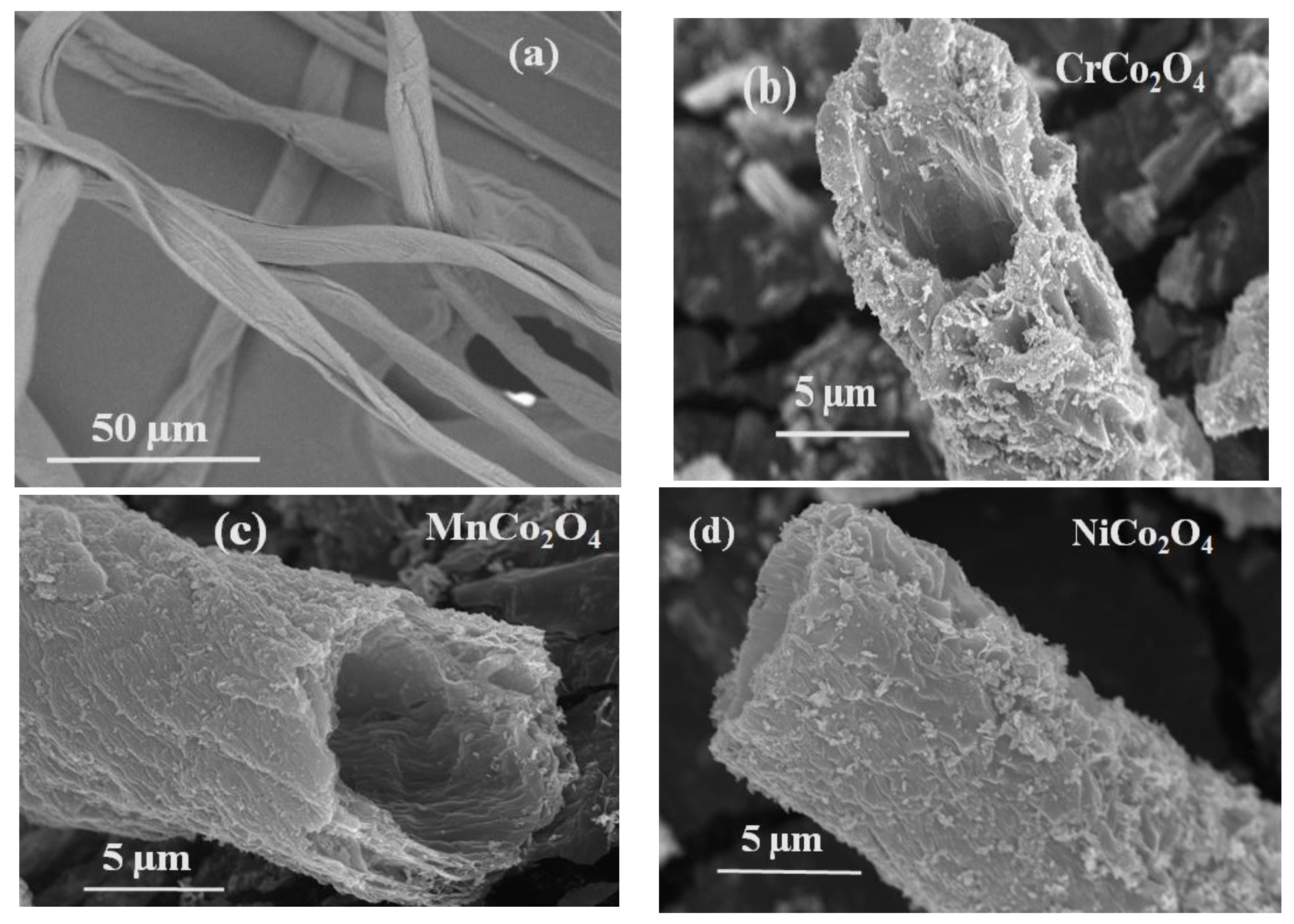
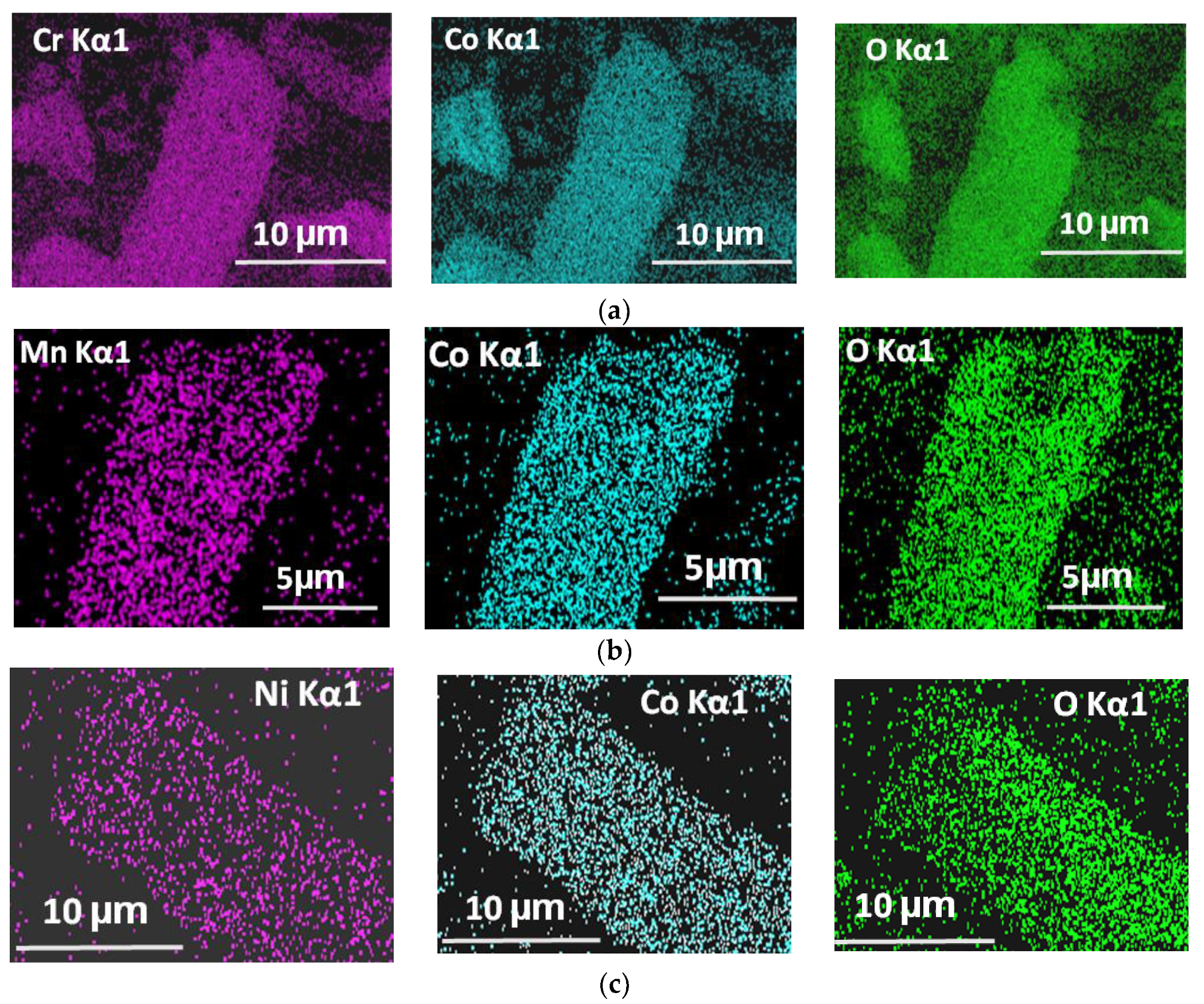
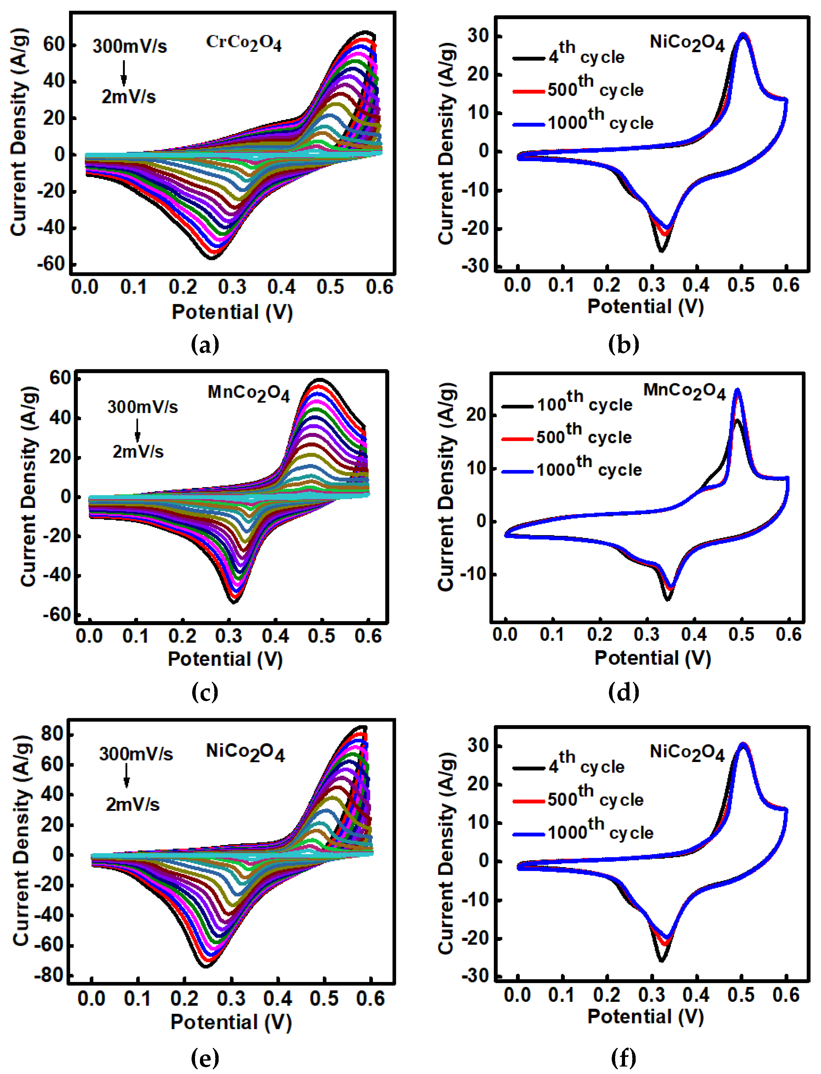
3. Results and Discussion
4. Conclusions
Author Contributions
Funding
Conflicts of Interest
References
- Suktha, P.; Chiochan, P.; Iamprasertkun, P.; Wutthiprom, J.; Phattharasupakun, N.; Suksomboon, M.; Kaewsongpol, T.; Sirisinudomkit, P.; Pettong, T.; Sawangphruk, M. High-performance supercapacitor of functionalized carbon fiber paper with high surface ionic and bulk electronic conductivity: Effect of organic functional groups. Electrochim. Acta 2015, 176, 504–513. [Google Scholar] [CrossRef]
- Sahoo, S.; Ratha, S.; Rout, C.S. Spinel NiCo2O4 nanorods for supercapacitor applications. Am. J. Eng. Appl. Sci. 2015, 8, 371–379. [Google Scholar] [CrossRef] [Green Version]
- El-Kady, M.F.; Strong, V.; Dubin, S.; Kaner, R.B. Laser scribing of high-performance and flexible graphene-based electrochemical capacitors. Science 2012, 335, 1326–1330. [Google Scholar] [CrossRef] [Green Version]
- Zhu, Y.; Murali, S.; Stoller, M.D.; Ganesh, K.J.; Cai, W.; Ferreira, P.J.; Pirkle, A.; Wallace, R.M.; Cychosz, K.A.; Thommes, M.; et al. Carbon-based supercapacitors produced by activation of graphene. Science 2011, 332, 1537–1541. [Google Scholar] [CrossRef] [Green Version]
- Pettong, T.; Iamprasertkun, P.; Krittayavathananon, A.; Sukha, P.; Sirisinudomkit, P.; Seubsai, A.; Chareonpanich, M.; Kongkachuichay, P.; Limtrakul, J.; Sawangphruk, M. High-performance asymmetric supercapacitors of MnCo2O4 nanofibers and N-doped reduced graphene oxide aerogel. ACS Appl. Mater. Interfaces 2016, 8, 34045–34053. [Google Scholar] [CrossRef] [PubMed]
- Fan, L.Z.; Qiao, S.; Song, W.; Wu, M.; He, X.; Qu, X. Effects of the functional groups on the electrochemical properties of ordered porous carbon for supercapacitors. Electrochim. Acta 2013, 105, 299–304. [Google Scholar] [CrossRef]
- Chen, X.L.; Li, W.S.; Tan, C.L.; Li, W.; Wu, Y.Z. Improvement in electrochemical capacitance of carbon materials by nitric acid treatment. J. Power Sources 2008, 184, 668–674. [Google Scholar] [CrossRef]
- Yang, Q.; Lu, Z.; Li, T.; Sun, X.; Liu, J. Hierarchical construction of core–shell metal oxide nanoarrays with ultrahigh areal capacitance. Nano Energy 2014, 7, 170–178. [Google Scholar] [CrossRef]
- Vilatela, J.J.; Eder, D. Nanocarbon composites and hybrids in sustainability: A review. ChemSusChem 2012, 5, 456–478. [Google Scholar] [CrossRef]
- Nguyen, T.; Montemor, M.D.F. Metal Oxide and Hydroxide–Based Aqueous Supercapacitors: From Charge Storage Mechanisms and Functional Electrode Engineering to Need-Tailored Devices. Adv. Sci. 2019, 6, 1801797. [Google Scholar] [CrossRef]
- Alqahtani, D.M.; Zequine, C.; Ranaweera, C.K.; Siam, K.; Kahol, P.K.; Poudel, T.P.; Mishra, S.R.; Gupta, R.K. Effect of metal ion substitution on electrochemical properties of cobalt oxide. J. Alloy. Compd. 2019, 771, 951–959. [Google Scholar] [CrossRef]
- Pendashteh, A.; Moosavifard, S.E.; Rahmanifar, M.S.; Wang, Y.; El-Kady, M.F.; Kaner, R.B.; Mousavi, M.F. Highly ordered mesoporous CuCo2O4 nanowires, a promising solution for high-performance supercapacitors. Chem. Mater. 2015, 27, 3919–3926. [Google Scholar] [CrossRef]
- Cheng, H.; Lu, Z.G.; Deng, J.Q.; Chung, C.Y.; Zhang, K.; Li, Y.Y. A facile method to improve the high rate capability of Co3O4 nanowire array electrodes. Nano Res. 2010, 3, 895–901. [Google Scholar] [CrossRef] [Green Version]
- Yan, D.; Zhang, H.; Chen, L.; Zhu, G.; Li, S.; Xu, H.; Yu, A. Biomorphic synthesis of mesoporous Co3O4 microtubules and their pseudocapacitive performance. ACS Appl. Mater. Interfaces 2014, 6, 15632–15637. [Google Scholar] [CrossRef] [PubMed]
- Sieber, H. Biomimetic synthesis of ceramics and ceramic composites. Mater. Sci. Eng. A 2005, 412, 43–47. [Google Scholar] [CrossRef]
- Habibi, Y.; Lucia, L.A.; Rojas, O.J. Cellulose nanocrystals: Chemistry, self-assembly, and applications. Chem. Rev. 2010, 110, 3479–3500. [Google Scholar] [CrossRef]
- Liu, Y.; Lv, B.; Li, P.; Chen, Y.; Gao, B.; Lin, B. Biotemplate-assisted hydrothermal synthesis of tubular porous Co3O4 with excellent charge-discharge cycle stability for supercapacitive electrodes. Mater. Lett. 2018, 210, 231–234. [Google Scholar] [CrossRef]
- Fan, T.X.; Chow, S.K.; Zhang, D. Biomorphic mineralization: From biology to materials. Prog. Mater. Sci. 2009, 54, 542–659. [Google Scholar] [CrossRef]
- Sotiropoulou, S.; Sierra-Sastre, Y.; Mark, S.S.; Batt, C.A. Biotemplated nanostructured materials. Chem. Mater. 2008, 20, 821–834. [Google Scholar] [CrossRef]
- Zhou, H.; Fan, T.; Zhang, D. Biotemplated materials for sustainable energy and environment: Current status and challenges. ChemSusChem 2011, 4, 1344–1387. [Google Scholar] [CrossRef]
- Shim, H.W.; Lim, A.H.; Kim, J.C.; Jang, E.; Seo, S.D.; Lee, G.H.; Kim, T.D.; Kim, D.W. Scalable one-pot bacteria-templating synthesis route toward hierarchical, porous-Co3O4 superstructures for supercapacitor electrodes. Sci. Rep. 2013, 3, 2325. [Google Scholar] [CrossRef] [PubMed] [Green Version]
- Han, L.; Yang, D.P.; Liu, A. Leaf-templated synthesis of 3D hierarchical porous cobalt oxide nanostructure as direct electrochemical biosensing interface with enhanced electrocatalysis. Biosens. Bioelectron. 2015, 63, 145–152. [Google Scholar] [CrossRef]
- Song, P.; Zhang, H.; Han, D.; Li, J.; Yang, Z.; Wang, Q. Preparation of biomorphic porous LaFeO3 by sorghum straw biotemplate method and its acetone sensing properties. Sens. Actuators B Chem. 2014, 196, 140–146. [Google Scholar] [CrossRef]
- Weatherspoon, M.R.; Cai, Y.; Crne, M.; Srinivasarao, M.; Sandhage, K.H. 3D Rutile Titania-Based Structures with Morpho Butterfly Wing Scale Morphologies. Angew. Chem. Int. Ed. 2008, 47, 7921–7923. [Google Scholar] [CrossRef] [PubMed]
- Yan, D.; Li, S.; Zhu, G.; Wang, Z.; Xu, H.; Yu, A. Synthesis and pseudocapacitive behaviors of biomorphic mesoporous tubular MnO2 templated from cotton. Mater. Lett. 2013, 95, 164–167. [Google Scholar] [CrossRef]
- Mondal, A.K.; Su, D.; Chen, S.; Ung, A.; Kim, H.S.; Wang, G. Mesoporous MnCo2O4 with a flake-like structure as advanced electrode materials for lithium-ion batteries and supercapacitors. Chem. A Eur. J. 2015, 21, 1526–1532. [Google Scholar] [CrossRef]
- Xu, J.; Sun, Y.; Lu, M.; Wang, L.; Zhang, J.; Tao, E.; Qian, J.; Liu, X. Fabrication of the porous MnCo2O4 nanorod arrays on Ni foam as an advanced electrode for asymmetric supercapacitors. Acta Mater. 2018, 152, 162–174. [Google Scholar] [CrossRef]
- Dong, Y.; Wang, Y.; Xu, Y.; Chen, C.; Wang, Y.; Jiao, L.; Yuan, H. Facile synthesis of hierarchical nanocage MnCo2O4 for high performance supercapacitor. Electrochim. Acta 2017, 225, 39–46. [Google Scholar] [CrossRef]
- Yang, J.; Cho, M.; Lee, Y. Synthesis of hierarchical NiCo2O4 hollow nanorods via sacrificial-template accelerate hydrolysis for electrochemical glucose oxidation. Biosens. Bioelectron. 2016, 75, 15–22. [Google Scholar] [CrossRef]
- Zhao, Z.; Geng, F.; Bai, J.; Cheng, H.M. Facile and controlled synthesis of 3D nanorods-based urchinlike and nanosheets-based flowerlike cobalt basic salt nanostructures. J. Phys. Chem. C 2007, 111, 3848–3852. [Google Scholar] [CrossRef]
- Jadhav, A.R.; Bandal, H.A.; Kim, H. NiCo2O4 hollow sphere as an efficient catalyst for hydrogen generation by NaBH4 hydrolysis. Mater. Lett. 2017, 198, 50–53. [Google Scholar] [CrossRef]
- Huang, W.; Lin, T.; Cao, Y.; Lai, X.; Peng, J.; Tu, J. Hierarchical NiCo2O4 hollow sphere as a peroxidase mimetic for colorimetric detection of H2O2 and glucose. Sensors 2017, 17, 217. [Google Scholar] [CrossRef] [Green Version]
- Pu, J.; Wang, J.; Jin, X.; Cui, F.; Sheng, E.; Wang, Z. Porous hexagonal NiCo2O4 nanoplates as electrode materials for supercapacitors. Electrochim. Acta 2013, 106, 226–234. [Google Scholar] [CrossRef]
- Park, J.; Shen, X.; Wang, G. Solvothermal synthesis and gas-sensing performance of Co3O4 hollow nanospheres. Sens. Actuators B Chem. 2009, 136, 494–498. [Google Scholar] [CrossRef]
- Lasheras, X.; Insausti, M.; Gil de Muro, I.; Garaio, E.; Plazaola, F.; Moros, M.; De Matteis, L.; M de la Fuente, J.; Lezama, L. Chemical synthesis and magnetic properties of monodisperse nickel ferrite nanoparticles for biomedical applications. J. Phys. Chem. C 2016, 120, 3492–3500. [Google Scholar] [CrossRef] [Green Version]
- Kuang, L.; Ji, F.; Pan, X.; Wang, D.; Chen, X.; Jiang, D.; Zhang, Y.; Ding, B. Mesoporous MnCo2O4 nanoneedle arrays electrode for high-performance asymmetric supercapacitor application. Chem. Eng. J. 2017, 315, 491–499. [Google Scholar] [CrossRef]
- Melot, B.C.; Tarascon, J.M. Design and preparation of materials for advanced electrochemical storage. Acc. Chem. Res. 2013, 46, 1226–1238. [Google Scholar] [CrossRef]
- Chen, K.; Xue, D. Materials chemistry toward electrochemical energy storage. J. Mater. Chem. A 2016, 4, 7522–7537. [Google Scholar] [CrossRef]
- Principles and Applications of Lithium Secondary Batteries; Park, J.K. (Ed.) John Wiley & Sons: Hoboken, NJ, USA, 2012. [Google Scholar]
- Zhang, Y.; Ma, M.; Yang, J.; Su, H.; Huang, W.; Dong, X. Selective synthesis of hierarchical mesoporous spinel NiCo2O4 for high-performance supercapacitors. Nanoscale 2014, 6, 4303–4308. [Google Scholar] [CrossRef]
- Yuan, C.; Wu, H.B.; Xie, Y.; Lou, X.W. Mixed transition-metal oxides: Design, synthesis, and energy-related applications. Angew. Chem. Int. Ed. 2014, 53, 1488–1504. [Google Scholar] [CrossRef]
- Zhang, L.L.; Zhao, X.S. Carbon-based materials as supercapacitor electrodes. Chem. Soc. Rev. 2009, 38, 2520–2531. [Google Scholar] [CrossRef]
- Zheng, Y.Z.; Ding, H.; Uchaker, E.; Tao, X.; Chen, J.F.; Zhang, Q.; Cao, G. Nickel-mediated polyol synthesis of hierarchical V2O5 hollow microspheres with enhanced lithium storage properties. J. Mater. Chem. A 2015, 3, 1979–1985. [Google Scholar] [CrossRef]
- Lithium Batteries: Science and Technology; Nazri, G.A.; Pistoia, G. (Eds.) Springer Science & Business Media: Berlin/Heidelberg, Germany, 2008. [Google Scholar]
- Li, J.; Wang, J.; Liang, X.; Zhang, Z.; Liu, H.; Qian, Y.; Xiong, S. Hollow MnCo2O4 submicrospheres with multilevel interiors: From mesoporous spheres to yolk-in-double-shell structures. ACS Appl. Mater. Interfaces 2013, 6, 24–30. [Google Scholar] [CrossRef] [PubMed] [Green Version]
- Bhojane, P.; Sen, S.; Shirage, P.M. Enhanced electrochemical performance of mesoporous NiCo2O4 as an excellent supercapacitive alternative energy storage material. Appl. Surf. Sci. 2016, 377, 376–384. [Google Scholar] [CrossRef]
- Jenkins, R.; Snyder, R.L. Introduction to X-ray Powder Diffractometry; (No. 543.427 JEN); Wiley: Hoboken, NJ, USA, 1996. [Google Scholar]
- Lin, H.K.; Chiu, H.C.; Tsai, H.C.; Chien, S.H.; Wang, C.B. Synthesis, characterization and catalytic oxidation of carbon monoxide over cobalt oxide. Catal. Lett. 2003, 88, 169–174. [Google Scholar] [CrossRef]
- Spencer, C.D.; Schroeer, D. Mössbauer study of several cobalt spinels using Co57 and Fe57. Phys. Rev. B 1974, 9, 3658. [Google Scholar] [CrossRef]
- Kurtulus, F.; Guler, H. A Simple Microwave-Assisted Route to Prepare Black Cobalt, Co3O4. Inorg. Mater. 2005, 41, 483–485. [Google Scholar] [CrossRef]
- St, G.C.; Stoyanova, M.; Georgieva, M.; Mehandjiev, D. Preparation and characterization of a higher cobalt oxide. Mater. Chem. Phys. 1999, 60, 39–43. [Google Scholar]
- Khairy, M.; Mousa, M. Synthesis of Ternary and Quaternary metal oxides based on Ni, Mn, Cu, and Co for high-performance Supercapacitor. J. Ovonic Res. 2019, 15, 181–198. [Google Scholar]
- Štěpánek, F.; Marek, M.; Adler, P.M. Modeling capillary condensation hysteresis cycles in reconstructed porous media. Aiche J. 1999, 45, 1901–1912. [Google Scholar] [CrossRef]
- Wang, R.; Li, Q.; Cheng, L.; Li, H.; Wang, B.; Zhao, X.S.; Guo, P. Electrochemical properties of manganese ferrite-based supercapacitors in aqueous electrolyte: The effect of ionic radius. Colloid. Surfaces A Physicochem. Eng. Aspects 2014, 457, 94–99. [Google Scholar] [CrossRef] [Green Version]
- Hou, L.; Yuan, C.; Yang, L.; Shen, L.; Zhang, F.; Zhang, X. Urchin-like Co3O4 microspherical hierarchical superstructures constructed by one-dimension nanowires toward electrochemical capacitors. Rsc Adv. 2011, 1, 1521–1526. [Google Scholar] [CrossRef]
- Yuan, C.; Yang, L.; Hou, L.; Shen, L.; Zhang, X.; Lou, X.W.D. Growth of ultrathin mesoporous Co3O4 nanosheet arrays on Ni foam for high-performance electrochemical capacitors. Energy Environ. Sci. 2012, 5, 7883–7887. [Google Scholar] [CrossRef]
- Adhikari, H.; Ghimire, M.; Ranaweera, C.K.; Bhoyate, S.; Gupta, R.K.; Alam, J.; Mishra, S.R. Synthesis and electrochemical performance of hydrothermally synthesized Co3O4 nanostructured particles in presence of urea. J. Alloy. Compd. 2017, 708, 628–638. [Google Scholar] [CrossRef]
- Sun, D.; He, L.; Chen, R.; Liu, Y.; Lv, B.; Lin, S.; Lin, B. Biomorphic composites composed of octahedral Co3O4 nanocrystals and mesoporous carbon microtubes templated from cotton for excellent supercapacitor electrodes. Appl. Surf. Sci. 2019, 465, 232–240. [Google Scholar] [CrossRef]
- Brousse, T.; Bélanger, D. A Hybrid Fe3O4 MnO2 Capacitor in Mild Aqueous Electrolyte. Electrochem. Solid-State Lett. 2003, 6, A244–A248. [Google Scholar] [CrossRef]
- Wang, S.Y.; Ho, K.C.; Kuo, S.L.; Wu, N.L. Investigation on capacitance mechanisms of Fe3O4 electrochemical capacitors. J. Electrochem. Soc. 2006, 153, A75–A80. [Google Scholar] [CrossRef]
- Tiruye, G.A.; Munoz-Torrero, D.; Palma, J.; Anderson, M.; Marcilla, R. All-solid state supercapacitors operating at 3.5 V by using ionic liquid based polymer electrolytes. J. Power Sources 2015, 279, 472–480. [Google Scholar] [CrossRef]
- Gao, F.; Shao, G.; Qu, J.; Lv, S.; Li, Y.; Wu, M. Tailoring of porous and nitrogen-rich carbons derived from hydrochar for high-performance supercapacitor electrodes. Electrochim. Acta 2015, 155, 201–208. [Google Scholar] [CrossRef]
- Selvam, M.; Srither, S.R.; Saminathan, K.; Rajendran, V. Chemically and electrochemically prepared graphene/MnO2 nanocomposite electrodes for zinc primary cells: A comparative study. Ionics 2015, 21, 791–799. [Google Scholar] [CrossRef]
- Tang, Y.; Liu, Y.; Yu, S.; Gao, F.; Zhao, Y. Comparative study on three commercial carbons for supercapacitor applications. Russ. J. Electrochem. 2015, 51, 77–85. [Google Scholar] [CrossRef]
- Sankar, K.V.; Selvan, R.K. Improved electrochemical performances of reduced graphene oxide based supercapacitor using redox additive electrolyte. Carbon 2015, 90, 260–273. [Google Scholar] [CrossRef]
- Sahu, V.; Shekhar, S.; Sharma, R.K.; Singh, G. Ultrahigh performance supercapacitor from lacey reduced graphene oxide nanoribbons. ACS Appl. Mater. Interfaces 2015, 7, 3110–3116. [Google Scholar] [CrossRef] [PubMed]
- Fic, K.; Platek, A.; Piwek, J.; Frackowiak, E. Sustainable materials for electrochemical capacitors. Mater. Today 2018, 21, 437–454. [Google Scholar] [CrossRef]
- Meher, S.K.; Justin, P.; Rao, G.R. Nanoscale morphology dependent pseudocapacitance of NiO: Influence of intercalating anions during synthesis. Nanoscale 2011, 3, 683–692. [Google Scholar] [CrossRef]
- Xiao, J.; Yang, S. Sequential crystallization of sea urchin-like bimetallic (Ni, Co) carbonate hydroxide and its morphology conserved conversion to porous NiCo2O4 spinel for pseudocapacitors. Rsc Adv. 2011, 1, 588–595. [Google Scholar] [CrossRef]
- Liu, X.Y.; Zhang, Y.Q.; Xia, X.H.; Shi, S.J.; Lu, Y.; Wang, X.L.; Gu, C.D.; Tu, J.P. Self-assembled porous NiCo2O4 hetero-structure array for electrochemical capacitor. J. Power Sources 2013, 239, 157–163. [Google Scholar] [CrossRef]
- Wang, X.; Han, X.; Lim, M.; Singh, N.; Gan, C.L.; Jan, M.; Lee, P.S. Nickel cobalt oxide-single wall carbon nanotube composite material for superior cycling stability and high-performance supercapacitor application. J. Phys. Chem. C 2012, 116, 12448–12454. [Google Scholar] [CrossRef]
- Liu, X.; Long, Q.; Jiang, C.; Zhan, B.; Li, C.; Liu, S.; Zhao, Q.; Huang, W.; Dong, X. Facile and green synthesis of mesoporous Co3O4 nanocubes and their applications for supercapacitors. Nanoscale 2013, 5, 6525–6529. [Google Scholar] [CrossRef]
- Zhu, Y.; Pu, X.; Song, W.; Wu, Z.; Zhou, Z.; He, X.; Lu, F.; Jing, M.; Tang, B.; Ji, X. High capacity NiCo2O4 nanorods as electrode materials for supercapacitor. J. Alloy. Compd. 2014, 617, 988–993. [Google Scholar] [CrossRef]
- Jokar, E.; Shahrokhian, S. Synthesis and characterization of NiCo2O4 nanorods for preparation of supercapacitor electrodes. J. Solid State Electrochem. 2015, 19, 269–274. [Google Scholar] [CrossRef]
- Veeramani, V.; Madhu, R.; Chen, S.M.; Sivakumar, M.; Hung, C.T.; Miyamoto, N.; Liu, S.B. NiCo2O4-decorated porous carbon nanosheets for high-performance supercapacitors. Electrochim. Acta 2017, 247, 288–295. [Google Scholar] [CrossRef]
- Zhai, Y.; Mao, H.; Liu, P.; Ren, X.; Xu, L.; Qian, Y. Facile fabrication of hierarchical porous rose-like NiCo2O4 nanoflake/MnCo2O4 nanoparticle composites with enhanced electrochemical performance for energy storage. J. Mater. Chem. A 2015, 3, 16142–16149. [Google Scholar] [CrossRef]
- Ghosh, D.; Giri, S.; Das, C.K. Hydrothermal synthesis of platelet β Co(OH)2 and Co3O4: Smart electrode material for energy storage application. Environ. Prog. Sustain. Energy 2014, 33, 1059–1064. [Google Scholar] [CrossRef]
- Yi, H.; Wang, H.; Jing, Y.; Peng, T.; Wang, X. Asymmetric supercapacitors based on carbon nanotubes@ NiO ultrathin nanosheets core-shell composites and MOF-derived porous carbon polyhedrons with super-long cycle life. J. Power Sources 2015, 285, 281–290. [Google Scholar] [CrossRef]
- Shen, B.; Guo, R.; Lang, J.; Liu, L.; Liu, L.; Yan, X. A high-temperature flexible supercapacitor based on pseudocapacitive behavior of FeOOH in an ionic liquid electrolyte. J. Mater. Chem. A 2016, 4, 8316–8327. [Google Scholar] [CrossRef]
- Augustyn, V.; Come, J.; Lowe, M.A.; Kim, J.W.; Taberna, P.L.; Tolbert, S.H.; Abruña, H.D.; Simon, P.; Dunn, B. High-rate electrochemical energy storage through Li+ intercalation pseudocapacitance. Nat. Mater. 2013, 12, 518. [Google Scholar] [CrossRef]
- Lindström, H.; Södergren, S.; Solbrand, A.; Rensmo, H.; Hjelm, J.; Hagfeldt, A.; Lindquist, S.E. Li+ ion insertion in TiO2 (anatase). 1. Chronoamperometry on CVD films and nanoporous films. J. Phys. Chem. B 1997, 101, 7710–7716. [Google Scholar] [CrossRef]
- Liu, T.C.; Pell, W.G.; Conway, B.E.; Roberson, S.L. Behavior of molybdenum nitrides as materials for electrochemical capacitors comparison with ruthenium oxide. J. Electrochem. Soc. 1998, 145, 1882–1888. [Google Scholar] [CrossRef]
- Wang, J.; Polleux, J.; Lim, J.; Dunn, B. Pseudocapacitive contributions to electrochemical energy storage in TiO2 (anatase) nanoparticles. J. Phys. Chem. C 2007, 111, 14925–14931. [Google Scholar] [CrossRef]
- Zhao, D.D.; Bao, S.J.; Zhou, W.J.; Li, H.L. Preparation of hexagonal nanoporous nickel hydroxide film and its application for electrochemical capacitor. Electrochem. Commun. 2007, 9, 869–874. [Google Scholar] [CrossRef]
- Sugimoto, W.; Iwata, H.; Yasunaga, Y.; Murakami, Y.; Takasu, Y. Preparation of ruthenic acid nanosheets and utilization of its interlayer surface for electrochemical energy storage. Angew. Chem. Int. Ed. 2003, 42, 4092–4096. [Google Scholar] [CrossRef] [PubMed]
- Li, L.; Zhang, Y.Q.; Liu, X.Y.; Shi, S.J.; Zhao, X.Y.; Zhang, H.; Ge, X.; Cai, G.F.; Gu, C.D.; Wang, X.L.; et al. One-dimension MnCo2O4 nanowire arrays for electrochemical energy storage. Electrochim. Acta 2014, 116, 467–474. [Google Scholar] [CrossRef]
- Hui, K.N.; San Hui, K.; Tang, Z.; Jadhav, V.V.; Xia, Q.X. Hierarchical chestnut-like MnCo2O4 nanoneedles grown on nickel foam as binder-free electrode for high energy density asymmetric supercapacitors. J. Power Sources 2016, 330, 195–203. [Google Scholar] [CrossRef] [Green Version]
- Shi, X.; Liu, Z.; Zheng, Y.; Zhou, G. Controllable synthesis and electrochemical properties of MnCo2O4 nanorods and microcubes. Colloids Surf. A: Physicochem. Eng. Asp. 2017, 522, 525–535. [Google Scholar] [CrossRef]
- Liu, S.; Hui, K.S.; Hui, K.N. 1 D hierarchical MnCo2O4 nanowire@ MnO2 sheet core–shell arrays on graphite paper as superior electrodes for asymmetric supercapacitors. ChemNanoMat 2015, 1, 593–602. [Google Scholar] [CrossRef] [Green Version]
- Venkatachalam, V.; Alsalme, A.; Alghamdi, A.; Jayavel, R. High performance electrochemical capacitor based on MnCo2O4 nanostructured electrode. J. Electroanal. Chem. 2015, 756, 94–100. [Google Scholar] [CrossRef]
- Zhu, Y.; Ji, X.; Yin, R.; Hu, Z.; Qiu, X.; Wu, Z.; Liu, Y. Nanorod-assembled NiCo2O4 hollow microspheres assisted by an ionic liquid as advanced electrode materials for supercapacitors. Rsc Adv. 2017, 7, 11123–11128. [Google Scholar] [CrossRef] [Green Version]
- Mao, J.W.; He, C.H.; Qi, J.Q.; Zhang, A.B.; Sui, Y.W.; He, Y.Z.; Meng, Q.K.; Wei, F.X. An Asymmetric Supercapacitor with Mesoporous NiCo2O4 Nanorod/Graphene Composite and N-Doped Graphene Electrodes. J. Electron. Mater. 2018, 47, 512–520. [Google Scholar] [CrossRef]
- Jabeen, N.; Xia, Q.; Yang, M.; Xia, H. Unique core–shell nanorod arrays with polyaniline deposited into mesoporous NiCo2O4 support for high-performance supercapacitor electrodes. Acs Appl. Mater. Interfaces 2016, 8, 6093–6100. [Google Scholar] [CrossRef]
- Srinivasan, V.; Weidner, J.W. Studies on the capacitance of nickel oxide films: Effect of heating temperature and electrolyte concentration. J. Electrochem. Soc. 2000, 147, 880–885. [Google Scholar] [CrossRef]
- Lin, C.; Ritter, J.A.; Popov, B.N. Characterization of sol-gel-derived cobalt oxide xerogels as electrochemical capacitors. J. Electrochem. Soc. 1998, 145, 4097–4103. [Google Scholar] [CrossRef] [Green Version]
- Zhi, M.; Xiang, C.; Li, J.; Li, M.; Wu, N. Nanostructured carbon–metal oxide composite electrodes for supercapacitors: A review. Nanoscale 2013, 5, 72–88. [Google Scholar] [CrossRef] [PubMed]
- Wu, Z.; Zhu, Y.; Ji, X. NiCo2O4-based materials for electrochemical supercapacitors. J. Mater. Chem. A 2014, 2, 14759–14772. [Google Scholar] [CrossRef]
- Bitla, Y.; Chin, Y.Y.; Lin, J.C.; Van, C.N.; Liu, R.; Zhu, Y.; Liu, H.J.; Zhan, Q.; Lin, H.J.; Chen, C.T.; et al. Origin of metallic behavior in NiCo 2 O 4 ferrimagnet. Sci. Rep. 2015, 5, 15201. [Google Scholar] [CrossRef] [Green Version]


| Sample | BET Surface Area(m2/g) | BJH Surface Area (m2/g) | BJH Avg. Pore Radius (nm) | BJH Avg. Pore Volume (cc/g) | Crystallite Size (nm) |
|---|---|---|---|---|---|
| CrCo2O4 | 34.4 | 46.9 | 1.135 | 0.106 | 10.57 |
| MnCo2O4 | 32.2 | 47.7 | 0.839 | 0.071 | 14.65 |
| NiCo2O4 | 18.9 | 31.8 | 1.129 | 0.039 | 19.97 |
| Co | C | O | Cr | Mn | Ni | |
|---|---|---|---|---|---|---|
| CrCo2O4 | 19.5 | 49.4 | 23.2 | 7.8 | ||
| MnCo2O4 | 33.1 | 34.6 | 23.5 | 8.8 | ||
| NiCo2O4 | 27.2 | 42.4 | 19.3 | 11.1 |
| CrCo2O4 | MnCo2O4 | NiCo2O4 | |
|---|---|---|---|
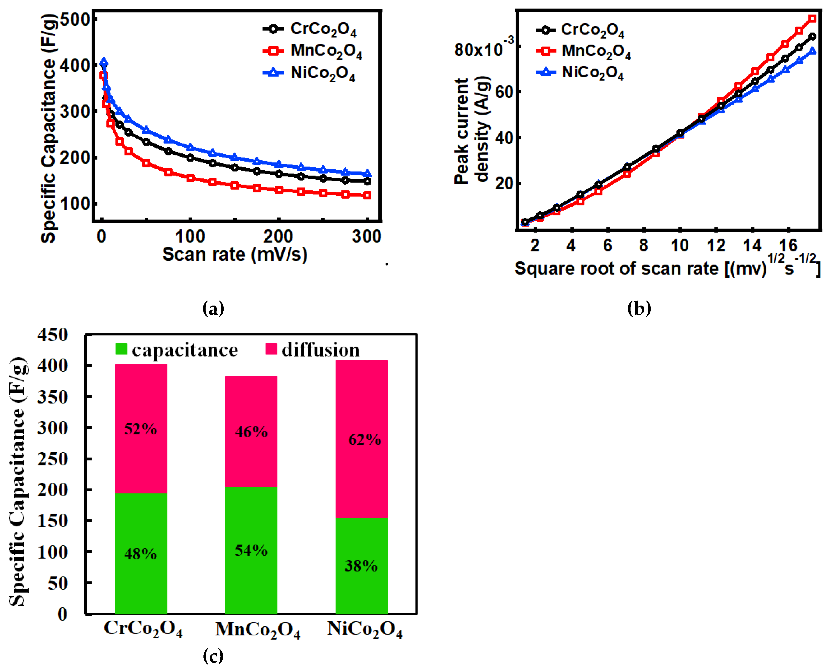
| Specific Capacitance at 2mv/s | 403.2 F/g | 378.1 F/g | 407.2 F/g |
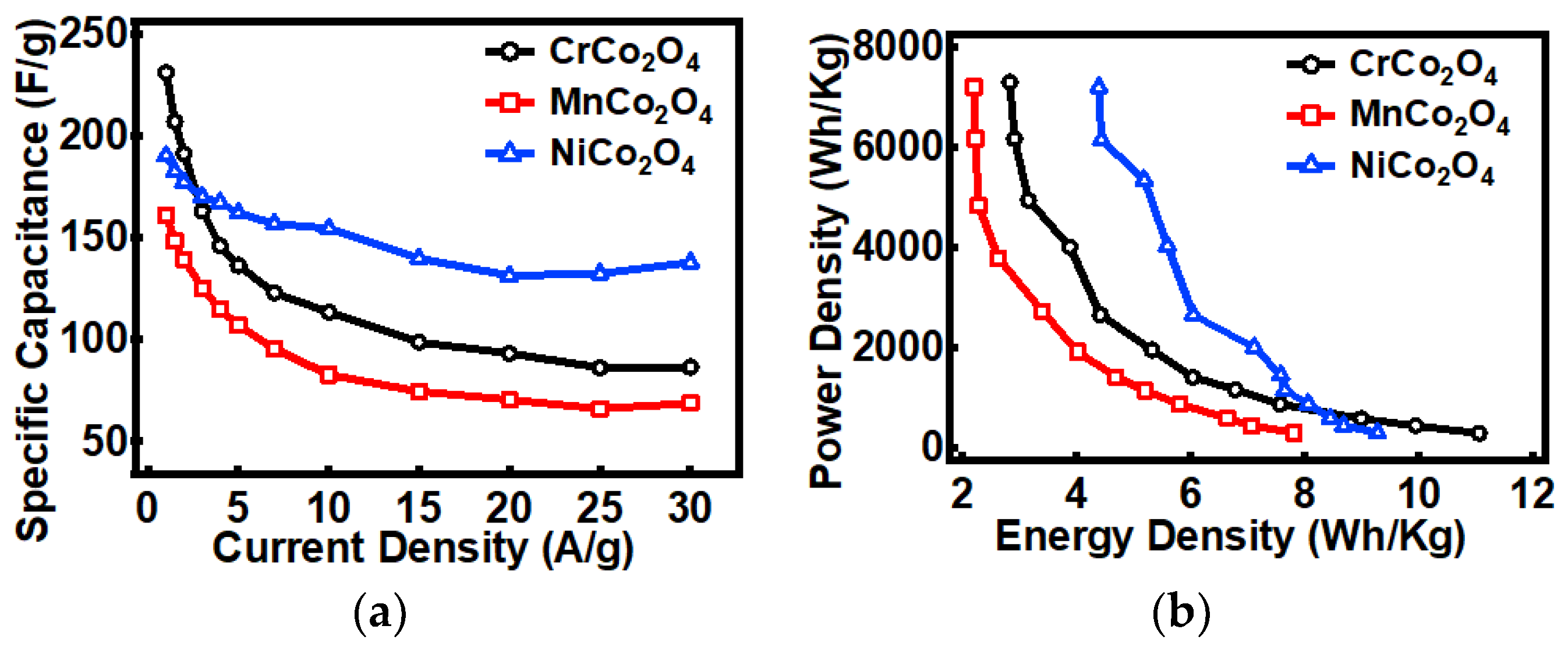
| Specific Capacitance at 1A/g | 231 F/g | 161 F/g | 190 F/g |
| Energy density | 11.1 Wh/Kg | 7.8 Wh/Kg | 9.3 Wh/Kg |
| Power density | 7287.34 W/Kg | 7195.33 W/Kg | 7186.12 W/Kg |
| Electrode Material | Electrolyte | Specific Capacitance | Energy Density | Power Density | Cyclic Performance (retention) | Ref. |
|---|---|---|---|---|---|---|
| MnCo2O4 nanofiber | 108 F/g at 10 A/g | 54 Wh/Kg | 9851 W/Kg | 85 % after 3000 cycles | [5] | |
| Nanorods MnCo2O4 | 1M KOH | 349.8 F/g at 1 A/g | 35.4 Wh/Kg | 225 W/Kg | 92.7% after 50 cycles | [86] |
| Nanoneedles MnCo2O4 | 6M KOH | 1535 F/g at 1 A/g | 35.4 Wh/Kg | 225 W/Kg | 94.3% after 12000 cycles | [87] |
| Nanorods MnCo2O4 | 2M KOH | 845.6 F/g at 1 A/g | 35.4 Wh/Kg | 225 W/Kg | 90.2% after 2000 cycles | [24] |
| Nanorods MnCo2O4 | 1M KOH | 308.3 F/g at 1 A/g | 55.5 Wh/Kg | 5400 W/Kg | 88.76% after 2000 cycles | [88] |
| MnCo2O4 nanowires @MnO2 | - | 2262 F/g at 1 A/g | 85.7 Wh/Kg | 800 W/Kg | - | [89] |
| Nanorods MnCo2O4 | 718 F/g at 0.5 A/g | - | - | 84 % after 1000 cycles | [90] | |
| NiCo2O4 nanorods | 2M KOH | 565 F/g at 1 A/g | - | - | 77.6% after 1000 cycles | [69] |
| RGO decorated nanorods bundle NiCo2O4 | 6M KOH | 1278F/g at 1 A/g | - | - | 95% after 1000 cycles | [84] |
| Nanorods assemble NiCo2O4 | 2M KOH | 764 F/g at 2 A/g | - | - | 101.7% after 1500 cycles | [91] |
| Nanorods NiCo2O4 | 2M KOH | 823 F/g at 0.823 A/g | 28.51 Wh/Kg | - | 101.7% after 1500 cycles | [2] |
| GO/ Nanorods NiCo2O4 | 1M KOH | 709.7 F/g at 1 A/g | 28 Wh/Kg | 8000 W/Kg | 94.3% after 5000 cycles | [92] |
| Nanorods NiCo2O4 | 2M KOH | 600 F/g at 5 A/g | - | - | 80% after 1500 cycles | [70] |
| Nanorods NiCo2O4 @PANI | 1M H2So4 | 901 F/g at 5 A/g | - | - | 91% after 3000 cycles | [93] |
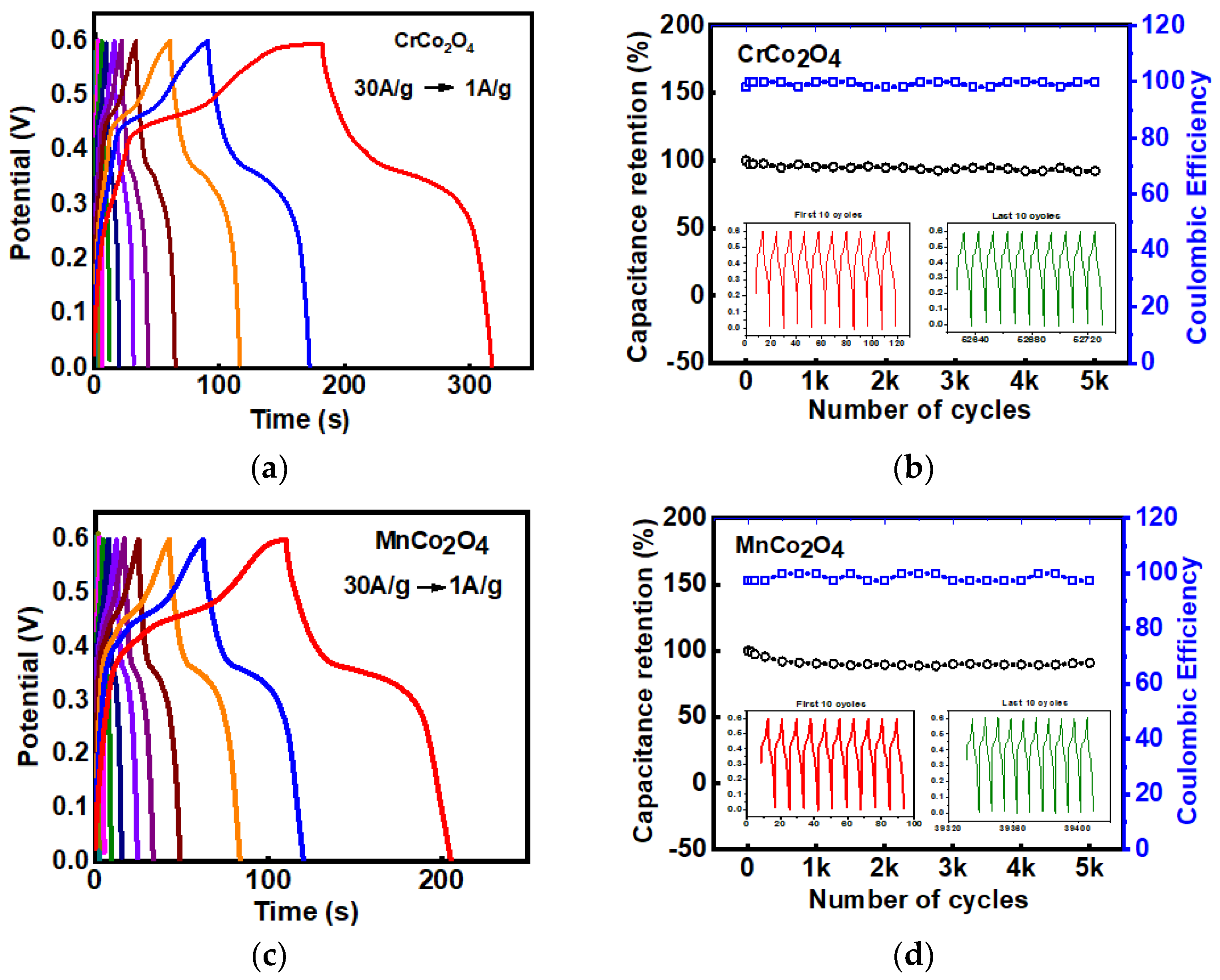
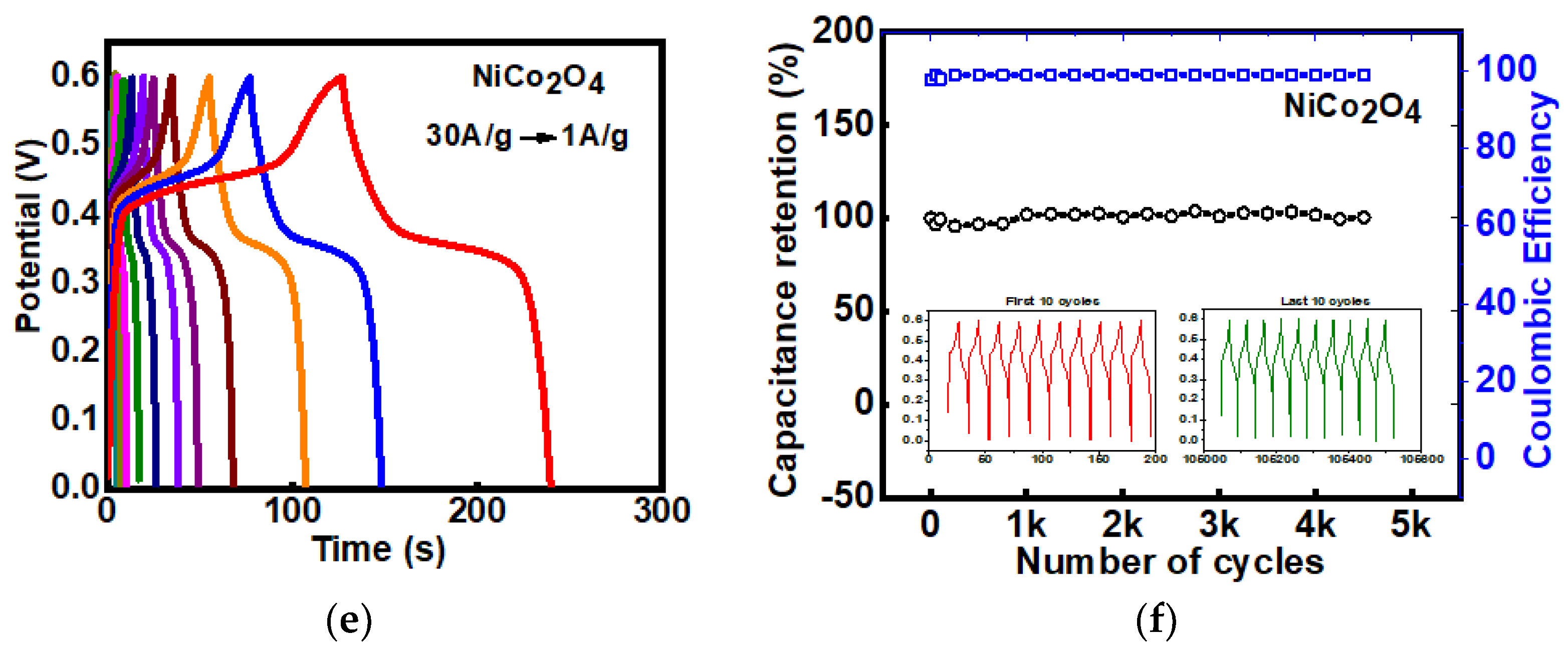
| Templated CrCo2O4, MnCo2O4, and NiCo2O4 microstructure | 3M KOH | 403.4, 378, and 407.2 F/g at 2mV/s and 231, 161, and 190 F/g at 1 A/g, respectively | 11.1, 7.8, and 9.3 Wh/kg, respectively | 7287.3, 7195.3, and 7186.1 W/Kg, respectively | 92% after 5000 cycles 91% after 5000 cycles and 100% after 5000 cycles, respectively. | [This work] |
© 2020 by the authors. Licensee MDPI, Basel, Switzerland. This article is an open access article distributed under the terms and conditions of the Creative Commons Attribution (CC BY) license (http://creativecommons.org/licenses/by/4.0/).
Share and Cite
Guragain, D.; Zequine, C.; Gupta, R.K.; Mishra, S.R. Facile Synthesis of Bio-Template Tubular MCo2O4 (M = Cr, Mn, Ni) Microstructure and Its Electrochemical Performance in Aqueous Electrolyte. Processes 2020, 8, 343. https://0-doi-org.brum.beds.ac.uk/10.3390/pr8030343
Guragain D, Zequine C, Gupta RK, Mishra SR. Facile Synthesis of Bio-Template Tubular MCo2O4 (M = Cr, Mn, Ni) Microstructure and Its Electrochemical Performance in Aqueous Electrolyte. Processes. 2020; 8(3):343. https://0-doi-org.brum.beds.ac.uk/10.3390/pr8030343
Chicago/Turabian StyleGuragain, Deepa, Camila Zequine, Ram K Gupta, and Sanjay R Mishra. 2020. "Facile Synthesis of Bio-Template Tubular MCo2O4 (M = Cr, Mn, Ni) Microstructure and Its Electrochemical Performance in Aqueous Electrolyte" Processes 8, no. 3: 343. https://0-doi-org.brum.beds.ac.uk/10.3390/pr8030343