Article

Effects of Mercury II on Cupriavidus metallidurans Strain MSR33 during Mercury Bioremediation under Aerobic and Anaerobic Conditions

by
Guillermo Bravo
1,
Paulina Vega-Celedón
1,
Juan Carlos Gentina
2 and
Michael Seeger
1,*
1
Molecular Microbiology and Environmental Biotechnology Laboratory, Department of Chemistry & Center of Biotechnology Daniel Alkalay Lowitt, Universidad Técnica Federico Santa María, Avenida España 1680, Valparaíso 2390123, Chile
2
School of Biochemical Engineering, Pontificia Universidad Católica de Valparaíso, Avenida Brasil 2085, Valparaíso 2362803, Chile
*
Author to whom correspondence should be addressed.
Submission received: 1 July 2020 / Revised: 21 July 2020 / Accepted: 23 July 2020 / Published: 25 July 2020
(This article belongs to the Special Issue Study of Biodegradation and Bioremediation)

Abstract

:
Mercury is a toxic element that harms organisms and disturbs biogeochemical cycles. Mercury bioremediation is based on the reduction of Hg (II) to Hg (0) by mercury-resistant bacteria. Cupriavidus metallidurans MSR33 possesses a broad-spectrum mercury resistance. This study aims to establish the effects of mercury on growth, oxygen uptake, and mercury removal parameters by C. metallidurans MSR33 in aqueous solution during aerobic and anaerobic mercury bioremediation. A new culture medium (GBC) was designed. The effects of mercury (II) (20 ppm) on growth parameters, oxygen uptake, and mercury removal were evaluated in GBC medium in a bioreactor (3 L) under aerobiosis. The anaerobic kinetics of mercury removal was evaluated by nitrogen replacement during mercury bioremediation in a bioreactor. Strain MSR33 reached a growth rate of µ = 0.43 h−1 in the bioreactor. Mercury inhibited oxygen uptake and bacterial growth; however, this inhibition was reversed after 5 h. Strain MSR33 was able to reduce Hg (II) under aerobic and anaerobic conditions, reaching, at 24 h, a metal removal of 97% and 71%, respectively. Therefore, oxygen was crucial for efficient mercury removal by this bacterium. Strain MSR33 was capable of tolerating the toxic effects of mercury (II) during aerobic bioremediation and recovered its metabolic activity.

1. Introduction

Mercury is a highly toxic metal for cells and is distributed in the water, soil, and air, due to human activities including mining and natural cycles [1,2,3,4]. Mercury presents in the Earth’s crust at concentrations between 20 and 150 ppb [4]. Until 2010, it was estimated that a cumulative total of 1540 (1060−2800) Gg of mercury had been released by human activities, 73% of which were released after 1850 [5]. The main sources of mercury pollution are mining (e.g., gold, copper), the chloralkali industry, sludge deposited in landfills, paints, disinfectants, pharmaceuticals, and seed-coat dressing, which mobilizes mercury into the water, soil, and atmosphere [3,4,5]. Mercury (II) is a highly toxic specie due to its high affinity to sulfhydryl and thioester groups of proteins, inactivating proteins of living organisms [2,6,7,8,9]. Due to its high toxicity, several technologies have been developed for the remediation of mercury. Physicochemical processes are efficient, but expensive at the industrial scale and not applicable in large polluted areas [10,11]. Bioremediation is an eco-friendly and low-cost strategy for the clean-up of polluted environments but is limited to a range of pollutant concentrations that are tolerated by microorganisms [12,13,14,15,16,17,18]. Bioremediation is based on the inoculation of microorganisms, especially bacteria and fungi, to mineralize or transform toxic compounds or elements into less toxic forms [12,13,14,15,16,17,18]. Mercury bioremediation by metal-resistant bacteria is mainly based on the reduction of Hg (II) to Hg (0) by proteins codified by the mer genes and the reducing power NADPH (Figure 1). Elemental mercury, Hg (0), is volatile due to its high vapor pressure and insoluble in water. Mercury removal strategies are based on the volatility and water insolubility of this heavy metal [19]. Diverse bacteria, including C. metallidurans strains CH34 and MSR33, Pseudomonas putida strains PpY101/pSR134, Spi3, Elb2 and KT2442, P. stutzeri strains Ibu8 and OX, P. aeruginosa Bro12, Aeromonas hydrophila, and Sphingomonas sp. SA2, reduce mercury under aerobic conditions [3,10,11,20,21,22,23,24]. Studies of mercury reduction under anaerobic conditions are scarce. P. stutzeri OX, Geobacter bemidjiensis Bem, and Geobacter sulfurreducens PCA reduce mercury under anaerobiosis [20,25,26].
C. metallidurans strains are highly resistant to heavy metals and are able to metabolize toxic organic pollutants such as toluene under aerobic and anaerobic conditions; therefore, specific strains have been applied in bioremediation [27,28,29,30]. C. metallidurans strain CH34 is a facultative anaerobe and heavy metal multi-resistant bacterium associated to the gold biogeochemical cycle [7,27,28]. Strain MSR33 is a transconjugant strain of wild type C. metallidurans CH34 that possesses increased resistance to heavy metals and organomercurial compounds [10,29]. Strain MSR33 exhibits 2.4-fold higher resistance to Hg (II), >16-fold higher resistance to methyl-Hg, and higher resistance to Cd (II), Co (II), and Ni (II) than strain CH34 [10,28,29]. Under aerobic conditions, strain MSR33 possesses a high resistance to mercury II (24 ppm), methyl-Hg (>17 ppm), and is capable of reducing Hg (II) and organomercurial compounds to Hg (0). C. metallidurans strain MSR33 has been used for the bioremediation of mercury-polluted aqueous solutions, achieving complete mercury removal in 2 h [10]. Oxygen uptake is a key parameter in bioprocesses such as bioremediation, where oxygen is the final electron acceptor in aerobic respiration. Aerial oxygen is incorporated into the process of mercury bioremediation for the displacement of gaseous mercury to the oxidizing trap [10,21,23,26]. The effects of mercury on oxygen uptake during the mercury bioremediation process have not been elucidated.
This study aims to establish the effects of mercury on growth, oxygen uptake, and mercury removal parameters by C. metallidurans MSR33 in aqueous solution in a bioreactor during aerobic and anaerobic mercury bioremediation. The strain MSR33 showed the ability to reduce mercury (II) under aerobic and anaerobic conditions. However, the presence of oxygen was crucial for efficient mercury removal. C. metallidurans MSR33 is capable of tolerating the toxic effects of mercury during mercury bioremediation and recovered its metabolic activity.

2. Materials and Methods

2.1. Chemicals

Succinate, HgCl2, H2SO4, NH4Cl, NaH2PO4 × 2H2O, KCl, FeSO4, HNO3, HCl, and NaOH were purchased from Merck (Darmstadt, Germany).

2.2. Culture Medium

A new culture medium for MSR33 strain was designed according to the nutritional requirements of a model microorganism [31]. The nutrient concentrations required in the culture medium were calculated. Theoretical yield (Yx/s) for each nutrient was determined. The concentration in excess (100%) of nutrients for 2 g L−1 of cellular biomass, except for the carbon and energy sources, was established to cover the theoretical values required by the cell. The components of GBC (Guillermo Bravo Cortés) medium are succinate (4 g L−1) as carbon and energy source, NH4Cl (1 g L−1) as nitrogen source, NaHPO4 × 2H2O (0.21 g L−1) as phosphorus and sodium sources, FeSO4 (0.2 g L−1) as iron and sulfur sources, and KCl (0.27 g L−1) as potassium source. The culture medium was adjusted to pH 7 with the addition of HCl (37%) and NaOH (10 M).

2.3. Biomass Determination

The biomass of strain MSR33 was assessed by measuring turbidity at 600 nm and using a curve of turbidity versus dry biomass concentration. For the determination of the cell dry weight, 25 mL of culture broth of strain MSR33 was collected and centrifuged in a Hettich model Rotina 380R centrifuge (Westfalia, Germany) at 3500× g for 10 min, removing the supernatant, and washing the cells three times with Milli-Q water. Cells were placed in a previously tared aluminum foil and dried in a Memmert oven (Schwabach, Germany) at 60 °C for 48 h. The initial and final mass difference was used to calculate the value of dry biomass weight. A correlation curve between turbidity and biomass dry weight was established.

2.4. Succinate Quantification

The succinate degradation was assessed by measuring succinate in GBC broth during the fermentation. The samples (300 µL) were centrifuged at 24,000× g for 10 min. The supernatant was passed through a 0.22 µm syringe filter, and the filtered solution samples (200 µL) were deposited in glass flasks for high performance liquid chromatography (HPLC) analysis. The succinate concentrations were analyzed according to a described protocol [32]. The samples (2 µL) were analyzed in an Agilent model 1260 Infinity Quaternary LC HPLC (Santa Clara, California, USA) equipped with a UV/IR detector using a BioRad Aminex HPX-97H column. The mobile phase was composed of H2SO4 (5 mM) with a flow rate of 0.6 mL min−1 at 45 °C.

2.5. Determination of Dissolved Oxygen Concentration

The dissolved oxygen concentration was determined using an oxygen optic sensor PreSens model Fibox 3 (Regensburg, Germany). The calibration of the equipment was carried out according to the manufacturer’s specifications. The determination of oxygen uptake rate by strain MSR33 was performed using the dynamic method of Humprey [33]. This method consists of the interruption of air supply during the exponential growth phase of strain MSR33, to reach 10% dissolved oxygen and its later supply to the fermentation. The slope obtained from the fall of oxygen concentration corresponds to the oxygen uptake rate by strain MSR33.

2.6. Mercury Determination

For the determination of Hg in aqueous samples, the AOAC 977.15 methodology was used with modifications [34]. The mercury quantification was carried out by cold vapor atomic absorption spectrometry using an atomic absorption spectrometer Agilent model 240AA series AA1110M032 with a hydride generation module (VGA 77) (Santa Clara, California, USA).

2.7. Kinetics of C. metallidurans MSR33 Growth

To increase the cellular biomass of strain MSR33, the growth kinetics were measured at different succinate concentrations (4, 8, and 12 g L−1), increasing the GBC medium concentrations 2 and 3 times. Batch experiments were performed in a stirred-tank bioreactor Applikon Biotechnology model Ez-control (Delft, The Netherlands) of 3 L total volume, equipped with a Rushton type turbine and pH and temperature controllers. MSR33 cells were grown in GBC medium (1 L fermentation volume) with agitation (500 rpm), aeration (air flow of 2 vvm) at pH 7 and 30 °C. Previously, MSR33 cells grown in Luria–Bertani medium until the late exponential phase were harvested and inoculated at 10% v v−1 in the fermentation volume. The specific growth rate (µ), cellular yield (Yx/s), cellular productivity (Qx), and oxygen uptake rate (Na) of strain MSR33 were assessed.

2.8. Effects of Mercury on Growth and Oxygen Uptake Rate of C. metallidurans Strain MSR33

The effects of mercury on growth and oxygen uptake rate of strain MSR33 were studied after the addition of mercury (II) (20 ppm) into the bioreactor during the exponential growth phase on succinate (8 g L−1) as sole carbon and energy source. Reduced mercury, Hg (0), generated by strain MSR33 was removed by air or nitrogen gas stripping and sparged into a solution of HNO3 (1 M), where Hg (0) was oxidized to Hg (II) [10]. The acidic solution was maintained in the trap for the capture of gaseous mercury.

2.9. Effects of Oxygen Availability on Mercury Reduction

The mercury reduction was assessed in the bioreactor under aerobic and anaerobic conditions. To establish the anaerobic conditions, air supply was replaced by nitrogen gas after strain MSR33 reached a cell mass of 1 g L−1, providing deoxygenation and the displacement of volatile mercury into the oxidizing trap [10,35]. A molecular nitrogen flow of 2 vvm was used. The kinetic evaluation of mercury removal was carried out during the exponential growth phase of strain MSR33 (biomass ~1.3 g cells L−1), by measuring the remaining total mercury concentration in the GBC culture broth.

3. Results

3.1. Culture Medium Design, Kinetics, and Operational Characterization of C. metallidurans MSR33 Growth

A new culture medium was designed for higher growth of C. metallidurans strain MSR33 (Figure 2). This culture medium was named GBC medium and is composed of succinate, ammonium chloride, phosphate (low concentration), and trace salts. The amount of carbon used for the culture medium was calculated from a carbon mass balance, expecting a theoretical biomass value of 2 g cells L−1 for 4 g L−1 succinate. However, the results show a lower biomass value (1.7 g cells L−1) than the theoretical biomass.
The kinetic and operational parameters of C. metallidurans MSR33 growth in the GBC medium were studied. The results show the dependence on succinate concentration of the growth of strain MSR33 in GBC medium (Figure 2a), validating the use of succinate as the sole carbon and energy source. The kinetics parameters obtained were the specific growth rate (µ) of 0.43 h−1, a doubling time (TD) of 1.61 h, a yield of biomass from the carbon and energy source (Yx/s) of 0.41 g cells g succinate−1, an oxygen uptake rate of 120 ppm oxygen h−1, a specific oxygen uptake rate of 60 mg O2 h−1 g cell−1, and a cellular productivity (Qx) of 0.24 g cells L−1 h−1.
To evaluate the kinetic behavior and the final biomass concentration in fermentation with a higher cell density, the carbon, and energy source of the GBC medium (succinate) was used two and three times concentrated (Figure 2b). When the succinate concentration of the medium GBC was doubled, the strain MSR33 exhibited a similar kinetic pattern and the biomass increased almost two-fold. When three times concentrated succinate was used, the biomass increased almost three-fold, but a delay at the beginning of the exponential phase was observed. Therefore, for mercury bioremediation assays, the GBC medium with the carbon source 2-fold concentrated (succinate 8 g L−1) was selected.

3.2. Effect of Mercury (II) on Bioremediation in Liquid Medium by C. metallidurans Strain MSR33

In the first approach, the effect of the addition of mercury to the culture broth on the growth of strain MSR33 was evaluated during 10 h (Figure 3). The addition of mercury (II) (20 ppm) during the exponential phase caused the inhibition of MSR33 growth and the interruption of its oxygen uptake. In contrast, in the absence of mercury (II), the oxygen is consumed during the complete period of cell growth.
In the second set of assays, the effects of mercury (II) (20 ppm) on growth kinetics and oxygen uptake were studied over a period of 26 h (Figure 4). The effects of mercury (II) on cellular growth and respiration were reversible. After a mercury (II) (20 ppm) pulse at the exponential phase, MSR33 cell growth was completely inhibited. However, 5 h after the addition of mercury (II), strain MSR33 reversed the inhibitory effects caused by mercury (II), re-starting the oxygen uptake and cell growth.

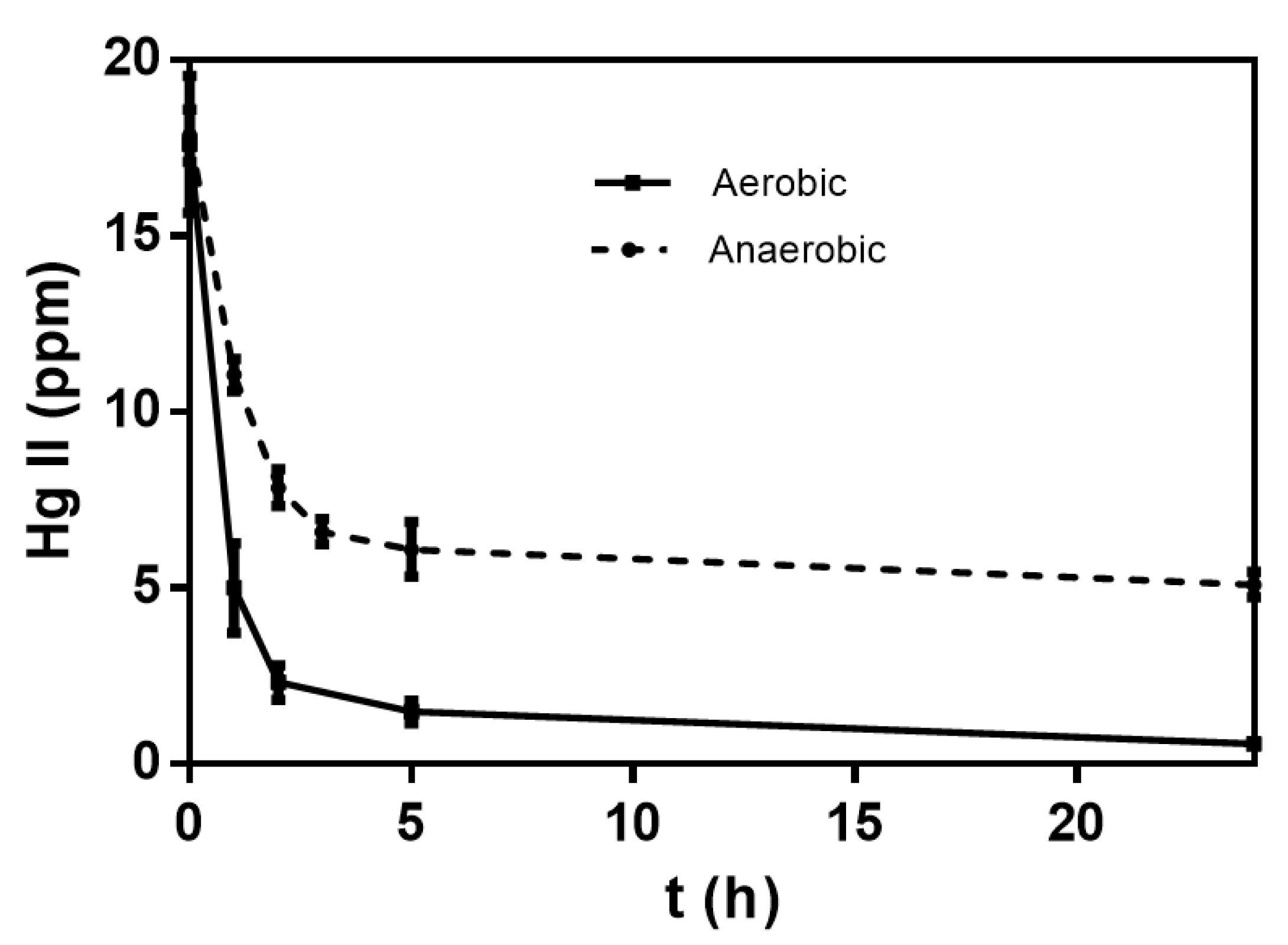
3.3. Effect of Oxygen Availability on Mercury (II) Reduction

To determine the effect of oxygen availability on the reduction of mercury (II) (20 ppm) by MSR33 cells, the reduction of mercury (II) under aerobic and anaerobic conditions was compared (Figure 5). Strain MSR33 was able to reduce Hg (II) under aerobic and anaerobic conditions, reaching a mercury removal after 24 h of 96.8% and 71.4%, respectively. The results indicate that the removal of mercury (II) by strain MSR33 after 24 h was higher under aerobic conditions than under anaerobiosis.

4. Discussion

This study determined the kinetic parameters of C. metallidurans MSR33 growth in a bioreactor (3 L) and the effect of mercury on mercury bioremediation by this strain. The new culture medium GBC for strain MSR33 growth was formulated in this study. Due to the complexing effect of phosphate on mercury, GBC medium was formulated with a low phosphate concentration. The low phosphate Tris-buffered mineral salts (LPTMS) medium has been used previously for C. metallidurans growth [10,28,29]. The LPTMS medium requires high concentrations of Tris salts buffer, which is not required for bioreactor cultures with automatic pH control. Succinate concentration was adjusted to energy and growth requirements of strain MSR33. The biomass difference between the theoretical (2 g cells L−1) and the experimental values (1.7 g cells L−1) may be attributed to factors such as carbon losses produced by the release of metabolites into the culture and higher maintenance energy requirements (Figure 2a).
The specific growth rate (µ) for C. metallidurans strain MSR33 obtained in this study is higher (8 times) than the µ reported previously [10]. Rojas et al. characterized the MSR33 growth in a 200 mL flask, whereas, in the present study, the strain was grown in a bioreactor of 3 L under controlled conditions of pH, agitation, and oxygen supply [10]. In this study, inhibition of MSR33 growth at high concentrations of succinate was observed (Figure 2b), suggesting an inhibition by the substrate. After 7 h of the growth kinetics, this inhibition was reversed. High succinate concentration has been reported to inhibit the succinate dehydrogenase [36], which may explain the initial inhibitory behavior observed with 12 g L−1 succinate. Strain MSR33 grown on succinate showed a lower specific oxygen uptake rate (60 mg O2 h−1 g cell−1) than E. coli NF790 grown on succinate (832 mg O2 h−1 g cell−1) but within the oxygen uptake range reported by E. coli K12 (29–739 mg O2 h−1 g cell−1) and Rhodococcus erythropolis IGTS8 (6.4–137 mg O2 h−1 g cell−1) [37,38].
In this study, mercury II (20 ppm) inhibited the growth and oxygen uptake of strain MSR33 (Figure 3 and Figure 4). Rojas et al. [10] observed that the addition of Hg II (8 ppm) during the exponential growth phase did not affect MSR33 cell growth. However, Hg II (8 ppm) stopped the growth of wild type strain C. metallidurans CH34. The minimum inhibitory concentration (MIC) of Hg (II) described under aerobic conditions for strains CH34 and MSR33 is 10 and 24 ppm, respectively [10,29]. The growth inhibition of strain MSR33 could be associated to the high mercury concentration (20 ppm) used in this study.
At the cellular level, the disruption of the oxygen uptake by mercury addition may be related to its effects on the respiratory chain. Mercury has been reported to alter membranes, reducing membrane transport and potential, affecting the respiratory chain, and producing oxidative stress [6,39]. Specifically, mercury affects sulfhydryl (cysteine) and thioester (methionine) residues of proteins and replaces other metals such as iron in metalloproteins, damaging the respiratory chain functioning at membranes in bacteria and eukaryotes [2,7,8,9,40]. Rojas et al. reported that Hg (II) (8 ppm) affects the membranes of C. metallidurans strain CH34, showing a fuzzy outer membrane [10]. C. metallidurans strains CH34 and MSR33 possess outer membrane and periplasmic sulfur-rich proteins such as CopA, CopB, CopC, CopK, and CopJ containing a significant number of methionine and cysteine amino acids, which has been proposed to be oxidized by heavy metals and participate in the reduction of gold (III) ions into Au (0) [7]. Hg (II) increases lipid peroxidation by the increase in H2O2 due to Fenton reactions in Shewanella oneidensis MR-1 under aerobic conditions, whereas lower lipoperoxidation was observed under anaerobiosis [41]. Mercury decreases the activity of photosystems cytochromes and quinones at the membrane in Rhodobacter sphaeroides [42]. Hg (II) decreases oxygen uptake and inhibits the electron transport chain and the oxidative phosphorylation in the mitochondria of fish liver cells [43].
Another factor that may contribute to the disruption of oxygen uptake and inhibition of growth by Hg (II) is the redirection of the NADPH cellular pool, from the synthesis of biomolecules and cell growth to the reduction of mercury and oxidative stress response. The reduction of Hg (II) to Hg (0) by strain MSR33 occurs in the cytosol where the mercury reductase (MerA) reduces Hg (II) into Hg (0) using 2 NADPH molecules [10,44]. The main damage caused by mercury is on the membranes but not in the cytoplasm [41]. The import of mercury (II) and its subsequent reduction are the key processes for detoxification. The exposure to Hg (II) induces the generation of reactive oxygen species (ROS) in bacteria and eukaryotes, which could decrease the NADPH levels in strain MSR33 even more [6,39,41,43]. Under oxidative stress, bacterial growth is arrested, whereas NADPH plays an important role for detoxifying ROS by the regeneration of reduced glutathione and thioredoxin, with a concomitant decrease in the NADPH pool [45,46]. A fast rerouting of the Embden–Meyerhof–Parnas pathway into the pentose phosphate pathway was observed in E. coli supplemented with glucose under oxidative stress, increasing the reduction rate of NADP+ to NADPH [47]. In our study, succinate was used as the sole carbon and energy source for strain MSR33 growth, suggesting a rerouting pathway for NADPH recycling. The pentose phosphate pathway, isocitrate dehydrogenase, malic enzyme, and transhydrogenases reactions are probably involved in NADPH pool regeneration in strain MSR33 under these aerobic conditions. On the other hand, higher expression of the mer genes under aerobic conditions was observed in Pseudomonas stutzeri OX than under anaerobiosis [20]. We postulate that under aerobiosis, mercury (II) in strain MSR33 causes membrane and protein damage that affects the respiratory chain and the oxidative phosphorylation, induces the broad-spectrum mer genes, and promotes the redirection of the NADPH reducing power to the mercury-detoxifying and oxidative stress response mechanisms (Figure 6).
In this study, the capability of strain MSR33 to re-establish the cell growth and the oxygen uptake after an initial inhibition by mercury (II) was observed, increasing the cellular biomass for mercury bioremediation. The response of strain MSR33 is based on the proteins encoded by the mer genes in the chromosome and pMOL28, pMOL30, and pTP6 plasmids that are associated with mercury resistance [10,29]. Strain MSR33 is capable of reducing Hg (II) and organomercurial compounds into a less toxic form (Hg 0), which enables detoxifying the environment surrounding the bacterium [26,44]. A high rate of mercury removal was observed after 1 h (Figure 5), indicating high mercury reduction by MSR33 MerA protein. The fast detoxifying response may be based on the mercury resistance genes’ redundancy in strain MSR33, which allows the bacterium to restore cell growth and oxygen uptake after the early inhibitory effects of mercury [10]. Due to the absence of oxygen uptake during exposure to mercury in a liquid medium, the kinetics of mercury removal were determined by strain MSR33 under aerobic conditions through the displacement of gases with an injection of air, and under anaerobic conditions by the injection of gaseous nitrogen (Figure 5). The results indicate that oxygen is required for an efficient removal of mercury, even though mercury inhibits oxygen uptake. Although, during aerobic mercury bioremediation, strain MSR33 stops consuming oxygen during initial mercury exposure, mercury probably induces the mercury reductase activity and the bacterium reroutes its metabolism towards the regeneration of the NADPH pool for the reduction of mercury (II) and to counteract ROS.
In this study, almost complete removal of mercury (II) (20 ppm) by C. metallidurans MSR33 was observed under aerobic conditions after 24 h. Rojas et al. [10] observed that strain MSR33 in the presence of thioglycolate reached a complete removal of mercury (II) (20 and 30 ppm) after 2 h in flasks of 250 mL (50 mL aqueous solution) with high aeration (6 vvm). In this study, the mercury removal after 24 h by strain MSR33 under aerobic conditions (97%) is higher than the 79% mercury (3.2 ppm) removal after 6 h by Sphingobium sp. SA2 and 88% of mercury (5 ppm) removal after 7 h by Pseudomonas stutzeri OX but similar to 92 to 98% mercury (40 ppm) removal after 24 h by P. putida PpY101/pSR134 [20,22,48].
Interestingly, the facultative anaerobe strain MSR33 was able to remove 71% Hg (II) (20 ppm) after 24 h under anaerobic conditions. The facultative anaerobe Pseudomonas stutzeri OX removes 84% mercury II (5 ppm) after 20 h under anaerobic conditions [20]. Anaerobic mercury reduction associated with methylation and demethylation by anaerobic obligate bacteria has been reported [49,50]. G. bemidjiensis Bem and G. sulfurreducens PCA reduce mercury at low concentrations (1 ppb) under anaerobic conditions [25,26]. Anaerobiosis favors the formation of inorganic mercury sulfide, decreases ROS levels, and significantly reduces the NADPH pool regeneration [45,51]. P. stutzeri OX exhibited higher tolerance to Hg (II) under anaerobiosis than under aerobic conditions; anaerobiosis affects Hg (II) transport into the cell and, therefore, also the expression of the mer genes [20]. It has been reported that the synthesis of MerA and MerB proteins in C. metallidurans MSR33 is strongly induced by mercury [10]. Therefore, a lower mercury reduction under anaerobic conditions by strain MSR33 may be explained by (i) a decreased Hg (II) transport into the cell compared to aerobic conditions, which leads to a lower expression of mer genes, and (ii) a lower NADPH pool, which negatively affects the reduction of mercury by MerA in the cytoplasm. However, the higher mercury removal rates during the first hours of mercury exposure under anaerobic conditions could be associated with microaerobic conditions that may be generated inside the reactor by oxygen remnants at the beginning of the anaerobic phase. In this study, mercury (II) removal was performed at concentrations close to the MIC [10]. Mercury concentrations higher than the MIC could irreversibly affect the growth and detoxifying activity of strain MSR33 under aerobic conditions. However, under anaerobic conditions, C. metallidurans strain MSR33 may tolerate higher mercury concentration than P. stutzeri OX [20].
Mercury inhibits the metabolic activity of C. metallidurans MSR33. However, strain MSR33 is capable of tolerating mercury during mercury bioremediation and recovered its metabolic activity. Notably, strain MSR33 is able to remove mercury in solution under anaerobic conditions. Under anaerobic conditions, toluene degradation by C. metallidurans CH34 using nitrate as a terminal electron acceptor in bioelectrochemical systems has been reported [30]. The results of our present study confirm the bioremediation capability of C. metallidurans strain MSR33 under anaerobic conditions. In this study, nitrate was not included in the composition of the culture medium. Interestingly, facultative anaerobic bacteria including mercury-reducing strains may use fumarate as the electron acceptor, where fumarate reductase catalyzes this final step in anaerobic respiration [46,49,52]. The gene encoding this enzyme was reported in the C. metallidurans genome [53], therefore, we propose that fumarate could be the electron acceptor under these anaerobic conditions. Further studies on anaerobic mercury reduction are required to understand the molecular and metabolic mechanisms involved in mercury removal by strain MSR33 under anaerobiosis.
The results of this study indicate that C. metallidurans MSR33 is an attractive biocatalyst for mercury bioremediation of polluted water such as mine groundwater and industrial wastewater under aerobic and anaerobic conditions.

5. Conclusions

The defined GBC culture medium was designed in this study for improved growth of strain MSR33, which is limited by succinate as the only carbon and energy source. The MSR33 growth rate in the GBC medium increased up to eight times compared to growth rate values reported by previous studies of this strain.
Mercury inhibited the growth and respiratory rate of strain MSR33 in liquid medium under aerobic conditions. However, the growth and respiration inhibitions were reversed after 5 h. Notably, strain MSR33 was able to remove mercury in a liquid medium under anaerobic conditions but higher removal of mercury was observed under aerobic conditions than under anaerobiosis.
This study suggests that in spite of the fact that mercury (II) harms C. metallidurans MSR33 metabolic activity, this strain is able to remove mercury from contaminated water and to recover its metabolic activity after 5 h. Therefore, C. metallidurans MSR33 may be useful for mercury bioremediation in polluted water under aerobic and anaerobic conditions.

Author Contributions

G.B., J.C.G., and M.S. conceived and designed the experiments; G.B. and P.V.-C. performed the experiments; G.B., P.V.-C., J.C.G., and M.S. analyzed the data; J.C.G. and M.S. contributed reagents, materials, and analysis tools; G.B., P.V.-C., J.C.G. and M.S. wrote the paper. All authors have read and agreed to the published version of the manuscript.

Funding

This research was funded by PhD Conicyt, PUCV and USM (G.B., P.V.-C.) fellowships, ANID PIA Ring Genomics and Applied Microbiology for Bioremediation and Bioproducts (GAMBIO) ACT172128 Chile (M.S., G.B., P.V.-C.), Fondequip EQM 170914 (M.S., G.B.), Fondecyt 1200756 (M.S., P.V.-C.), PIIC USM (G.B., P.V.-C.), and USM (M.S., P.V.-C., G.B.) grants.

Acknowledgments

Fermentation Laboratory, School of Biochemical Engineering, Pontificia Universidad Católica de Valparaíso.

Conflicts of Interest

The authors declare no conflict of interest.

References

  1. Nieboer, E.; Richardson, D.H. The replacement of the nondescript term ‘heavy metals’ by a biologically and chemically significant classification of metal ions. Environ. Pollut. Ser. B Chem. Phys. 1980, 1, 3–26. [Google Scholar] [CrossRef]
  2. Tagliafierro, L.; Officioso, A.; Sorbo, S.; Basile, A.; Manna, C. The protective role of olive oil hydroxytyrosol against oxidative alterations induced by mercury in human erythrocytes. Food Chem. Toxicol. 2015, 82, 59–63. [Google Scholar] [CrossRef] [PubMed]
  3. Yang, L.; Zhang, Y.; Wang, F.; Luo, Z.; Guo, S.; Strähle, U. Toxicity of mercury: Molecular evidence. Chemosphere 2020, 245, 125586. [Google Scholar] [CrossRef] [PubMed]
  4. Patra, M.; Sharma, A. Mercury toxicity in plants. Bot. Rev. 2000, 66, 379–422. [Google Scholar] [CrossRef]
  5. Streets, D.G.; Horowitz, H.M.; Jacob, D.J.; Lu, Z.; Levin, L.; Ter Schure, A.F.H.; Sunderland, E.M. Total mercury released to the environment by human activities. Environ. Sci. Technol. 2017, 51, 5969–5977. [Google Scholar] [CrossRef] [PubMed]
  6. Spiller, H.A. Rethinking mercury: The role of selenium in the pathophysiology of mercury toxicity. Clin. Toxicol. 2017, 56, 313–326. [Google Scholar] [CrossRef] [PubMed]
  7. Montero-Silva, F.; Durán, N.; Seeger, M. Synthesis of extracellular gold nanoparticles using Cupriavidus metallidurans CH34 cells. IET Nanobiotechnol. 2018, 12, 40–46. [Google Scholar] [CrossRef]
  8. Xu, F.F.; Imlay, J.A. Silver(I), mercury (II), cadmium (II), and zinc (II) target exposed enzymic iron-sulfur clusters when they toxify Escherichia coli. Appl. Environ. Microbiol. 2012, 78, 3614–3621. [Google Scholar] [CrossRef] [Green Version]
  9. Lemire, J.A.; Harrison, J.J.; Turner, R.J. Antimicrobial activity of metals: Mechanisms, molecular targets and applications. Nat. Rev. Microbiol. 2013, 11, 371–384. [Google Scholar] [CrossRef]
  10. Rojas, L.A.; Yañez, C.; González, M.; Lobos, S.; Smalla, K.; Seeger, M. Characterization of the metabolically modified heavy metal-resistant Cupriavidus metallidurans strain MSR33 generated for mercury bioremediation. PLoS ONE 2011, 6, e17555. [Google Scholar] [CrossRef] [Green Version]
  11. McCarthy, D.; Edwards, G.C.; Gustin, M.S.; Care, A.; Miller, M.B.; Sunna, A. An innovative approach to bioremediation of mercury contaminated soils from industrial mining operations. Chemosphere 2017, 184, 694–699. [Google Scholar] [CrossRef] [PubMed]
  12. Hernández, M.; Morgante, V.; Ávila, M.; Villalobos, P.; Millares, P.; González, M.; Seeger, M. Novel s-triazine-degrading bacteria isolated from agricultural soils of central Chile for herbicide bioremediation. Electron. J. Biotechnol. 2008, 11, 5–6. [Google Scholar] [CrossRef] [Green Version]
  13. Seeger, M.; Hernández, M.; Mendez, V.; Ponce, B.; Córdova, M.; González, M. Bacterial degradation and bioremediation of chlorinated herbicides and biphenyls. J. Soil Sci. Plant Nutr. 2010, 10, 320–332. [Google Scholar] [CrossRef]
  14. Altimira, F.; Yáñez, C.; Bravo, G.; González, M.; Rojas, L.A.; Seeger, M. Characterization of copper-resistant bacteria and bacterial communities from copper-polluted agricultural soils of central Chile. BMC Microbiol. 2012, 12, 193. [Google Scholar] [CrossRef] [PubMed] [Green Version]
  15. Romero-Silva, M.J.; Méndez, V.; Agulló, L.; Seeger, M. Genomic and functional analyses of the gentisate and protocatechuate ring-cleavage pathways and related 3-hydroxybenzoate and 4-hydroxybenzoate peripheral pathways in Burkholderia xenovorans LB400. PLoS ONE 2013, 8, e56038. [Google Scholar] [CrossRef] [PubMed] [Green Version]
  16. Fuentes, S.; Barra, B.; Caporaso, J.G.; Seeger, M. From rare to dominant: A fine-tuned soil bacterial bloom during petroleum hydrocarbon bioremediation. Appl. Environ. Microbiol. 2015, 82, 888–896. [Google Scholar] [CrossRef] [Green Version]
  17. Aguirre, A.; Bernal, P.; Maureira, D.; Ramos, N.; Vásquez, J.; Urrutia, H.; Gentina, J.C.; Aroca, G. Biofiltration of trimethylamine in biotrickling filter inoculated with Aminobacter aminovorans. Electron. J. Biotechnol. 2018, 33, 63–67. [Google Scholar] [CrossRef]
  18. Orellana, R.; Macaya, C.; Bravo, G.; Dorochesi, F.; Cumsille, A.; Valencia, R.; Rojas, C.; Seeger, M. Living at the frontiers of life: Extremophiles in Chile and their potential for bioremediation. Front. Microbiol. 2018, 9, 2309. [Google Scholar] [CrossRef]
  19. Zhang, Z.; Wu, J.; Liu, D. Co3O4/g-C3N4 hybrids for gas-phase Hg0 removal at low temperature. Processes 2019, 7, 279. [Google Scholar] [CrossRef] [Green Version]
  20. Schaefer, J.; Letowski, J.; Barkay, T. Mer-mediated resistance and volatilization of Hg (II) under anaerobic conditions. Geomicrobiol. J. 2002, 19, 87–102. [Google Scholar] [CrossRef]
  21. Deckwer, W.-D.; Becker, F.U.; Ledakowicz, S.; Wagner-Döbler, I. Microbial removal of ionic mercury in a three-phase fluidized bed reactor. Environ. Sci. Technol. 2004, 38, 1858–1865. [Google Scholar] [CrossRef] [PubMed]
  22. Mahbub, K.; Krishnan, K.; Megharaj, M.; Naidu, R. Bioremediation potential of a highly mercury resistant bacterial strain Sphingobium SA2 isolated from contaminated soil. Chemosphere 2016, 144, 330–337. [Google Scholar] [CrossRef] [PubMed]
  23. Dash, H.R.; Basu, S.; Das, S. Evidence of mercury trapping in biofilm-EPS and mer operon-based volatilization of inorganic mercury in a marine bacterium Bacillus cereus BW-201B. Arch. Microbiol. 2016, 199, 445–455. [Google Scholar] [CrossRef]
  24. Wang, X.; He, Z.; Luo, H.; Zhang, M.; Zhang, D.; Pan, X.; Gadd, G.M. Multiple-pathway remediation of mercury contamination by a versatile selenite-reducing bacterium. Sci. Total Environ. 2018, 615, 615–623. [Google Scholar] [CrossRef] [PubMed] [Green Version]
  25. Hu, H.; Lin, H.; Zheng, W.; Tomanicek, S.J.; Johs, A.; Feng, X.; Elias, D.A.; Liang, L.; Gu, B. Oxidation and methylation of dissolved elemental mercury by anaerobic bacteria. Nat. Geosci. 2013, 6, 751–754. [Google Scholar] [CrossRef]
  26. Lu, X.; Liu, Y.; Johs, A.; Zhao, L.; Wang, T.; Yang, Z.; Lin, H.; Elias, D.A.; Pierce, E.M.; Liang, L.; et al. Anaerobic mercury methylation and demethylation by Geobacter bemidjiensis Bem. Environ. Sci. Technol. 2016, 50, 4366–4373. [Google Scholar] [CrossRef] [PubMed]
  27. Mergeay, M.; Nies, D.; Schlegel, H.G.; Gerits, J.; Charles, P.; Van Gijsegem, F. Alcaligenes eutrophus CH34 is a facultative chemolithotroph with plasmid-bound resistance to heavy metals. J. Bacteriol. 1985, 162, 328–334. [Google Scholar] [CrossRef] [Green Version]
  28. Alviz-Gazitua, P.; Fuentes-Alburquenque, S.; Rojas, L.A.; Turner, R.; Guiliani, N.; Seeger, M. The response of Cupriavidus metallidurans CH34 to cadmium involves inhibition of the initiation of biofilm formation, decrease in intracellular c-di-GMP levels, and a novel metal regulated phosphodiesterase. Front. Microbiol. 2019, 10, 1499. [Google Scholar] [CrossRef]
  29. Millacura, F.A.; Janssen, P.J.; Monsieurs, P.; Janssen, A.; Provoost, A.; Van Houdt, R.; Rojas, L.A. Unintentional genomic changes endow Cupriavidus metallidurans with an augmented heavy-metal resistance. Genes 2018, 9, 551. [Google Scholar] [CrossRef] [Green Version]
  30. Espinoza-Tofalos, A.; Daghio, M.; González, M.; Papacchini, M.; Franzetti, A.; Seeger, M. Toluene degradation by Cupriavidus metallidurans CH34 in nitrate-reducing conditions and in bioelectrochemical systems. FEMS Microbiol. Lett. 2018, 365, fny119. [Google Scholar] [CrossRef]
  31. Zhang, J.; Greasham, R. Chemically defined media for commercial fermentations. Appl. Microbiol. Biotechnol. 1999, 51, 407–421. [Google Scholar] [CrossRef]
  32. Acevedo, A.; Conejeros, R.; Aroca, G. Ethanol production improvement driven by genome-scale metabolic modeling and sensitivity analysis in Scheffersomyces stipites. PLoS ONE 2017, 12, e0180074. [Google Scholar] [CrossRef] [PubMed] [Green Version]
  33. Casas López, J.L.; Rodríguez Porcel, E.M.; Oller Alberola, I.; Ballesteros Martín, M.M.; Sánchez Pérez, J.; Fernández Sevilla, J.M.; Chisti, Y. Simultaneous determination of oxygen consumption rate and volumetric oxygen transfer coefficient in pneumatically agitated bioreactors. Ind. Eng. Chem. Res. 2006, 45, 1167–1171. [Google Scholar] [CrossRef]
  34. AOAC Official Method 977.15 Mercury in Fish Alternative Flameless Atomic Absorption Spectrophotometric Method First Action 1977 Final Action 1978. Available online: http://www.aoacofficialmethod.org/index.php?main_page=product_info&cPath=1&products_id=2383 (accessed on 24 July 2020).
  35. Attalah, S.; Waller, P.; Steichen, S.; Gao, S.; Brown, C.; Ogden, K.; Brown, J. Application of deoxygenation-aeration cycling to control the predatory bacterium Vampirovibrio chlorellavorus in Chlorella sorokiniana cultures. Algal Res. 2019, 39, 101427. [Google Scholar] [CrossRef]
  36. Cao, Y.; Zhang, R.; Sun, C.; Cheng, T.; Liu, Y.; Xian, M. Fermentative succinate production: An emerging technology to replace the traditional petrochemical processes. BioMed Res. Int. 2013, 2013, 1–12. [Google Scholar] [CrossRef] [Green Version]
  37. Andersen, K.B.; Von Meyenburg, K. Are growth rates of Escherichia coli in batch cultures limited by respiration? J. Bacteriol. 1980, 144, 114–123. [Google Scholar] [CrossRef] [Green Version]
  38. Garcia-Ochoa, F.; Gomez, E.; Santos, V.E.; Merchuk, J.C. Oxygen uptake rate in microbial processes: An overview. Biochem. Eng. J. 2010, 49, 289–307. [Google Scholar] [CrossRef]
  39. Belyaeva, E.A.; Sokolova, T.V.; Emelyanova, L.V.; Zakharova, I.O. Mitochondrial electron transport chain in heavy metal-induced neurotoxicity: Effects of cadmium, mercury, and copper. Sci. World J. 2012, 2012, 136063. [Google Scholar] [CrossRef] [Green Version]
  40. Norambuena, J.; Wang, Y.; Hanson, T.; Boyd, J.M.; Barkay, T. Low-molecular-weight thiols and thioredoxins are important players in Hg (II) resistance in Thermus thermophilus HB27. Appl. Environ. Microbiol. 2018, 84, e01931-17. [Google Scholar] [CrossRef] [Green Version]
  41. Wang, Y.; Robison, T.; Wiatrowski, H. The impact of ionic mercury on antioxidant defenses in two mercury-sensitive anaerobic bacteria. BioMetals 2013, 26, 1023–1031. [Google Scholar] [CrossRef]
  42. Asztalos, E.; Sipka, G.; Kis, M.; Trotta, M.; Maróti, P. The reaction center is the sensitive target of the mercury (II) ion in intact cells of photosynthetic bacteria. Photosynth. Res. 2012, 112, 129–140. [Google Scholar] [CrossRef] [PubMed]
  43. Mieiro, C.; Pardal, M.; Duarte, A.C.; Pereira, E.; Palmeira, C. Impairment of mitochondrial energy metabolism of two marine fish by in vitro mercuric chloride exposure. Mar. Pollut. Bull. 2015, 97, 488–493. [Google Scholar] [CrossRef] [PubMed]
  44. Dash, H.R.; Das, S. Bioremediation of mercury and the importance of bacterial mer genes. Int. Biodeterior. Biodegrad. 2012, 75, 207–213. [Google Scholar] [CrossRef]
  45. Shimizu, K.; Matsuoka, Y. Redox rebalance against genetic perturbations and modulation of central carbon metabolism by the oxidative stress regulation. Biotechnol. Adv. 2019, 37, 107441. [Google Scholar] [CrossRef] [PubMed]
  46. Bergkessel, M.; Basta, D.; Newman, D.K. The physiology of growth arrest: Uniting molecular and environmental microbiology. Nat. Rev. Microbiol. 2016, 14, 549–562. [Google Scholar] [CrossRef]
  47. Christodoulou, D.; Kuehne, A.; Estermann, A.; Fuhrer, T.; Lang, P.F.; Sauer, U. Reserve flux capacity in the pentose phosphate pathway by NADPH binding is conserved across kingdoms. iScience 2019, 19, 1133–1144. [Google Scholar] [CrossRef] [Green Version]
  48. Okino, S.; Iwasaki, K.; Yagi, O.; Tanaka, H. Development of a biological mercury removal-recovery system. Biotechnol. Lett. 2000, 22, 783–788. [Google Scholar] [CrossRef]
  49. Colombo, M.J.; Ha, J.; Reinfelder, J.R.; Barkay, T.; Yee, N. Oxidation of Hg (0) to Hg (II) by diverse anaerobic bacteria. Chem. Geol. 2014, 363, 334–340. [Google Scholar] [CrossRef]
  50. Ma, M.; Du, H.; Wang, D. Mercury methylation by anaerobic microorganisms: A review. Crit. Rev. Environ. Sci. Technol. 2019, 49, 1893–1936. [Google Scholar] [CrossRef]
  51. Ercal, N.; Gurer-Orhan, H.; Aykin-Burns, N. Toxic metals and oxidative stress Part I: Mechanisms involved in metal-induced oxidative damage. Curr. Top. Med. Chem. 2001, 1, 529–539. [Google Scholar] [CrossRef]
  52. Maklashina, E.; Berthold, D.A.; Cecchini, G. Anaerobic expression of Escherichia coli succinate dehydrogenase: Functional replacement of fumarate reductase in the respiratory chain during anaerobic growth. J. Bacteriol. 1998, 180, 5989–5996. [Google Scholar] [CrossRef] [PubMed] [Green Version]
  53. Janssen, P.J.; Van Houdt, R.; Moors, H.; Monsieurs, P.; Morin, N.; Michaux, A.; Benotmane, M.A.; Leys, N.; Vallaeys, T.; Lapidus, A.; et al. The complete genome sequence of Cupriavidus metallidurans strain CH34, a master survivalist in harsh and anthropogenic environments. PLoS ONE 2010, 5, e10433. [Google Scholar] [CrossRef] [PubMed]
Figure 1. Mercury (II) and organomercurial compounds resistance mechanism of C. metallidurans MSR33. Mercury (II) and organomercurials compounds resistance is codified by mer genes in the chromosome and the plasmids pMOL30, pMOL28, and pTP6 of strain MSR33. Hg (II) is reduced to Hg (0) by mercuric reductase MerA using reducing power NADPH. MerB catalyzes the protonolysis of the carbon–mercury bond in organomercurials. MerP and MerT are proteins involved in the transport of mercury (II) inside the cell. MerG is a periplasmic protein involved in the importing of phenylmercury. Figure adapted from Rojas et al. [10].
Processes 08 00893 g001
Figure 2. Growth and succinate degradation of C. metallidurans MSR33 in GBC medium. (a) Growth of strain MSR33 on succinate 4 g L−1. (b) Effect of increased succinate concentration on the growth kinetics of C. metallidurans MSR33. The kinetics was obtained from a batch reactor with aeration (2 vvm), pH 7, 30 °C and agitation (500 rpm). The assays were performed in triplicate. Bars indicate the standard deviation.
Processes 08 00893 g002
Figure 3. Effects of the addition of mercury (II) on the kinetics of cell growth and oxygen uptake by C. metallidurans MSR33. In these assays, Hg (II) (20 ppm) was added during the exponential phase at 6 h of growth onset by C. metallidurans MSR33. The assays were performed in duplicate. Bars indicate the standard deviation.
Processes 08 00893 g003
Figure 4. Effect of mercury (II) (20 ppm) on the growth and the respiration of C. metallidurans MSR33. In these assays, Hg (II) (20 ppm) was added at the exponential growth phase. The assays were performed in duplicate. Bars indicate the standard deviation.
Processes 08 00893 g004
Figure 5. Kinetics of mercury (II) (20 ppm) removal by C. metallidurans MSR33 from polluted aqueous solution under aerobic and anaerobic conditions. Hg (II) removal was evaluated during the exponential growth phase (1.3 g cells L−1), using gas flows (2 vvm) of air and nitrogen gas for aerobic and anaerobic conditions, respectively. The assays were performed in duplicate. Bars indicate the standard deviation.
Processes 08 00893 g005
Figure 6. Proposed aerobic mercury detoxifying mechanisms in C. metallidurans MSR33. Mercury in presence of oxygen generate reactive oxygen species (ROS) through the electron transport chain and Fenton reactions. ROS activate oxidative stress response mechanisms that induce the rerouting of the metabolism, increasing the NADPH pool (blue letters) through the pentose phosphate pathway, malic enzyme, isocitrate dehydrogenase and transhydrogenase reactions (light blue squares). Mercury under aerobic conditions induces mercury detoxifying mechanisms through the expression of the mer genes. The mercuric reductase MerA reduces Hg (II) into Hg (0) using 2 NADPH. The NADPH pool is consumed during mercury reduction and oxidative stress response mechanisms. Abbreviations: SUC, succinate; SUCD, succinate dehydrogenase; FUM, fumarate; MAL, malate; OAA, oxaloacetate; CIT, citrate; ICIT, isocitrate; KG, α-ketoglutarate; SucCoA, succinylCoA; AcCoA, acetylCoA; PYR, pyruvate; PEP, phosphoenolpyruvate; G6P, glucose-6-phosphate; 6PGL, 6-phosphogluconolactone; 6PG, 6-phosphogluconate; Ru5P, Ribulose-5-phosphate; GAP, glyceraldehyde-3-phosphate.
Processes 08 00893 g006

