
Digital Twins for Tissue Culture Techniques—Concepts, Expectations, and State of the Art


Institute of Bioprocess and Biosystems Engineering, Hamburg University of Technology, 21073 Hamburg, Germany
*
Author to whom correspondence should be addressed.
Processes 2021, 9(3), 447; https://0-doi-org.brum.beds.ac.uk/10.3390/pr9030447
Submission received: 12 January 2021
/
Revised: 24 February 2021
/
Accepted: 25 February 2021
/
Published: 2 March 2021
(This article belongs to the Section Biological Processes and Systems)
Abstract
:Techniques to provide in vitro tissue culture have undergone significant changes during the last decades, and current applications involve interactions of cells and organoids, three-dimensional cell co-cultures, and organ/body-on-chip tools. Efficient computer-aided and mathematical model-based methods are required for efficient and knowledge-driven characterization, optimization, and routine manufacturing of tissue culture systems. As an alternative to purely experimental-driven research, the usage of comprehensive mathematical models as a virtual in silico representation of the tissue culture, namely a digital twin, can be advantageous. Digital twins include the mechanistic of the biological system in the form of diverse mathematical models, which describe the interaction between tissue culture techniques and cell growth, metabolism, and the quality of the tissue. In this review, current concepts, expectations, and the state of the art of digital twins for tissue culture concepts will be highlighted. In general, DT’s can be applied along the full process chain and along the product life cycle. Due to the complexity, the focus of this review will be especially on the design, characterization, and operation of the tissue culture techniques.
1. Introduction
Tissue culture refers to a micro-structured cell culture under in vitro conditions that take into account the three-dimensionality (3D) and the physiology of the tissue. Areas of application cover basic research (e.g., stem cell research, tumor models), pharmacology and toxicity tests, regenerative medicine (tissue engineering for implantation), as well as in vitro meat, among others. Techniques for tissue cultivation have undergone significant changes during the last decades, and 3D cell culture systems, e.g., for interacting cells and organoids, 3D cell co-cultures, organ/body-on-chip, have become available [1].
As tissue constructs are nowadays applied routinely, e.g., for pharmacological and toxicity tests or for the generation of implantable tissue, the requirements regarding quantity and quality of the generated constructs have significantly increased [2], driven further by quality standards of regulatory bodies such as FDA or EMA [3]. As most protocols and procedures have originally been established in research labs often based on trial and error without defined quality criteria, the transfer to routine, standardized applications is troublesome. Hence, in silico methods such as Digital Twins (DT) have been suggested for a more profound development, characterization, and routine manufacturing of 3D tissue culture systems, comprised under the novel paradigm of “developmental engineering” [4,5,6]. Furthermore, in silico methods are intensively investigated to replace animal testing, as they are in compliance with the 3R principles (i.e., reduction, refinement, and replacement). [7,8,9,10].
According to El Saddik [11], DTs are referred to as “digital replications of living as well as non-living entities that enable data to be seamlessly transmitted between the physical and virtual worlds”. Prominent examples of successful DTs can be found in the Apollo missions or in aircraft construction. In the Industry 4.0 era, DTs are routinely assigned as the heart of the digital factory and as core concepts of digital transformation strategies. In medicine, under the catchphrase “The Virtual Physiological Human (VPH)”, also referred to as “in silico medicine”, the use of individualized, physiology-based computer simulations is targeted in all aspects of prevention, diagnosis, prognostic evaluation, treatment of diseases, and development of biomedical products. While DT-based concepts for production of biopharmaceuticals are already being used in industrial practice or tested by pharmaceutical companies for the development, optimization, and operation of production systems for biopharmaceuticals (recently reviewed in [12,13]), there are so far only a few DT-approaches, mostly for the generation of implantable tissues as Advanced Therapy Medical Products (ATMPs) or tissue-specific test systems for new pharmaceutics or toxicity tests [5,6,14,15,16,17,18,19]. This is probably also due to insufficiently precisely formulated expectations and knowledge with regard to the possibilities, but also the limits of model-based DT concepts in a community that works mainly on a data basis.
In the beginning of this review, an overview of tissue culture systems, followed by an introduction to the model-based DT-concepts with a special focus on tissue culture, mainly concerning Tissue Engineering as ATMP and test systems will be presented. Subsequently, the types of mathematical models applied for DT’s and various examples for model-based/model-assisted design and operation will be discussed. Finally, conclusions will be drawn concerning the state of the art and future expectation for digital twins for tissue culture concepts.
2. Overview Tissue Culture Systems
In the following chapter, tissue culture systems are introduced focusing on general aspects first followed by culture systems for cell expansion. Then, different culture systems for tissue generation and maturation are discussed and considerations for their design and operation are reviewed.
2.1. General Aspects
Tissue culture can be described as the culture of living cells within micro-assembled devices and supports that create a growth environment mimicking the native tissue as closely as possible [20,21,22]. Unlike 2D monolayer cell culture, 3D tissue culture is regarded as much more suitable to mimic in vivo cells behaviors and organization (morphology and physiology) [23]. The ultimate goal is to create a vital tissue with organ specific structure and functionality considering the complexity of the cellular microenvironment in vivo [24,25,26]. Among others, the following aspects were identified to be of major relevance for tissue culture: Extracellular matrix (ECM) components and stiffness regulating cell differentiation, proliferation, and metabolic cellular functions [2,24,25,26,27] and complex cell–cell and cell–matrix interactions (e.g., heterogeneous cell populations including stromal cells, varying cell proliferation zones (quiescent vs. replicating)) [2,28,29,30]. A heterogeneous cellular microenvironment could be important to reflect in vivo conditions with gradients of biomolecular concentrations, e.g., areas of hypoxia, pH gradients, soluble signal gradients, and differential nutrient and metabolic waste transport [2,31,32]. Additionally, flow-induced dynamic effects and mechanical stimuli [33,34,35] and interactions between multiple organs [36] need to be considered. The required degree of complexity is determined by the specific demands.
The workflow for creating 3D tissue cultures can be divided as shown in Figure 1. In the following, the techniques and devices applied for cell expansion and tissue generation and maturation are briefly discussed, focusing on the required engineering knowledge. Tasks that can be addressed by a mathematical description are discussed in Section 4.
Devices used for cell expansion and generation of tissue constructs shall be categorized at this point because they are used to some extent for different purposes. First of all, a distinction can be made between static and dynamic cultivation systems. In static systems, the fluid and/or the cellular tissue construct are not moved. The success of static culture is often limited by mass transfer (e.g., supply of oxygen or nutrients or growth factors, removal of waste products), as this relies on a diffusion mechanism only [37]. Furthermore, they are usually not equipped with on-line sensors for monitoring of e.g., temperature, pH, or dissolved oxygen.
These drawbacks can be overcome by dynamic cultivation concepts, where cells/tissue constructs are maintained under flow conditions and/or mechanical stimulation. Dynamic culture systems are usually grouped under the term “bioreactor”, a device in which biological or biochemical processes develop under a dynamic, closely monitored, and tightly controlled environment [38,39,40]. Bioreactors on different scales are applied for cell expansion or generation of 3D tissue constructs.
2.2. Culture Systems for Cell Expansion
Cell proliferation is the first step in creating a tissue culture. On a smaller scale, static culture dishes (e.g., well plates, T-flasks, stacked plate systems, mostly single-use) are mostly used for cell expansion [41]. Since these only allow the cell number to be increased by a factor of about 10, multiple subcultures are required, which is considered to be one of the main causes of cell dedifferentiation. As most of these culture systems are operated in an incubator to ensure adequate conditions with respect to temperature, pH, and oxygen supply, it is challenging to monitor and control these parameters. Even if it is well known that oxygen concentration within the body varies for different tissues (e.g., well vascularized tissues with higher oxygen concentration, cartilage tissue with lower oxygen concentration), these effects are hardly considered during practical application. A comprehensive mathematical characterization of the systems would help to detect conditions that are not optimal. [42] Similar to oxygen, cells are exposed to mostly constant nutrient concentrations in vivo. However, as medium in culture flasks are usually exchanged batch-wise, cell experience a seesaw behavior of nutrient concentrations causing a detrimental environment to the cells.
As plates and flasks allow for a limited expansion only, scalable bioreactor systems based on matrices to provide cell attachment have been established. These cover hollow fiber bioreactors, bioreactors for macrocarriers (e.g., fixed bed) [43], meander type bioreactors [44], and, in particular, microcarriers (reviewed by [45]) for use in stirred tank reactors, [45] either re-usable or single use. [43,44,46,47,48] A further alternative is carrier-free cultivation in 3D aggregates or spheroids, or encapsulation, e.g., in alginate or in hydrogels. These techniques are often used to induce a lineage specific differentiation of stem cells. With respect to the expansion of cells, e.g., MSCs, there is conflicting evidence in the literature with regard to the usefulness of these techniques (reviewed by [41]), even if this technique has been successfully adopted for expansion of human pluripotent stem cells [47].
There is strong evidence that the growth and differentiation of tissue cells, especially stem cells, are affected by several types of mechanical forces including stretch, strain, compression, and shear stress, especially in the mostly turbulent environment within a stirred bioreactor. This aspect is discussed more in detail in Section 4.2.
2.3. Culture Systems for Tissue Generation and Maturation
2.3.1. Non-Scaffold–Based Organoid Culture
Scaffold-free organoid culture methods rely on the self-aggregation of cells in specialized culture plates, when a surface is not available for cell attachment and cells tend to form cellular spheroids. [1,21,48]. Techniques used to generate spheroids comprise hanging drop microplates, low adhesion plates with ultra-low attachment coating that promotes spheroid formation, micropatterned plates that allow for microfluidic cell culture, and magnetic cell levitation where cells are preloaded with magnetic nanoparticles that are kept floating within an externally applied magnetic field. [2,29] Due to the scaffold-free nature, spheroids can be readily imaged by microscopic techniques, e.g., light, fluorescence, and confocal microscopy. Furthermore, most of the applied techniques are well suited for high-throughput screening [2,21,49].
Spheroids, and in particular multicellular spheroids, recapitulate tissue specific cell–cell contacts or cell–matrix interactions. Therefore, they can mimic gradients with respect to oxygen, nutrients, waste products, or pH as well as diffusion of regulatory molecules. Therefore, they have been extensively been used for e.g., modelling solid tumor growth and metastasis studies as well as in therapeutic studies [48,50,51,52].
2.3.2. Scaffold-Based 3D Cell Culture
Scaffold-based culture technologies provide a physical support mimicking key properties of the extracellular matrix for cells to attach, aggregate migrate, and fill the interstices within the scaffold to form 3D, organotypic cultures. Scaffolds range from simple mechanical structures to ECM-like matrices. In the best case, the scaffold provides an organotypic environment [2,38,53,54]. Depending on the specific application, physical factors such as porosity, stiffness, and stability in culture as well as biological properties such as cell compatibility or adherence have to be considered [2]. Furthermore, growth factors, hormones, or other biologically active molecules can be encapsulated to enhance cell proliferation or to promote a specific cell phenotype [2]. Scaffold-based culture is mainly intended for use in more sophisticated culture devices such as organ-chips or specific bioreactor systems (see below).
Scaffolds can be of biological origin or synthetic. If intended for transplantation, they should be biocompatible and biodegradable [2]. Three individual groups of biomaterials, inorganic/ceramic materials (mainly used for bone regeneration), synthetic polymers and natural polymers as well as composites are therefore used in the fabrication of scaffolds for tissue engineering (for review see [38,54,55]).
A special variant of scaffolds are hydrogels, networks formed from dilute polymer chains with given structure and properties, obtained either by intermolecular or by interfibrillar crosslinks. Hydrogels may come from natural sources (e.g., collagen, fibrin or Matrigel) or can be synthetic, with the possibility of mixing different hydrogel materials to obtain hybrid hydrogels possessing new physical and biological properties. Hydrogels can be functionalized with defined adhesive moieties, include proteolytic sites, and encapsulate growth factors [2,54,56].
2.3.3. Microfluidic 3D Cell Culture
Microfluidic 3D cell culture techniques (“organ-on-a-chip” (OC) or “multi-organ-on-a-chip” (MOC)) intend to contribute an additional level of complexity by introducing a controlled perfusive flow aspect to the cellular environment, allowing for continuous nutrition and oxygen introduction as well as waste removal through culture medium [20,57,58]. As microfluidic techniques provide structures in the micrometer range, the typical length scale of in vivo microenvironment, it is an ideal tool for recreating the cellular microenvironment. Furthermore, on-line sensors (e.g., temperature, pH, dissolved oxygen, impedance, and fluorescence) can be integrated for monitoring the physiological state of the culture. These techniques allow for the mechanistic investigation of the dynamics of multi-organ interactions, various disease models, or drug activities. [59] Successfully established OC and MOC systems comprise skin, blood vessel, liver, gastrointestinal tract, blood–brain-barrier, lung, kidney, and heart equivalents, among others, as well as combinations thereof ([60,61,62,63,64,65,66,67,68], reviewed by [36]). Due to the higher complexity, OC- and MOC-techniques allow for high-contend but medium-throughput-investigations.
2.3.4. Tissue-Specific Bioreactors
Whereas microfluidic techniques are a powerful tool for drug and substance testing, they can hardly be applied for the generation of tissue-intended implants and organ-support. Here the complex task is to recreate the required in vivo microenvironment on a scale appropriate for implantation by providing optimal conditions for co-culture of cells of different phenotypes, cell–cell interactions, vascularization, shear and mechano-stimulation, etc. [37,69,70,71,72,73,74,75]. Furthermore, any scaffold used for cell seeding has to be biodegradable with respect to later implantation.
These bioreactors used in this respect should enable the control of environmental conditions such as oxygen tension, pH, temperature, and shear stress, as well as allowing aseptic operation (e.g., feeding and sampling). Furthermore, a bioreactor system should allow for automated processing steps, especially during the routine manufacture of tissues for clinical application. Furthermore, specific key criteria for tissue constructs based on cells and scaffolds must also be met, including the proliferation of cells, the seeding of cells onto macroporous scaffolds, nutrient (particularly oxygen) supply within the resulting tissue, and mechanical stimulation of the developing tissues. Various studies have shown that mechanical stimulation (e.g., mechanical compression, hydrodynamic pressure, and fluid flow, which are important modulators of cell physiology) can have a positive impact on tissue formation, particularly in the context of musculoskeletal tissue engineering, cartilage formation, and cardiovascular tissues [76].
Historically, most of these techniques were introduced before the invention of microfluidic techniques. First developments focused on culture systems developed mainly for cultivation of mammalian cells for production of biopharmaceuticals, which were adapted for cultivation of tissue cells in three-dimensional structures (e.g., spinner and shake flasks, membrane-based systems such as hollow-fiber reactors, or fluidized- and fixed-bed reactors). Because these systems are capable of supporting culture of large 3D cell agglomerates, but hardly mimic the in vivo microenvironment, numerous culture systems are designed especially for tissue engineering mimicking the special demands of a three-dimensional tissue (including mechano-stimulation) have been developed. Unfortunately, the majority of these have been custom-made, with only very few having been commercialized [69,76].
A great step toward tissue-like structures and vascularization was the invention of decellularization techniques, a process to isolate the extracellular matrix (ECM) of a tissue from its inhabiting cells, leaving an ECM scaffold of the original tissue, which can be used in artificial organ and tissue regeneration (for review see [69]).
2.4. Considerations for Design and Operation
For appropriate design and operation of 3D tissue culture, not only is a profound biological understanding of the tissue/organ of interest required, but also an extensive „engineering“ knowledge comprising design of special culture systems (bioreactors) (mass transfer, shear effects, etc.), design of special materials for cell immobilization (biocompatible, biodegradable), mass transfer problems (oxygen and nutrient supply, removal of toxic metabolites), as well as control and cultivation strategies (including Operator Training Systems).
Even if it is common sense that effects of such engineering parameters on biological cultures are worth to be thoroughly investigated, so far, they have been addressed in only a few studies. The combined consideration of biological and engineering effects especially can be improved. A solution is provided by emerging mathematical modelling and computational tools, e.g., for analyzing oxygen, carbon dioxide, and nutrient and metabolism waste material transports. Discovering the optimal engineering parameters is crucial not only to reduce the cost and time of experiments, but also to enhance efficacy and functionality of the tissue construct. [22,77]
3. Digital Twins: Concept and Advantages for Tissue Cultures
Historically, a DT was defined as a computational entity of a machine (e.g., airplane, spacecraft, car manufacturing) used as an engineering handle to deal with the increased complexity of these machines (reviewed in [78,79]). Universally applied definitions are still scarce and not sufficient, since DTs are differently defined in industry sectors. Barricelli et al. (2019) investigated the evolvement of the DT concept in literature and defined them rather broad:
“DTs can be defined as (physical and/or virtual) machines or computer-based models that are simulating, emulating, mirroring, or “twinning” the life of a physical entity, which may be an object, a process, a human, or a human-related feature.” [78].
Most of the recent efforts were made in the field of mechanical engineering and aircraft design, since the object of investigation is well known and based on physical laws (e.g., material specifications, mechanical properties). Moreover, modern design processes of machines and manufacturing sides are made using computer-aided design software, which can be the basis for a DT in the respective field. A major overcame obstacle to implement DTs was the continuous increase in computational power, enabling Big Data methods, such as Artificial Intelligence (AI) or data mining techniques.
In the past years, the DT concept is more and more discussed in medical-related fields, such as the design and optimization of manufacturing processes for biopharmaceuticals, the development of personalized medicine, and for the characterization, design, and optimizing of 3D tissue culture systems [15,80,81,82,83]. Cell culture is more suitable for high-throughput screening than using laboratory animals, even if the interactions between the organs in an organism are not considered. There are indications that organ-like behavior of the cell culture leads to more meaningful results when the mode of action of a new drug is to be tested and conclusions about its pharmacokinetics and pharmacodynamics have to be drawn [84,85]. Additionally, there are various 3D organ models for the respective entry point and place of action of the substance, for example for the skin, liver, pancreas, or the lungs [86]. Meanwhile, it is common sense that proper development, utilization, and data interpretation in the above-mentioned applications requires in silico mathematical modelling to address the tremendous biological and technical complexity. A transient from a technology-driven science-focused field toward a patient-driven, manufacturing-focused one can be seen, driven by innovations at the interface between biology and technology, including robust biological building blocks, precise biomanufacturing technologies, advanced analytical tools, and in silico models [15].
The main challenge in tissue culture is the translation of biological knowledge on complex cell and tissue behavior into a predictive and robust engineering process including proper analytical tools and predictive control methods. This involves the quantification and optimizing of tissue engineering production processes and the investigation of the influence of the cultivation environment/environmental conditions in vitro on the quality of the cultivated tissue, especially an increasing understanding of the pathophysiology, design treatment strategies, and an appropriate quality control. [87] In the author’s opinion, a DT-based framework is seen as a knowledge-based process development and engineering strategy, for which the main advantages are:
- A comprehensive summary of knowledge of the investigated experimental system into a computational system, with the linkage of physical laws and metabolic understanding to design and evaluate culture systems, also at different scales. This increases the level of knowledge about the process steadily through reiteration processes.
- Increasing understanding of the process and its influence on cell growth, phenotyping, epigenetic criteria, prognostic markers, etc., in many process steps.
- Usage of next-generation computational tools to improve the understanding of the cultivation concepts, e.g., optimizers, learning algorithms.
- Decrease of the development cost for experimental design to define fast and efficient cell expansion, resulting in accelerated time to clinic.
- Support of regulatory documentation within serial/routine manufacturing to treat individual patients with customized medications.
- Evolving of the DT during the lifecycle of the medical product, including the prediction of defects and capturing platform knowledge and transferability along the product life cycle with donor-specific manufacturing processes.
- Evaluation, screening, and virtual testing of new configurations/settings prior to experiments.
It should be noticed that the benefits of the application of DTs heavily rely on their intended use and that the above-described points are only valid for the design, characterization, and operation of the culture system and the respective cultivation process. As can be seen in Figure 2, potential areas of the application of DTs can be found over the whole lifecycle of tissue engineered products.
Starting with the genes, cells, or the tissue, the expansion process is developed and optimized, and the manufacturing plant is designed, qualified, and installed accordingly. Then, the process in the respective scale needs to be optimized and scale-up needs to be validated [88,89]. The DTs act as an engineering tool for process design and optimization and to design special culture systems considering e.g., mass transfer and shear effects. Furthermore, tailored materials for cell immobilization considering mass transfer problems could be focused on (e.g., biodegradability, oxygen supply). To address such tasks, the DT should be able to describe all or parts of the mechanical integrity including the microstructure and the biostability and biochemical activity of the cultured tissue. Furthermore, different cellular properties, like cell differentiation, proliferation, metabolism, and cell and surface interactions might be considered by the DT.
Within these steps, an operator training system (OTS) including a DT and capturing the main in vivo and in vitro reactions is beneficial (see list of advantages above) [90,91]. OTSs offer the possibility to train future reactor operators and bioprocess engineers in a very practical way without carrying out the real process. Furthermore, they offer enormous potential for the development of control strategies. After manufacturing of the tissue culture, a DT can assist in the value chain to support the delivery and sales up to the clinical or industrial application [12,13].
4. Mathematical Models as Core of a DT
The general difference of DTs is the structure of the underlying algorithms and mathematical models (i.e., modelling paradigm), which are specific to their intended use [92]. No off-the-shelf software tool is so far commercially available since the complexity of the used mathematical model rely on the different number of measures, the available data, and the computational power. Exemplarily, the investigation of fluid flows in 3D cultivation systems by computational fluid dynamics (CFD) requires different models than the automated and computer-guided screening of drugs including big data analytics [93,94]. The type and the degree of complexity of the core mathematical model(s) are key for the function and usability of a DT. In this way, the model should describe the real phenomena as accurately as needed with a clear focus on simplicity and adaptability. The model evolves over time, e.g., starting with a simple structure and well-known effects and adapting stepwise to newly available data and new observations [83,95,96]. In the beginning, the parameters considered should have biological significance and should be determinable by standard experiments to avoid falls conclusions and compromise between the accuracy of the model and the required experimental effort for the determination of the parameters needs to be agreed on for each application. Table 1 summarizes references for specific tasks addressed by mathematical modelling. Specific examples of tissue culture systems for drug/substance testing are discussed in the next sections.
In the following, different model classes are introduced and their potential application in DTs is highlighted in a general manner. In the author’s opinion, the most prominent model classes are First-Principles or mechanistic models, Compartmental Models, and Artificial Intelligence, as schematically shown in Figure 3.
4.1. First-Principle or Mechanistic Models
These models are based on fundamental physical/chemical laws and a sound understanding of the investigated system [96,216]. Therefore, they have mostly based on well-known material or energy balances and derived from this first assumption/proposition, i.e., first principles. These balances can then be mathematically transferred and adapted to the modeled system (e.g., mass input into a system equals mass in the system plus mass out) and different other material characteristics can be added. The main advantage of these models is the extrapolation capacity. Since the underlying relationships are well-known from scratch, future states can be predicted beforehand and model parameters are meaningful. Besides physical laws, chemical and biochemical reactions belong to this model class [212].
In the field of medical engineering, first principle-derived models are used to describe the biological functions of tissue and single cells. Hence, the complexity of biological systems is very large with thousands of reactions involved, which are not understood in every detail, and first principle/mechanistic models can then be used to describe known growth characteristics (e.g., cell growth based on consumed glucose). Most published models consider the “bio”-phase using heuristically derived linear/non-linear mathematical expressions, which are distinguished according to the considered complexity of the model (engineering approach, reviewed in [92]). Among other classes, the distinction in segregated and non-segregated models gaining more importance in the last years. In non-segregated models, the biological system is considered equally with the same physiological, morphological, and genetic identity [92,96]. Novel efforts were made to explain segregated biological classes describing individual cellular behavior, such as different cell cycle phases, their interaction, and impact on cell growth and cell culture quality in systems biology (i.e., population-balanced modeling) [128,217].
4.2. Compartmental Models
In Compartment Models, the investigated system is separated into individual parts, i.e., compartments, with defined sub-models, their shape, and their interactions [218]. The most prominent example is Computational Fluid Dynamic (CFD) simulations predicting fluid flow behavior in 3D shapes and constructions. In the beginning, the geometry of the object is defined with a computer system and the fluid volume is split into discrete cells/elements, known as mesh. Different forms and shapes of the mesh elements (e.g., cubes) can be used and meshing is not limited to just one form so that also combinations can be used.
Next, the physical model is defined. Mostly, the core of CFD is the Navier–Stokes fluid momentum balance describing the fluid motion, enthalpy, heat radiation, and energy conversion (please see [219] for more information). Then, the boundary and initial conditions need to be defined. Exemplarily, the fluid movement at the wall and temperature changes are mostly neglected, which strongly reduces the balance equations. Next, the simulations are initiated, and the data are examined and post-processed afterward. It should be noticed that meshing and definition of boundary conditions heavily involve heuristics and expert knowledge, and mistakes may lead to questionable outcomes [220]. Using CFD, the following phenomena could be investigated and used to design and understand 3D cell expansion processes:
- Heterogeneous distribution of components, such as growth factors and oxygen.
- Gradients in the applied system, e.g., pH gradients.
- Whole system hydrodynamic characteristics, such as average fluid flow.
- Local-distributed hydrodynamic characteristics, e.g., shear stress.
- Stationary and time-resolved fluid flow characteristics.
The disadvantages are the high computational power required to solve complex geometries and the strong dependency of the quality of prediction on the mesh and boundary conditions. CFD is especially important for tissue engineering constructs, in which a set of complex phenomena such as hydrodynamics, nutrient transfer, cell growth, and matrix deposition have to be taken into account. Parameters such as fluid velocity, oxygen tension, and stress, which are difficult to determine experimentally, can be derived from CFD simulations and related to first-principle kinetics (e.g., segregated metabolic responses) [219].
4.3. Artificial Intelligence
Artificial Intelligence is broadly seen as theoretical and practical computer systems, which perform with human-like intelligent behavior without human intervention [221]. Mostly, two branches are distinguished: Virtual AI and physical AI. In virtual AI, mathematical algorithms are used for data analysis and computational learning, known as Machine Learning. Common classes of Machine learning are supervised, unsupervised, and reinforcement learning [222]. Supervised learning uses previous examples and a predefined output value/function to train the computer system. Modell parameter estimation based on experimental data is a common example, where the input is the experimental data, and the algorithms predict the model parameters to fit the data. As output, the goodness of regression is calculated (i.e., reward, judgment, output, error function) and sent back to the algorithm for training [79]. Unsupervised Machine Learning targets the identification of hidden structures/clusters in data without a reward function commonly used in cluster analysis and pattern recognition applications. Exemplarily, Lopez et al. applied unsupervised machine learning to cluster patients based on genetic properties and clinical outcomes to classify and identify high-risk patients [93]. In Reinforcement Machine learning, the algorithms learn based on a cumulative reward function, which can include positive and/or negative feedback, like training tricks to an animal. The agent (i.e., a self-guiding computer program) is allowed to perform its own actions to maximize the reward and is organizing itself to do so. It can be used to discover adaptive personalized therapies and to control robots in robotic-assisted surgery [223,224,225,226].
4.4. Hybrid Approaches
In hybrid modelling, different model sub-classes (e.g., mentioned above) are summarized into one combined model. Mostly, the structured understanding obtained in first-principle or mechanistic models are the backbone of a hybrid model describing the main investigated phenomena [227]. Effects, which cannot be expressed by the mechanistic equations or which are not understood so far, are then incorporated by data-driven machine learning tools or artificial intelligence [228]. Therefore, the model can be used to describe complex systems in real time applications (e.g., model-based online control of cell expansion) or as an intermediate step in obtaining a systematic understanding of the modelled system.
5. Efforts toward “Digital Twin” Concepts for Tissue Culture
As summarized in Table 1, numerous studies have been performed using mathematical models for tissue models, especially CFD simulations. It would be beyond the scope of this review to discuss all this work in detail. The intention of the following section is to show that the complexity of the transition from modelling to DT is fluent and includes an increasing complexity. Therefore, examples were selected, which show on the one hand the potential, and on the other hand, the still-existing limitations of model assisted/DT-concepts for 3D tissue culture.
5.1. Expansion of Stem and Immune Cells
A decisive step for the therapeutic use of stem and immune cells is the increase in the number of cells. In some cases, expansion factors in the range of 500–1000 are required here without affecting the differentiation behavior of the cells [229]. As already mentioned in Section 2.2, a widely applied method for the expansion of these mostly adherent cells is cultivation on microcarriers in suspension reactors, e.g., stirred reactors. However, pilot and industrial-scale reactors (e.g., starting from 2 L up to a few m3) are characterized by very heterogeneous flow conditions and concentration environmental gradients, such as inhomogeneous distributions of dissolved oxygen or pH [230,231,232,233]. There is strong evidence that the growth and differentiation of tissue cells, especially stem cells, are affected by several types of mechanical forces including stretch, strain, compression, and shear stress [41]. Stem cells react to flow effects, mainly caused by shear stress, not only through changes in growth behavior up to cell death, but also, among other, by a shear induced differentiation. Flow-induced shear stress can be both stimulating and harmful to the cells. A large number of studies have dealt with the question of how a targeted “lineage”-specific differentiation of stem cells can be induced [234,235,236,237]. For the expansion of stem cells, an undesired “pre-differentiation” during cultivation is to be regarded as disadvantageous. This requires the least possible variation of the lineage-specific differentiation, while the genetic and epigenetic stability of the cells must be kept under control [238]. To ensure this, the micro-environment that affects the cells must be characterized in a bioreactor and kept as similar as possible during the “up-scaling” of the process [239,240].
In the complex, mostly turbulent environment e.g., in a stirred bioreactor, the local shear rate varies within the vessel and it therefore is very difficult to associate cellular effects (cell damage, differentiation, etc.) with the magnitude of the prevailing shear rate or the associated shear stress.
Flow-induced shear stress can be detrimental or stimulating to the behavior of tissue cells. Cell damage in microcarrier cultures was studied very intensively for permanent or established cell lines in the 1980s and 1990s. Especially for stem cells, it was shown that these cells are affected by shear stress below damaging levels. With respect to cell expansion, preferably without significant unwanted differentiation, a narrow band of shear stress levels is essential. Turbulent flow in stirred bioreactors, however, represents a scenario that is not characterized by a narrow band of shear stress levels. The effect of broadband stresses associated with the turbulence in stirred bioreactors on preservation of the physiological state of stem cells is still poorly understood. Although these can be easily described using modern CFD methods, a reliable link between flow-related data and cellular effects is still missing, especially a detailed examination of the micro-environment affecting the cells (flow, oxygen concentration, etc.) (reviewed by [41]).
A further severe problem with the expansion of stem cells on microcarriers can be seen in the formation of aggregates that can be several millimeters in size (Note: Diameter of the microcarrier is approximately 200 µm) [45]. It is obvious that cells in these aggregates are only very poorly supplied mainly with oxygen, so that, in extreme cases, necrotic zones develop with massive cell death. In addition, the influence of large oxygen gradients on the physiological state of the cells has to be considered [241]. Oxygen limitation in stem cell aggregates in stirred suspension cultures is described by Wu et al. [242] using a time-dependent reaction-diffusion model that was linked to a “population balance” model. Again, a link to cellular effects is still missing.
5.2. Bioreactors for Liver Tissue Engineering
Using the example of bioreactors intended as bioartificial liver to develop enhanced bioartificial liver support systems to study liver cell metabolism or as an alternative to animal tests for drug screening or toxicity assessment in vitro [243,244,245], it can be shown how mathematical modelling can help to improve understanding, design, and operation of the system. The ideal biological liver substitute should perform most or all of the liver-specific detoxification, synthetic, and biotransformation functions, many of which are hardly or not at all known. The design of liver-bioreactors poses special technical challenges with respect to liver cell physical-chemical requirements and the scale, as the liver is a highly structured organ with many distinct cell sub-populations and a sophisticated network of blood vessels. Specific phenomena such as “liver zonation”, apparently regulated by gradients of oxygen, hormones, and ECM composition, and the structure function relationship, among others.
Typically, liver constructs are engineered in vitro by culturing liver (or liver-like) cells in/on synthetic scaffolds, which provide the template for cell adhesion, re-arrangement, proliferation, and development. To meet the specifications discussed above, it has been suggested to pattern the biological substitutes after the liver micro-architecture. This comprehensive approach has been realized for organs-on-a-chip´s (see chapter 2.3) on a small scale. However, as for extracorporal liver support systems, at least 500g cells are supposed to be required, and most culture techniques used in this respect focus just on maintaining the hepatic functions relevant to stop the progression of acute liver failure [42,246]. Most bioreactors used as a bioartificial liver support system are membrane bioreactors (for review compare [55,245,246,247]), mainly to prevent a cellular immune response. These mostly polymeric permselective membranes act as selective barriers that compartmentalize the cells in an immuno-privileged compartment (i.e., the cell compartment) by preventing immune-competent cells and proteins from crossing their wall, yet they are freely permeable to the nutrients and metabolites necessary for cell survival and metabolism. Engineering challenges addressed by mathematical modelling comprise mainly nutrient and oxygen supply to cells, and the control of biochemical signals gradients and concentrations. Special attention is made to the transport of oxygen, often limiting cell functions and tissue growth in vitro. Just by using mass transfer equations for the mostly diffusive external, transmembrane, and internal transport, linked to cell metabolism, optimal conditions with respect to the composition of gas phase, membrane properties (especially membrane thickness), flow velocities, an internal distance of membranes, etc., it is possible to provide an optimal design of the complex bioreactor system, as shown by the groups of Gerlach and Catapano [246].
5.3. Permeation and Diffusion in Cultivated 3D Skin Models
Characterization of native skin or cultured 3D skin models with respect to permeability plays an important role in the development and testing of pharmaceuticals and cosmetics. Extensive efforts have been dedicated to determining the key parameters describing permeability and diffusion. Whereas respective methods are well established for native skin biopsies [248], only few are available for 3D skin models. The need to evaluate skin permeation, test cosmetic products, and screen topically applied compounds is evident. Since animal experiments are under massive debate, ethical and regulatory issues, but also severe differences between animal and human data, pushed the development and commercialization of diverse in vitro skin models [249]. Human skin equivalents (HSEs) can be categorized into two main groups: The epidermis-only and full-thickness models. Even if huge progress has been made over the years and a number of skin models are commercially available [250], the barrier function between artificial and human skin can especially differ, so permeation and diffusion investigation in this area are indispensable.
A number of methods for the determination of diffusion and permeability parameters have been developed for large biopsies (e.g., the Franz diffusion cell, Fluorescence recovery after photobleaching (FRAP), Fourier-transform-infrared (FTIR) spectroscopy, tape stripping [251]). These together provide a detailed characterization of diffusion and permeation effects within the skin. However, most of them can hardly be adapted to skin tissue models used in drug and substance testing. Here, usually small culture devices, e.g., 12- or 96-well plates or even Multi-organ-chips are preferred, as they allow for handling of a large number of samples in parallel or investigation of multi-organ interactions. Furthermore, most of the methods mentioned above require treatment of the sample in one or the other way. Therefore, they do not allow to determine the time-depending changes of diffusion and permeation in vitro.
Hsu et al. [148,251,252] developed an alternative method based on skin tissue cultures in a membrane insert system. By this, the permeation coefficient of substances, especially permeation of substances with a high molecular weight, through a skin-tissue barrier can be determined. The diffusion coefficient is estimated via parameter optimization performed in COMSOL Multiphysics. This method can be used to investigate changes in the permeation behavior of the skin model during the cultivation or it can also be adapted for other systems, which use membrane insert Systems, e.g., Multi-Organ-Chips [156]. Furthermore, different diffusion mechanisms, e.g., diffusion described by Fick’s law or abnormal diffusion, can be included. Furthermore, the simulation is an attractive tool to support the experiments. On the one hand, it can be used to understand physical phenomena and to reduce experimental effort. On the other hand, it is modular and can be integrated into a more complex system to support permeation studies.
5.4. An in Silico Strategy for 3D Тumor Мodels
Identification of new drug targets and biomarker profiles for innovative, personalized new treatment strategies against cancer require an in-depth understanding of signaling networks and their changes upon drug response. Tissue culture techniques offer tumor models mimicking the tumor microenvironment and providing multiple read-out options to reflect the clinical situation. The group of Walles et al. [253,254] combined a 3D in vitro model for lung carcinoma based on a decellularized tissue matrix to provide a complex microenvironment for cell growth and an in silico model to identify dependencies in signaling networks involved in proliferation, apoptosis, and invasion. The combined in vitro and in silico model represents allow to identify biomarker profiles for targeted therapies.
5.5. Digital Strategies from Bench to Bedside
Digital, model-based strategies have already been developed for a variety of tissue engineering processes. This is illustrated using the example of the extensive work of the working group of Geris et al. (reviewed in [14,115]). Using skeletal tissue engineering as a case study, a number of mathematical models at various stages of use between bench and bedside are discussed and ranging from pure data-driven models to models built on known mechanisms and first principles. Generally, in all these models, the aim is firstly to understand the biological process at hand and secondly to design strategies in silico to enhance the desired in vitro or in vivo behavior. The shown examples cover the cellular regulation of the growth plate, a system approach to the design of new biomaterials, 3D neotissue growth modelling for understanding and optimization of the bioreactor process, and on-line monitoring techniques to determine critical quality attributes. This comprehensive approach underpins that the tools toward a digital transformation of development and manufacturing of tissue engineering products as ATMP are at hand and the main goal for the future will be to link these in a comprehensive, digital strategy.
6. Conclusions
In this contribution, the concepts, expectations, and the state of the art of DTs were reviewed for tissue culture techniques with a special focus on their design, characterization, and operation. In the authors’ opinion, mathematical modelling and its incorporation into a digital twin has the strong potential to consider a high number of different data and knowledge sources and to use these to gain an improved understanding of tissue culture techniques, especially for ATMPs for human use as well as for drug and toxicity studies [12,13]. This is featured by new capabilities via digitalization and “Internet of things” (IoT). The possibility to access, sort, and incorporate a vast amount of information and experience, not only on the same system but also on related systems and processes, will provide new efficiencies for tissue culture techniques. A digital twin-assisted process development framework enables knowledge-based and donor specific test systems and/or manufacturing processes for ATMPs.
This digital transformation will be stimulated with novel sensors, enhancement of computational power and AI algorithms, as well as versatile data management approaches [216]. Data can be gathered along the full development and application chain and along the product life cycle. Upon that, models can be generated, even automatically [212,217]. With carefully planned experiments, enhanced quality analytics, and an in-depth determination of critical process parameters, mathematical models (factor analysis, etc.) will help to make development more efficient and faster and predict properties that could endanger the safety of the end product as well as make changes dynamically to mitigate those risks. The models can be integrated into process control systems and as knowledge management tools in quality systems and can be adapted as a platform to novel tasks. By this, a general approach for a science and cultivation process-based cell expansion and tissue manufacturing system with decreased costs and accelerated time to clinic can be industrially implemented.
However, there is much to be done to make this vision possible. An integrated data to knowledge approach, integrated in process and software platforms along both the process chains (e.g., for ATMP´s) and the product life cycle is needed. To conduct this ambition, the following enablers are required: Monitoring and Process Analytical Technology (PAT), Knowledge Management, Data Integrity, Data Science, and mathematical models to achieve process and platform understanding [89,255,256]. A particular challenge compared to production of biopharmaceuticals is seen in the fact that case-specific, personalized processing is required, especially for ATMPs. Beside these technical challenges, researchers and scientists are required who are trained in an interdisciplinary and inter-sectoral manner, covering classical biochemical engineering, real industrial applications in ATMP cell cultivation, integrated processing, digitalization solutions, and advanced analytics.
Further research is needed to closely link the real-world tissue cultures and the respective virtual DT´s to regulatory guidelines, especially the new Good Manufacturing Guideline (GMP) on ATMP with the new concept, that initial development data are required “to be used for subsequent phases of development” (ICH Q10). Even though these tools are partly established for production of biopharmaceuticals and encouraged by the FDA, they have hardly been applied yet to manufacturing of ATMPs. A first approach in this respect was suggested [257]. The digital twin approach can fit into existing regulatory guidelines, but it is likely that both industry and government agencies will insist on gaining experience and confidence with this radically new approach for designing and controlling bioprocesses [258]. Taken together, an embedded framework of which the DT is the core element will improve tissue culture techniques significantly in the future.
Author Contributions
Both authors contributed equally to this contribution. All authors have read and agreed to the published version of the manuscript.
Funding
Publishing fees supported by Funding Programme *Open Access Publishing* of Hamburg University of Technology (TUHH).
Institutional Review Board Statement
Not applicable.
Informed Consent Statement
Not applicable.
Data Availability Statement
No new data were created or analyzed in this study. Data sharing is not applicable to this article.
Conflicts of Interest
The authors declare no conflict of interest.
References
- Dhaliwal, A. Three Dimensional Cell Culture: A Review. Mater. Methods 2012, 2. [Google Scholar] [CrossRef]
- Langhans, S.A. Three-Dimensional in Vitro Cell Culture Models in Drug Discovery and Drug Repositioning. Front. Pharmacol. 2018, 9. [Google Scholar] [CrossRef]
- Detela, G.; Lodge, A. EU Regulatory Pathways for ATMPs: Standard, Accelerated and Adaptive Pathways to Marketing Authorisation. Mol. Ther. Methods Clin. Dev. 2019, 13, 205–232. [Google Scholar] [CrossRef] [Green Version]
- Geris, L. In Vivo, In Vitro, In Silico: Computational Tools for Product and Process Design in Tissue Engineering. In Computational Modeling in Tissue Engineering; Geris, L., Ed.; Springer: Berlin/Heidelberg, Germany, 2013; pp. 1–15. [Google Scholar] [CrossRef] [Green Version]
- Hoffman, T.; Khademhosseini, A.; Langer, R. Chasing the Paradigm: Clinical Translation of 25 Years of Tissue Engineering. Tissue Eng. Part. A 2019, 25, 679–687. [Google Scholar] [CrossRef] [PubMed]
- Ingber, D.E.; van Mow, C.; Butler, D.; Niklason, L.; Huard, J.; Mao, J.; Yannas, I.; Kaplan, D.; Vunjak-Novakovic, G. Tissue engineering and developmental biology: Going biomimetic. Tissue Eng. 2006, 12, 3265–3283. [Google Scholar] [CrossRef] [PubMed]
- Lang, A.; Volkamer, A.; Behm, L.; Röblitz, S.; Ehrig, R.; Schneider, M.; Geris, L.; Wichard, J.; Buttgereit, F. In silico methods—Computational alternatives to animal testing. ALTEX 2018, 35, 124–126. [Google Scholar] [CrossRef] [Green Version]
- Madden, J.C.; Enoch, S.J.; Paini, A.; Cronin, M.T.D. A Review of In Silico Tools as Alternatives to Animal Testing: Principles, Resources and Applications. Altern. Lab. Anim. 2020, 48, 146–172. [Google Scholar] [CrossRef] [PubMed]
- Passini, E.; Britton, O.J.; Lu, H.R.; Rohrbacher, J.; Hermans, A.N.; Gallacher, D.J.; Greig, R.J.H.; Bueno-Orovio, A.; Rodriguez, B. Human In Silico Drug Trials Demonstrate Higher Accuracy than Animal Models in Predicting Clinical Pro-Arrhythmic Cardiotoxicity. Front. Physiol. 2017, 8, 668. [Google Scholar] [CrossRef] [Green Version]
- Doke, S.K.; Dhawale, S.C. Alternatives to animal testing: A review. Saudi Pharm. J. 2015, 23, 223–229. [Google Scholar] [CrossRef] [Green Version]
- El Saddik, A. Digital Twins: The Convergence of Multimedia Technologies. IEEE Multimed. 2018, 25, 87–92. [Google Scholar] [CrossRef]
- Herwig, C.; Pörtner, R.; Möller, J. Advances in Biochemical Engineering/Biotechnology: Digital Twins—Tools and Concepts for Smart Biomanufacturing; Springer: Berlin/Heidelberg, Germany, 2021. [Google Scholar] [CrossRef]
- Herwig, C.; Pörtner, R.; Möller, J. Advances in Biochemical Engineering/Biotechnology: Digital Twins—Applications for Design and Optimization of Bioprocesses; Springer: Berlin/Heidelberg, Germany, 2021. [Google Scholar] [CrossRef]
- Geris, L.; Lambrechts, T.; Carlier, A.; Papantoniou, I. The future is digital: In silico tissue engineering. Curr. Opin. Biomed. Eng. 2018, 6, 92–98. [Google Scholar] [CrossRef]
- Geris, L.; Papantoniou, I. The Third Era of Tissue Engineering: Reversing the Innovation Drivers. Tissue Eng. Part. A 2019, 25, 821–826. [Google Scholar] [CrossRef] [PubMed]
- Lenas, P.; Moos, M.; Luyten, F.P. Developmental engineering: A new paradigm for the design and manufacturing of cell-based products. Part I: From three-dimensional cell growth to biomimetics of in vivo development. Tissue Eng. Part. B Rev. 2009, 15. [Google Scholar] [CrossRef] [Green Version]
- Lenas, P.; Luyten, F.P. An Emerging Paradigm in Tissue Engineering: From Chemical Engineering to Developmental Engineering for Bioartificial Tissue Formation through a Series of Unit Operations that Simulate the In Vivo Successive Developmental Stages. Ind. Eng. Chem. Res. 2011, 50, 482–522. [Google Scholar] [CrossRef]
- Lenas, P.; Moos, M.; Luyten, F.P. Developmental engineering: A new paradigm for the design and manufacturing of cell-based products. Part II: From genes to networks: Tissue engineering from the viewpoint of systems biology and network science. Tissue Eng. Part. B Rev. 2009, 15, 395–422. [Google Scholar] [CrossRef] [PubMed] [Green Version]
- Swiss Federal Laboratories for Materials Science and Technology. Digital Twin for Personalized Therapies. Available online: https://medicalxpress.com/news/2019-07-digital-twin-personalized-therapies.html (accessed on 1 March 2021).
- Huh, D.; Hamilton, G.A.; Ingber, D.E. From 3D cell culture to organs-on-chips. Trends Cell Biol. 2011, 21, 745–754. [Google Scholar] [CrossRef] [PubMed] [Green Version]
- Haycock, J.W. 3D Cell Culture: A Review of Current Approaches and Techniques; Humana Press: Totowa, NJ, USA, 2010. [Google Scholar] [CrossRef]
- Archer, R.; Williams, D.J. Why tissue engineering needs process engineering. Nat. Biotechnol. 2005, 23. [Google Scholar] [CrossRef]
- Elveflow. Introduction about 3D Cell Culture. Available online: https://www.elveflow.com/microfluidic-reviews/organs-on-chip-3d-cell-culture/3d-cell-culture-methods-and-applications-a-short-review/ (accessed on 1 March 2021).
- Hynes, R.O.; Naba, A. Overview of the matrisome—An inventory of extracellular matrix constituents and functions. Cold Spring Harb. Perspect. Biol. 2012, 4, a004903. [Google Scholar] [CrossRef] [PubMed] [Green Version]
- Bonnans, C.; Chou, J.; Werb, Z. Remodelling the extracellular matrix in development and disease. Nat. Rev. Mol. Cell Biol. 2014, 15, 786–801. [Google Scholar] [CrossRef]
- Mouw, J.K.; Ou, G.; Weaver, V.M. Extracellular matrix assembly: A multiscale deconstruction. Nat. Rev. Mol. Cell Biol. 2014, 15, 771–785. [Google Scholar] [CrossRef] [PubMed]
- Handorf, A.M.; Zhou, Y.; Halanski, M.A.; Li, W.-J. Tissue stiffness dictates development, homeostasis, and disease progression. Organogenesis 2015, 11, 1–15. [Google Scholar] [CrossRef] [Green Version]
- Abbott, A. Cell culture: Biology’s new dimension. Nature 2003, 424, 870–872. [Google Scholar] [CrossRef] [PubMed]
- Larson, B. 3D Cell Culture: A Review of Current Techniques. Available online: https://www.biotek.com/resources/white-papers/3d-cell-culture-a-review-of-current-techniques/ (accessed on 1 March 2021).
- Lovitt, C.J.; Shelper, T.B.; Avery, V.M. Advanced cell culture techniques for cancer drug discovery. Biology 2014, 3, 345–367. [Google Scholar] [CrossRef] [PubMed] [Green Version]
- Keenan, T.M.; Folch, A. Biomolecular gradients in cell culture systems. Lab. Chip 2008, 8, 34–57. [Google Scholar] [CrossRef]
- Chouaib, S.; Noman, M.Z.; Kosmatopoulos, K.; Curran, M.A. Hypoxic stress: Obstacles and opportunities for innovative immunotherapy of cancer. Oncogene 2017, 36, 439–445. [Google Scholar] [CrossRef] [PubMed] [Green Version]
- Young, E.W.K.; Simmons, C.A. Macro- and microscale fluid flow systems for endothelial cell biology. Lab. Chip 2010, 10, 143–160. [Google Scholar] [CrossRef]
- Knothe Tate, M.L.; Knothe, U. An ex vivo model to study transport processes and fluid flow in loaded bone. J. Biomech. 2000, 33, 247–254. [Google Scholar] [CrossRef]
- Rosivall, L.; Mirzahosseini, S.; Toma, I.; Sipos, A.; Peti-Peterdi, J. Fluid flow in the juxtaglomerular interstitium visualized in vivo. Am. J. Physiol. Ren. Physiol. 2006, 291, F1241–F1247. [Google Scholar] [CrossRef] [PubMed] [Green Version]
- Lee, J.B.; Sung, J.H. Organ-on-a-chip technology and microfluidic whole-body models for pharmacokinetic drug toxicity screening. Biotechnol. J. 2013, 8, 1258–1266. [Google Scholar] [CrossRef] [PubMed]
- Selden, C.; Fuller, B. Role of Bioreactor Technology in Tissue Engineering for Clinical Use and Therapeutic Target Design. Bioengineering 2018, 5, 32. [Google Scholar] [CrossRef] [Green Version]
- O’Brien, F.J. Biomaterials & scaffolds for tissue engineering. Mater. Today 2011, 14, 88–95. [Google Scholar] [CrossRef]
- De León, S.E.; Pupovac, A.; McArthur, S.L. Three-Dimensional (3D) cell culture monitoring: Opportunities and challenges for impedance spectroscopy. Biotechnol. Bioeng. 2020, 117, 1230–1240. [Google Scholar] [CrossRef]
- Voronov, R.S.; VanGordon, S.B.; Shambaugh, R.L.; Papavassiliou, D.V.; Sikavitsas, V.I. 3D tissue-engineered construct analysis via conventional high-resolution microcomputed tomography without X-ray contrast. Tissue Eng Part. C Methods 2013, 19, 327–335. [Google Scholar] [CrossRef] [PubMed]
- Jossen, V.; Pörtner, R.; Kaiser, S.C.; Kraume, M.; Eibl, D.; Eibl, R. Mass Production of Mesenchymal Stem Cells—Impact of Bioreactor Design and Flow Conditions on Proliferation and Differentiation. In Cells and Biomaterials in Regenerative Medicine; Eberli, D., Ed.; InTech: London, UK, 2014. [Google Scholar] [CrossRef] [Green Version]
- Catapano, G. Bioreactors for Bioartificial Organs. In Cell and Tissue Reaction Engineering; Eibl, R., Ed.; Springer: Berlin/Heidelberg, Germany, 2009; pp. 279–313. [Google Scholar] [CrossRef]
- Eibl, R. Cell and Tissue Reaction Engineering; Springer: Berlin/Heidelberg, Germany, 2009. [Google Scholar] [CrossRef]
- Bröker, K.; Sinelnikov, E.; Gustavus, D.; Schumacher, U.; Pörtner, R.; Hoffmeister, H.; Lüth, S.; Dammermann, W. Mass Production of Highly Active NK Cells for Cancer Immunotherapy in a GMP Conform Perfusion Bioreactor. Front. Bioeng. Biotechnol. 2019, 7, 194. [Google Scholar] [CrossRef] [Green Version]
- Jossen, V.; Schirmer, C.; Mostafa Sindi, D.; Eibl, R.; Kraume, M.; Pörtner, R.; Eibl, D. Theoretical and Practical Issues That Are Relevant When Scaling Up hMSC Microcarrier Production Processes. Stem Cells Int. 2016, 2016, 4760414. [Google Scholar] [CrossRef] [Green Version]
- Eibl, R.; Eibl, D. Single-Use Technology in Biopharmaceutical Manufacture, 2nd ed.; Wiley: Hoboken, NJ, USA, 2019. [Google Scholar]
- Kropp, C.; Massai, D.; Zweigerdt, R. Progress and challenges in large-scale expansion of human pluripotent stem cells. Process. Biochem. 2017, 59, 244–254. [Google Scholar] [CrossRef] [Green Version]
- Takahashi, T. Organoids for Drug Discovery and Personalized Medicine. Annu. Rev. Pharmacol. Toxicol. 2019, 59, 447–462. [Google Scholar] [CrossRef]
- Ivanov, D.P.; Parker, T.L.; Walker, D.A.; Alexander, C.; Ashford, M.B.; Gellert, P.R.; Garnett, M.C. Multiplexing spheroid volume, resazurin and acid phosphatase viability assays for high-throughput screening of tumour spheroids and stem cell neurospheres. PLoS ONE 2014, 9, e103817. [Google Scholar] [CrossRef] [Green Version]
- Kunz-Schughart, L.A.; Freyer, J.P.; Hofstaedter, F.; Ebner, R. The use of 3-D cultures for high-throughput screening: The multicellular spheroid model. pdf. J. Biomol. Screen. 2004, 9, 273–285. [Google Scholar] [CrossRef] [PubMed] [Green Version]
- Joseph, J.S.; Malindisa, S.T.; Ntwasa, M. Two-Dimensional (2D) and Three-Dimensional (3D) Cell Culturing in Drug Discovery. In Cell Culture; Mehanna, R.A., Ed.; IntechOpen: London, UK, 2019. [Google Scholar] [CrossRef] [Green Version]
- Lv, D.; Hu, Z.; Lu, L.; Lu, H.; Xu, X. Three-dimensional cell culture: A powerful tool in tumor research and drug discovery (Review). Oncol. Lett. 2017. [Google Scholar] [CrossRef] [Green Version]
- Zhao, P.; Gu, H.; Mi, H.; Rao, C.; Fu, J.; Turng, L.-S. Fabrication of scaffolds in tissue engineering: A review. Front. Mech. Eng. 2017, 13, 107–119. [Google Scholar] [CrossRef]
- Pina, S.; Ribeiro, V.P.; Paiva, O.C.; Correlo, V.M.; Oliveira, J.M.; Reis, R.L. Tissue engineering scaffolds. In Handbook of Tissue Engineering Scaffolds; Elsevier: Amsterdam, The Netherlands, 2019; Volume 1, pp. 165–185. [Google Scholar] [CrossRef]
- Zhao, L.-F.; Pan, X.-P.; Li, L.-J. Key challenges to the development of extracorporeal bioartificial liver support systems. Hepatobiliary Pancreat. Dis. Int. 2012, 11, 243–249. [Google Scholar] [CrossRef]
- Li, J.; Chen, M.; Fan, X.; Zhou, H. Recent advances in bioprinting techniques: Approaches, applications and future prospects. J. Transl. Med. 2016, 14, 271. [Google Scholar] [CrossRef] [Green Version]
- Huh, D.; Kim, H.J.; Fraser, J.P.; Shea, D.E.; Khan, M.; Bahinski, A.; Hamilton, G.A.; Ingber, D.E. Microfabrication of human organs-on-chips. Nat. Protoc. 2013, 8, 2135–2157. [Google Scholar] [CrossRef] [PubMed]
- Bhatia, S.N.; Ingber, D.E. Microfluidic organs-on-chips. Nat. Biotechnol. 2014, 32, 760–772. [Google Scholar] [CrossRef] [PubMed]
- Wu, Q.; Liu, J.; Wang, X.; Feng, L.; Wu, J.; Zhu, X.; Wen, W.; Gong, X. Organ-on-a-chip: Recent breakthroughs and future prospects. Biomed. Eng. Online 2020, 19, 9. [Google Scholar] [CrossRef] [Green Version]
- Barbulovic-Nad, I.; Au, S.H.; Wheeler, A.R. A microfluidic platform for complete mammalian cell culture. Lab. Chip 2010, 10, 1536–1542. [Google Scholar] [CrossRef] [PubMed]
- Castiaux, A.D.; Spence, D.M.; Martin, R.S. Review of 3D cell culture with analysis in microfluidic systems. Anal. Methods 2019, 11, 4220–4232. [Google Scholar] [CrossRef] [PubMed] [Green Version]
- Materne, E.-M.; Maschmeyer, I.; Lorenz, A.K.; Horland, R.; Schimek, K.M.S.; Busek, M.; Sonntag, F.; Lauster, R.; Marx, U. The multi-organ chip—A microfluidic platform for long-term multi-tissue coculture. J. Vis. Exp. 2015, e52526. [Google Scholar] [CrossRef] [Green Version]
- Van den Berg, A.; Mummery, C.L.; Passier, R.; van der Meer, A.D. Personalised organs-on-chips: Functional testing for precision medicine. Lab. Chip 2019, 19, 198–205. [Google Scholar] [CrossRef] [Green Version]
- Liu, M.C.; Tai, Y.-C. A 3-D microfluidic combinatorial cell array. Biomed. Microdevices 2011, 13, 191–201. [Google Scholar] [CrossRef] [PubMed]
- Wikswo, J.P.; Curtis, E.L.; Eagleton, Z.E.; Evans, B.C.; Kole, A.; Hofmeister, L.H.; Matloff, W.J. Scaling and systems biology for integrating multiple organs-on-a-chip. Lab. Chip 2013, 13, 3496–3511. [Google Scholar] [CrossRef] [PubMed]
- Yang, J.-W.; Shen, Y.-C.; Lin, K.-C.; Cheng, S.-J.; Chen, S.-L.; Chen, C.-Y.; Kumar, P.V.; Lin, S.-F.; Lu, H.-E.; Chen, G.-Y. Organ-on-a-Chip: Opportunities for Assessing the Toxicity of Particulate Matter. Front. Bioeng. Biotechnol. 2020, 8, 519. [Google Scholar] [CrossRef] [PubMed]
- Young, E.W.K. Cells, tissues, and organs on chips: Challenges and opportunities for the cancer tumor microenvironment. Integr. Biol. 2013, 5, 1096–1109. [Google Scholar] [CrossRef]
- Zhang, B.; Korolj, A.; Lai, B.F.L.; Radisic, M. Advances in organ-on-a-chip engineering. Nat. Rev. Mater. 2018, 3, 257–278. [Google Scholar] [CrossRef]
- Hansmann, J.; Groeber, F.; Kahlig, A.; Kleinhans, C.; Walles, H. Bioreactors in tissue engineering—Principles, applications and commercial constraints. Biotechnol. J. 2013, 8, 298–307. [Google Scholar] [CrossRef]
- Plunkett, N.; O’Brien, F.J. Bioreactors in tissue engineering. Technol. Health Care 2011, 19, 55–69. [Google Scholar] [CrossRef] [Green Version]
- Ahmed, S.; Chauhan, V.M.; Ghaemmaghami, A.M.; Aylott, J.W. New generation of bioreactors that advance extracellular matrix modelling and tissue engineering. Biotechnol. Lett. 2019, 41, 1–25. [Google Scholar] [CrossRef] [PubMed] [Green Version]
- Ravichandran, A.; Liu, Y.; Teoh, S.-H. Review: Bioreactor design towards generation of relevant engineered tissues: Focus on clinical translation. J. Tissue Eng. Regen. Med. 2018, 12, e7–e22. [Google Scholar] [CrossRef]
- Martin, I.; Smith, T.; Wendt, D. Bioreactor-based roadmap for the translation of tissue engineering strategies into clinical products. Trends Biotechnol. 2009, 27. [Google Scholar] [CrossRef] [Green Version]
- Pörtner, R.; Nagel-Heyer, S.; Goepfert, C.; Adamietz, P.; Meenen, N.M. Bioreactor design for tissue engineering. J. Biosci. Bioeng. 2005, 100, 235–245. [Google Scholar] [CrossRef] [Green Version]
- Volkmer, E.; Otto, S.; Polzer, H.; Saller, M.; Trappendreher, D.; Zagar, D.; Hamisch, S.; Ziegler, G.; Wilhelmi, A.; Mutschler, W.; et al. Overcoming hypoxia in 3D culture systems for tissue engineering of bone in vitro using an automated, oxygen-triggered feedback loop. J. Mater. Sci Mater. Med. 2012, 23, 2793–2801. [Google Scholar] [CrossRef] [PubMed]
- Pörtner, R.; Giese, C. An Overview on Bioreactor Design, Prototyping and Process. Control for Reproducible Three-Dimensional Tissue Culture; Wiley: Hoboken, NJ, USA, 2006. [Google Scholar] [CrossRef] [Green Version]
- Salehi-Nik, N.; Amoabediny, G.; Pouran, B.; Tabesh, H.; Shokrgozar, M.A.; Haghighipour, N.; Khatibi, N.; Anisi, F.; Mottaghy, K.; Zandieh-Doulabi, B. Engineering parameters in bioreactor’s design: A critical aspect in tissue engineering. Biomed. Res. Int. 2013, 2013, 762132. [Google Scholar] [CrossRef] [PubMed]
- Barricelli, B.R.; Casiraghi, E.; Fogli, D. A Survey on Digital Twin: Definitions, Characteristics, Applications, and Design Implications. IEEE Access 2019, 7, 167653–167671. [Google Scholar] [CrossRef]
- Portela, R.M.C.; Varsakelis, C.; Richelle, A.; Giannelos, N.; Pence, J.; Dessoy, S.; Stosch, M. von. When Is an In Silico Representation a Digital Twin? A Biopharmaceutical Industry Approach to the Digital Twin Concept. Adv. Biochem. Eng. Biotechnol. 2020. [Google Scholar] [CrossRef]
- Del Sol, A.; Thiesen, H.J.; Imitola, J.; Carazo Salas, R.E. Big-Data-Driven Stem Cell Science and Tissue Engineering: Vision and Unique Opportunities. Cell Stem Cell 2017, 20, 157–160. [Google Scholar] [CrossRef] [PubMed] [Green Version]
- Smiatek, J.; Jung, A.; Bluhmki, E. Towards a Digital Bioprocess Replica: Computational Approaches in Biopharmaceutical Development and Manufacturing. Trends Biotechnol. 2020, 38, 1141–1153. [Google Scholar] [CrossRef] [PubMed]
- Björnsson, B.; Borrebaeck, C.; Elander, N.; Gasslander, T.; Gawel, D.R.; Gustafsson, M.; Jörnsten, R.; Lee, E.J.; Li, X.; Lilja, S.; et al. Digital twins to personalize medicine. Genome Med. 2019, 12. [Google Scholar] [CrossRef] [PubMed] [Green Version]
- Moser, A.; Kuchemüller, K.B.; Deppe, S.; Hernández Rodríguez, T.; Frahm, B.; Pörtner, R.; Hass, V.C.; Möller, J. Model-assisted DoE software: Optimization of growth and Biocatalysis in Saccharomyces cerevisiae bioprocesses. Bioprocess. Biosyst. Eng. 2021. [Google Scholar] [CrossRef]
- Dellaquila, A. Organ-on-Chip Models vs. Standard In Vitro and Vivo Systems for Drug Testing. Available online: https://www.elveflow.com/microfluidic-reviews/organs-on-chip-3d-cell-culture/organ-on-chip-models-in-vitro-in-vivo-systems-drug-testing/ (accessed on 1 March 2021).
- Goh, J.-Y.; Weaver, R.J.; Dixon, L.; Platt, N.J.; Roberts, R.A. Development and use of in vitro alternatives to animal testing by the pharmaceutical industry 1980–2013. Toxicol. Res. 2015, 4, 1297–1307. [Google Scholar] [CrossRef] [Green Version]
- Kitaeva, K.V.; Rutland, C.S.; Rizvanov, A.A.; Solovyeva, V.V. Cell Culture Based In Vitro Test Systems for Anticancer Drug Screening. Front. Bioeng. Biotechnol. 2020, 8, 322. [Google Scholar] [CrossRef] [PubMed] [Green Version]
- Haddadi, M. In vitro ADME Screening Instead of In Vivo Studies in Preclinical Safety. Biomed. J. Sci. Tech. Res. 2020, 24. [Google Scholar] [CrossRef]
- Möller, J.; Kuchemüller, K.B.; Steinmetz, T.; Koopmann, K.S.; Pörtner, R. Model-assisted Design of Experiments as a concept for knowledge-based bioprocess development. Bioprocess. Biosyst. Eng. 2019, 42, 867–882. [Google Scholar] [CrossRef] [PubMed]
- Möller, J.; Hernández Rodríguez, T.; Müller, J.; Arndt, L.; Kuchemüller, K.B.; Frahm, B.; Eibl, R.; Eibl, D.; Pörtner, R. Model uncertainty-based evaluation of process strategies during scale-up of biopharmaceutical processes. Comput. Chem. Eng. 2020, 134, 106693. [Google Scholar] [CrossRef]
- Appl, C.; Moser, A.; Baganz, F.; Hass, V.C. Digital Twins for Bioprocess Control Strategy Development and Realisation. Adv. Biochem. Eng. Biotechnol. 2020. [Google Scholar] [CrossRef]
- Hass, V.C. Operator Training Simulators for Bioreactors. In Bioreactors: Design, Operation and Novel Applications; Mandenius, C.-F., Ed.; Wiley: Hoboken, NJ, USA, 2016; pp. 453–486. [Google Scholar] [CrossRef]
- Kuchemüller, K.B.; Pörtner, R.; Möller, J. Digital Twins and Their Role in Model-Assisted Design of Experiments. Adv. Biochem. Eng. Biotechnol. 2020. [Google Scholar] [CrossRef]
- Lopez, C.; Tucker, S.; Salameh, T.; Tucker, C. An unsupervised machine learning method for discovering patient clusters based on genetic signatures. J. Biomed. Inform. 2018, 85, 30–39. [Google Scholar] [CrossRef] [PubMed]
- Zhao, F.; Melke, J.; Ito, K.; van Rietbergen, B.; Hofmann, S. A multiscale computational fluid dynamics approach to simulate the micro-fluidic environment within a tissue engineering scaffold with highly irregular pore geometry. Biomech. Model. Mechanobiol. 2019, 18, 1965–1977. [Google Scholar] [CrossRef] [PubMed] [Green Version]
- Möller, J.; Bhat, K.; Riecken, K.; Pörtner, R.; Zeng, A.-P.; Jandt, U. Process-induced cell cycle oscillations in CHO cultures: Online monitoring and model-based investigation. Biotechnol. Bioeng. 2019, 116, 2931–2943. [Google Scholar] [CrossRef] [PubMed] [Green Version]
- Moser, A.; Appl, C.; Brüning, S.; Hass, V.C. Mechanistic Mathematical Models as a Basis for Digital Twins. Adv. Biochem. Eng. Biotechnol. 2020. [Google Scholar] [CrossRef]
- Lemon, G.; Sjoqvist, S.; Lim, M.L.; Feliu, N.; Firsova, A.B.; Amin, R.; Gustafsson, Y.; Stuewer, A.; Gubareva, E.; Haag, J.; et al. The Use of Mathematical Modelling for Improving the Tissue Engineering of Organs and Stem Cell Therapy. Curr. Stem Cell Res. Ther. 2016, 11, 666–675. [Google Scholar] [CrossRef] [PubMed] [Green Version]
- Almeida, H.A.; da Silva Bártolo, P.J. Mathematical Modeling of 3D Tissue Engineering Constructs. In 3D Printing and Biofabrication; Ovsianikov, A., Yoo, J., Mironov, V., Eds.; Springer International Publishing: Berlin/Heidelberg, Germany, 2017; pp. 1–30. [Google Scholar] [CrossRef]
- Burova, I.; Wall, I.; Shipley, R.J. Mathematical and computational models for bone tissue engineering in bioreactor systems. J. Tissue Eng. 2019, 10, 2041731419827922. [Google Scholar] [CrossRef] [Green Version]
- Vetsch, J.R.; Müller, R.; Hofmann, S. The evolution of simulation techniques for dynamic bone tissue engineering in bioreactors. J. Tissue Eng. Regen. Med. 2015, 9, 903–917. [Google Scholar] [CrossRef] [PubMed] [Green Version]
- Sun, W.; Lal, P. Recent development on computer aided tissue engineering—A review. Comput. Methods Programs Biomed. 2002, 67, 85–103. [Google Scholar] [CrossRef] [Green Version]
- Zhang, H.; Zhou, L.; Zhang, W. Control of scaffold degradation in tissue engineering: A review. Tissue Eng. Part. B Rev. 2014, 20, 492–502. [Google Scholar] [CrossRef] [PubMed]
- Díaz-Zuccarini, V.; Lawford, P.V. An in-silico future for the engineering of functional tissues and organs. Organogenesis 2010, 6, 245–251. [Google Scholar] [CrossRef] [Green Version]
- Mencattini, A.; Mattei, F.; Schiavoni, G.; Gerardino, A.; Businaro, L.; Di Natale, C.; Martinelli, E. From Petri Dishes to Organ on Chip Platform: The Increasing Importance of Machine Learning and Image Analysis. Front. Pharmacol. 2019, 10, 100. [Google Scholar] [CrossRef]
- Meneses, J.C.; Silva, J.R.; Fernandes, S.; Datta, A.; Castelo Ferreira, F.; Moura, C.; Amado, S.; Alves, N.; Pascoal-Faria, P. A Multimodal Stimulation Cell Culture Bioreactor for Tissue Engineering: A Numerical Modelling Approach. Polymers 2020, 12, 940. [Google Scholar] [CrossRef] [PubMed] [Green Version]
- Patrachari, A.R.; Podichetty, J.T.; Madihally, S.V. Application of computational fluid dynamics in tissue engineering. J. Biosci. Bioeng. 2012, 114, 123–132. [Google Scholar] [CrossRef] [PubMed]
- Semple, J.L.; Woolridge, N.; Lumsden, C.J. In vitro, in vivo, in silico: Computational systems in tissue engineering and regenerative medicine. Tissue Eng. 2005, 11, 341–356. [Google Scholar] [CrossRef] [PubMed]
- Setty, Y. In-silico models of stem cell and developmental systems. Theor. Biol. Med. Model. 2014, 11, 1. [Google Scholar] [CrossRef] [Green Version]
- Singh, H.; Hutmacher, D.W. Bioreactor studies and computational fluid dynamics. Adv. Biochem. Eng. Biotechnol. 2009, 112, 231–249. [Google Scholar] [CrossRef] [PubMed]
- Van de Waterbeemd, H.; Gifford, E. ADMET in silico modelling: Towards prediction paradise? Nat. Rev. Drug Discov. 2003, 2, 192–204. [Google Scholar] [CrossRef] [PubMed]
- Díaz-Zuccarini, V.; Narracott, A.J.; Burriesci, G.; Zervides, C.; Rafiroiu, D.; Jones, D.; Hose, D.R.; Lawford, P.V. Adaptation and development of software simulation methodologies for cardiovascular engineering: Present and future challenges from an end-user perspective. Philos. Trans. Ser. A Math. Phys. Eng. Sci. 2009, 367, 2655–2666. [Google Scholar] [CrossRef] [PubMed] [Green Version]
- Sanz-Herrera, J.A.; Reina-Romo, E. Continuum Modeling and Simulation in Bone Tissue Engineering. Appl. Sci. 2019, 9, 3674. [Google Scholar] [CrossRef] [Green Version]
- Geris, L.; Gerisch, A.; Schugart, R.C. Mathematical modeling in wound healing, bone regeneration and tissue engineering. Acta Biotheor. 2010, 58, 355–367. [Google Scholar] [CrossRef]
- Medical Device Innovation Consortium. Computational Modeling and Simulation (CM&S). Available online: https://mdic.org/program/computational-modeling-and-simulation-cms/ (accessed on 1 March 2021).
- Geris, L. Computational Modeling in Tissue Engineering; Springer: Berlin/Heidelberg, Germany, 2013. [Google Scholar] [CrossRef] [Green Version]
- Loessner, D.; Little, J.P.; Pettet, G.J.; Hutmacher, D.W. A multiscale road map of cancer spheroids—Incorporating experimental and mathematical modelling to understand cancer progression. J. Cell Sci. 2013, 126, 2761–2771. [Google Scholar] [CrossRef] [PubMed] [Green Version]
- Bull, J.A.; Mech, F.; Quaiser, T.; Waters, S.L.; Byrne, H.M. Mathematical modelling reveals cellular dynamics within tumour spheroids. PLoS Comput. Biol. 2020, 16, e1007961. [Google Scholar] [CrossRef]
- Chen, Z.; Zou, Y. A multiscale model for heterogeneous tumor spheroid in vitro. Math. Biosci. Eng. 2018, 15, 361–392. [Google Scholar] [CrossRef] [PubMed] [Green Version]
- Cui, X.; Dini, S.; Dai, S.; Bi, J.; Binder, B.J.; Green, J.E.F.; Zhang, H. A mechanistic study on tumour spheroid formation in thermosensitive hydrogels: Experiments and mathematical modelling. RSC Adv. 2016, 6, 73282–73291. [Google Scholar] [CrossRef]
- Galle, J.; Hoffmann, M.; Krinner, A. Mesenchymal Stem Cell Heterogeneity and Ageing In Vitro: A Model Approach. In Computational Modeling in Tissue Engineering; Geris, L., Ed.; Springer: Berlin/Heidelberg, Germany, 2013; pp. 183–205. [Google Scholar] [CrossRef]
- Jeong, D.; Yun, A.; Kim, J. Mathematical model and numerical simulation of the cell growth in scaffolds. Biomech. Model. Mechanobiol. 2012, 11, 677–688. [Google Scholar] [CrossRef] [PubMed]
- Loerakker, S.; Ristori, T. Computational modeling for cardiovascular tissue engineering: The importance of including cell behavior in growth and remodeling algorithms. Curr. Opin. Biomed. Eng. 2020, 15, 1–9. [Google Scholar] [CrossRef]
- Marusić, M.; Bajzer, Z.; Freyer, J.P.; Vuk-Pavlović, S. Analysis of growth of multicellular tumour spheroids by mathematical models. Cell Prolif. 1994, 27, 73–94. [Google Scholar] [CrossRef] [PubMed]
- Michel, T.; Fehrenbach, J.; Lobjois, V.; Laurent, J.; Gomes, A.; Colin, T.; Poignard, C. Mathematical modeling of the proliferation gradient in multicellular tumor spheroids. J. Theor. Biol. 2018, 458, 133–147. [Google Scholar] [CrossRef] [Green Version]
- Mokhtari-Jafari, F.; Amoabediny, G.; Haghighipour, N.; Zarghami, R.; Saatchi, A.; Akbari, J.; Salehi-Nik, N. Mathematical modeling of cell growth in a 3D scaffold and validation of static and dynamic cultures. Eng. Life Sci. 2016, 16, 290–298. [Google Scholar] [CrossRef]
- Stein, A.M.; Demuth, T.; Mobley, D.; Berens, M.; Sander, L.M. A mathematical model of glioblastoma tumor spheroid invasion in a three-dimensional in vitro experiment. Biophys. J. 2007, 92, 356–365. [Google Scholar] [CrossRef] [PubMed] [Green Version]
- Wallace, D.I.; Guo, X. Properties of tumor spheroid growth exhibited by simple mathematical models. Front. Oncol. 2013, 3, 51. [Google Scholar] [CrossRef] [PubMed] [Green Version]
- Möller, J.; Korte, K.; Pörtner, R.; Zeng, A.-P.; Jandt, U. Model-based identification of cell-cycle-dependent metabolism and putative autocrine effects in antibody producing CHO cell culture. Biotechnol. Bioeng. 2018, 115, 2996–3008. [Google Scholar] [CrossRef] [PubMed]
- Casasco, A.; Cornaglia, A.I.; Riva, F.; Casasco, M.; Calligaro, A. Cell kinetic analysis in artificial skin using immunochemical methods. pdf laden, sonst Fernleihe. Methods Mol. Biol. 2005, 289, 229–238. [Google Scholar] [PubMed]
- Devarapalli, M.; Lawrence, B.J.; Madihally, S.V. Modeling nutrient consumptions in large flow-through bioreactors for tissue engineering. pdf, Papier. Biotechnol. Bioeng. 2009, 103, 1003–1015. [Google Scholar] [CrossRef] [PubMed] [Green Version]
- Kino-Oka, M.; Maeda, Y.; Yamamoto, T.; Sugawara, K.; Taya, M. A kinetic modeling of chondrocyte culture for manufacture of tissue-engineered cartilage. J. Biosci. Bioeng. 2005, 99, 197–207. [Google Scholar] [CrossRef]
- Magrofuoco, E.; Elvassore, N.; Doyle, F.J. Theoretical analysis of insulin-dependent glucose uptake heterogeneity in 3D bioreactor cell culture. Biotechnol. Prog. 2012, 28, 833–845. [Google Scholar] [CrossRef] [PubMed]
- Beheshtizadeh, N. An In-Silico Study of Cartilage Tissue Engineering and Its Primary Developmental Growth Factors in a Systems Biology Approach; Research Square: Durham, NC, USA, 2020. [Google Scholar] [CrossRef]
- Israelowitz, M.; Weyand, B.; Rizvi, S.W.H.; Gille, C.; von Schroeder, H.P. Protein Modelling and Surface Folding by Limiting the Degrees of Freedom. In Computational Modeling in Tissue Engineering; Geris, L., Ed.; Springer: Berlin/Heidelberg, Germany, 2013; pp. 19–45. [Google Scholar] [CrossRef]
- Cincotti, A.; Fadda, S. Modelling the Cryopreservation Process of a Suspension of Cells: The Effect of a Size-Distributed Cell Population. In Computational Modeling in Tissue Engineering; Geris, L., Ed.; Springer: Berlin/Heidelberg, Germany, 2013; pp. 145–181. [Google Scholar] [CrossRef]
- Coy, R.; Al-Badri, G.; Kayal, C.; O’Rourke, C.; Kingham, P.J.; Phillips, J.B.; Shipley, R.J. Combining in silico and in vitro models to inform cell seeding strategies in tissue engineering. J. R. Soc. Interface 2020, 17, 20190801. [Google Scholar] [CrossRef] [PubMed] [Green Version]
- Kahlig, A.; Hansmann, J.; Groeber, F.; Schwarz, T.; Weyhmuller, J.; Illig, A.; Kleinhans, C.; Walles, H. In silico Approaches for the Identification of Optimal Culture Condition for Tissue Engineered Bone Substitutes. Curr. Anal. Chem. 2012, 9, 16–28. [Google Scholar] [CrossRef]
- Sengers, B.G.; Please, C.P.; Taylor, M.; Oreffo, R.O.C. Experimental-computational evaluation of human bone marrow stromal cell spreading on trabecular bone structures. Ann. Biomed. Eng. 2009, 37, 1165–1176. [Google Scholar] [CrossRef] [PubMed]
- Abberger, T.; Jennings, P.; Mirlach, A.; Prajczer, S.; Pfaller, W. Application of a population balance model to a perfusion in vitro toxicity system. Toxicol. Vitr. 2006, 20, 1213–1224. [Google Scholar] [CrossRef]
- Arbabi, V.; Pouran, B.; Weinans, H.; Zadpoor, A.A. Multiphasic modeling of charged solute transport across articular cartilage: Application of multi-zone finite-bath model. J. Biomech. 2016, 49, 1510–1517. [Google Scholar] [CrossRef] [PubMed]
- Atrux-Tallau, N.; Le, T.H.B.; Denis, A.; Padois, K.; Zahouani, H.; Haftek, M.; Falson, F.; Pirot, F. Simultaneous characterization of oxygen transport into and through porcine skin exposed to oxygen-saturated water. Ski. Pharm. Physiol. 2009, 22, 210–217. [Google Scholar] [CrossRef] [PubMed]
- Bernardo, F.P.; Saraiva, P.M. A theoretical model for transdermal drug delivery from emulsions and its dependence upon formulation. J. Pharm. Sci. 2008, 97, 3781–3809. [Google Scholar] [CrossRef]
- Braun, R.D.; Beatty, A.L. Modeling of oxygen transport across tumor multicellular layers. Microvasc. Res. 2007, 73, 113–123. [Google Scholar] [CrossRef] [PubMed] [Green Version]
- Buchwald, P.; Bodor, N. A simple, predictive, structure-based skin permeability model. J. Pharm. Pharm. 2001, 53, 1087–1098. [Google Scholar] [CrossRef] [PubMed]
- Değím, T.; Hadgraft, J.; Ilbasmiş, S.; Ozkan, Y. Prediction of skin penetration using artificial neural network (ANN) modeling. J. Pharm. Sci. 2003, 92, 656–664. [Google Scholar] [CrossRef]
- Dureja, H.; Tiwary, A.K.; Gupta, S. Simulation of skin permeability in chitosan membranes. Int. J. Pharm. 2001, 213, 193–198. [Google Scholar] [CrossRef]
- He, N.; Warner, K.S.; Higuchi, W.I.; Li, S.K. Model analysis of flux enhancement across hairless mouse skin induced by chemical permeation enhancers. Int. J. Pharm. 2005, 297, 9–21. [Google Scholar] [CrossRef] [PubMed]
- Hsu, H.-H.; Kracht, J.-K.; Harder, L.E.; Rudnik, K.; Lindner, G.; Schimek, K.; Marx, U.; Pörtner, R. A Method for Determination and Simulation of Permeability and Diffusion in a 3D Tissue Model in a Membrane Insert System for Multi-well Plates. J. Vis. Exp. 2018. [Google Scholar] [CrossRef] [PubMed] [Green Version]
- Ibrahim, R.; Kasting, G.B. Improved method for determining partition and diffusion coefficients in human dermis. J. Pharm. Sci. 2010, 99, 4928–4939. [Google Scholar] [CrossRef] [PubMed]
- Kushner, J.; Deen, W.; Blankschtein, D.; Langer, R. First-principles, structure-based transdermal transport model to evaluate lipid partition and diffusion coefficients of hydrophobic permeants solely from stratum corneum permeation experiments. J. Pharm. Sci. 2007, 96, 3236–3251. [Google Scholar] [CrossRef]
- Leedale, J.A.; Kyffin, J.A.; Harding, A.L.; Colley, H.E.; Murdoch, C.; Sharma, P.; Williams, D.P.; Webb, S.D.; Bearon, R.N. Multiscale modelling of drug transport and metabolism in liver spheroids. Interface Focus 2020, 10, 20190041. [Google Scholar] [CrossRef] [Green Version]
- Lian, G.; Chen, L.; Han, L. An evaluation of mathematical models for predicting skin permeability. J. Pharm. Sci. 2008, 97, 584–598. [Google Scholar] [CrossRef]
- Mitragotri, S. Modeling skin permeability to hydrophilic and hydrophobic solutes based on four permeation pathways. J. Control. Release 2003, 86, 69–92. [Google Scholar] [CrossRef]
- Nugroho, A.K.; Della Pasqua, O.; Danhof, M.; Bouwstra, J.A. Compartmental modeling of transdermal iontophoretic transport: I. In vitro model derivation and application. Pharm. Res. 2004, 21, 1974–1984. [Google Scholar] [CrossRef]
- Sasaki, H.; Matsuoka, F.; Yamamoto, W.; Kojima, K.; Honda, H.; Kato, R. Image-Based Cell Quality Assessment: Modeling of Cell Morphology and Quality for Clinical Cell Therapy. In Computational Modeling in Tissue Engineering; Geris, L., Ed.; Springer: Berlin/Heidelberg, Germany, 2013; pp. 207–226. [Google Scholar] [CrossRef]
- Schimek, K.; Hsu, H.-H.; Boehme, M.; Kornet, J.J.; Marx, U.; Lauster, R.; Pörtner, R.; Lindner, G. Bioengineering of a Full-Thickness Skin Equivalent in a 96-Well Insert Format for Substance Permeation Studies and Organ-On-A-Chip Applications. Bioengineering 2018, 5, 43. [Google Scholar] [CrossRef] [Green Version]
- Shanbhag, S.; Woo Lee, J.; Kotov, N. Diffusion in three-dimensionally ordered scaffolds with inverted colloidal crystal geometry. Biomaterials 2005, 26, 5581–5585. [Google Scholar] [CrossRef] [PubMed]
- Sohail, A.; Maqbool, K.; Asif, A.; Ahmad, H. Numerical Modelling of Effective Diffusivity in Bone Tissue Engineering. Int. J. Agric. Biosyst. Eng. 2015. [Google Scholar] [CrossRef]
- Sugibayashi, K.; Todo, H.; Oshizaka, T.; Owada, Y. Mathematical model to predict skin concentration of drugs: Toward utilization of silicone membrane to predict skin concentration of drugs as an animal testing alternative. Pharm. Res. 2010, 27, 134–142. [Google Scholar] [CrossRef] [PubMed]
- Krause, A.L.; Beliaev, D.; van Gorder, R.A.; Waters, S.L. Lattice and continuum modelling of a bioactive porous tissue scaffold. Math. Med. Biol. 2019, 36, 325–360. [Google Scholar] [CrossRef] [PubMed]
- Lacroix, D.; Planell, J.A.; Prendergast, P.J. Computer-aided design and finite-element modelling of biomaterial scaffolds for bone tissue engineering. Philos. Trans. Ser. A Math. Phys. Eng. Sci. 2009, 367. [Google Scholar] [CrossRef] [PubMed]
- Lawrence, B.J.; Devarapalli, M.; Madihally, S.V. Flow dynamics in bioreactors containing tissue engineering scaffolds. pdf. Biotechnol. Bioeng. 2009, 102, 935–947. [Google Scholar] [CrossRef] [PubMed]
- Lesman, A.; Blinder, Y.; Levenberg, S. Modeling of flow-induced shear stress applied on 3D cellular scaffolds: Implications for vascular tissue engineering. Biotechnol. Bioeng. 2009. [Google Scholar] [CrossRef]
- Maes, F.; van Ransbeeck, P.; van Oosterwyck, H.; Verdonck, P. Modeling fluid flow through irregular scaffolds for perfusion bioreactors. Biotechnol. Bioeng. 2009, 103, 621–630. [Google Scholar] [CrossRef] [PubMed]
- O’Dea, R.D.; Osborne, J.M.; El Haj, A.J.; Byrne, H.M.; Waters, S.L. The interplay between tissue growth and scaffold degradation in engineered tissue constructs. J. Math. Biol. 2013, 67, 1199–1225. [Google Scholar] [CrossRef] [Green Version]
- Pfeiler, T.W.; Sumanasinghe, R.D.; Loboa, E.G. Finite element modeling of 3D human mesenchymal stem cell-seeded collagen matrices exposed to tensile strain. J. Biomech. 2008, 41, 2289–2296. [Google Scholar] [CrossRef] [Green Version]
- Porter, B.; Zauel, R.; Stockman, H.; Guldberg, R.; Fyhrie, D. 3-D computational modeling of media flow through scaffolds in a perfusion bioreactor. J. Biomech. 2005, 38, 543–549. [Google Scholar] [CrossRef] [PubMed]
- Raimondi, M.T.; Boschetti, F.; Falcone, L.; Migliavacca, F.; Remuzzi, A.; Dubini, G. The effect of media perfusion on three-dimensional cultures of human chondrocytes: Integration of experimental and computational approaches. Biorheology 2004, 41, 401–410. [Google Scholar]
- Scheiner, S.; Komlev, V.S.; Gurin, A.N.; Hellmich, C. Multiscale Mathematical Modeling in Dental Tissue Engineering: Toward Computer-Aided Design of a Regenerative System Based on Hydroxyapatite Granules, Focussing on Early and Mid-Term Stiffness Recovery. Front. Physiol. 2016, 7, 383. [Google Scholar] [CrossRef] [Green Version]
- Stylianopoulos, T.; Aksan, A.; Barocas, V.H. A structural, kinetic model of soft tissue thermomechanics. Biophys. J. 2008, 94, 717–725. [Google Scholar] [CrossRef] [Green Version]
- Velasco, M.A.; Narváez-Tovar, C.A.; Garzón-Alvarado, D.A. Design, materials, and mechanobiology of biodegradable scaffolds for bone tissue engineering. Biomed. Res. Int. 2015, 2015, 729076. [Google Scholar] [CrossRef]
- Cioffi, M.; Küffer, J.; Ströbel, S.; Dubini, G.; Martin, I.; Wendt, D. Computational evaluation of oxygen and shear stress distributions in 3D perfusion culture systems: Macro-scale and micro-structured models. J. Biomech. 2008, 41, 2918–2925. [Google Scholar] [CrossRef]
- Coletti, F.; Macchietto, S.; Elvassore, N. Mathematical Modeling of Three-Dimensional Cell Cultures in Perfusion Bioreactors. Ind. Eng. Chem. Res. 2006, 45, 8158–8169. [Google Scholar] [CrossRef]
- Hutmacher, D.W.; Singh, H. Computational fluid dynamics for improved bioreactor design and 3D culture. Trends Biotechnol. 2008, 26. [Google Scholar] [CrossRef] [PubMed]
- Mareels, G.; Poyck, P.P.C.; Eloot, S.; Chamuleau, R.A.F.M.; Verdonck, P.R. Three-dimensional numerical modeling and computational fluid dynamics simulations to analyze and improve oxygen availability in the AMC bioartificial liver. Ann. Biomed. Eng. 2006, 34, 1729–1744. [Google Scholar] [CrossRef] [PubMed] [Green Version]
- Mohebbi-Kalhori, D.; Behzadmehr, A.; Doillon, C.J.; Hadjizadeh, A. Computational modeling of adherent cell growth in a hollow-fiber membrane bioreactor for large-scale 3-D bone tissue engineering. J. Artif. Organs 2012, 15, 250–265. [Google Scholar] [CrossRef]
- Noble, C.; Choe, J.; Uthamaraj, S.; Deherrera, M.; Lerman, A.; Young, M. In Silico Performance of a Recellularized Tissue Engineered Transcatheter Aortic Valve. J. Biomech. Eng. 2019. [Google Scholar] [CrossRef] [PubMed] [Green Version]
- Nokhbatolfoghahaei, H.; Bohlouli, M.; Adavi, K.; Paknejad, Z.; Rezai, R.M.; Khani, M.M.; Salehi-Nik, N.; Khojasteh, A. Computational modeling of media flow through perfusion-based bioreactors for bone tissue engineering. Proc. Inst. Mech. Eng. Part. H. J. Eng. Med. 2020, 234. [Google Scholar] [CrossRef]
- Pierre, J.; Oddou, C. Engineered bone culture in a perfusion bioreactor: A 2D computational study of stationary mass and momentum transport. Comput. Methods Biomech. Biomed. Engin. 2007, 10, 429–438. [Google Scholar] [CrossRef] [PubMed]
- Radisic, M.; Deen, W.; Langer, R.; Vunjak-Novakovic, G. Mathematical model of oxygen distribution in engineered cardiac tissue with parallel channel array perfused with culture medium containing oxygen carriers. Am. J. Physiol. Heart Circ. Physiol. 2005, 288, H1278–H1289. [Google Scholar] [CrossRef] [PubMed]
- Shakhawath Hossain, M.; Bergstrom, D.J.; Chen, X.B. A mathematical model and computational framework for three-dimensional chondrocyte cell growth in a porous tissue scaffold placed inside a bi-directional flow perfusion bioreactor. Biotechnol. Bioeng. 2015, 112, 2601–2610. [Google Scholar] [CrossRef]
- Song, K.; Wang, H.; Zhang, B.; Lim, M.; Liu, Y.; Liu, T. Numerical simulation of fluid field and in vitro three-dimensional fabrication of tissue-engineered bones in a rotating bioreactor and in vivo implantation for repairing segmental bone defects. Cell Stress Chaperones 2013, 18, 193–201. [Google Scholar] [CrossRef] [PubMed] [Green Version]
- Spencer, T.J.; Hidalgo-Bastida, L.A.; Cartmell, S.H.; Halliday, I.; Care, C.M. In silico multi-scale model of transport and dynamic seeding in a bone tissue engineering perfusion bioreactor. Biotechnol. Bioeng. 2013, 110, 1221–1230. [Google Scholar] [CrossRef]
- Williams, K.A.; Saini, S.; Wick, T.M. Computational fluid dynamics modeling of steady-state momentum and mass transport in a bioreactor for cartilage tissue engineering. Biotechnol. Prog. 2002, 18, 951–963. [Google Scholar] [CrossRef] [PubMed]
- Xue, R.; Chung, B.; Tamaddon, M.; Carr, J.; Liu, C.; Cartmell, S.H. Osteochondral tissue coculture: An in vitro and in silico approach. Biotechnol. Bioeng. 2019, 116, 3112–3123. [Google Scholar] [CrossRef] [PubMed] [Green Version]
- Babalola, O.M.; Bonassar, L.J. Parametric finite element analysis of physical stimuli resulting from mechanical stimulation of tissue engineered cartilage. J. Biomech. Eng. 2009, 131, 61014. [Google Scholar] [CrossRef] [PubMed]
- Breuls, R.G.M.; Sengers, B.G.; Oomens, C.W.J.; Bouten, C.V.C.; Baaijens, F.P.T. Predicting local cell deformations in engineered tissue constructs: A multilevel finite element approach. J. Biomech. Eng. 2002, 124, 198–207. [Google Scholar] [CrossRef]
- Raimondi, M.T.; Boschetti, F.; Falcone, L.; Fiore, G.B.; Remuzzi, A.; Marinoni, E.; Marazzi, M.; Pietrabissa, R. Mechanobiology of engineered cartilage cultured under a quantified fluid-dynamic environment. Biomech. Modeling Mechanobiol. 2002, 1, 69–82. [Google Scholar] [CrossRef] [PubMed]
- Riehl, B.D.; Park, J.-H.; Kwon, I.K.; Lim, J.Y. Mechanical stretching for tissue engineering: Two-dimensional and three-dimensional constructs. Tissue Eng. Part B Rev. 2012, 18, 288–300. [Google Scholar] [CrossRef] [PubMed] [Green Version]
- Van Griensven, M.; Diederichs, S.; Roeker, S.; Boehm, S.; Peterbauer, A.; Wolbank, S.; Riechers, D.; Stahl, F.; Kasper, C. Mechanical Strain Using 2D and 3D Bioreactors Induces Osteogenesis: Implications for Bone Tissue Engineering. Adv. Biochem. Eng. Biotechnol. 2009, 112, 95–123. [Google Scholar] [CrossRef] [PubMed]
- Salinas, E.Y.; Hu, J.C.; Athanasiou, K. A Guide for Using Mechanical Stimulation to Enhance Tissue-Engineered Articular Cartilage Properties. Tissue Eng. Part B Rev. 2018, 345–358. [Google Scholar] [CrossRef] [PubMed]
- Li, K.; Zhang, C.; Qiu, L.; Gao, L.; Zhang, X. Advances in Application of Mechanical Stimuli in Bioreactors for Cartilage Tissue Engineering. Tissue Eng. Part B Rev. 2017, 399–411. [Google Scholar] [CrossRef] [PubMed]
- Ravalli, S.; Szychlinska, M.A.; Lauretta, G.; Musumeci, G. New Insights on Mechanical Stimulation of Mesenchymal Stem Cells for Cartilage Regeneration. Appl. Sci. 2020, 10, 2927. [Google Scholar] [CrossRef]
- Bowers, H.J.; Fannin, E.E.; Thomas, A.; Weis, J.A. Characterization of multicellular breast tumor spheroids using image data-driven biophysical mathematical modeling. Sci. Rep. 2020, 10, 11583. [Google Scholar] [CrossRef] [PubMed]
- Cioffi, M.; Boschetti, F.; Raimondi, M.T.; Dubini, G. Modeling evaluation of the fluid-dynamic microenvironment in tissue-engineered constructs: A micro-CT based model. Biotechnol. Bioeng. 2006, 93, 500–510. [Google Scholar] [CrossRef] [PubMed]
- Jaecques, S.V.N.; van Oosterwyck, H.; Muraru, L.; van Cleynenbreugel, T.; de Smet, E.; Wevers, M.; Naert, I.; Vander Sloten, J. Individualised, micro CT-based finite element modelling as a tool for biomechanical analysis related to tissue engineering of bone. Biomaterials 2004, 25, 1683–1696. [Google Scholar] [CrossRef]
- Jin, W.; Spoerri, L.; Haass, N.K.; Simpson, M.J. Mathematical Model of Tumour Spheroid Experiments with Real-Time Cell Cycle Imaging. bioRxiv 2020. [Google Scholar] [CrossRef]
- Wang, T.-F.; Kasting, G.B.; Nitsche, J.M. A multiphase microscopic diffusion model for stratum corneum permeability. II. Estimation of physicochemical parameters, and application to a large permeability database. J. Pharm. Sci. 2007, 96, 3024–3051. [Google Scholar] [CrossRef] [PubMed]
- Lemon, G.; Howard, D.; Tomlinson, M.J.; Buttery, L.D.; Rose, F.R.; Waters, S.L.; King, J.R. Mathematical modelling of tissue-engineered angiogenesis. Math. Biosci. 2009, 221. [Google Scholar] [CrossRef] [PubMed]
- Lemon, G.; King, J.R.; Macchiarini, P. Mathematical Modelling of Regeneration of a Tissue-Engineered Trachea. In Computational Modeling in Tissue Engineering; Geris, L., Ed.; Springer: Berlin/Heidelberg, Germany, 2013; pp. 405–439. [Google Scholar] [CrossRef]
- Lesage, R.; Kerkhofs, J.; Geris, L. Computational Modeling and Reverse Engineering to Reveal Dominant Regulatory Interactions Controlling Osteochondral Differentiation: Potential for Regenerative Medicine. Front. Bioeng. Biotechnol. 2018, 6, 165. [Google Scholar] [CrossRef] [PubMed] [Green Version]
- Weiss, H.E.; Roberts, S.J.; Schrooten, J.; Luyten, F.P. A semi-autonomous model of endochondral ossification for developmental tissue engineering. Tissue Eng. Part A 2012, 18, 1334–1343. [Google Scholar] [CrossRef] [PubMed]
- Peiffer, V.; Gerisch, A.; Vandepitte, D.; van Oosterwyck, H.; Geris, L. A hybrid bioregulatory model of angiogenesis during bone fracture healing. Biomech. Modeling Mechanobiol. 2011, 10, 383–395. [Google Scholar] [CrossRef] [PubMed]
- Geris, L.; Gerisch, A.; Sloten, J.V.; Weiner, R.; Oosterwyck, H.V. Angiogenesis in bone fracture healing: A bioregulatory model. J. Theor. Biol. 2008, 251. [Google Scholar] [CrossRef] [PubMed]
- Evans, D.J.; Lawford, P.V.; Gunn, J.; Walker, D.H.; Smallwood, R.H.; Chopard, B.; Krafczyk, M.; Bernsdorf, J.; Hoekstra, A. The application of multiscale modelling to the process of development and prevention of stenosis in a stented coronary artery. Philos. Trans. Ser. A Math. Phys. Eng. Sci. 2008, 366. [Google Scholar] [CrossRef]
- Keshavarzian, M.; Meyer, C.A.; Hayenga, H.N. In Silico Tissue Engineering: A Coupled Agent-Based Finite Element Approach. Tissue Eng. Part C Methods 2019, 25, 641–654. [Google Scholar] [CrossRef] [PubMed] [Green Version]
- Blaauboer, B.J. The integration of data on physico-chemical properties, in vitro-derived toxicity data and physiologically based kinetic and dynamic as modelling a tool in hazard and risk assessment. A commentary. Toxicol. Lett. 2003, 138, 161–171. [Google Scholar] [CrossRef]
- Christian Maass, C. Integrating Organ-on-Chips & In Silico Models for Translational Pharmacology Applications. Available online: https://www.certara.com/blog/integrating-organ-on-chips-in-silico-models-for-translational-pharmacology-applications/ (accessed on 1 March 2021).
- Chenel, M.; Bouzom, F.; Aarons, L.; Ogungbenro, K. Drug-drug interaction predictions with PBPK models and optimal multiresponse sampling time designs: Application to midazolam and a phase I compound. Part 1: Comparison of uniresponse and multiresponse designs using PopDes. J. Pharm. Pharm. 2008, 35, 635–659. [Google Scholar] [CrossRef] [PubMed]
- Herkenne, C.; Naik, A.; Kalia, Y.N.; Hadgraft, J.; Guy, R.H. Ibuprofen transport into and through skin from topical formulations: In vitro-in vivo comparison. pdf. J. Invest. Derm. 2007, 127, 135–142. [Google Scholar] [CrossRef] [PubMed]
- The American Society of Mechanical Engineers. Assessing Credibility of Computational Modeling through Verification and Validation: Application to Medical Devices; ASME: New York, NY, USA, 2018; ISBN 9780791872048. [Google Scholar]
- Abt, V.; Barz, T.; Cruz-Bournazou, M.N.; Herwig, C.; Kroll, P.; Möller, J.; Pörtner, R.; Schenkendorf, R. Model-based tools for optimal experiments in bioprocess engineering. Curr. Opin. Chem. Eng. 2018, 22, 244–252. [Google Scholar] [CrossRef]
- Casey, F.P.; Baird, D.; Feng, Q.; Gutenkunst, R.N.; Waterfall, J.J.; Myers, C.R.; Brown, K.S.; Cerione, R.A.; Sethna, J.P. Optimal experimental design in an epidermal growth factor receptor signalling and down-regulation model. IET Syst. Biol. 2007, 1, 190–202. [Google Scholar] [CrossRef] [PubMed] [Green Version]
- Faller, D.; Klingmüller, U.; Timmer, J. Simulation Methods for Optimal Experimental Design in Systems Biology. Simulation 2003, 79, 717–725. [Google Scholar] [CrossRef]
- Thorne, B.C.; Bailey, A.M.; Peirce, S.M. Combining experiments with multi-cell agent-based modeling to study biological tissue patterning. Brief. Bioinform. 2007, 8, 245–257. [Google Scholar] [CrossRef] [PubMed]
- Gargalo, C.L.; de las Heras, S.C.; Jones, M.N.; Udugama, I.; Mansouri, S.S.; Krühne, U.; Gernaey, K.V. Towards the Development of Digital Twins for the Bio-Manufacturing Industry; Springer: Berlin/Heidelberg, Germany, 2020. [Google Scholar] [CrossRef]
- Kroll, P.; Hofer, A.; Stelzer, I.V.; Herwig, C. Workflow to set up substantial target-oriented mechanistic process models in bioprocess engineering. Process. Biochem. 2017, 62, 24–36. [Google Scholar] [CrossRef]
- Morris, P.D.; Narracott, A.; von Tengg-Kobligk, H.; Soto, D.A.S.; Hsiao, S.; Lungu, A.; Evans, P.; Bressloff, N.W.; Lawford, P.V.; Hose, D.R.; et al. Computational fluid dynamics modelling in cardiovascular medicine. Heart 2015, 102, 18–28. [Google Scholar] [CrossRef] [PubMed] [Green Version]
- Jossen, V.; Eibl, D.; Eibl, R. Numerical Methods for the Design and Description of In Vitro Expansion Processes of Human Mesenchymal Stem Cells. Adv. Biochem. Eng. Biotechnol. 2020. [Google Scholar] [CrossRef]
- Berg, P.; Saalfeld, S.; Voß, S.; Beuing, O.; Janiga, G. A review on the reliability of hemodynamic modeling in intracranial aneurysms: Why computational fluid dynamics alone cannot solve the equation. Neurosurg. Focus 2019, 47, E15. [Google Scholar] [CrossRef] [Green Version]
- Hamet, P.; Tremblay, J. Artificial intelligence in medicine. Metab. Clin. Exp. 2017, 69S. [Google Scholar] [CrossRef]
- Alanazi, H.O.; Abdullah, A.H.; Qureshi, K.N. A Critical Review for Developing Accurate and Dynamic Predictive Models Using Machine Learning Methods in Medicine and Health Care. J. Med. Syst. 2017, 41, 69. [Google Scholar] [CrossRef] [PubMed]
- Petersen, B.K.; Yang, J.; Grathwohl, W.S.; Cockrell, C.; Santiago, C.; An, G.; Faissol, D.M. Deep Reinforcement Learning and Simulation as a Path Toward Precision Medicine. J. Comput. Biol. 2019, 26, 597–604. [Google Scholar] [CrossRef] [PubMed]
- Esteva, A.; Robicquet, A.; Ramsundar, B.; Kuleshov, V.; DePristo, M.; Chou, K.; Cui, C.; Corrado, G.; Thrun, S.; Dean, J. A guide to deep learning in healthcare. Nat. Med. 2019, 25, 24–29. [Google Scholar] [CrossRef] [PubMed]
- Raden, N. Digital Twins for Personalized Medicine—A Critical Assessment. Available online: https://diginomica.com/digital-twins-personalized-medicine-critical-assessment (accessed on 1 March 2021).
- Bruynseels, K.; Santoni de Sio, F.; van den Hoven, J. Digital Twins in Health Care: Ethical Implications of an Emerging Engineering Paradigm. Front. Genet. 2018, 9, 31. [Google Scholar] [CrossRef]
- Von Stosch, M.; Davy, S.; Francois, K.; Galvanauskas, V.; Hamelink, J.-M.; Luebbert, A.; Mayer, M.; Oliveira, R.; O’Kennedy, R.; Rice, P.; et al. Hybrid modeling for quality by design and PAT-benefits and challenges of applications in biopharmaceutical industry. Biotechnol. J. 2014, 9, 719–726. [Google Scholar] [CrossRef] [PubMed] [Green Version]
- Von Stosch, M.; Oliveira, R.; Peres, J.; Feyo de Azevedo, S. Hybrid semi-parametric modeling in process systems engineering: Past, present and future. Comput. Chem. Eng. 2014, 60, 86–101. [Google Scholar] [CrossRef] [Green Version]
- Pörtner, R.; Parida, S.K.; Schaffer, C.; Hoffmeister, H. Landscape of Manufacturing Process of ATMP Cell Therapy Products for Unmet Clinical Needs. In Stem Cells in Clinical Practice and Tissue Engineering; Sharma, R., Ed.; InTechOpen: London, UK, 2018. [Google Scholar] [CrossRef] [Green Version]
- Brunner, M.; Braun, P.; Doppler, P.; Posch, C.; Behrens, D.; Herwig, C.; Fricke, J. The impact of pH inhomogeneities on CHO cell physiology and fed-batch process performance—Two-compartment scale-down modelling and intracellular pH excursion. Biotechnol. J. 2017, 12. [Google Scholar] [CrossRef] [PubMed]
- Gao, Y.; Ray, S.; Dai, S.; Ivanov, A.R.; Abu-Absi, N.R.; Lewis, A.M.; Huang, Z.; Xing, Z.; Borys, M.C.; Li, Z.J.; et al. Combined metabolomics and proteomics reveals hypoxia as a cause of lower productivity on scale-up to a 5000-liter CHO bioprocess. Biotechnol. J. 2016, 11, 1190–1200. [Google Scholar] [CrossRef] [PubMed]
- Schmitz, J.; Noll, T.; Grünberger, A. Heterogeneity Studies of Mammalian Cells for Bioproduction: From Tools to Application. Trends Biotechnol. 2019, 37, 645–660. [Google Scholar] [CrossRef] [PubMed]
- Lovett, M.; Rockwood, D.; Baryshyan, A.; Kaplan, D.L. Simple modular bioreactors for tissue engineering: A system for characterization of oxygen gradients, human mesenchymal stem cell differentiation, and prevascularization. Tissue Eng. Part C Methods 2010, 16, 1565–1573. [Google Scholar] [CrossRef] [PubMed] [Green Version]
- Burdick, J.A.; Vunjak-Novakovic, G. Engineered microenvironments for controlled stem cell differentiation. Tissue Eng. Part A 2009, 15, 205–219. [Google Scholar] [CrossRef] [PubMed]
- Anderson, E.J.; Kaliyamoorthy, S.; Iwan, J.; Alexander, D.; Knothe, T.M.L. Nano-microscale models of periosteocytic flow show differences in stresses imparted to cell body and processes. Ann. Biomed. Eng. 2005, 33. [Google Scholar] [CrossRef]
- Zhang, H.; Dai, S.; Bi, J.; Liu, K.-K. Biomimetic three-dimensional microenvironment for controlling stem cell fate. Interface Focus 2011, 1, 792–803. [Google Scholar] [CrossRef] [PubMed] [Green Version]
- Titmarsh, D.; Hidalgo, A.; Turner, J.; Wolvetang, E.; Cooper-White, J. Optimization of flowrate for expansion of human embryonic stem cells in perfusion microbioreactors. Biotechnol. Bioeng. 2011, 108, 2894–2904. [Google Scholar] [CrossRef] [PubMed]
- Liovic, P.; Šutalo, I.D.; Stewart, R.; Glattauer, V.; Meagher, L. Fluid flow and stresses on microcarriers in spinner flask bioreactors. In Proceedings of the Ninth International Conference on CFD in the Minerals and Process Industries, Melbourne, Australia, 10–12 December 2012. [Google Scholar]
- Bratt-Leal, A.M.; Carpenedo, R.L.; McDevitt, T.C. Engineering the embryoid body microenvironment to direct embryonic stem cell differentiation. Biotechnol. Prog. 2009, 25, 43–51. [Google Scholar] [CrossRef] [PubMed] [Green Version]
- Chen, A.; Ting, S.; Seow, J.; Reuveny, S.; Oh, S. Considerations in designing systems for large scale production of human cardiomyocytes from pluripotent stem cells. Stem Cell Res. Ther. 2014, 5, 12. [Google Scholar] [CrossRef] [PubMed] [Green Version]
- Sart, S.; Agathos, S.N.; Li, Y. Process engineering of stem cell metabolism for large scale expansion and differentiation in bioreactors. Biochem. Eng. J. 2014, 84, 74–82. [Google Scholar] [CrossRef]
- Wu, J.; Rostami, M.R.; Cadavid Olaya, D.P.; Tzanakakis, E.S. Oxygen transport and stem cell aggregation in stirred-suspension bioreactor cultures. PLoS ONE 2014, 9, e102486. [Google Scholar] [CrossRef] [PubMed] [Green Version]
- García Martínez, J.J.; Bendjelid, K. Artificial liver support systems: What is new over the last decade? Ann. Intensive Care 2018, 8. [Google Scholar] [CrossRef] [PubMed] [Green Version]
- Tandon, R.; Froghi, S. Artificial liver support systems. J. Gastroenterol. Hepatol. 2020. [Google Scholar] [CrossRef] [PubMed]
- He, Y.-T.; Qi, Y.-N.; Zhang, B.-Q.; Li, J.-B.; Bao, J. Bioartificial liver support systems for acute liver failure: A systematic review and meta-analysis of the clinical and preclinical literature. World J. Gastroenterol. 2019, 25, 3634–3648. [Google Scholar] [CrossRef]
- Catapano, G.; Gerlach, J.C. Bioreactors for Liver Tissue Engineering. Topics in Tissue Engineering. 2007, Volume 3. Ashammakhi, N., Reis, R., Chiellini, E., Eds. Available online: https://www.oulu.fi/spareparts/ebook_topics_in_t_e_vol3/abstracts/catapano_01.pdf (accessed on 1 March 2021).
- Struecker, B.; Raschzok, N.; Sauer, I.M. Liver support strategies: Cutting-edge technologies. Nat. Rev. Gastroenterol. Hepatol. 2014, 11, 166–176. [Google Scholar] [CrossRef]
- Meyer, S.; Peters, N.; Mann, T.; Wolber, R.; Pörtner, R.; Nierle, J. In vitro efficacy and release study with anti-inflammatory drugs incorporated in adhesive transdermal drug delivery systems. J. Pharm. Sci. 2014, 103, 1142–1148. [Google Scholar] [CrossRef] [PubMed]
- Basketter, D.; Jírova, D.; Kandárová, H. Review of skin irritation/corrosion Hazards on the basis of human data: A regulatory perspective. Interdiscip. Toxicol. 2012, 5, 98–104. [Google Scholar] [CrossRef] [PubMed]
- Mathes, S.H.; Ruffner, H.; Graf-Hausner, U. The use of skin models in drug development. Adv. Drug Deliv. Rev. 2014, 69–70, 81–102. [Google Scholar] [CrossRef] [PubMed]
- Hsu, H.-H.; Schimek, K.; Marx, U.; Pörtner, R. Measurement and Simulation of Permeation and Diffusion in Native and Cultivated Tissue Constructs. In Biomaterials in Regenerative Medicine; Dobrzański, L.A., Ed.; IntechOpen: London, UK, 2018. [Google Scholar] [CrossRef] [Green Version]
- Hsu, H.-H. Charakterisierung und Numerische Simulation an Hautmodellen in Einem Multi-Organ-Chip. Ph.D. Thesis, Technische Universität Hamburg, Hamburg, Germany, 2019. [Google Scholar]
- Göttlich, C.; Müller, L.C.; Kunz, M.; Schmitt, F.; Walles, H.; Walles, T.; Dandekar, T.; Dandekar, G.; Nietzer, S.L. A Combined 3D Tissue Engineered In Vitro/In Silico Lung Tumor Model for Predicting Drug Effectiveness in Specific Mutational Backgrounds. J. Vis. Exp. 2016, e53885. [Google Scholar] [CrossRef] [Green Version]
- Stratmann, A.T.; Fecher, D.; Wangorsch, G.; Göttlich, C.; Walles, T.; Walles, H.; Dandekar, T.; Dandekar, G.; Nietzer, S.L. Establishment of a human 3D lung cancer model based on a biological tissue matrix combined with a Boolean in silico model. Mol. Oncol. 2014, 8, 351–365. [Google Scholar] [CrossRef]
- Sinner, P.; Daume, S.; Herwig, C.; Kager, J. Usage of Digital Twins Along a Typical Process Development Cycle. Adv. Biochem. Eng. Biotechnol. 2020. [Google Scholar] [CrossRef]
- Arndt, L.; Wiegmann, V.; Kuchemüller, K.B.; Baganz, F.; Pörtner, R.; Möller, J. Model-based workflow for scale-up of process strategies developed in miniaturized bioreactor systems. Biotechnol. Prog. 2020. [Google Scholar] [CrossRef]
- Stacey, A.J.; Cheeseman, E.A.; Glen, K.E.; Moore, R.L.L.; Thomas, R.J. Experimentally integrated dynamic modelling for intuitive optimisation of cell based processes and manufacture. Biochem. Eng. J. 2018, 132, 130–138. [Google Scholar] [CrossRef]
- Scheper, T.; Beutel, S.; McGuinness, N.; Heiden, S.; Oldiges, M.; Lammers, F.; Reardon, K.F. Digitalization and Bioprocessing: Promises and Challenges. Adv. Biochem. Eng. Biotechnol. 2020. [Google Scholar] [CrossRef]
Figure 1.
Overview of cell culture techniques and devices used along the workflow of tissue generation for different applications.
Figure 1.
Overview of cell culture techniques and devices used along the workflow of tissue generation for different applications.

Figure 2.
Potential areas for Digital Twins (DTs) in tissue culture.

Figure 3.
Overview about established model classes as part of a DT for tissue culture systems. A: Flow chamber bioreactor B: Labelled L929 cells C: Porcine Chondrocytes on Cytodex 3 (Pictures kindly provided by Mr. Roman Jupitz (A), Dr.-Ing. Grit Blume (B), Mrs.Christiane Schaffer, all Hamburg University of Technology).


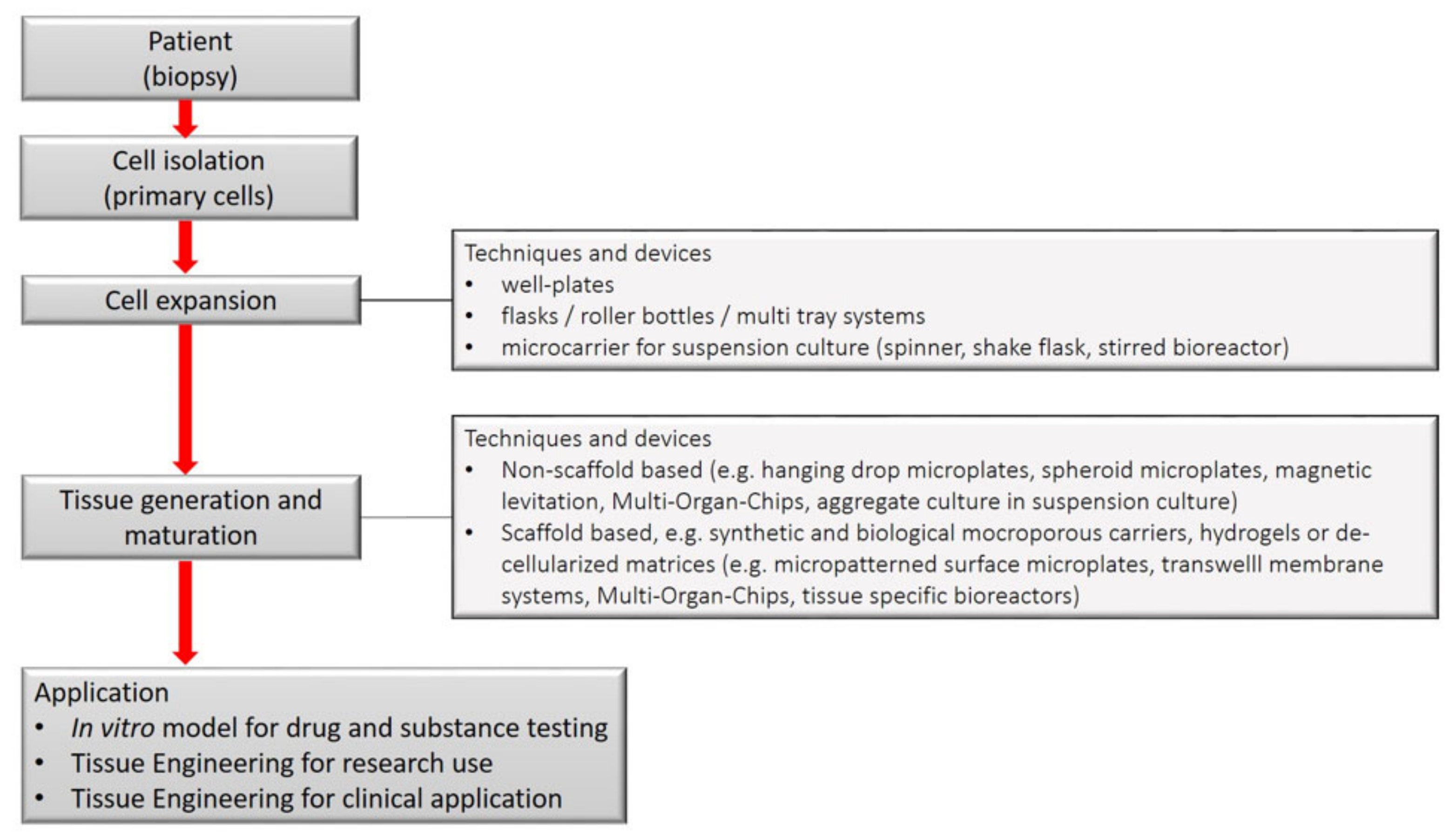
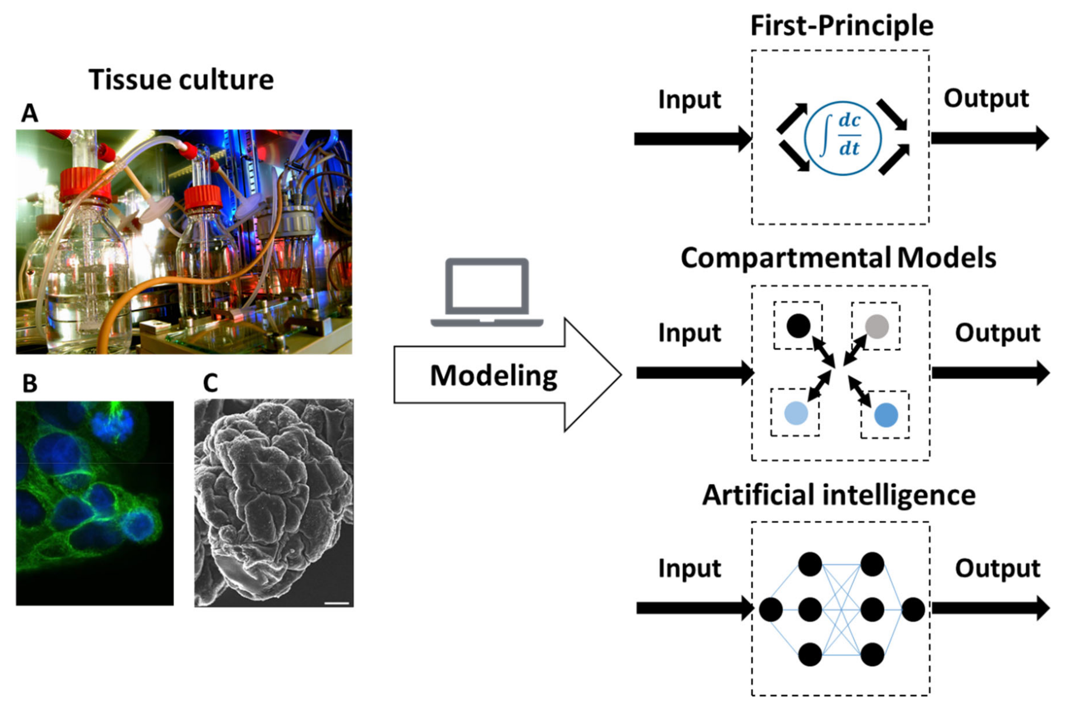
Table 1.
References for specific tasks addressed by mathematical modelling.
| Tasks Addressed by Mathematical Modelling | References |
|---|---|
| General Reviews | [97,98,99,100,101,102,103,104,105,106,107,108,109,110,111,112,113,114,115,116] |
| Cell growth, morphology, heterogeneity, dynamics | [117,118,119,120,121,122,123,124,125,126,127,128] |
| Cell kinetics | [129,130,131,132] |
| Systems biology | [133,134] |
| Cell handling, seeding, culture conditions | [135,136,137,138] |
| Penetration, permeation, diffusion | [139,140,141,142,143,144,145,146,147,148,149,150,151,152,153,154,155,156,157,158,159] |
| Scaffolds-fluid flow, shear, mass transfer | [112,160,161,162,163,164,165,166,167,168,169,170,171] |
| Bioreactor-fluid flow, shear, mass transfer | [45,99,172,173,174,175,176,177,178,179,180,181,182,183,184,185] |
| Mechanical stimulation | [186,187,188,189,190,191,192,193] |
| Image-based modelling | [194,195,196,197,198] |
| Tissue specific effects | [199,200,201,202,203,204,205,206] |
| Pharmacology | [207,208,209,210] |
| Handling of models | [96,211] |
| Model-assisted experimental design | [88,92,212,213,214,215] |
Publisher’s Note: MDPI stays neutral with regard to jurisdictional claims in published maps and institutional affiliations. |
© 2021 by the authors. Licensee MDPI, Basel, Switzerland. This article is an open access article distributed under the terms and conditions of the Creative Commons Attribution (CC BY) license (http://creativecommons.org/licenses/by/4.0/).
Share and Cite
MDPI and ACS Style
Möller, J.; Pörtner, R. Digital Twins for Tissue Culture Techniques—Concepts, Expectations, and State of the Art. Processes 2021, 9, 447. https://0-doi-org.brum.beds.ac.uk/10.3390/pr9030447
AMA Style
Möller J, Pörtner R. Digital Twins for Tissue Culture Techniques—Concepts, Expectations, and State of the Art. Processes. 2021; 9(3):447. https://0-doi-org.brum.beds.ac.uk/10.3390/pr9030447
Chicago/Turabian StyleMöller, Johannes, and Ralf Pörtner. 2021. "Digital Twins for Tissue Culture Techniques—Concepts, Expectations, and State of the Art" Processes 9, no. 3: 447. https://0-doi-org.brum.beds.ac.uk/10.3390/pr9030447
Note that from the first issue of 2016, this journal uses article numbers instead of page numbers. See further details here.