Modification of Lattice Structures and Mechanical Properties of Metallic Materials by Energetic Ion Irradiation and Subsequent Thermal Treatments


Abstract
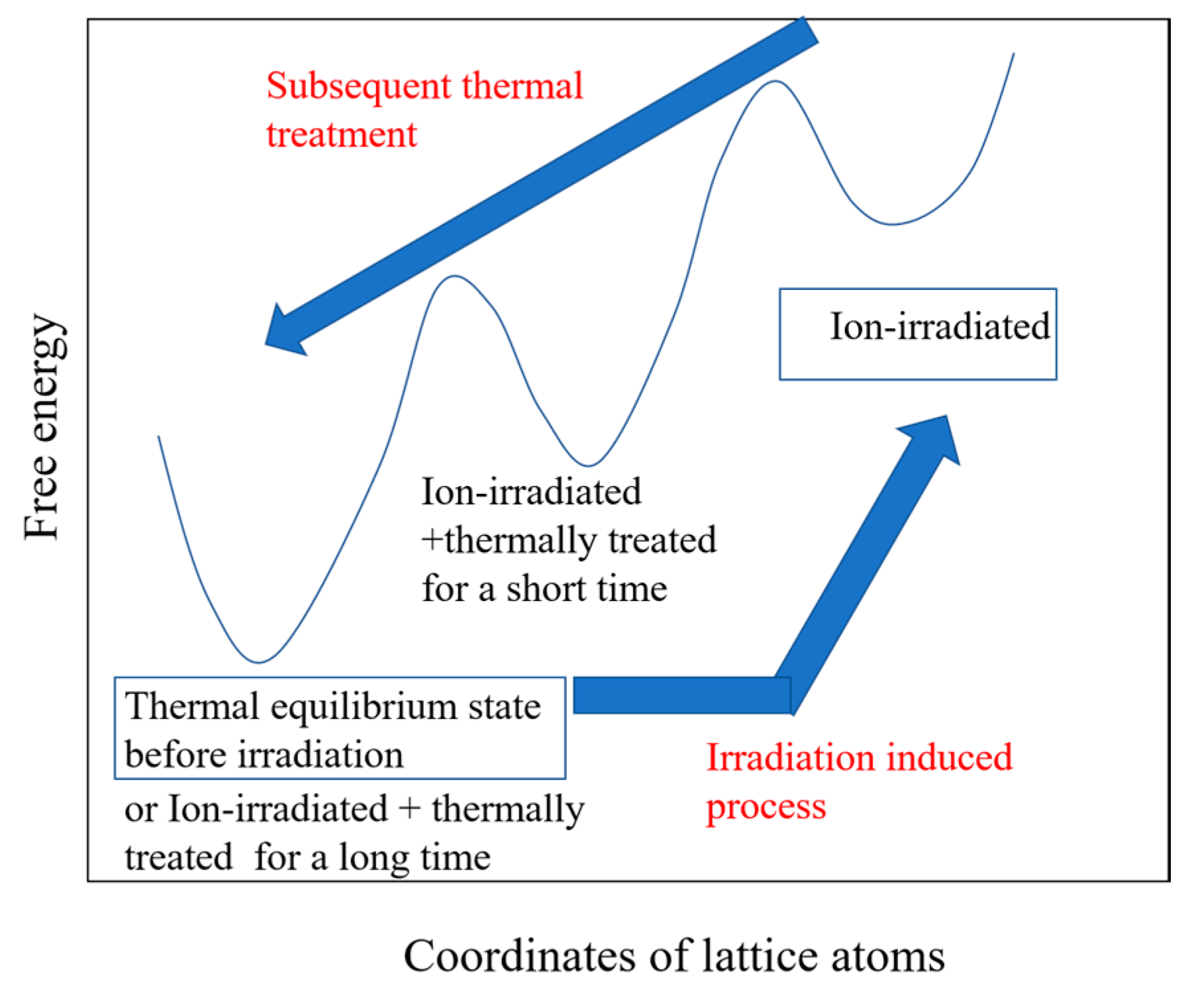
:1. Introduction
2. Modification of the Microstructures and the Surface Hardness of Dilute Aluminum Alloys
2.1. Experimental Procedure
2.2. Results and Discussion
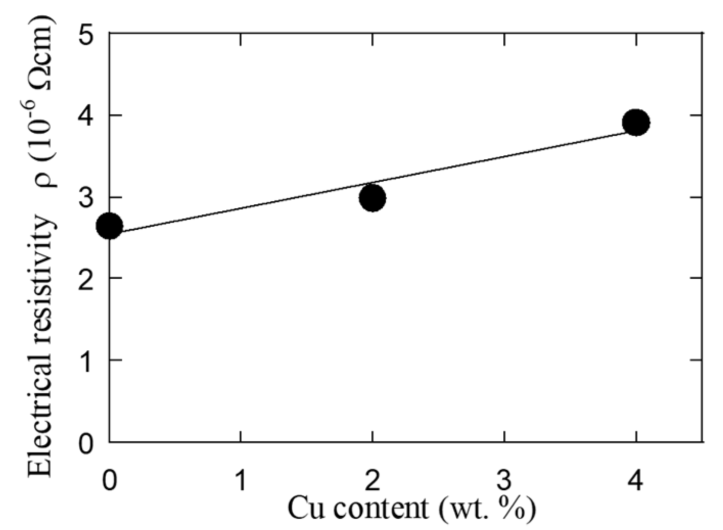
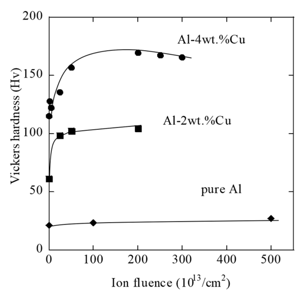
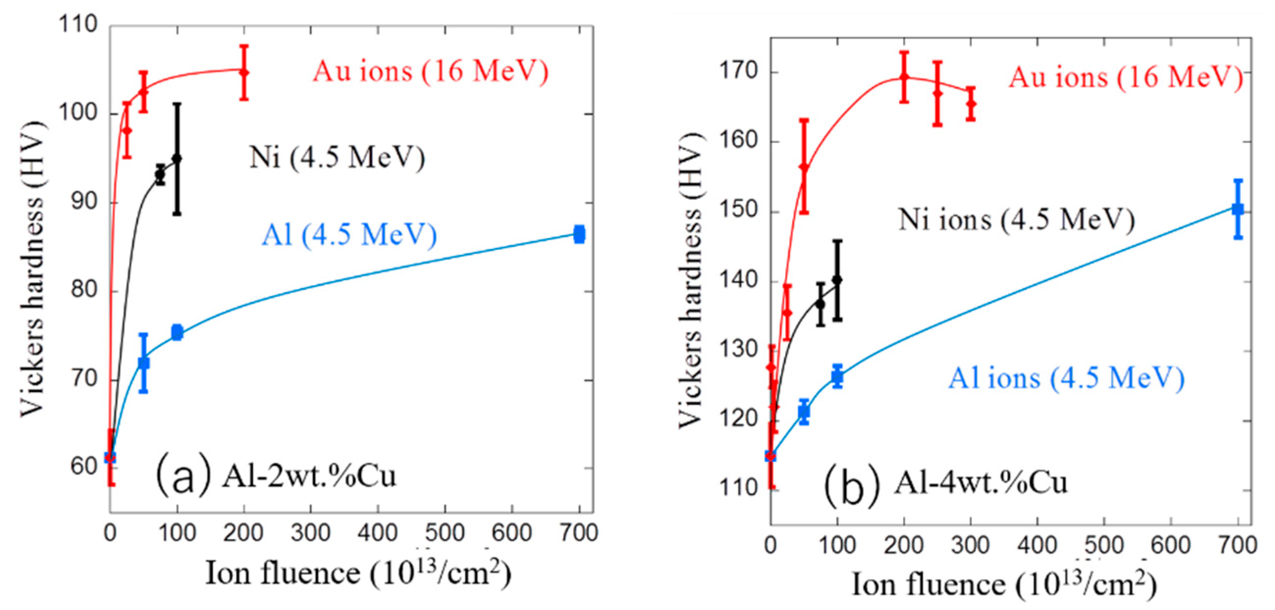
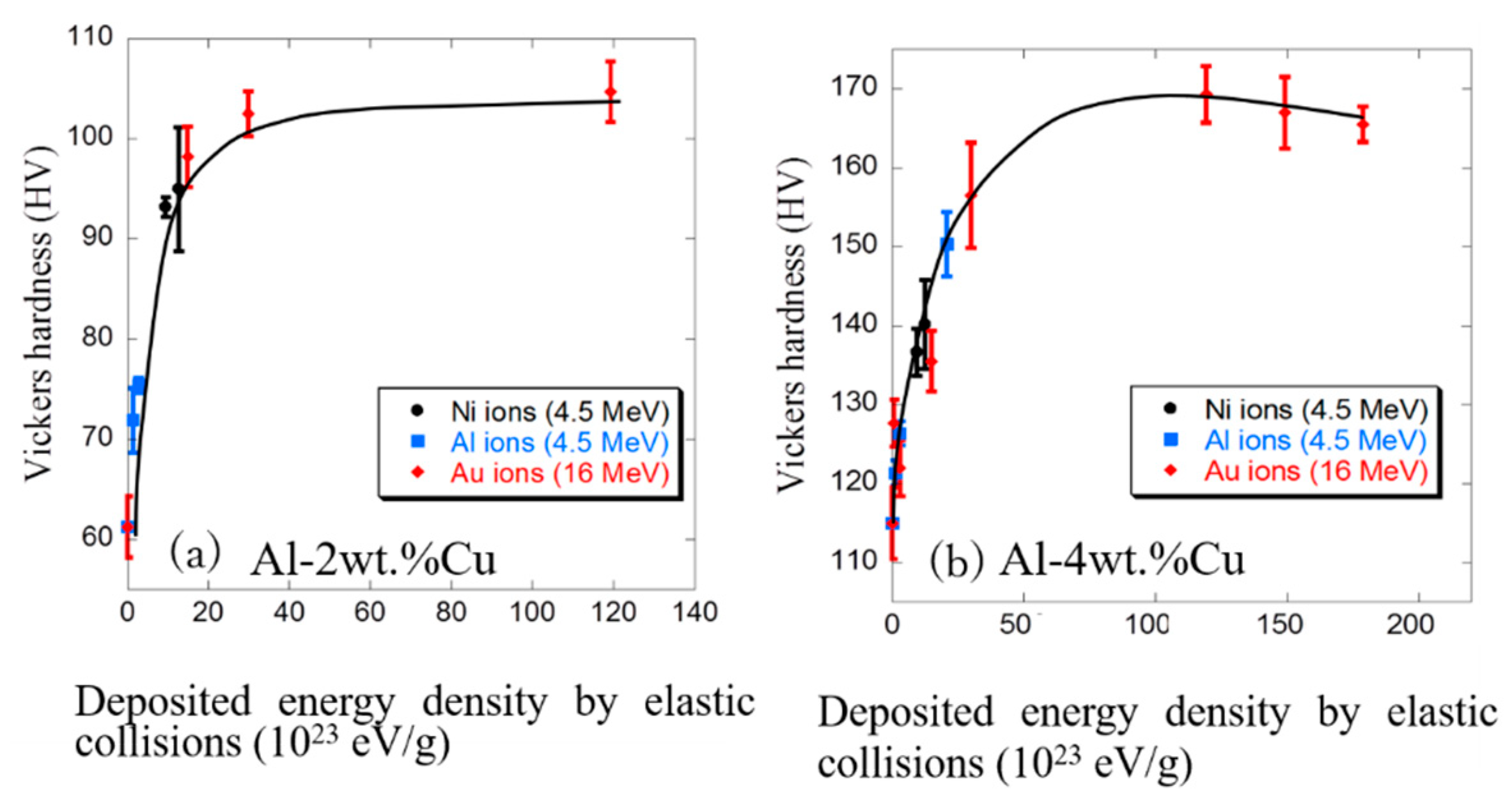
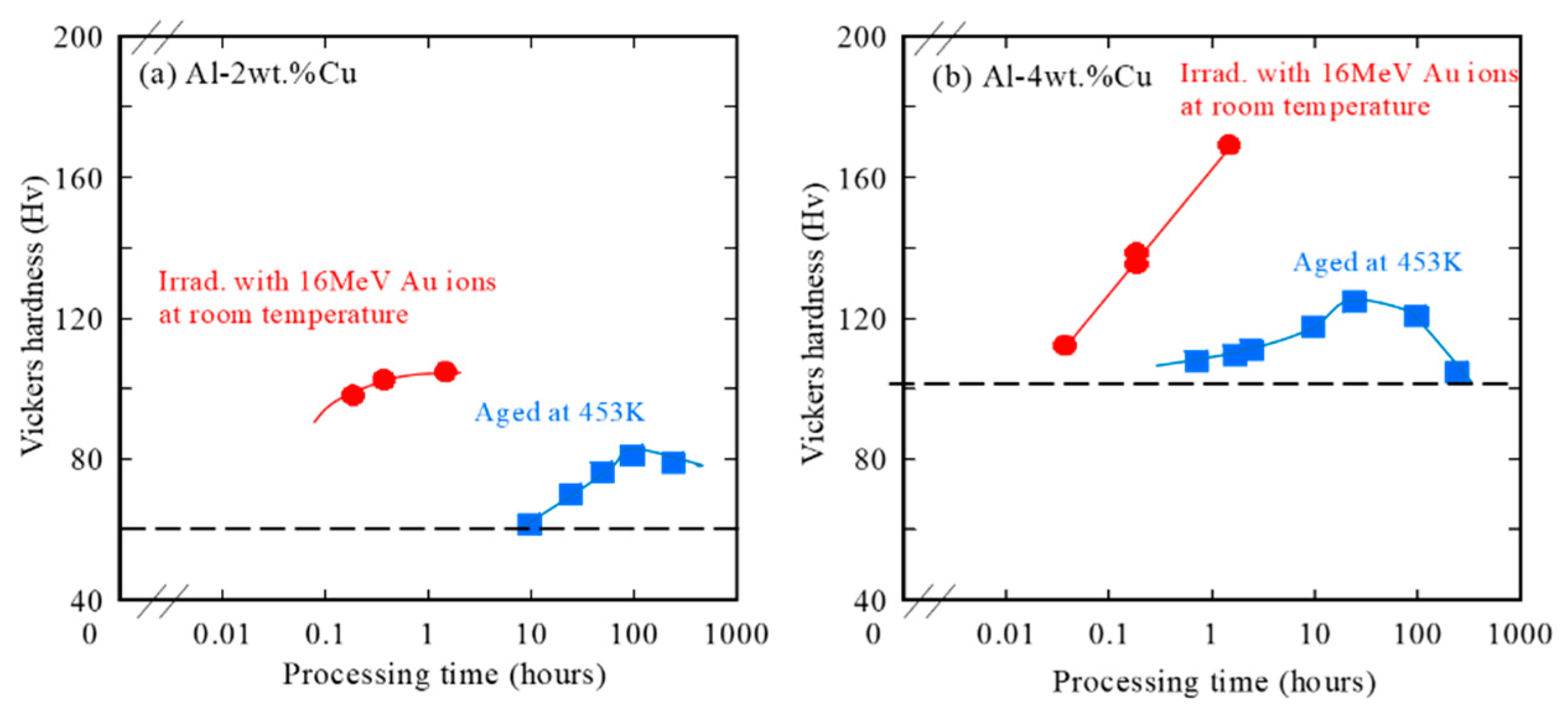
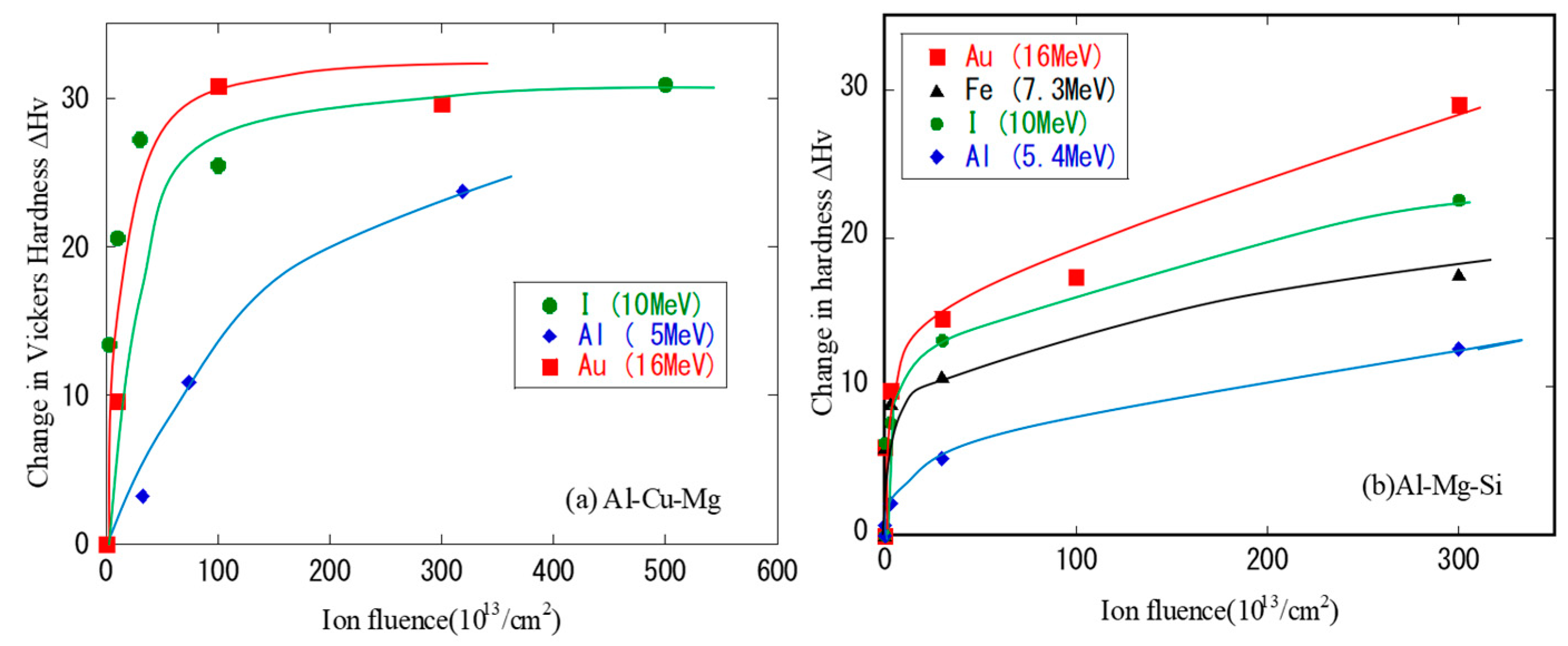
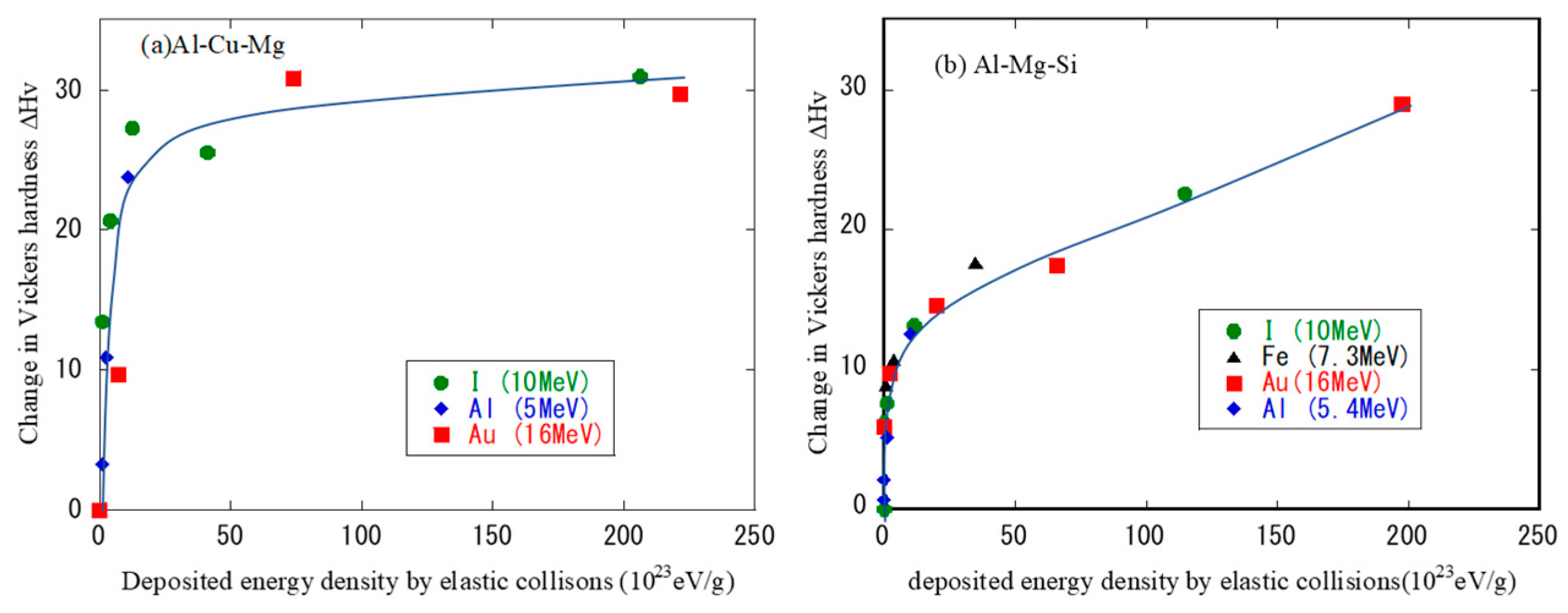
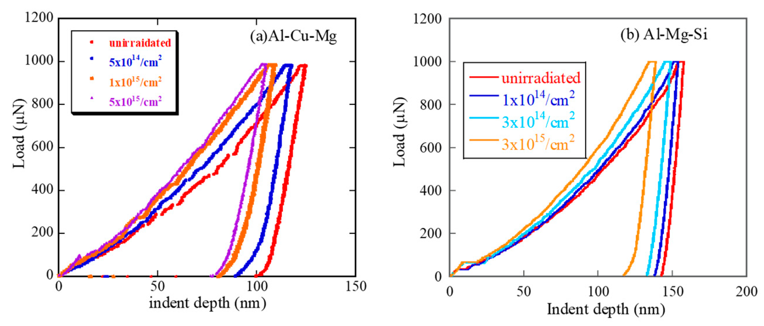
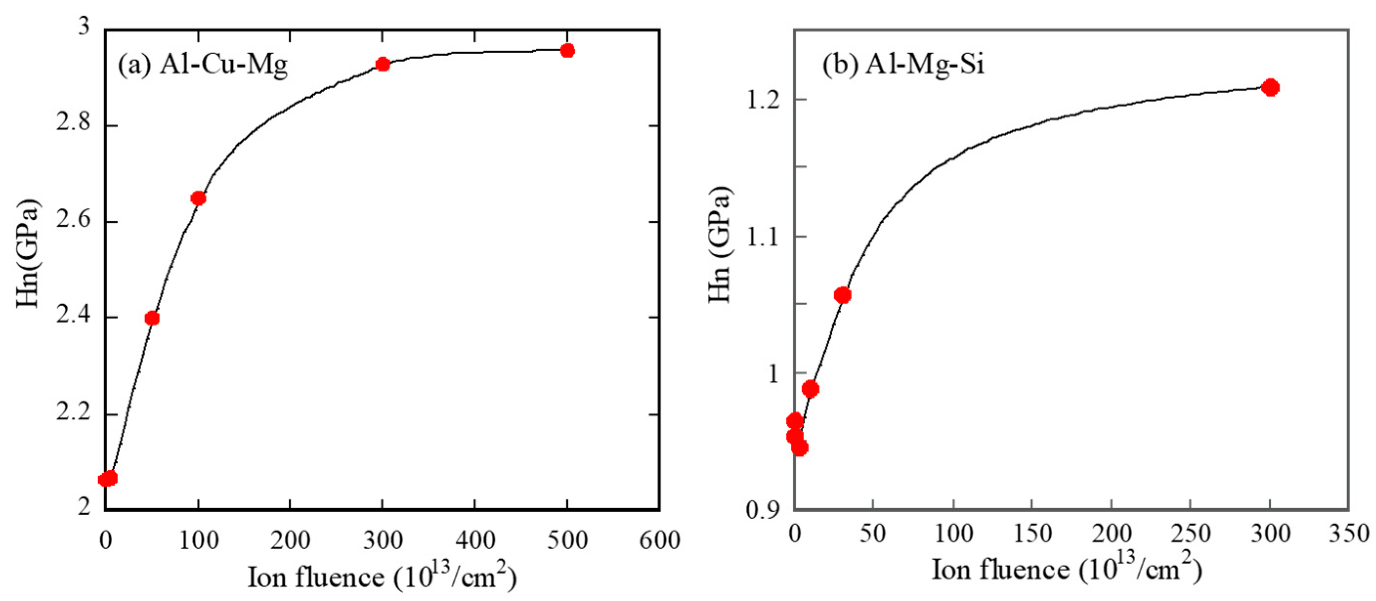
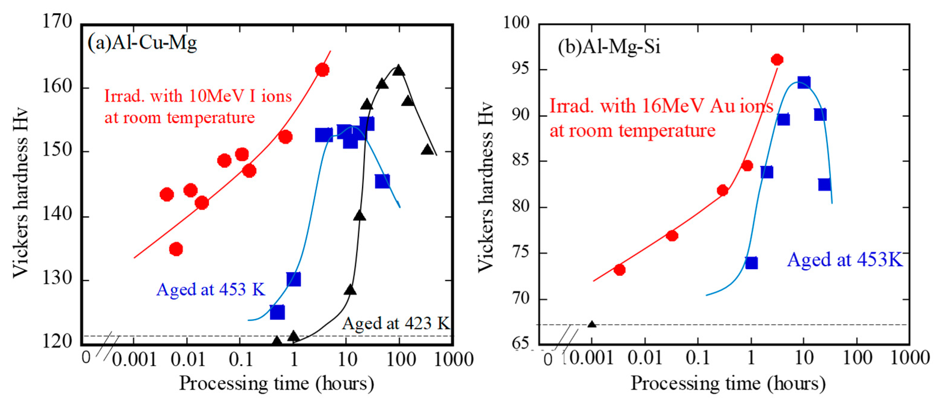
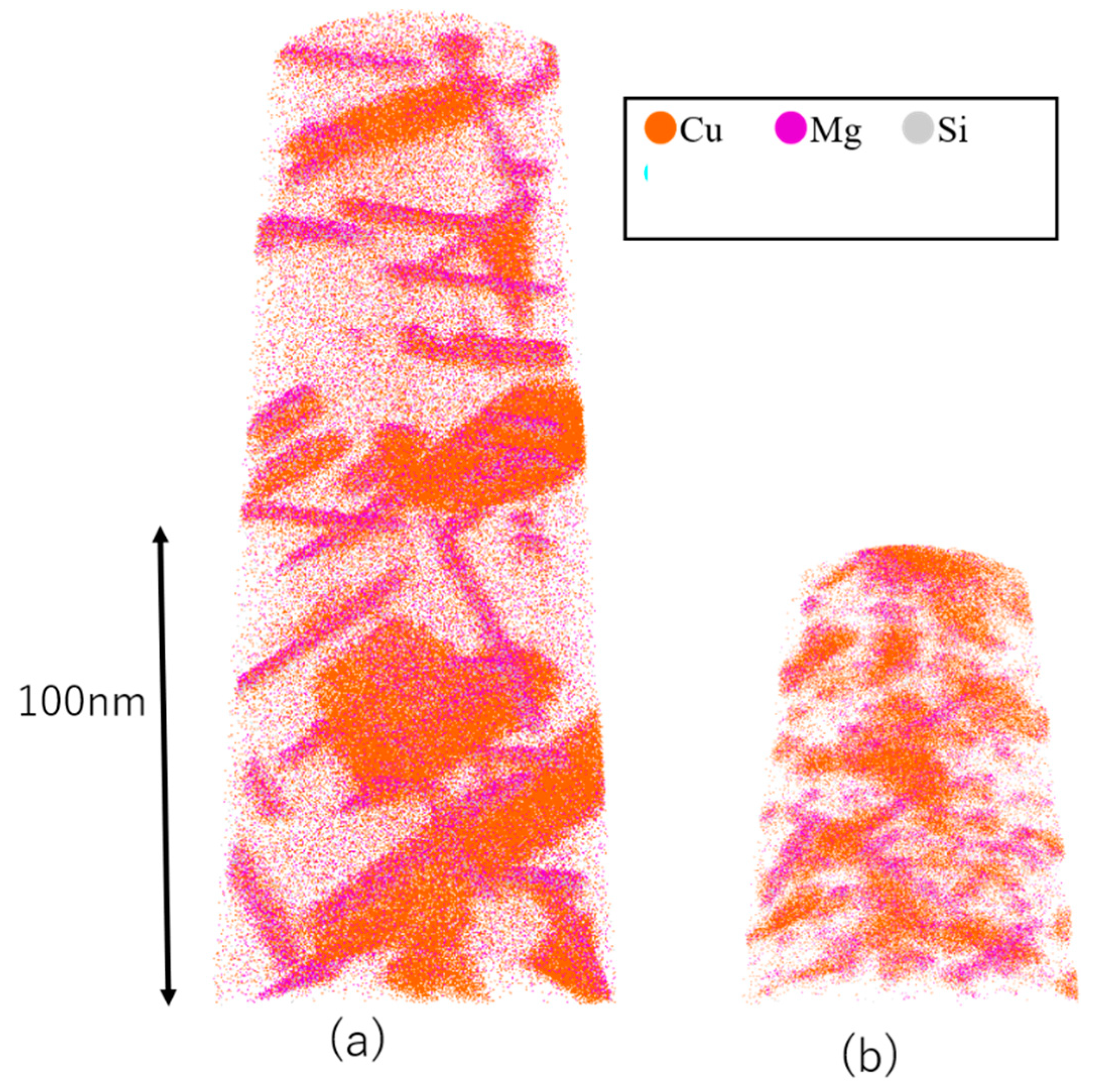
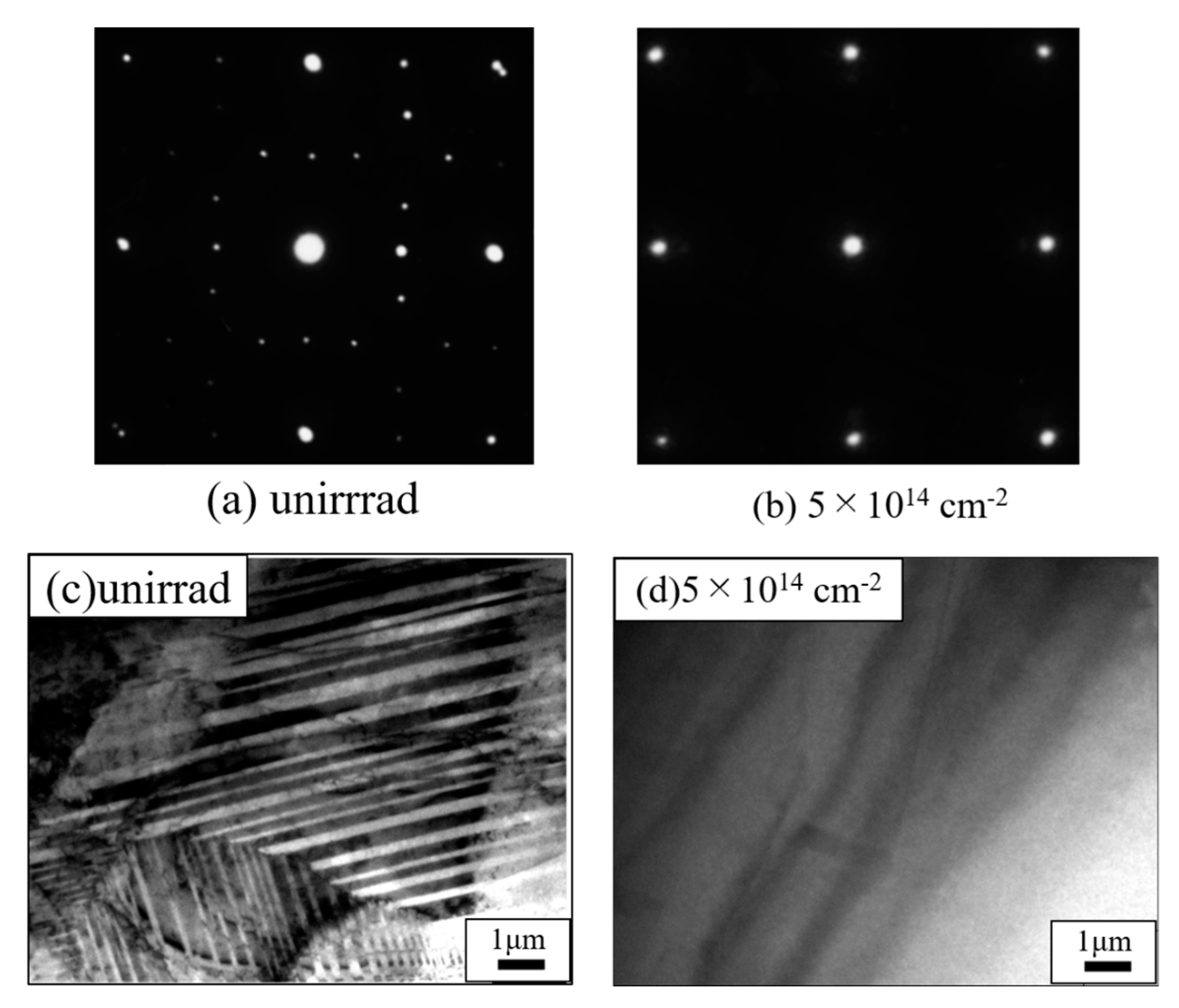
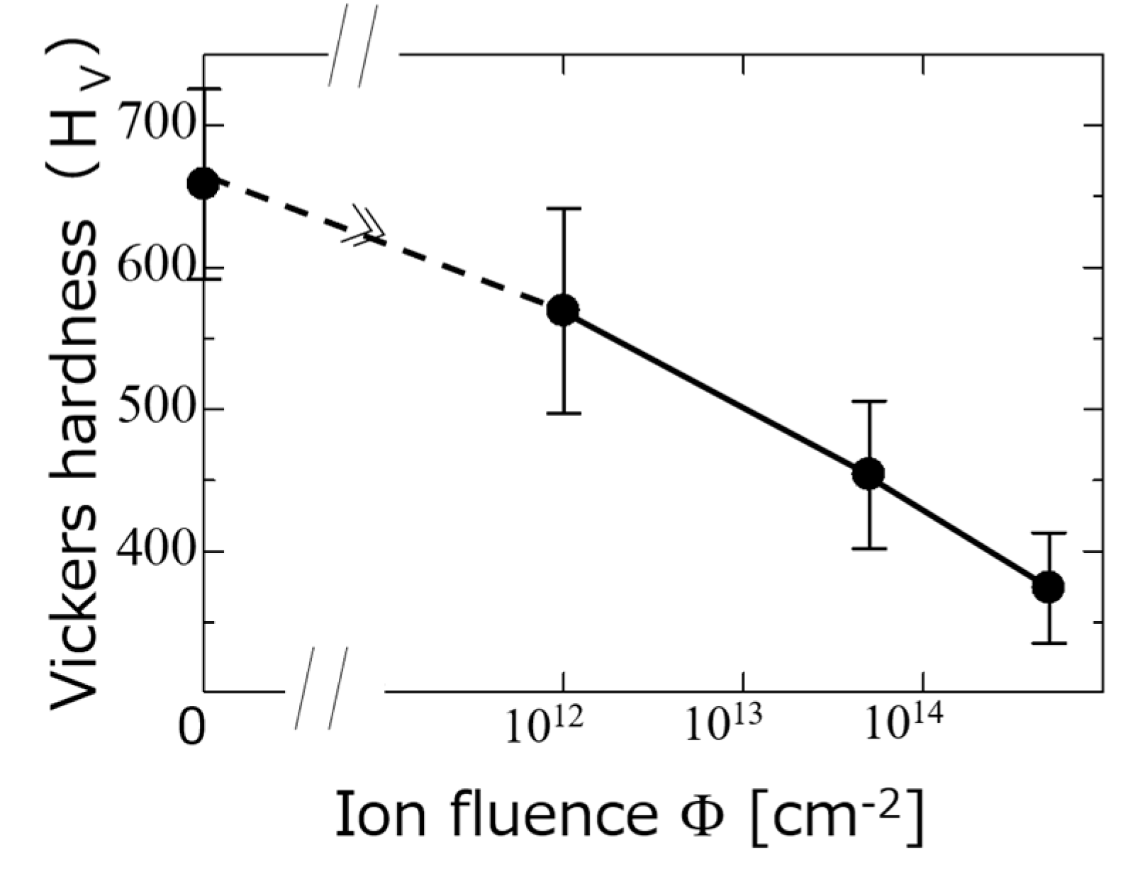
2.2.1. Effects of Ion Irradiation on the Microstructure and Surface Hardness of the Dilute Aluminum Alloys
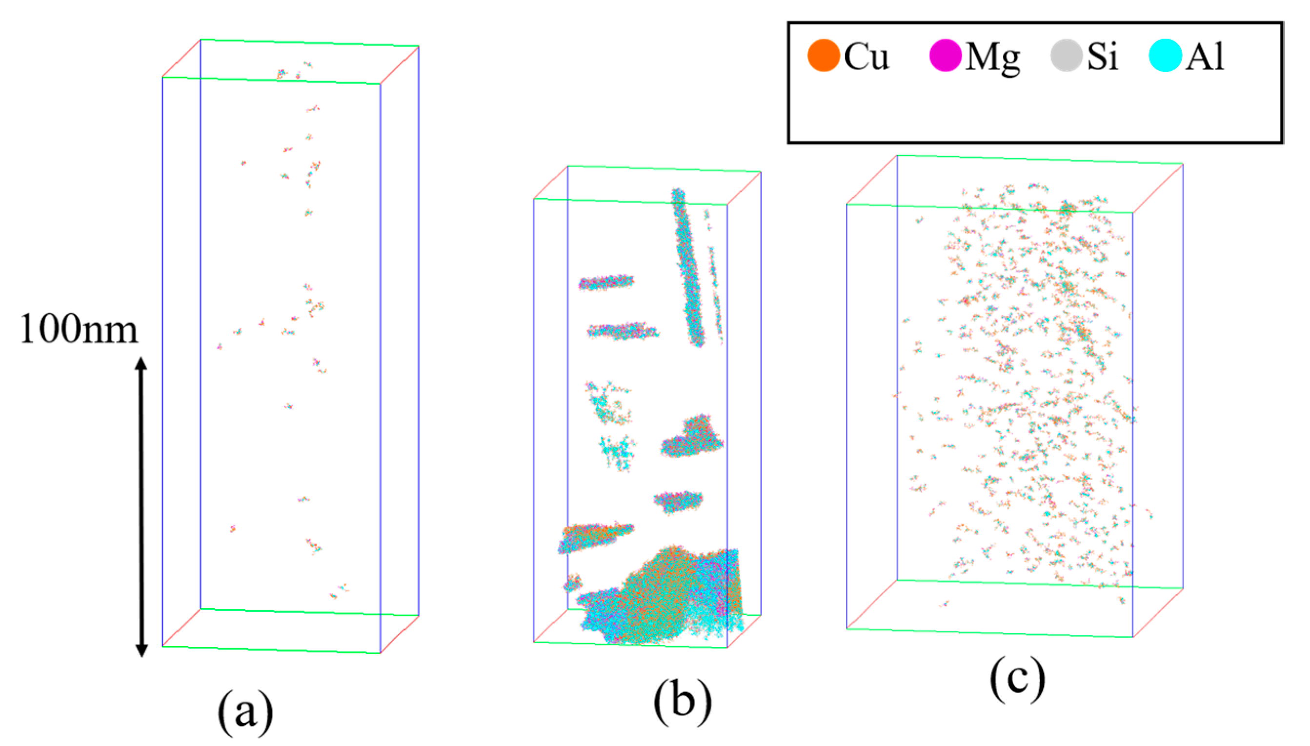
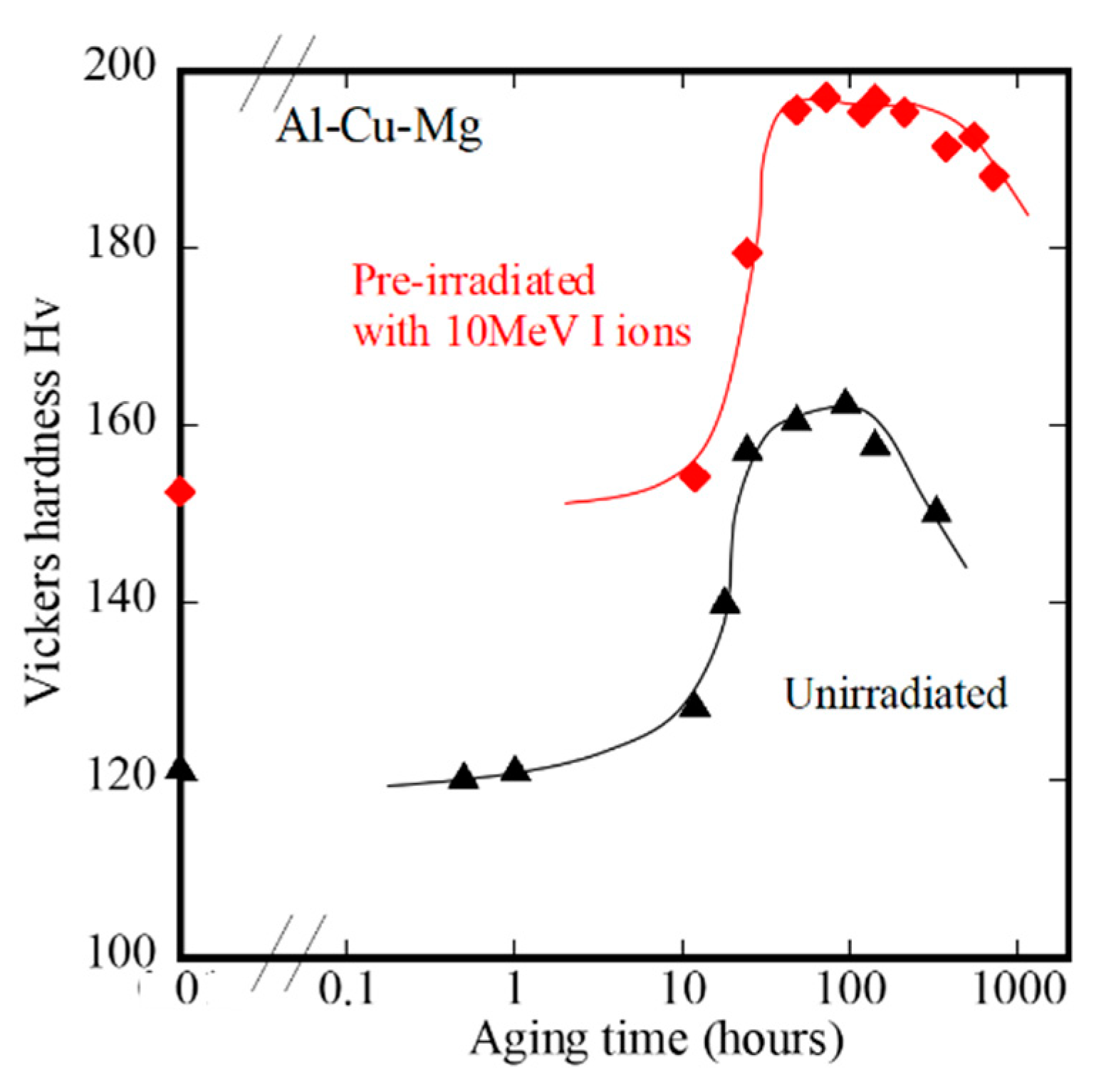
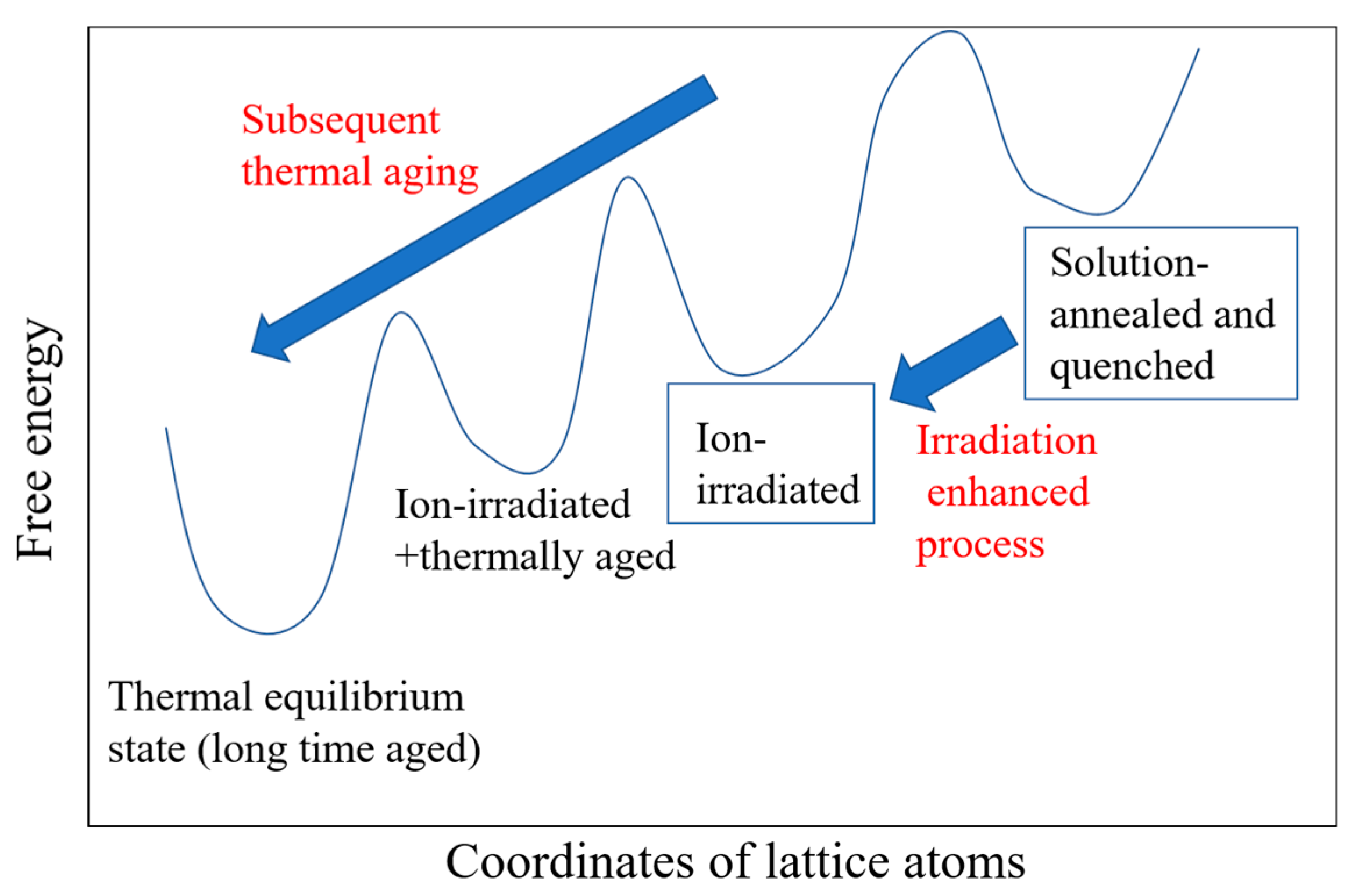
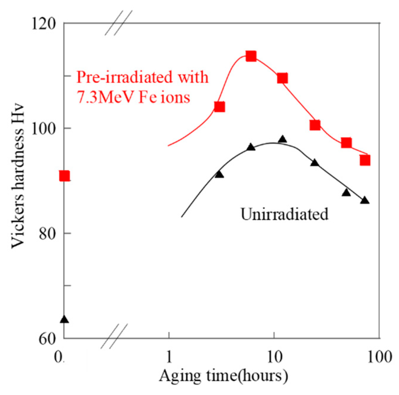
2.2.2. Effect of Subsequent Thermal Treatments after the Irradiation on the Microstructures and Hardness of the Dilute Aluminum Alloys
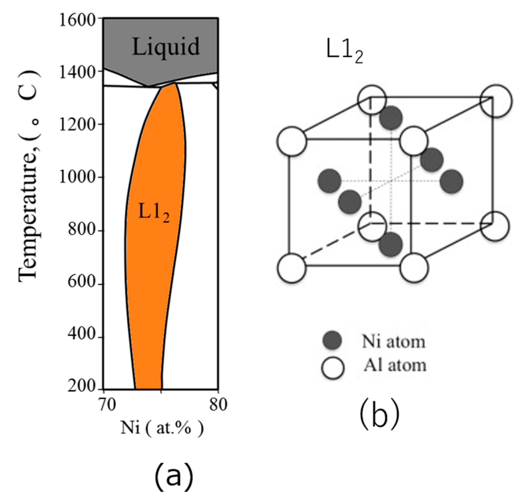
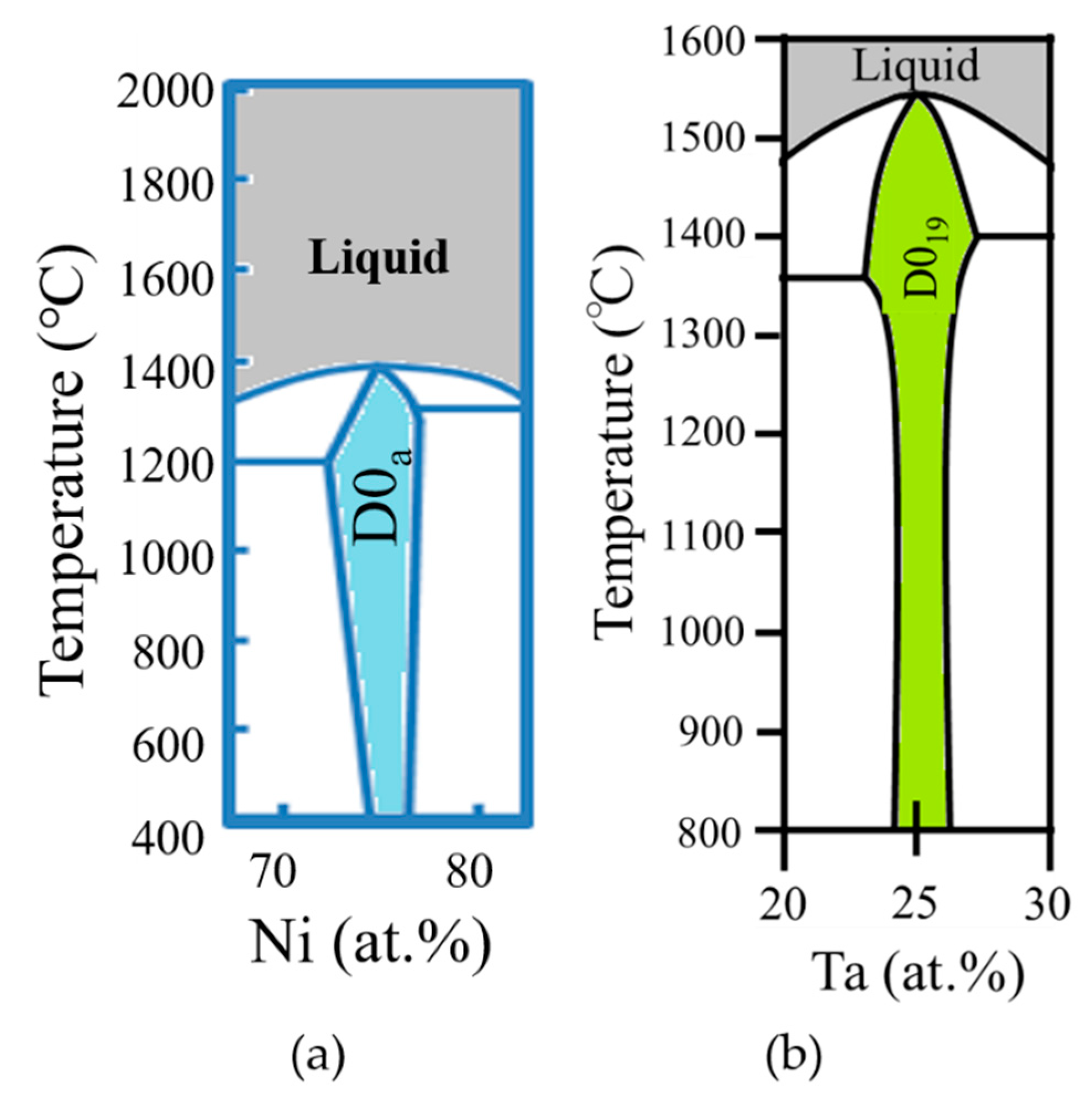
3. Modification of Lattice Structures and Surface Hardness of Ni-Based Intermetallic Compounds
3.1. Experimental Procedure
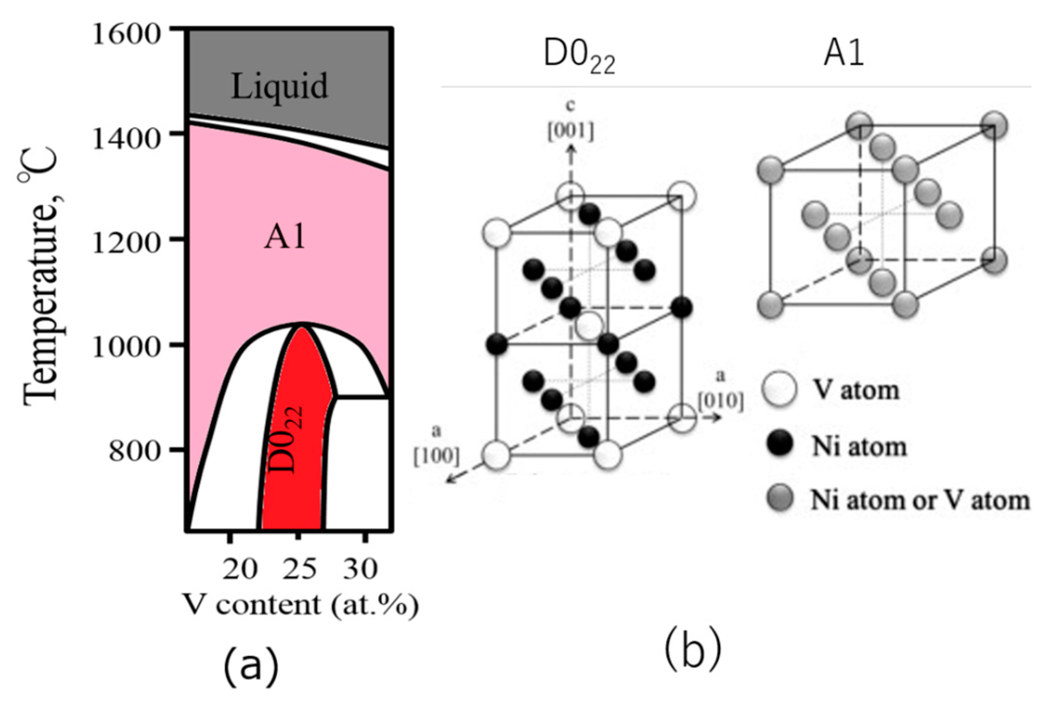
3.2. Results and Discussion
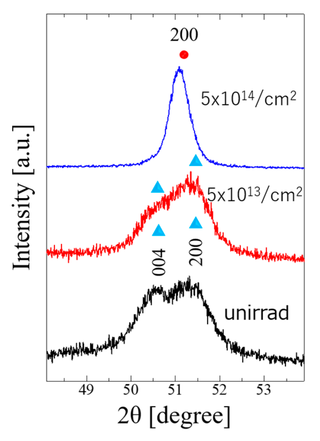
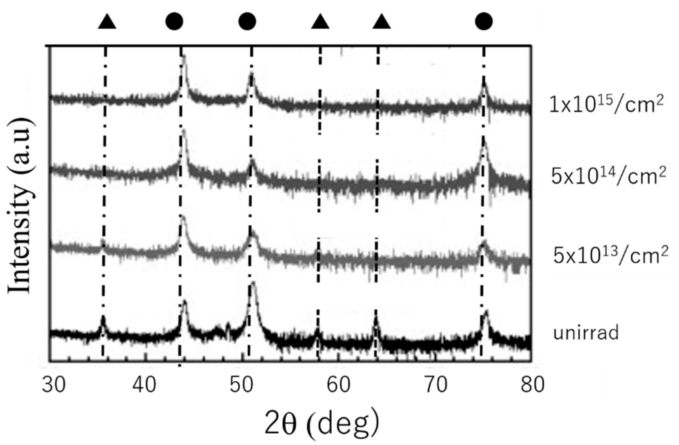
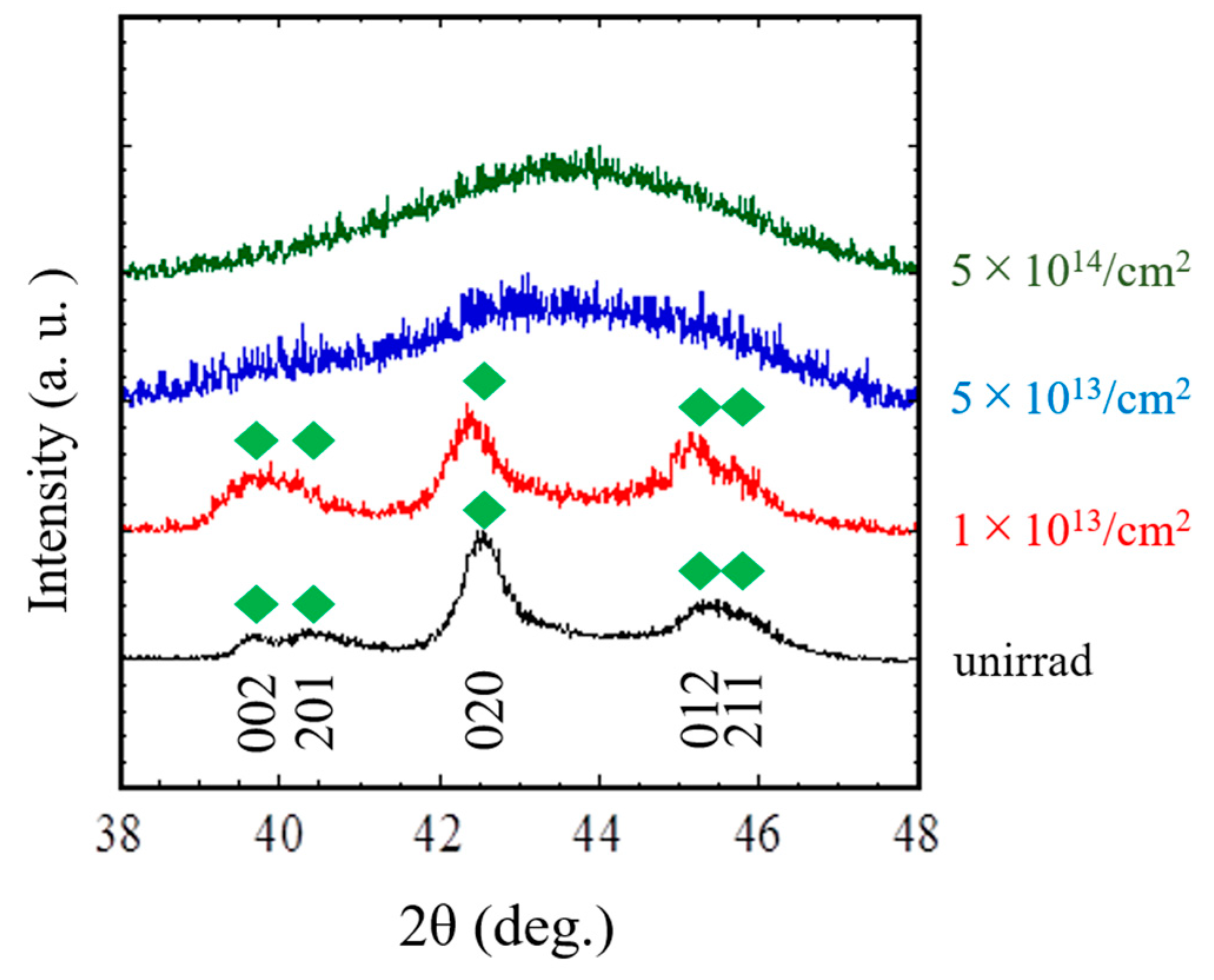
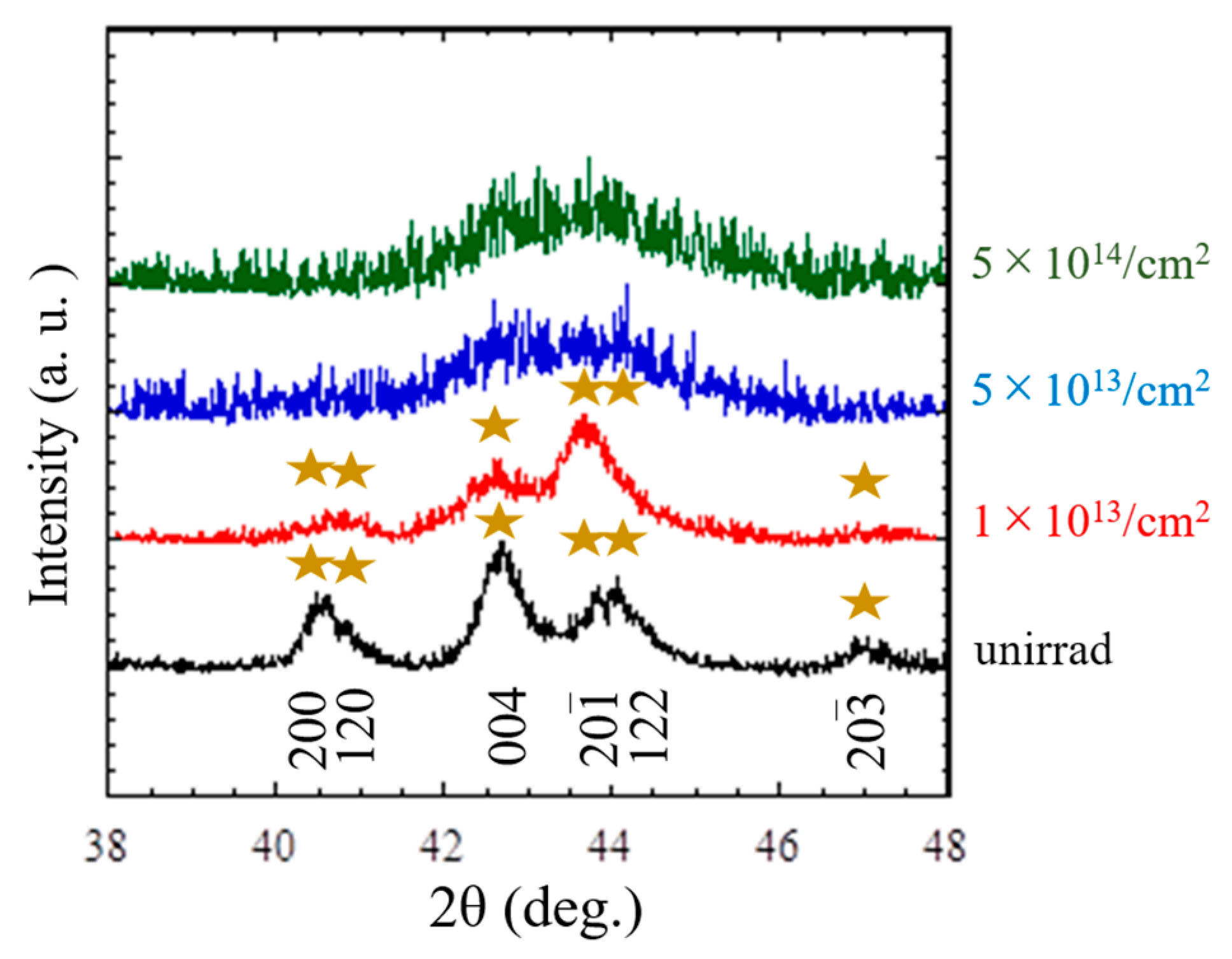
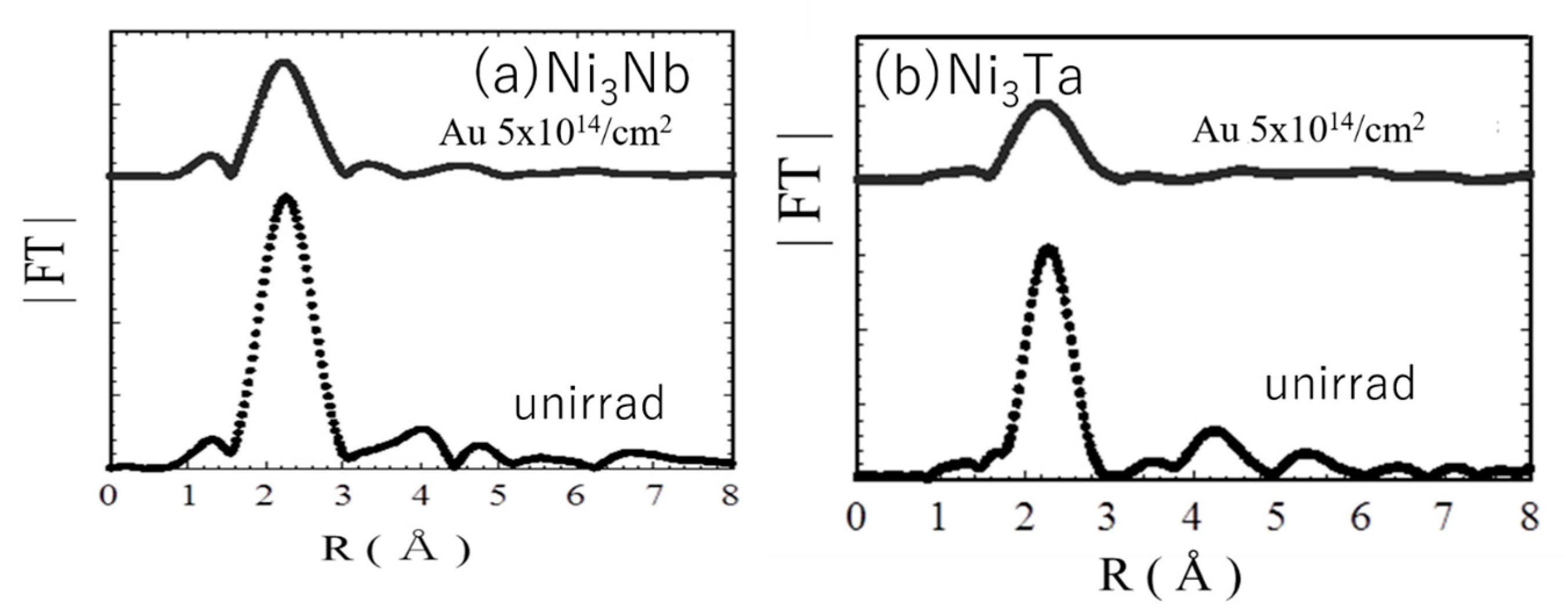
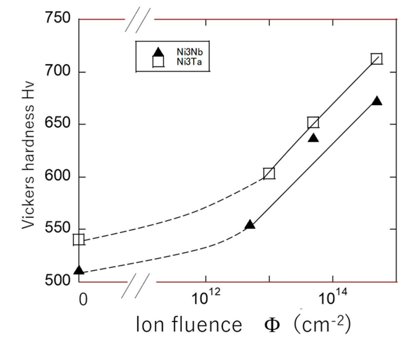
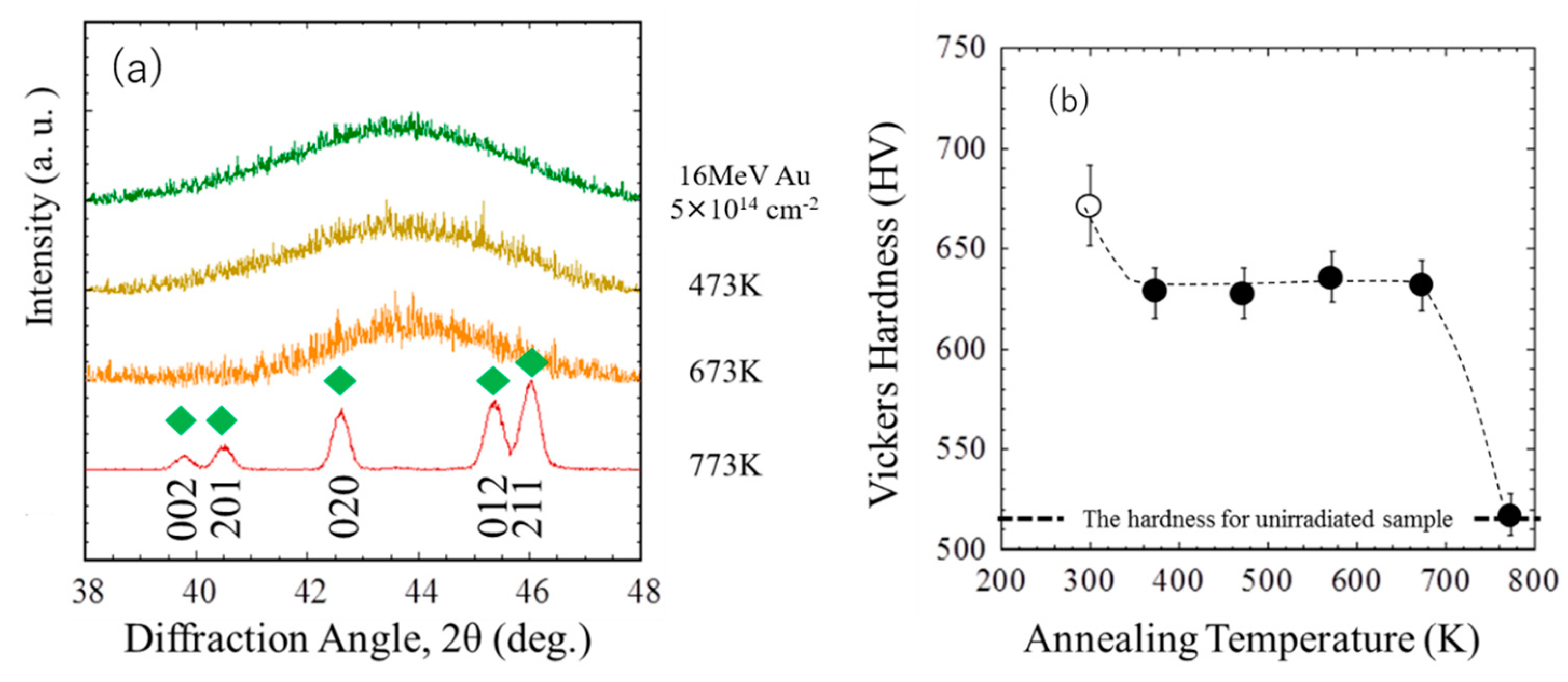
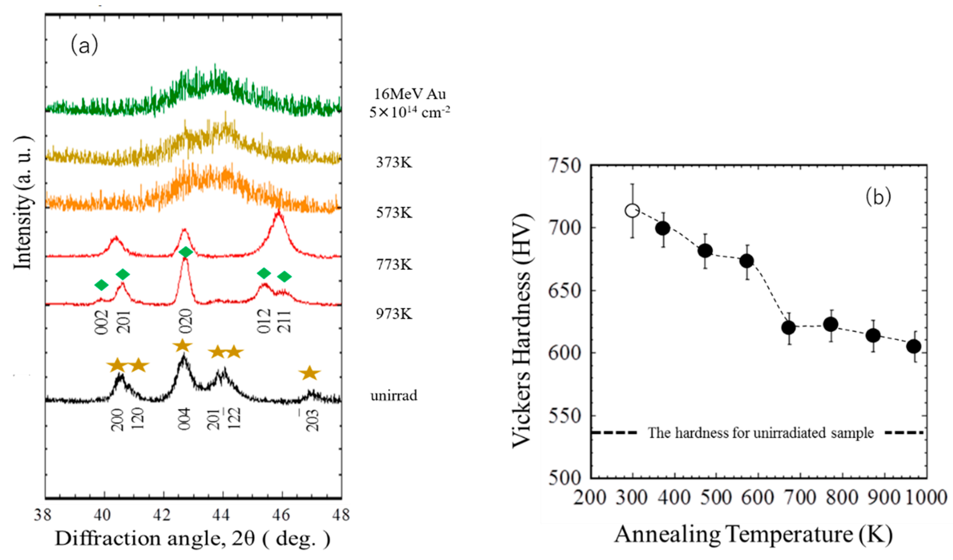
3.2.1. Effects of 16 MeV Au Ion Irradiation on the Lattice Structure and Surface Hardness of the Ni-Based Intermetallic Compounds
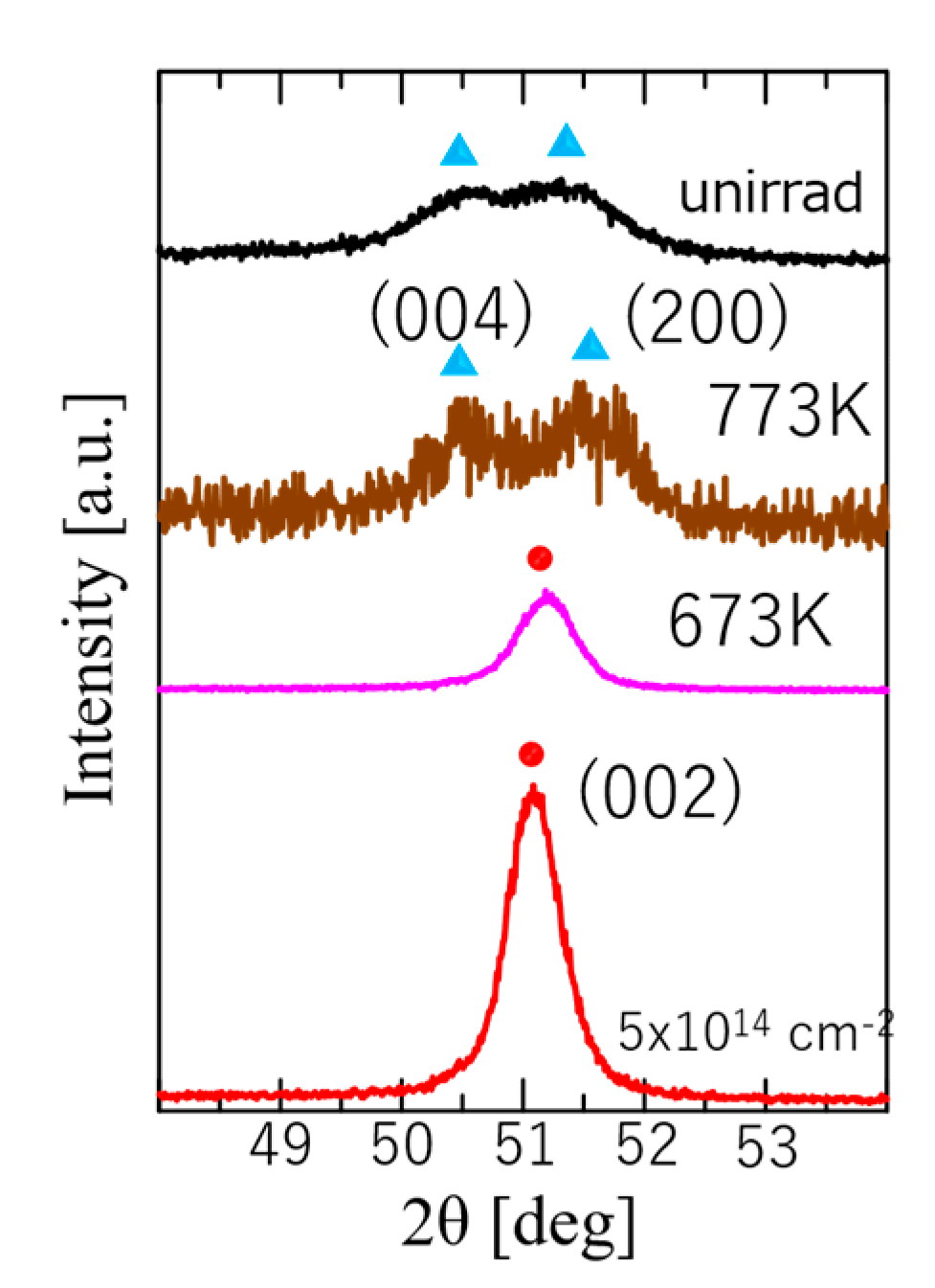
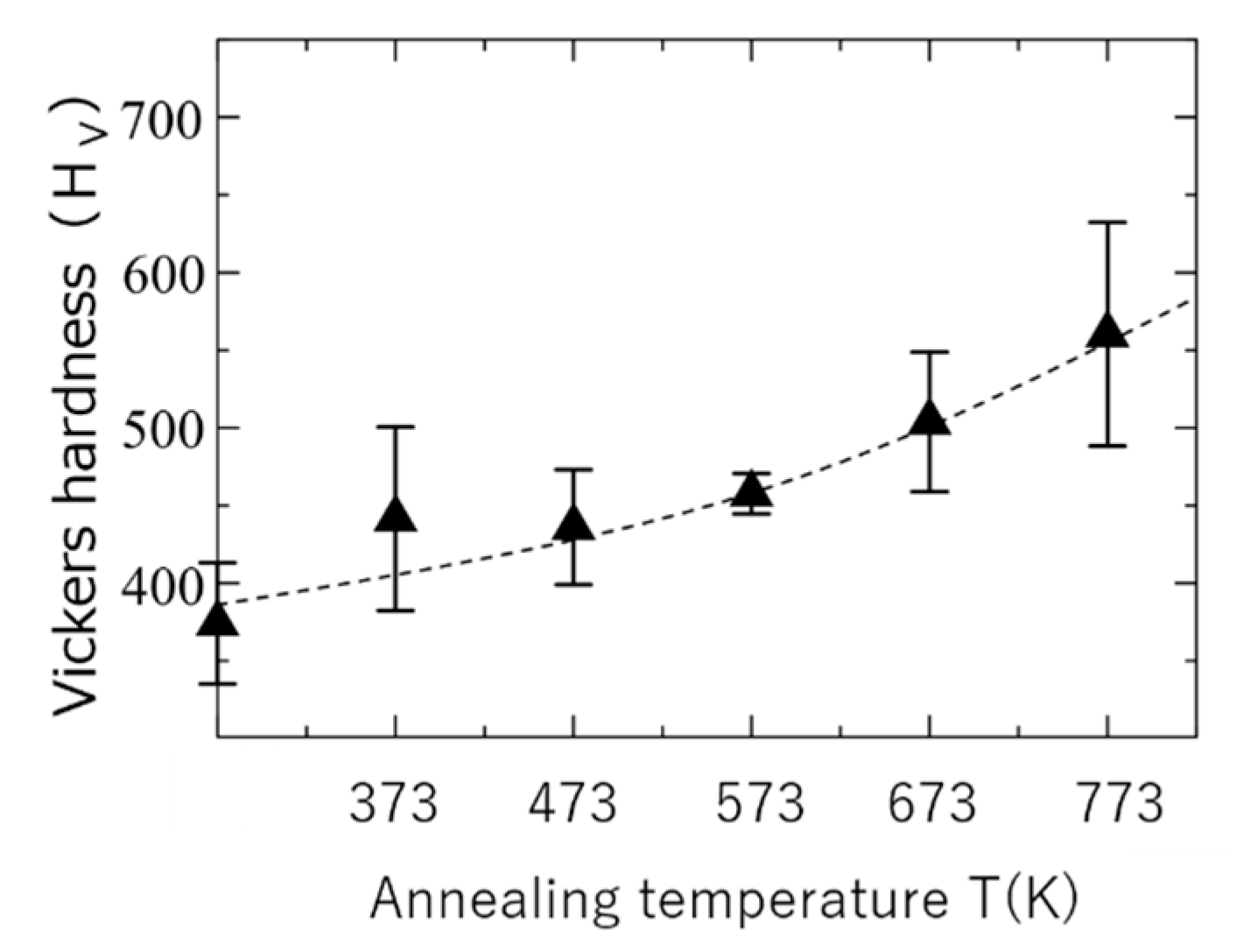
3.2.2. Effect of Subsequent Thermal Treatments after the Irradiation on the Lattice Structures and Hardness of the Ni-Based Intermetallic Compounds
4. Industrial Application of Energetic Ion Irradiation to the Control of Surface Hardness of Metallic Alloys
5. Summary
Author Contributions
Funding
Acknowledgments
Conflicts of Interest
References
- Lehmann, C. Interaction of Radiation with Solids and Elementary Defect Production; North-Holland Publishing Company: Amsterdam, The Netherlands, 1977. [Google Scholar]
- Harish Kumar, H.G. High Energy Electron Irradiation Effects in Polymers; LAP LAMBERT Academic Publishing: Deutschland, Germany, 2011. [Google Scholar]
- Ashworth, V.; Grant, W.A.; Procter, R.P.M. (Eds.) Ion Implantation into Metals; Pergamon Press: Oxford, UK, 1982. [Google Scholar]
- Rimini, E. Ion Implantation: Basics to Device Fabrication; Springer: Berlin/Heidelberg, Germany, 1995. [Google Scholar]
- Was, G.S. Fundamentals of Radiation Materials Science, Metals and Alloys; Springer: Berlin/Heidelberg, Germany, 2007. [Google Scholar]
- Imaizumi, M.; Nakamura, T.; Takamoto, T.; Ohshima, T.; Tajima, M. Radiation degradation characteristics of component subcells in inverted metamorphic triple-junction solar cells irradiated with electrons and protons. Prog. Photovolt. Res. Appl. 2017, 25, 161–174. [Google Scholar] [CrossRef]
- Mitsuda, T.; Kobayashi, I.; Kosugi, S.; Fujita, N.; Saitoh, Y.; Hori, F.; Semboshi, S.; Kaneno, Y.; Nishida, K.; Soneda, N.; et al. Hardening of Al-Cu-Mg alloy by energetic ion irradiation. J. Nucl. Mater. 2011, 408, 201–204. [Google Scholar] [CrossRef]
- Mitsuda, T.; Kobayashi, I.; Kosugi, S.; Fujita, N.; Saitoh, Y.; Hori, F.; Semboshi, S.; Kaneno, Y.; Nishida, K.; Soneda, N.; et al. Hardness modification of aluminum-alloys by means of energetic ion irradiation and subsequent thermal aging. Nucl. Instrum. Methods B 2012, 272, 49–52. [Google Scholar] [CrossRef]
- Ueyama, D.; Saitoh, Y.; Hori, F.; Kaneno, Y.; Nishida, K.; Dohi, K.; Soneda, N.; Semboshi, S.; Iwase, A. Effects of energetic heavy ion irradiation on hardness of Al-Mg-Si alloys. Nucl. Instrum. Methods B 2013, 314, 107–111. [Google Scholar] [CrossRef]
- Hashimoto, A.; Kaneno, Y.; Matsui, T.; Saitoh, Y.; Iwase, A. Non-thermal equilibrium crystal structure and Vickers hardness of FeRh intermetallic compound irradiated with energetic heavy ions. Trans. Mater. Res. Soc. Jpn. 2013, 38, 329–331. [Google Scholar] [CrossRef] [Green Version]
- Hashimoto, A.; Kaneno, Y.; Semboshi, S.; Yoshizaki, H.; Saitoh, Y.; Iwase, A. Effect of high temperature annealing on non-thermal equilibrium phases induced by energetic ion irradiation in FeRh and Ni3V intermetallic compounds. Jpn. J. Appl. Phys. 2014, 53, 05FC08. [Google Scholar] [CrossRef]
- Ueyama, D.; Semboshi, S.; Saitoh, Y.; Ishikawa, N.; Nishida, K.; Soneda, N.; Hori, F.; Iwase, A. Hardening induced by energetic electron beam for Cu-Ti alloys. Jpn. J. Appl. Phys. 2014, 53, 05FC04. [Google Scholar] [CrossRef]
- Hashimoto, A.; Kaneno, Y.; Semboshi, S.; Yoshizaki, H.; Saitoh, Y.; Okamoto, Y.; Iwase, A. Microstructure evolution and hardness change in ordered Ni3V intermetallic alloy by energetic ion irradiation. Nucl. Instrum. Methods B 2014, 338, 72–76. [Google Scholar] [CrossRef]
- Ueyama, D.; Semboshi, S.; Saitoh, Y.; Hori, F.; Nishida, K.; Soneda, N.; Iwase, A. Modification of microstructure and hardness of Cu-Ti alloy by means of energetic ion beam irradiation. Nucl. Instrum. Methods B 2014, 341, 53–57. [Google Scholar] [CrossRef]
- Yoshizaki, H.; Hashimoto, A.; Kaneno, Y.; Semboshi, S.; Saitoh, Y.; Okamoto, Y.; Iwase, A. Modification of surface hardness for dual two-phase Ni3Al-Ni3V intermetallic compound by using energetic ion beam and subsequent thermal treatment. Nucl. Instrum. Methods B 2015, 345, 22–26. [Google Scholar] [CrossRef]
- Yoshizaki, H.; Hashimoto, A.; Kaneno, Y.; Semboshi, S.; Hori, F.; Saitoh, Y.; Iwase, A. Energetic ion beam induced crystal phase transformation and resulting hardness change in Ni3Al intermetallic compound. Nucl. Instrum Methods B 2015, 354, 287–291. [Google Scholar] [CrossRef]
- Ueyama, D.; Saitoh, Y.; Ishikawa, N.; Ohmura, T.; Semboshi, S.; Hori, F.; Iwase, A. Hardness modification of Al-Mg-Si alloy by using energetic ion beam irradiation. Nucl. Instrum. Methods B 2015, 351, 1–5. [Google Scholar] [CrossRef]
- Kojima, H.; Yoshizaki, H.; Kaneno, Y.; Semboshi, S.; Hori, F.; Saitoh, Y.; Okamoto, Y.; Iwase, A. Lattice structure transformation and change in surface hardness of Ni3Nb and Ni3Ta intermetallic compounds induced by energetic ion beam irradiation. Nucl. Istrum. Methods B 2016, 372, 72–77. [Google Scholar] [CrossRef]
- Mayumi, R.; Semboshi, S.; Okamoto, Y.; Saitoh, Y.; Yoshiie, T.; Iwase, A. Radiation enhanced precipitation of solute atoms in AlCu binary alloys—Energetic ion irradiation experiment and computer simulation. Trans. Mater. Res. Soc. Jpn. 2017, 42, 9–14. [Google Scholar] [CrossRef] [Green Version]
- Kojima, H.; Kaneno, Y.; Ochi, M.; Semboshi, S.; Hori, F.; Saitoh, Y.; Ishikawa, N.; Okamoto, Y.; Iwase, A. Ion species/energy dependence of irradiation-induced lattice structure transformation and surface hardness of Ni3Nb and Ni3Ta intermetallic compounds. Mater. Trans. 2017, 58, 739–748. [Google Scholar] [CrossRef] [Green Version]
- Kojima, H.; Ochi, M.; Kaneno, Y.; Semboshi, S.; Hori, F.; Saitoh, Y.; Iwase, A. Thermal stability of energetic ion irradiation induced amorphization for Ni3Nb and Ni3Ta intermetallic compounds. Trans. Mat. Res. Soc. Jpn. 2017, 42, 41–45. [Google Scholar] [CrossRef] [Green Version]
- Ochi, M.; Kojima, H.; Fukuda, K.; Kaneno, Y.; Semboshi, S.; Hori, F.; Saitoh, Y.; Iwase, A. Thermal stability of irradiation-induced metastable lattice structures in NiTi intermetallic compound. Trans. Mater. Res. Soc. Jpn. 2018, 43, 53–56. [Google Scholar] [CrossRef]
- Ochi, M.; Kojima, H.; Hori, F.; Kaneno, Y.; Semboshi, S.; Saitoh, Y.; Okamoto, Y.; Ishikawa, N.; Iwase, A. Effect of elastic collisions and electronic excitation on lattice structure of NiTi bulk intermetallic compound irradiated with energetic ions. Nucl. Instrum. Methods B 2018, 427, 14–19. [Google Scholar] [CrossRef]
- Fukuzumi, M.; Chimi, Y.; Ishikawa, N.; Ono, F.; Komatsu, S.; Iwase, A. Swift heavy ion induced magnetic phase transition of FeRh alloy. Nucl. Instrum. Methods B 2005, 230, 269–273. [Google Scholar] [CrossRef]
- Iwase, A.; Fukuzumi, M.; Zushi, Y.; Suzuki, M.; Takagaki, M.; Kawamura, N.; Chimi, Y.; Ishikawa, N.; Mizuki, J.; Ono, F. Study on irradiation-induced magnetic transition in FeRh alloys by means of Fe K-edge XMCD spectroscopy. Nucl. Instrum. Methods B 2007, 256, 429–433. [Google Scholar] [CrossRef]
- Zushi, Y.; Fukuzumi, M.; Chimi, Y.; Ishikawa, N.; Ono, F.; Iwase, A. Ion-species dependence of swift heavy ion induced ferromagnetism of Fe-50 at.% Rh alloy at low temperatures. Nucl. Instrum. Methods B 2007, 256, 434–437. [Google Scholar] [CrossRef]
- Kosugi, S.; Fujita, N.; Zushi, Y.; Matsui, T.; Ishikawa, N.; Saitoh, Y.; Iwase, A. Modification of magnetic properties of FeRh intermetallic compounds by energetic ion beam bombardment. Nucl. Instrum. Methods. B 2009, 267, 1612–1615. [Google Scholar] [CrossRef]
- Nao, F.; Matsui, T.; Kosugi, S.; Satoh, T.; Saitoh, Y.; Takano, K.; Koka, M.; Kamiya, T.; Seki, S.; Iwase, A. Micronmeter-sized magnetic patterning of FeRh films using an energetic ion microbeam. Jpn. J. Appl. Phys. 2010, 49, 060211. [Google Scholar]
- Kosugi, S.; Matsui, T.; Ishikawa, N.; Itou, M.; Sakurai, Y.; Aikoh, K.; Shimizu, K.; Tahara, Y.; Hori, F.; Iwase, A. Study on ion-irradiation-induced ferromagnetism in FeRh intermetallic compound by means of magnetic Compton scattering. J. Appl. Phys. 2011, 109, 07B737. [Google Scholar] [CrossRef]
- Aikoh, K.; Tohki, A.; Matsui, T.; Iwase, A.; Satoh, T.; Takano, K.; Kohka, M.; Saitoh, Y.; Kamiya, T.; Ohkochi, T.; et al. MFM and PEEM observation of micrometer-sized magnetic dot arrays fabricated by ion-microbeam irradiation in FeRh thin films. J. Synchrotron Rad. 2012, 19, 223–226. [Google Scholar] [CrossRef] [PubMed] [Green Version]
- Shimizu, K.; Kosugi, S.; Tahara, Y.; Yasunaga, K.; Kaneta, Y.; Ishikawa, N.; Hori, F.; Matsui, T.; Iwase, A. Change in magnetic properties induced by swift heavy ion irradiation in CeO2. Nucl. Instrum. Mathods B 2012, 286, 291–294. [Google Scholar] [CrossRef]
- Tohki, A.; Aikoh, K.; Shinoda, R.; Ohkochi, T.; Kotsugi, M.; Nakamura, T.; Kinoshita, T.; Iwase, A.; Matsui, T. X-ray magnetic circular dichroism photoemission electron microscopy of focused ion beam-induced magnetic patterns on iron-rhodium surfaces. Nucl. Instrum. Methods B 2013, 302, 51–54. [Google Scholar] [CrossRef]
- Koide, T.; Saitoh, Y.; Sakamaki, M.; Amemiya, K.; Iwase, A.; Matsui, T. Change in magnetic and structural properties of FeRh thin films by gold cluster ion beam irradiation with the energy of 1.67MeV/atom. J. Appl. Phys. 2014, 115, 17B722. [Google Scholar] [CrossRef]
- Kishino, T.; Shinoda, R.; Shimizu, K.; Saitoh, Y.; Ishikawa, N.; Okamoto, Y.; Hori, F.; Matsui, T.; Iwase, A. Effect of 10 MeV iodine ion irradiation on the magnetic properties and lattice structure of CeO2. Jpn. J. Appl. Phys. 2014, 53, 05FC07. [Google Scholar] [CrossRef]
- Koide, T.; Satoh, T.; Kohka, M.; Saitoh, Y.; Kamiya, T.; Ohkouchi, T.; Kotsugi, M.; Kinoshita, T.; Nakamura, T.; Iwase, A.; et al. Magnetic patterning of FeRh thin films by energetic light ion microbeam irradiation. Jpn. J. Appl. Phys. 2014, 53, 05FC06. [Google Scholar] [CrossRef]
- Soma, R.; Saitoh, Y.; Sakamaki, M.; Amemiya, K.; Iwase, A.; Matsui, T. Irradiation effect on magnetic properties of FeRh thin films with energetic C60 cluster ion beam. AIP Adv. 2018, 8, 056433. [Google Scholar] [CrossRef]
- Soma, R.; Iwase, A.; Saitoh, Y.; Matsui, T. Directional magnetic modification of iron rhodium compound by ion irradiation and annealing. Mater. Trans. 2019, 60, 476–478. [Google Scholar] [CrossRef]
- Fukuda, K. Clustering of metal atoms by high energy ion implantation in oxides and the effects on magnetic and optical properties. In Proceedings of the 27th International Conference on Atomic Collisions in Solids (ICACS 27), Lanzhou, China, 24–29 July 2016. [Google Scholar]
- Ziegler, J. SRIM—The Stopping and Range of Ions in Matter. Available online: http://www/srim.org/ (accessed on 5 January 2020).
- Iwase, A.; Sasaki, S.; Iwata, T.; Nihira, T. Anomalous reduction of stage-I recovery in nickel irradiated with heavy ions in the energy range 100–120 MeV. Phys. Rev. Lett. 1987, 58, 2450–2453. [Google Scholar] [CrossRef] [PubMed]
- Dunlop, A.; Lesueur, D.; Legrand, P.; Dammak, H.; Dural, J. Effects induced by high electronic excitations in pure metals: A detailed study in iron. Nucl. Instrum. Methods B 1994, 90, 330–338. [Google Scholar] [CrossRef]
- Tobita, T.; Nakagawa, S.; Takeuchi, T.; Suzuki, M.; Ishikawa, N.; Chimi, Y.; Saitoh, Y.; Soneda, N.; Nishida, K.; Ishino, S.; et al. Effects of irradiation induced Cu clustering on Vickers hardness and electrical resistivity of Fe-Cu model alloys. J. Nucl. Mater. 2014, 452, 241–247. [Google Scholar] [CrossRef]
- Russel, K.C.; Brown, L.M. A dispersion strengthening model based on differing elastic moduli applied to the iron-copper system. Acta Metall. 1972, 20, 969–974. [Google Scholar] [CrossRef]
- Shibata, T.; Kawanishi, M.; Nagahora, J.; Inoue, A.; Masumoto, T. High specific strength of extruded Mg-Al-Ge alloys produced by rapid solidification processing. Mater. Sci. Eng. 1994, A179/A180, 632–636. [Google Scholar] [CrossRef]
- Zhang, T.; Inoue, A. Mechanical properties of Zr-Ti-Al-Ni-Cu bulk amorphous sheets prepared by squeeze casting. Mater. Trans. 1998, 39, 1230–1237. [Google Scholar] [CrossRef] [Green Version]
- Nicolas, A.; Ferrero, E.E.; Martens, K.; Barrat, J.-L. Deformation and flow of amorphous solids: An updated review of mesoscale elastoplastic models. arXiv 2018, arXiv:1708.09194v4. [Google Scholar]


| Cu | Mg | Mn | Si | Fe | Zn | Cr | Al | |
|---|---|---|---|---|---|---|---|---|
| Al-Cu-Mg | 3.5–4.5 | 0.4–0.8 | 0.4–1.0 | 0.2–0.8 | 0.7 | 0.25 | 0.1 | balance |
| Al-Mg-Si | 0.1 | 0.35–0.8 | 0.03 | 0.3–0.7 | 0.5 | 0.1 | 0.03 | balance |
| Alloys | Ion Species and Energies | |||
|---|---|---|---|---|
| Al-2wt %Cu | 4.5 MeV Al | 4.5 MeV Ni | 16 MeV Au | |
| Al-4wt %Cu | ||||
| Al-Cu-Mg | 5 MeV Al | 10 MeV I | 16 MeV Au | |
| Al-Mg-Si | 5.4 MeV Al | 7.3 MeV Fe | 10 MeV I | 16 MeV Au |
© 2020 by the authors. Licensee MDPI, Basel, Switzerland. This article is an open access article distributed under the terms and conditions of the Creative Commons Attribution (CC BY) license (http://creativecommons.org/licenses/by/4.0/).
Share and Cite
Iwase, A.; Hori, F. Modification of Lattice Structures and Mechanical Properties of Metallic Materials by Energetic Ion Irradiation and Subsequent Thermal Treatments. Quantum Beam Sci. 2020, 4, 17. https://0-doi-org.brum.beds.ac.uk/10.3390/qubs4010017
Iwase A, Hori F. Modification of Lattice Structures and Mechanical Properties of Metallic Materials by Energetic Ion Irradiation and Subsequent Thermal Treatments. Quantum Beam Science. 2020; 4(1):17. https://0-doi-org.brum.beds.ac.uk/10.3390/qubs4010017
Chicago/Turabian StyleIwase, Akihiro, and Fuminobu Hori. 2020. "Modification of Lattice Structures and Mechanical Properties of Metallic Materials by Energetic Ion Irradiation and Subsequent Thermal Treatments" Quantum Beam Science 4, no. 1: 17. https://0-doi-org.brum.beds.ac.uk/10.3390/qubs4010017