Case Report

Fatal Rattlesnake Envenomation in Northernmost Brazilian Amazon: A Case Report and Literature Overview

by
Jilvando M. Medeiros
1,
Isadora S. Oliveira
2,
Isabela G. Ferreira
2,
Gabriel Melo Alexandre-Silva
1,
Felipe A. Cerni
2,
Umberto Zottich
1 and
Manuela B. Pucca
1,*
1
Medical School, Federal University of Roraima, Boa Vista 69310-000, RR, Brazil
2
Department of BioMolecular Sciences, School of Pharmaceutical Sciences of Ribeirão Preto, University of São Paulo, Ribeirão Preto 14049-900, SP, Brazil
*
Author to whom correspondence should be addressed.
Submission received: 30 January 2020 / Revised: 1 April 2020 / Accepted: 5 April 2020 / Published: 8 April 2020

Abstract

:
Snakebite envenomations are classified as Category A Neglected Tropical Diseases by the World Health Organization. In Brazil, 405 snake species are distributed among 11 families, with the genera Bothrops and Crotalus being the most studied and main responsible for severe and lethal envenomations. In the country, Crotalus genus (i.e., rattlesnakes) is represented by Crotalus durissus species, showing seven different subspecies distributed along the country, including Crotalus durissus ruruima, which inhabits Roraima, the Brazilian nothermost state from Amazon forest. Here, we report a fatal case of a severe envenomation following a rattlesnake bite. The patient presented classic crotalic neurological signs and symptoms such as ptosis, drooling of saliva, sluggishness, macroscopic hematuria, and oliguria, which evolved to acute kidney failure (AKF) and hemodynamic instability. Although the patient was treated with the specific antivenom therapy, the severe envenomation resulted in three cardiac arrests and death of the victim in less than 38 h. This study discusses the causes of the patient death, the features of rattlesnake venom-induced AKF, and shows evidences that the Brazilian crotalic antivenom should be improved to treat rattlesnake envenomations caused by C. d. ruruima venom in Roraima state.

1. Introduction

Every year, an estimated 4.5 million people are bitten by venomous snakes, resulting in death for more than 100,000 people and around 400,000 life-long disfigurements or disabilities [1]. Snakebites most affect populations from tropical and underdeveloped countries, and the majority of the victims are unable to get the treatment that could save them from death or permanent disfigurement [1,2]. Snakebite envenomation was recognized by its official addition to the list of Category A Neglected Tropical Diseases by the World Health Organization (WHO) in 2017 [3], whilst it is still under a hidden health crises.
In Brazil, there are 405 snake species distributed in 11 different families [4]. The venomous snakes correspond to 15% of the species and belongs to two families: Viperidae, represented by the genera Bothrops, Crotalus, and Lachesis; and Elapidae, represented by Micrurus and Leptomicrurus genera [5].
Brazilian rattlesnake is represented only by Crotalus durissus species, which is divided into six subspecies: C. d. dryinas (Amapá), C. d. terrificus (South), C. d. cascavella (Northeast), C. d. ruruima (North), C. d. marajoensis (Marajó Island), and C. d. collilineatus (Midwest, North of Minas Gerais, and São Paulo). Although there is a previous study reporting C. d. trigonicus in Roraima state [6], the literature lacks research articles containing updated information, and our group has only encountered C. d. ruruima in the state in the last years. Roraima has nearly 600,000 inhabitants, with more than 23% of the population living in rural areas and forests (~40% of the state is considered Indian areas). The rich vegetation due to rainy season, the abundant flora and fauna, and a scattered population using paths traversing rural areas or forests makes people from Roraima particularly prone to snakebite envenomations [7]. Indeed, epidemiological data demonstrate that Roraima has the highest incidence of snakebite in the country (84.4/100,000 population), followed by Amazonas with (52.2/100,000 population) [8]. Moreover, there are still other important factors that made snakebites in Roraima a huge and neglected problem and often life-threatening disease. (1) Records of patients treated by traditional methods are missing from official databased statistics and deaths reported at the hamlet level or indigenous areas are mostly not sent on to ministry headquarters, resulting in several unreported cases [7,9]; (2) the scarcity of antivenoms in the state is frequent [10]; (3) there is a fragile health system, lacking doctors and medical supplies [11]; (4) there are difficulties to the patients quickly access the health centers [12]; (5) the recent struggle of Venezuelan migrants (more than 100,000) also increase the snakebite accidents [13]; (6) and the physicians and health staff are mostly not well trained to manage snakebite envenomations [14]. Based on that, the Roraima snakebite problem was recently recognized by the Global Snakebite Initiative [1] and an education program founded by the Hamish Ogston Foundation will be implemented in 2020 to improve the snakebite management in the state—from first aid through to hospital care.
The major toxic and lethal effects of Crotalus durissus spp. venoms are associated to crotoxin, a heterodimer toxin composed by the noncovalent association of a basic subunit (CB), with phospholipase A2 (PLA2) activity, and an acidic subunit (CA) that lacks enzymatic activity, i.e., crotapotin. CB is responsible for the myotoxic and neurotoxic actions induced by crotoxin and the CA is recognized as a chaperone of CB [15,16,17,18]. Among all the subspecies of rattlesnakes in Brazil, C. d. ruruima is the one with the highest protein levels of crotoxin (82.7%) [19,20]. The subspecies is exclusively found in the northern area of Roraima, Brazil, an in southern of Venezuela (bordering Roraima). Moreover, C. d. ruruima is the second subspecies responsible for snakebites in the state of Roraima, leading to lethal, neurotoxic, coagulant, and myotoxic effects in the victims [21,22]. Two different types of C. d. ruruima venom have been described, i.e., white and yellow, according to their colour appearance. The highest toxicities were observed for white venoms, with different results for coagulant and edematogenic activities. On the other hand, crotamine activity was only observed in yellow venoms, which is usually found in the snakes from the region surrounding Pacaraima, RR, Brazil [19].
The crotalic envenomations can be classified as mild (1), moderate (2), and severe (3), depending on their clinical manifestations. (1) The mild envenomation is characterized by the presence of discrete neurotoxic effects despite the lack of myalgia and altered urine color; (2) the moderate envenomation presents discrete neurotoxic manifestations, myalgia, and myoglobinuria; (3) during the severe envenomation, the neurotoxic signs and symptoms are evident and intense, such as myasthenic facies, muscle weakness, intense myalgia, together with the remarkable dark urine, oliguria or anuria [23,24]. The effective treatment of rattlesnake envenomations is performed with specific antivenoms, which consist of hyperimmune antibodies from animals immunized with specific snake venoms [25]. The present study will report and discuss a fatal case of snakebite in Roraima state of Brazil, a region inhabits by the rattlesnake C. d. ruruima.

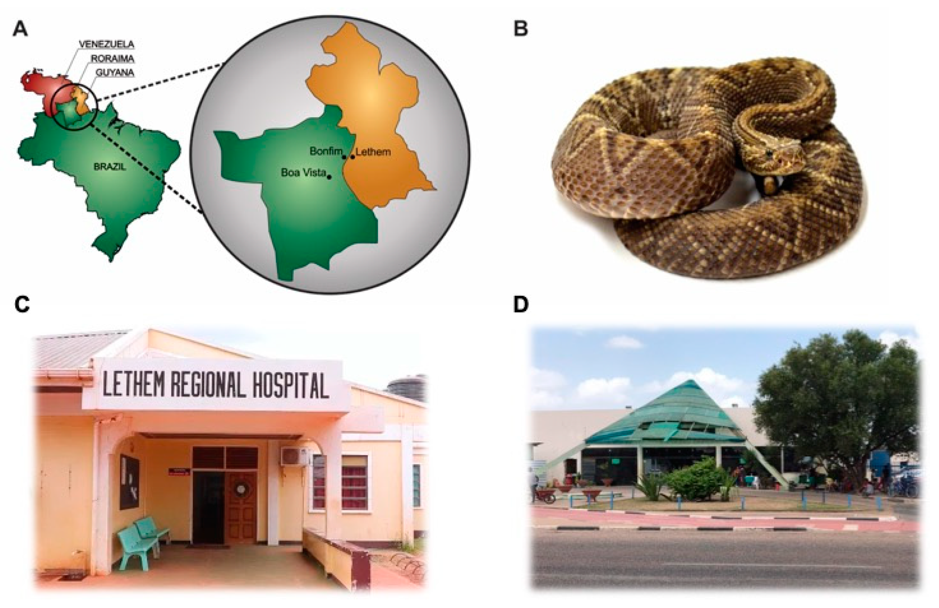
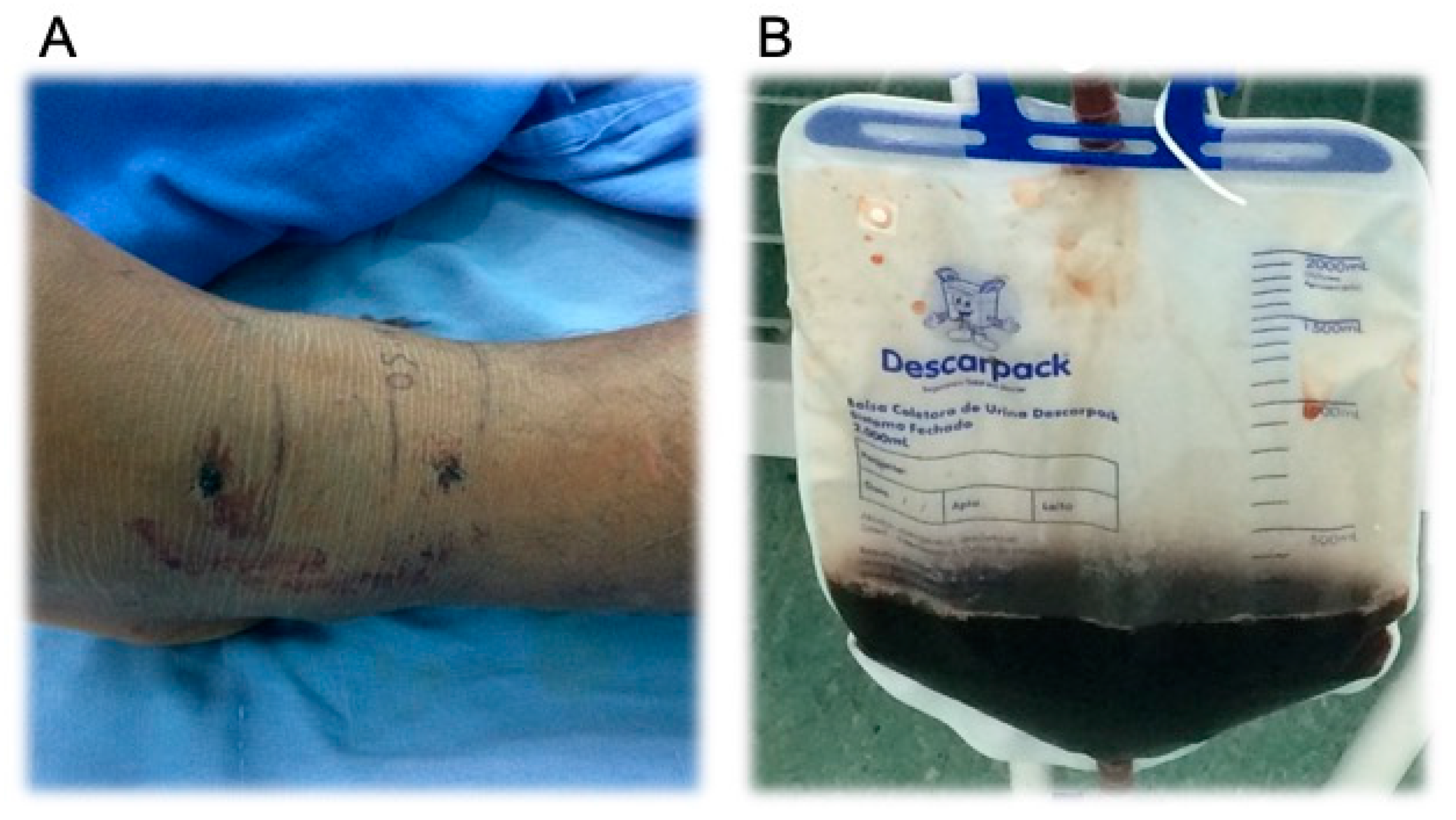
2. Case Presentation Section
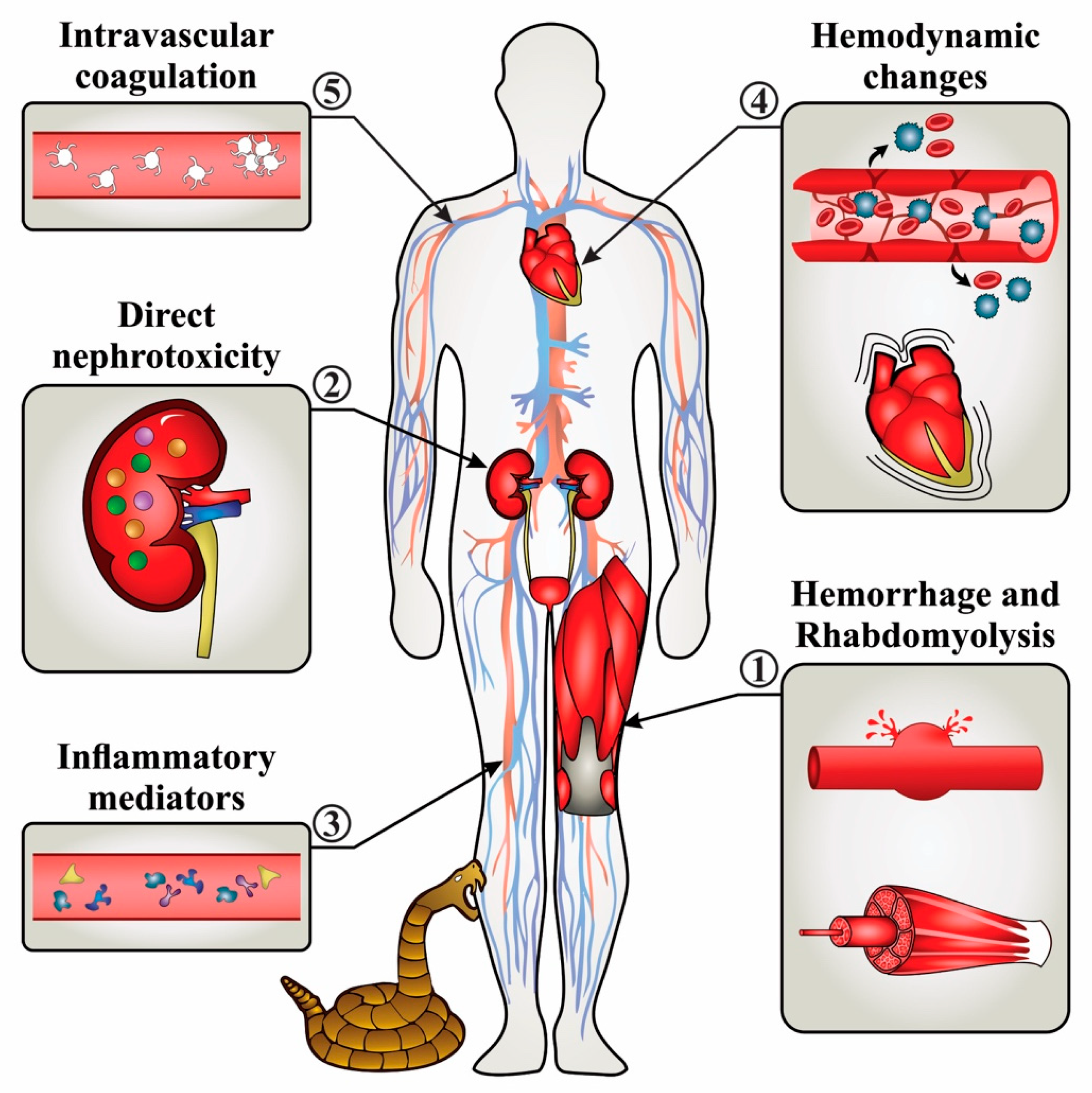
A 34-year-old male, single, from the city of Bonfim, a city located in the Mideast of the state of Roraima in Brazil (Figure 1A), was bitten by a snake on his left ankle during his work activities bordering Guyana at 10:20 am on 5 September 2018 (day 0). The patient identified the snake as a rattlesnake (Figure 1B).
Since the snakebite happened in a rural area near Guyana border, the patient first sought to medical assistance at Lethem Regional Hospital in Lethem, Guyana (Figure 1C). The patient arrived in the hospital 3 h after the snakebite. At hospital admission, the symptoms were moderate local pain, mouth dryness, and blurred vision. Moreover, the patient presented signs of neurotoxicity such as ptosis, drooling of saliva, and sluggishness. Local examination showed two deep bleeding fang marks, local redness, bruising, and swelling. On the other hand, the patient had a fair general condition (e.g., eupneic, hypoxic, anicteric, acyanotic, and hydrated), arterial blood pressure of 139/89 mmHg, heart rate of 108 beats/min, respiratory rate of 26 incursions/min, and temperature of 36.1 °C. The case was reported as a rattlesnake envenomation. The patient received intravenous (iv) injection of 200 mg of hydrocortisone and 10 mg of piriton intravenously (iv) to avoid allergic reactions, and 70 mg of diclofenac anti-inflammatory. Due to the lack of the specific serum in Lethem and laboratorial follow-up, the victim was referred to the Hospital Geral de Roraima (HGR) in Boa Vista city, Roraima, Brazil (Figure 1D).
Patient arrived in HGR at 8:04 pm, nearly 10 h upon the snakebite. After clinical examination, the physician verified early stages of a truly severe rattlesnake envenomation, as the patient presented neurotoxic facies, bilateral ptosis, ophthalmoplegia, edema at the biting site (Figure 2A), and macroscopic hematuria following oliguria (Figure 2B).
The patient was immediately transferred to an intensive care unit and received adequate heart rate and blood pressure monitoring, pulse oximetry, and oxygen, following iv hydration with 1.5 L of physiological solution (0.9%) supplemented with hydrocortisone (200 mg) and metamizole (1 g), to avoid further allergic reactions and pain, respectively. Finally, sixteen ampoules of crotalic antivenom were administered (iv) to the patient. Antibiotic therapy with cephalothin (1 g each 6 h) was prescribed, and a gamut of laboratorial tests was requested (Table 1).
On day 1 (6 September 2018) the patient developed hemodynamic instability with platelet counts less than 20,000/μL (i.e., indicating intravascular coagulation), demanding ventilatory support and vasoactive drugs in order to maintain blood pressure. Arterial blood gas evidenced severe metabolic acidosis, hyponatremia, hypocalcemia, and a bad gas exchange (ratio PaO2/FiO2 = 239). Moreover, the high serum levels of creatinine and urea, together with rhabdomyolysis evidenced by myoglobinuria, and oliguria, indicated the development of an acute kidney injury (AKI). In order to control such alterations, ventilatory parameters were adjusted and sodium bicarbonate and calcium gluconate were administered (iv) for controlling acidosis. However, the patient persisted with hemodynamic instability, culminating in 3 cardiac arrests. Unfortunately, the medical team did not obtain success in reverting the third consecutive arrest and the patient passed away at 11:30 pm of day 1.
The study was conducted in accordance with the Declaration of Helsinki, and the protocol was approved by the local Research Ethic Committee under protocol numbers CAAE 70659917.3.0000.5302 and CAAE70651217.7.0000.5302. Written informed consent was obtained from the patient.

3. Discussion

In Brazil, pit vipers and rattlesnakes are responsible for the highest number and the most severe cases of envenomations. In 2017, Brazilian epidemiological studies demonstrated 2484 snakebites caused by Crotalus spp. and 20,093 caused by Bothrops spp. On the other hand, crotalic envenomations presented the highest rates of lethality (0.7%) in comparison to bothropic envenomations (0.3%) [8].
Crotalic envenomations can cause local manifestations such as mild pain, edema, erythema, and paresthesia [24]. Systemic manifestations commonly appear in the first hours after the bite and are characterized by neurological effects including myasthenic facies, which are evidenced by palpebral ptosis (uni or bilateral), facial muscle flaccidity, pupillary diameter alteration, ophthalmoplegia, blurred vision, and diplopia [26]. Furthermore, venom-induced myotoxicity produces systemic skeletal muscle damage (rhabdomyolysis), leading to the release of enzymes and myoglobin, which are excreted in the urine. Thus, a classical sign of severe crotalic envenomation is the dark urine due to the elimination of myoglobin [27]. In this report, the patient presented most of literature classical symptoms of crotalic envenomation (Table 2), which confirmed the information provided by the victim.
One of the main complications and cause of death caused by rattlesnakes is the acute kidney failure (AKF), which can occur in the first 48 h after snakebite [35]. Indeed, the patient of the study presented myoglobinuria during the first 24 h. AKF is mainly diagnosed through high levels of serum creatinine and oliguria or anuria, albeit other parameters can also support kidney’s injury such as high levels of urea, hematuria, proteinuria, hypocalcemia, and hyponatremia [36]. Thus, the victim of the study developed an AKF. Although the pathogenesis of crotalic venom-induced AKF is not completely elucidated, researches correlate it to rhabdomyolysis, renal vasoconstriction, intravascular hemolysis, disseminated intravascular coagulation, and direct tubular cell toxicity caused by the venom [35,37,38]. Figure 3 presents a literature overview of AKF induced by rattlesnake venoms.
Moreover, our patient also presented other important alterations, such as the abnormally low levels of thrombocytes (i.e., thrombocytopenia). It is known that the treatment of rattlesnake envenomation with crotalic specific antivenom may not reverse the associated thrombocytopenia [39]. Interestingly, in a clinical study with 24 patients envenomed with rattlesnake venom, only the two fatal cases presented thrombocytopenia [40].
The increasing number of leukocytes, probably caused by neutrophilia, indicated that the patient was under an inflammatory response. Similarly, a study with 107 patients demonstrated that the neutrophil/leucocytes ratio was significantly increased in patients that developed complications and needed a longer hospitalization [41]. Although the glucose levels were higher than the reference range (133.26 mg/dL or ~7.3 mmol/L), the patient was not under fasting conditions and the levels known to indicate risk factor for high-grade envenomations following viper bites is 7.7 mmol/L [42].
Additional important biochemical parameters such as creatine kinase (CK) and creatine kinase MB isoenzyme (CK-MB) were not requested by the responsible physician from HGR, which could be very important to evaluate the cardiac status of the patient. For instance, during rattlesnake envenomation CK increase is early, with peak maximum elevation within the first 24 h after the accident. On the other hand, LDH level was examined. LDH increase is low and gradual, representing an important parameter for late diagnosis of severe crotalic envenomation [23].
Cardiac arrests are not prominent features of snakebite [43], whilst there are few clinical studies reporting that [44,45,46,47,48]. Although the venom mechanism of myocardial damage is not elucidated, it is known that increasing levels of blood potassium levels can be responsible for the cardiac arrest shortly after the bite [49]. In our case, patient potassium levels were inside the normal reference range. On the other hand, disseminated intravascular coagulation promotes spontaneous formation of clots large enough to block circulation, which may block arteries to supply vital organs, such as the heart, causing myocardial infarction [50]. In this case, arrest can be a delayed effect following snakebite envenomation, which has been reported even 10 days after the accident [40].
The Brazilian crotalic antivenom (Cav) is produced by horse immunization with C. d. terrificus and C. d. collilineatus venoms (1:1), being species not found in the North of Brazil [12,51]. Cav is manufactured with 10 mL solution containing specific and purified heterologous F(ab)2 antibodies [52]. Besides the high percentage (more than 70%) of non-neutralizing antibodies (for review see [53]), the remaining 30% of Cav antibodies may have several non-specific antibodies for the Roraima’s rattlesnake venom. Various omics studies have demonstrated the high venom variability between Crotalus durissus subspecies added to the intraspecies and geographic venom differences. In fact, the venom of Roraima’s rattlesnake subspecies C. d. ruruima presents low levels of metalloproteinase (2.9%), but higher levels of crotoxin (82.7%) in comparison to C. d. terrificus, which presents 4.8% and 59.5%, respectively [19]. On the basis of the above, the Brazilian Cav may not present high efficacy in neutralizing rattlesnake envenomation in Roraima state. Our case report supports this fact, since the victim died even after receiving 16 ampoules of Cav. In addition, other unpublished clinical data from our research group have already observed the low efficiency of Brazilian Cav antivenom in other rattlesnake bite patients in Roraima, where several victims developed long-term effects following envenomation, such as chronic kidney disease with indication to undergo dialysis.
Figure 3. Mechanisms of acute kidney failure (AKF) induced by crotalic venom. AKF occurs in consequence to the acute kidney injury (AKI), which is defined by a sudden decrease in kidney function. Snake toxins trigger several mechanisms to induce AKF. (1) Venom-induced hemorrhage and rhabdomyolysis, which results in hematuria and myoglobinuria, induce direct tubulotoxicity, renal tubular obstruction, and secondary decreased of glomerular filtration [54,55,56]. This kidney injury is also referred to as pigment-induced nephropathy [57]. (2) Direct effects of snake venom toxins on kidneys (i.e., direct nephrotoxicity) are reported as well [58]; however the precise mechanism of injury remain to be elucidated. (3) Inflammatory mediators (i.e., IL-6, TNF-α, and IL-1β cytokines, prostaglandins, leukotrienes, and proteins from complement system such as C3a, C4a, and C5a) are also involved in the pathogenesis of AKI [59,60]. (4) Hemodynamic changes, such as hypotension, and hypovolemia caused by venom compounds (e.g., phospholipases A2, natriuretic peptides, serine proteases, and vascular endothelial factor), decrease vascular resistance, increase cardiac output, and increase renal vascular resistance, with the latter causing hypovolemia [61]. (5) Coagulation activation, specifically disseminated intravascular coagulation, results in thrombotic microangiopathy, which decreases renal blood flow and contributes to AKI [62].
Reports 03 00009 g003
The Brazilian Ministry of Health recommends the administration of 5 ampoules of Cav for mild, 10 ampoules for moderate, and 20 ampoules for severe crotalic envenomations [23]. Our patient did not receive the recommended 20 vials for severe envenomations due to the limited antivenom available at that moment. Lack of sufficient antivenoms is a globally issue [63] and, in Brazil, the growing financial limitations of public institutions and governments had an extremely negative impact on antivenom production. Nevertheless, other clinical studies with severe crotalic envenomation showed success even with 12 vials of crotalic antivenom therapy, which was administered 7 h after the snakebite [64].
The timeframe of the snakebite episode and the antivenom therapy is also an important issue. Clinical studies have demonstrated that victims that received antivenoms in the first hours after the bite have a far better prognosis. A case report regarding C. d. terrificus severe envenomation demonstrated that the efficacy of an antivenom was better and faster when the patient received it 25 min after the snakebite (17 ampoules of Antivipmyn TRI® antivenom still in the ambulance on the way to the hospital), since the victim showed significant improvement with normal vital signs immediately after finishing antivenom administration (after 5–6 h of admission time) [65]. On the other hand, another study performed with Australian snakebite victims demonstrated that independent of the set time of the initiation of the antivenom therapy (before or after 6 h of snakebite), the serum was unable to inhibit venom-induced coagulopathy [66]. In addition, an interesting study demonstrated that levels of serum creatinine concentration on admission and neurotoxicity following snakebite were strong predictors of mortality among in-hospital envenomation patients [67]. Indeed, our patient had neurotoxic alterations and high creatinine levels (1.65 mg/dL) on day 0.
Whilst our patient received the specific therapy, i.e., antivenom, there are other medical approaches that may also be critical during severe snakebite cases. Since myoglobin precipitates at pH 6.5, to avoid kidney damage by myoglobinuria, it is recommended to perform osmotic diuresis and urine alkalization in the patient [29], but no diuretic drug was prescribed to the patient (e.g., mannitol or furosemide). On the other hand, the patient received infusion of sodium bicarbonate aiming to adjust his blood acidosis. Unfortunately, the patient died before a new round of laboratory tests, and we could not analyze if the acidosis was controlled. However, due to the well-established AKF, a recommended treatment for the patient should have been dialysis (hemodialysis or peritoneal dialysis), which should be continued until irreversibility of the underlying lesion has been proved beyond doubt [68,69], although it was not performed.
While the relationship between acidosis and cardiac arrest needs further investigation, it is reported that lactate accounts for 50% of the metabolic acidosis and consequent acidemia seen in arrested patients [70], and our patient presents a pH 6.7 and high levels of lactate. Despite this, it is hard to predict if a different medical management protocol could have changed the victims’ negative outcome. Actually, we believe that the physician in charge of the patient did not have enough time to make different approaches, since the envenomation quickly resulted in death in less than 16 h after hospitalization in HGR.
Rattlesnake case reports, as well as current reviews [23,35], have been alerting the medical community about the great diversity during case evolution. Pardal et al. (2007) reported a moderate case of a 19-year-old boy who was bitten by a rattlesnake and sought medical attention 12 h after the accident, due to the appearance of classic symptoms. The patient received a total of 14 ampoules of antivenom, evolved well, and was discharged 5 days after the accident [24]. Nishioka and co-authors (2000) also showed a case of a 38-year-old patient bitten by a rattlesnake who presented classic symptoms received 12 ampoules of antivenom and was discharged; however, the patient returned to the hospital a week later due to a bacterial infection [64]. On the other hand, Baum and colleagues (2019) described a severe case of a herpetologist (56-year-old) who was bitten by a rattlesnake. Although the patient received administration of antivenom (Antivipmyn-TRI®), he presented respiratory failure and required mechanical ventilation. Fortunately, the patient was discharged 55 h after the accident [65]. Azevedo-Marques and colleagues (1987) reported three cases of patients of different ages (27, 24, and 9 years old) bitten by rattlesnakes. All of them showed the classic signs of crotalic envenomation, including dark urine, requiring the administration of antivenom. Only one of them developed acute renal failure [27]. Unfortunately, the envenomation evolution of our patient was different from the other presented cases, whilst he received the antivenom therapy.
Based on the above, it is extremely difficult to determine the physio-pathological aspects that culminated in our patient death. Actually, we assume that multiple factors were responsible for causing the victim’s death: (1) the time frame between the snakebite and the antivenom administration; (2) the use of an unspecific antivenom, due to the lack of C. d. ruruima venom for horse immunization during antivenom manufacture; (3) the low dose of antivenom (16 rather than 20 vials); (4) the venom-induced AKT; (5) and the cardiac arrests.
Our clinical case supports the idea that new protocols to manufacture the Brazilian crotalic antivenom are required in aspects to produce a heterologous and polyclonal antivenom effective to neutralize all Crotalus durissus subspecies venoms from the country. Therefore, this article strengthens the necessity of the production of an effective antivenom able to neutralize the C. d. ruruima venom, a neglected subspecies found in Roraima: the northernmost, the poorest (with the lowest GDP—Gross Domestic Product), and with the highest snakebite incidence of Brazil [7]. In addition, our case report can also assist the local medical community with rattlesnake envenomations, showing how a case developing irreversible envenomation effects (e.g., AKF and arrest) can be treated since antivenom may not be effective and first aid measures need to be the focus.

Author Contributions

Investigation, J.M.M., U.Z., and M.B.P.; writing—original draft preparation, M.B.P., J.M.M., I.S.O., G.M.A.-S., and I.G.F.; figures and review, F.A.C.; supervision and project administration, M.B.P. All authors have read and agreed to the published version of the manuscript.

Funding

We thank Conselho Nacional de Desenvolvimento Científico e Tecnológico (CNPq, The National Council for Scientific and Technological Development, scholarship to MBP no. 307155/2017-0, and scholarship to J.M.M no. 144195/2017-8); Fundação de Amparo à Pesquisa do Estado de São Paulo (FAPESP, São Paulo Research Foundation; scholarship to FAC no. 2017/14035-1, and scholarship to ISO no. 2017/03580-9), and the Coordenação de Aperfeiçoamento de Pessoal de Nível Superior - Brasil (CAPES, Finance Code 001, scholarship to IGF).

Acknowledgments

We thank the physicians and nurses of Hospital Geral de Roraima (HGR), who have made the commitment to better quality care for the patient. We also thanks Alessandra Galvão Martins to be very helpful and professional in assisting us with patient data.

Conflicts of Interest

The authors declare no conflict of interest.

References

  1. Williams, D.J.; Faiz, M.A.; Abela-Ridder, B.; Ainsworth, S.; Bulfone, T.C.; Nickerson, A.D.; Habib, A.G.; Junghanss, T.; Fan, H.W.; Turner, M.; et al. Strategy for a globally coordinated response to a priority neglected tropical disease: Snakebite envenoming. PLoS Negl. Trop. Dis. 2019, 13, e0007059. [Google Scholar] [CrossRef] [PubMed] [Green Version]
  2. Gutiérrez, J.M.; Williams, D.; Fan, H.W.; Warrell, D.A. Snakebite envenoming from a global perspective: Towards an integrated approach. Toxicon 2010, 56, 1223–1235. [Google Scholar] [CrossRef] [PubMed]
  3. Chippaux, J.-P. Snakebite envenomation turns again into a neglected tropical disease! J. Venom. Anim. Toxins Incl. Trop. Dis. 2017, 23, 38. [Google Scholar] [CrossRef] [PubMed]
  4. Costa, H.C.; Bérnils, R.S. Répteis do Brasil e suas Unidades Federativas: Lista de espécies. Herpetol. Bras. 2018, 7, 11–57. [Google Scholar]
  5. Cardoso, J.L.C.; França, F.O.S.; Wen, F.H.; Málaque, C.M.S.; Haddad, V., Jr. Animais peçonhentos no Brasil: Biologia, clínica e terapêutica dos acidentes. Rev. Inst. Med. Trop. São Paulo 2003, 45, 338. [Google Scholar] [CrossRef]
  6. Harris, H.S.; Simmons, R.S. A new subspecies of Crotalus durissus (serpentes: Crolalidae) from the Rupununisavanna of Southewestern Guyana. Memórias Inst. Butantan 1976, 40, 305–311. [Google Scholar]
  7. Souza, W.M.P.; Alexandre-Silva, G.; Cerni, F.A.; Oliveira, I.S.; Zottich, U.; Bassoli, B.K.; Pucca, M.B. Envenomings caused by venomous animals in Roraima: A neglected health problem in the Brazil’s Northernmost state. TCR 2019, 3, 1–8. [Google Scholar] [CrossRef]
  8. DATASUS TabNet Win32 3.0: Acidentes Por Animais Peçomentos. Notificações Registradas no Sistema de Informação de Agravos de Notificação, Brasil. Available online: http://tabnet.datasus.gov.br/cgi/tabcgi.exe?sinannet/cnv/animaisbr.def (accessed on 16 January 2020).
  9. Nascimento, S.P. do Aspectos epidemiológicos dos acidentes ofídicos ocorridos no Estado de Roraima, Brasil, entre 1992 e 1998. Cad. Saúde Pública 2000, 16, 271–276. [Google Scholar] [CrossRef] [Green Version]
  10. Alirol, E.; Lechevalier, P.; Zamatto, F.; Chappuis, F.; Alcoba, G.; Potet, J. Antivenoms for Snakebite Envenoming: What Is in the Research Pipeline? PLoS Negl. Trop. Dis. 2015, 9, e0003896. [Google Scholar] [CrossRef] [Green Version]
  11. Luna, W.F.; Ávila, B.T.; Brazão, C.F.F.; Freitas, F.P.D.P.; Cajado, L.C.D.S.; Bastos, L.O.D.A.; Luna, W.F. Project More Doctors for Brazil in remote areas of the state of Roraima: Relationship between doctors and the Special Supervision Group. Interface Comun. Saúde Educ. 2019, 23, e180029. [Google Scholar] [CrossRef]
  12. Wen, F.H.; Monteiro, W.M.; da Silva, A.M.M.; Tambourgi, D.V.; da Silva, I.M.; Sampaio, V.S.; dos Santos, M.C.; Sachett, J.; Ferreira, L.C.L.; Kalil, J.; et al. Snakebites and scorpion stings in the Brazilian Amazon: Identifying research priorities for a largely neglected problem. PLoS Negl. Trop. Dis. 2015, 9, e0003701. [Google Scholar]
  13. The Struggle of Venezuelan Migrants and Asylum Seekers in Northern Brazil|MSF. Available online: https://www.msf.org/struggle-venezuelan-migrants-and-asylum-seekers-northern-brazil (accessed on 30 March 2020).
  14. Magalhães, S.F.V.; Peixoto, H.M.; Moura, N.; Monteiro, W.M.; de Oliveira, M.R.F. Snakebite envenomation in the Brazilian Amazon: A descriptive study. Trans. R. Soc. Trop. Med. Hyg. 2019, 113, 143–151. [Google Scholar] [CrossRef] [PubMed]
  15. Rübsamen, K.; Breithaupt, H.; Habermann, B. Biochemistry and pharmacology of the crotoxin complex. Naunyn Schmiedebergs Arch. Pharmak. 1971, 270, 274–288. [Google Scholar] [CrossRef]
  16. Hendon, R.A.; Fraenkel-Conrat, H. Biological roles of the two components of crotoxin. Proc. Natl. Acad. Sci. (USA) 1971, 68, 1560–1563. [Google Scholar] [CrossRef] [PubMed] [Green Version]
  17. Faure, G.; Bon, C. Several isoforms of crotoxin are present in individual venoms from the South American rattlesnake Crotalus durissus terrificus. Toxicon 1987, 25, 229–234. [Google Scholar] [CrossRef]
  18. Breithaupt, H. Enzymatic characteristics of Crotalus phospholipase A2 and the crotoxin complex. Toxicon 1976, 14, 221–233. [Google Scholar] [CrossRef]
  19. Calvete, J.J.; Sanz, L.; Cid, P.; de la Torre, P.; Flores-Díaz, M.; Dos Santos, M.C.; Borges, A.; Bremo, A.; Angulo, Y.; Lomonte, B.; et al. Snake venomics of the Central American rattlesnake Crotalus simus and the South American Crotalus durissus complex points to neurotoxicity as an adaptive paedomorphic trend along Crotalus dispersal in South America. J. Proteome Res. 2010, 9, 528–544. [Google Scholar] [CrossRef]
  20. de Carvalho, A.E.Z.; Giannotti, K.; Junior, E.L.; Matsubara, M.; Santos, M.C.D.; Fortes-Dias, C.L.; Teixeira, C. Crotalus durissus ruruima snake venom and a phospholipase A2 isolated from this venom elicit macrophages to form lipid droplets and synthesize inflammatory lipid mediators. J. Immunol. Res. 2019, 2019, 2745286. [Google Scholar] [CrossRef] [Green Version]
  21. Santos, M.C. dos Characterization of the biological activities of the Crotalus durissus ruruima yellow and white venoms compared with Crotalus durissus terrificus venom: Neutralizing effect of the antivenoms against Crotalus durissus terrificus venom. J. Venom. Anim. Toxins 1996, 2, 163. [Google Scholar] [CrossRef]
  22. Nascimento, S.P. Epidemiological characteristics of snake bites in the state of Roraima, Brazil, 1992–1998. Cad. Saude Publica 2000, 16, 271–276. [Google Scholar] [CrossRef] [Green Version]
  23. Frare, B.T.; Silva Resende, Y.K.; Dornelas, B.D.C.; Jorge, M.T.; Souza Ricarte, V.A.; Alves, L.M.; Izidoro, L.F.M. Clinical, laboratory, and therapeutic aspects of Crotalus durissus (South American rattlesnake) victims: A literature review. Biomed Res. Int. 2019, 2019, 1345923. [Google Scholar] [CrossRef] [PubMed] [Green Version]
  24. Pardal, P.P.D.O.; Silva, C.L.Q.D.; Hoshino, S.D.S.N.; Pinheiro, M.D.F.R. Acidente por cascavel (Crotalus sp) em Ponta de Pedras, Ilha do Marajó, Pará-Relato de caso. Rev. Para. Med. 2007, 21, 69–73. [Google Scholar] [CrossRef]
  25. WHO. Guidelines for the Production, Control and Regulation of Snake Antivenom Immunoglobulins. Available online: https://www.who.int/bloodproducts/snake_antivenoms/snakeantivenomguide/en/ (accessed on 22 January 2020).
  26. Azevedo-Marques, M.M.; Cupo, P.; Hering, S.E. Acidentes por animais peçonhentos: Serpentes peçonhentas. Medicina 2003, 36, 480–489. [Google Scholar] [CrossRef] [Green Version]
  27. Azevedo-Marques, M.M.; Hering, S.E.; Cupo, P. Evidence that Crotalus durissus terrificus (South American rattlesnake) envenomation in humans causes myolysis rather than hemolysis. Toxicon 1987, 25, 1163–1168. [Google Scholar] [CrossRef]
  28. Bucaretchi, F.; Capitani, E.M.D.; Hyslop, S.; Mello, S.M.; Fernandes, C.B.; Bergo, F.; Nascimento, F.B.P. Compartment syndrome after South American rattlesnake (Crotalus durissus terrificus) envenomation. Clin. Toxicol. 2014, 2, 639–641. [Google Scholar] [CrossRef]
  29. Pinho, F.M.O.; Pereira, I.D. Ofidismo. Rev. Assoc. Médica Bras. 2001, 47, 24–29. [Google Scholar] [CrossRef]
  30. Bucaretchi, F.; Herrera, S.R.F.; Hyslop, S.; Baracat, E.C.E.; Vieira, R.J. Snakebites by Crotalus durissus ssp in children in Campinas, São Paulo, Brazil. Rev. Inst. Med. Trop. Sao Paulo 2002, 44, 133–138. [Google Scholar] [CrossRef]
  31. Madey, J.J.; Price, A.B.; Dobson, J.V.; Stickler, D.E.; McSwain, S.D. Facial diplegia, pharyngeal paralysis, and ophthalmoplegia after a timber rattlesnake envenomation. Pediatr. Emerg. Care 2013, 29, 1213–1216. [Google Scholar] [CrossRef]
  32. Bush, S.P.; Jansen, P.W. Severe rattlesnake envenomation with anaphylaxis and rhabdomyolysis. Ann. Emerg. Med. 1995, 25, 845–848. [Google Scholar] [CrossRef]
  33. Jorge, M.T.; Ribeiro, L.A. Epidemiologia e quadro clínico do acidente por cascavel sul-americana (Crotalus durissus). Rev. Inst. Med. Trop. São Paulo 1992, 34, 347–354. [Google Scholar] [CrossRef]
  34. Pinho, F.M.O.; Zanetta, D.M.T.; Burdmann, E.A. Acute renal failure after Crotalus durissus snakebite: A prospective survey on 100 patients. Kidney Int. 2005, 67, 659–667. [Google Scholar] [CrossRef] [PubMed] [Green Version]
  35. Albuquerque, P.L.M.M.; Jacinto, C.N.; Silva, G.B.; Lima, J.B.; Veras, M.D.S.B.; Daher, E.F. Acute kidney injury caused by Crotalus and Bothrops snake venom: A review of epidemiology, clinical manifestations and treatment. Rev. Inst. Med. Trop. Sao Paulo 2013, 55, 295–301. [Google Scholar] [CrossRef] [PubMed] [Green Version]
  36. Moysés-Neto, M.; Guimarães, F.M.; Ayoub, F.H.; Vieira-Neto, O.M.; Costa, J.A.C.; Dantas, M. Acute renal failure and hypercalcemia. Ren. Fail. 2006, 28, 153–159. [Google Scholar] [CrossRef] [PubMed]
  37. Drews, R.E.; Weinberger, S.E. Thrombocytopenic disorders in critically Ill patients. Am. J. Respir. Crit. Care Med. 2000, 162, 347–351. [Google Scholar] [CrossRef] [Green Version]
  38. Kohli, H.S.; Sakhuja, V. Snake bites and acute renal failure. Saudi J. Kidney Dis. Transpl. 2003, 14, 165–176. [Google Scholar]
  39. Odeleye, A.A.; Presley, A.E.; Passwater, M.E.; Mintz, P.D. Rattlesnake venom-induced thrombocytopenia. Ann. Clin. Lab. Sci. 2004, 34, 467–470. [Google Scholar]
  40. Sano-Martins, I.S.; Tomy, S.C.; Campolina, D.; Dias, M.B.; de Castro, S.C.B.; de Sousa-e-Silva, M.C.C.; Amaral, C.F.S.; Rezende, N.A.; Kamiguti, A.S.; Warrell, D.A.; et al. Coagulopathy following lethal and non-lethal envenoming of humans by the South American rattlesnake (Crotalus durissus) in Brazil. QJM 2001, 94, 551–559. [Google Scholar] [CrossRef] [Green Version]
  41. Elbey, B.; Baykal, B.; Yazgan, Ü.C.; Zengin, Y. The prognostic value of the neutrophil/lymphocyte ratio in patients with snake bites for clinical outcomes and complications. Saudi J. Biol. Sci. 2017, 24, 362–366. [Google Scholar] [CrossRef] [Green Version]
  42. Claudet, I.; Grouteau, E.; Cordier, L.; Franchitto, N.; Bréhin, C. Hyperglycemia is a risk factor for high-grade envenomations after European viper bites (Vipera spp.) in children. Clin. Toxicol. 2016, 54, 34–39. [Google Scholar] [CrossRef]
  43. Gaballa, M.; Taher, T.; Brodin, L.A.; van der Linden, J.; O’Reilly, K.; Hui, W.; Brass, N.; Cheung, P.K.; Grip, L. Images in cardiovascular medicine. Myocardial infarction as a rare consequence of a snakebite: Diagnosis with novel echocardiographic tissue Doppler techniques. Circulation 2005, 112, e140–e142. [Google Scholar] [CrossRef]
  44. Johnston, M.A.; Fatovich, D.M.; Haig, A.D.; Daly, F.F.S. Successful resuscitation after cardiac arrest following massive brown snake envenomation. Med. J. Aust. 2002, 177, 646–649. [Google Scholar] [CrossRef]
  45. Dhaliwal, U. Cortical blindness: An unusual sequela of snake bite. Indian J. Ophthalmol. 1999, 47, 191. [Google Scholar] [PubMed]
  46. Gomes, R.A.F.; Cantarelli, F.L.; Vieira, F.A.; Macedo, A.R.A., Jr.; Gouveia, M.M.D.A.; Feitosa, A.D.D.M. Myocardial infarction after snake bite. Int. J. Cardiovasc. Sci. 2018, 31, 79–81. [Google Scholar] [CrossRef]
  47. Brown, R.; Dewar, H.A. Heart damage following Adder bite in England. Br. Heart J. 1965, 27, 144–147. [Google Scholar] [CrossRef] [Green Version]
  48. Upadhyaya, A.C.; Murthy, G.L.; Sahay, R.K.; Srinivasan, V.R.; Shantaram, V. Snake bite presenting as acute myocardial infarction, ischaemic cerebrovascular accident, acute renal failure and disseminated intravascular coagulopathy. J. Assoc. Physicians India 2000, 48, 1109–1110. [Google Scholar] [PubMed]
  49. Hifumi, T.; Sakai, A.; Kondo, Y.; Yamamoto, A.; Morine, N.; Ato, M.; Shibayama, K.; Umezawa, K.; Kiriu, N.; Kato, H.; et al. Venomous snake bites: Clinical diagnosis and treatment. J. Intensiv. Care 2015, 3, 16. [Google Scholar] [CrossRef] [PubMed] [Green Version]
  50. Kini, R.M. Anticoagulant proteins from snake venoms: Structure, function and mechanism. Biochem. J. 2006, 397, 377–387. [Google Scholar] [CrossRef] [PubMed] [Green Version]
  51. de Oliveira, I.S.; Pucca, M.B.; Sampaio, S.V.; Arantes, E.C. Antivenomic approach of different Crotalus durissus collilineatus venoms. J. Venom. Anim. Toxins Incl. Trop. Dis. 2018, 24, 34. [Google Scholar] [CrossRef] [PubMed]
  52. Instituto Butantan Soro Anticrotalico Instituto Butantan. Available online: https://consultaremedios.com.br/ (accessed on 22 January 2020).
  53. Laustsen, A.H.; Gutiérrez, J.M.; Knudsen, C.; Johansen, K.H.; Bermúdez-Méndez, E.; Cerni, F.A.; Jürgensen, J.A.; Ledsgaard, L.; Martos-Esteban, A.; Øhlenschlæger, M.; et al. Pros and cons of different therapeutic antibody formats for recombinant antivenom development. Toxicon 2018, 146, 151–175. [Google Scholar] [CrossRef]
  54. Ponraj, D.; Gopalakrishnakone, P. Renal lesions in rhabdomyolysis caused by Pseudechis australis snake myotoxin. Kidney Int. 1997, 51, 1956–1969. [Google Scholar] [CrossRef] [Green Version]
  55. Tracz, M.J.; Alam, J.; Nath, K.A. Physiology and pathophysiology of heme: Implications for kidney disease. J. Am. Soc. Nephrol. 2007, 18, 414–420. [Google Scholar] [CrossRef] [PubMed]
  56. Sitprija, V.; Sitprija, S. Renal effects and injury induced by animal toxins. Toxicon 2012, 60, 943–953. [Google Scholar] [CrossRef] [PubMed]
  57. Dineshkumar, T.; Dhanapriya, J.; Murugananth, S.; Surendar, D.; Sakthirajan, R.; Rajasekar, D.; Balasubramaniyan, T.; Gopalakrishnan, N. Snake envenomation-induced acute interstitial nephritis. J. Integr. Nephrol. 2018, 5, 14. [Google Scholar]
  58. Martins, A.M.C.; Monteiro, H.S.A.; Júnior, E.O.G.; Menezes, D.B.; Fonteles, M.C. Effects of Crotalus durissus cascavella venom in the isolated rat kidney. Toxicon 1998, 36, 1441–1450. [Google Scholar] [CrossRef]
  59. Voronov, E.; Apte, R.N.; Sofer, S. The systemic inflammatory response syndrome related to the release of cytokines following severe envenomation. J. Venom. Anim. Toxins 1999, 5, 5–33. [Google Scholar] [CrossRef]
  60. Kinsey, G.R.; Li, L.; Okusa, M.D. Inflammation in acute kidney injury. NEE 2008, 109, e102–e107. [Google Scholar] [CrossRef]
  61. Sakwiwatkul, K.; Chaiyabutr, N.; Sitprija, V. Renal function following sea snake venom (Lapemis hardwicki) administration in dogs treated with sodium bicarbonate solution. J. Nat. Toxins 2002, 11, 111–121. [Google Scholar]
  62. Isbister, G.K.; Little, M.; Cull, G.; McCoubrie, D.; Lawton, P.; Szabo, F.; Kennedy, J.; Trethewy, C.; Luxton, G.; Brown, S.G.A.; et al. Thrombotic microangiopathy from Australian brown snake (Pseudonaja) envenoming. Intern. Med. J. 2007, 37, 523–528. [Google Scholar] [CrossRef]
  63. Gutiérrez, J.M. Global availability of antivenoms: The relevance of public manufacturing laboratories. Toxins 2019, 11, 5. [Google Scholar] [CrossRef] [Green Version]
  64. Nishioka, S.D.A.; orge, M.T.; Silveira, P.V.; Ribeiro, L.A. South American rattlesnake bite and soft-tissue infection: Report of a case. Rev. Soc. Bras. Med. Trop. 2000, 33, 401–402. [Google Scholar] [CrossRef] [Green Version]
  65. Baum, R.A.; Bronner, J.; Akpunonu, P.D.S.; Plott, J.; Bailey, A.M.; Keyler, D.E. Crotalus durissus terrificus (viperidae; crotalinae) envenomation: Respiratory failure and treatment with antivipmyn TRI® antivenom. Toxicon 2019, 163, 32–35. [Google Scholar] [CrossRef]
  66. Isbister, G.K.; Duffull, S.B.; Brown, S.G.A. Failure of antivenom to improve recovery in Australian snakebite coagulopathy. QJM 2009, 102, 563–568. [Google Scholar] [CrossRef] [PubMed]
  67. Kalantri, S.; Singh, A.; Joshi, R.; Malamba, S.; Ho, C.; Ezoua, J.; Morgan, M. Clinical predictors of in-hospital mortality in patients with snake bite: A retrospective study from a rural hospital in central India. Trop. Med. Int. Health 2006, 11, 22–30. [Google Scholar] [CrossRef] [PubMed]
  68. Chugh, K.S.; Pal, Y.; Chakravarty, R.N.; Datta, B.N.; Mehta, R.; Sakhuja, V.; Mandal, A.K.; Sommers, S.C. Acute Renal Failure Following Poisonous Snakebite. Am. J. Kidney Dis. 1984, 4, 30–38. [Google Scholar] [CrossRef]
  69. Danzig, L.E.; Abels, G.H. Hemodialysis of Acute Renal Failure Following Rattlesnake Bite, with Recovery. JAMA 1961, 175, 136–137. [Google Scholar] [CrossRef]
  70. Makino, J.; Uchino, S.; Morimatsu, H.; Bellomo, R. A quantitative analysis of the acidosis of cardiac arrest: A prospective observational study. Crit. Care 2005, 9, R357–R362. [Google Scholar] [CrossRef] [Green Version]
Figure 1. (A) Snakebite and therapy locations. Left panel shows Roraima, the northernmost state of Brazil, bordering Guyana and Venezuela. The map also shows the cities of Bonfim (Brazilian city and the region of the snakebite), Lethem (Guyana city and local of the first medical care), and Boa Vista (capital of Roraima state and the local of the Hospital that patient received the specific therapy). (B) Crotalus durissus ruruima. The rattlesnake species found in Roraima state and bordering areas with Guyana and Venezuela. Photo kindly provided by Anderson Maciel Rocha. (C) Lethem Regional Hospital. Photo from the Department of Public Information website, Lethem, Guyana, 2019. (D) Hospital Geral de Roraima, Boa Vista, RR, Brazil. Photo from Jilvando M. Medeiros, 2020.
Reports 03 00009 g001
Figure 2. Patient signs following 10 h of snakebite. (A) Local examination showed two deep fang marks, swelling, and bleeding. (B) Urinary drainage bag showing macroscopic myoglobinuria, a black-colored urine.
Reports 03 00009 g002
Table 1. Patient laboratory tests following rattlesnake bite.
AnalytesDaysReference Range &
01
Hemoglobin16.3 *15.213.5–18.0 g/dL
Hematocrit46.745.740.0%–50.0%
Leucocytes16,810 *15,920 *4000–10,000 cells/µL
Neutrophils-94.00 *50.0%–70.0%
Platelets158,00016,000 *150,000–400,000/µL
PT14.2 *13.510.0–14.0 s
PTT32.236.125.0–39.0 s
Na+139.0133.0 *135.0–145.0 mmol/L
K+4.03.83.5–5.1 mmol/L
Ca2+0.68 *0.72 *1.17–1.32 mmol/L
Cl100.0107.098.0–107.0 mmol/L
Glucose133.26 *-60.0–99.0 mg/dL
Urea58.71 *-15–40 mg/dL
Creatinine1.65 *-0.7–1.4 mg/dL
ALT466.6 *-5.0–48.8 U/mL
AKP56.21-27.0–100.0 mg/dL
γGT20.5-12.0–45.0 U/L
LDH151.52 *-200.0–480.0 U/L
CRP4.56-0.0–8.0 mg/L
Lactate35.53 *-4.5–19.8 mg/dL
* Means that results were different from the reference range. & Reference range from the Laboratório Central de Roraima (LACEM-HGR), Boa Vista, Roraima, Brazil. AKP: alkaline phosphatase; ALT: alanine aminotransferase; AST: aspartate aminotransferase; Ca2+: calcium; CK-MB: MB fraction of creatine kinase; Cl: chloride; CPK: Creatinophosphokinase; CRP: C-reactive protein; γGT: gammaglutamyltranspeptidase; K+: potassium; LDH: lactate dehydrogenase; Na+: sodium; PT: prothrombin time; PTT: partial thromboplastin time.
Table 2. Patient clinical presentation.
Patient Signs & SymptomsReference
Local pain[28]
Local redness, bruising, and edema[28]
Mouth dryness[29]
Bilateral ptosis[30]
Ophthalmoplegia[31]
Salivation[30]
Sluggishness[32]
Myoglobinuria[33]
Oliguria/Anuria[34]

