


Characterization of the Plastic Scintillator Detector System Exradin W2 in a High Dose Rate Flattening-Filter-Free Photon Beam
 , ,
, ,
Abstract
:1. Introduction
2. Materials and Methods
3. Results
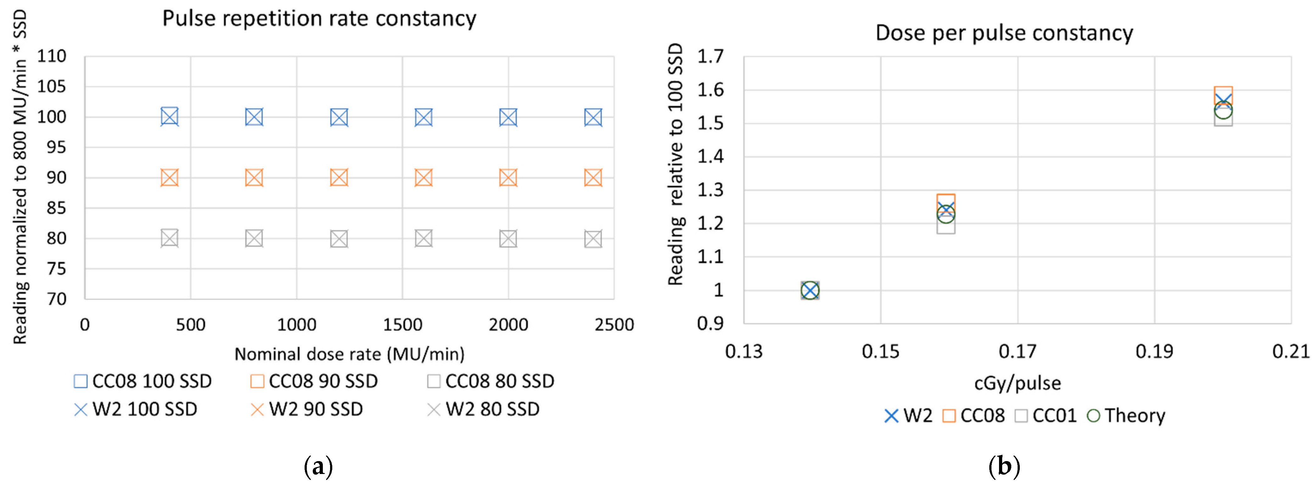
3.1. Pion and Ppol
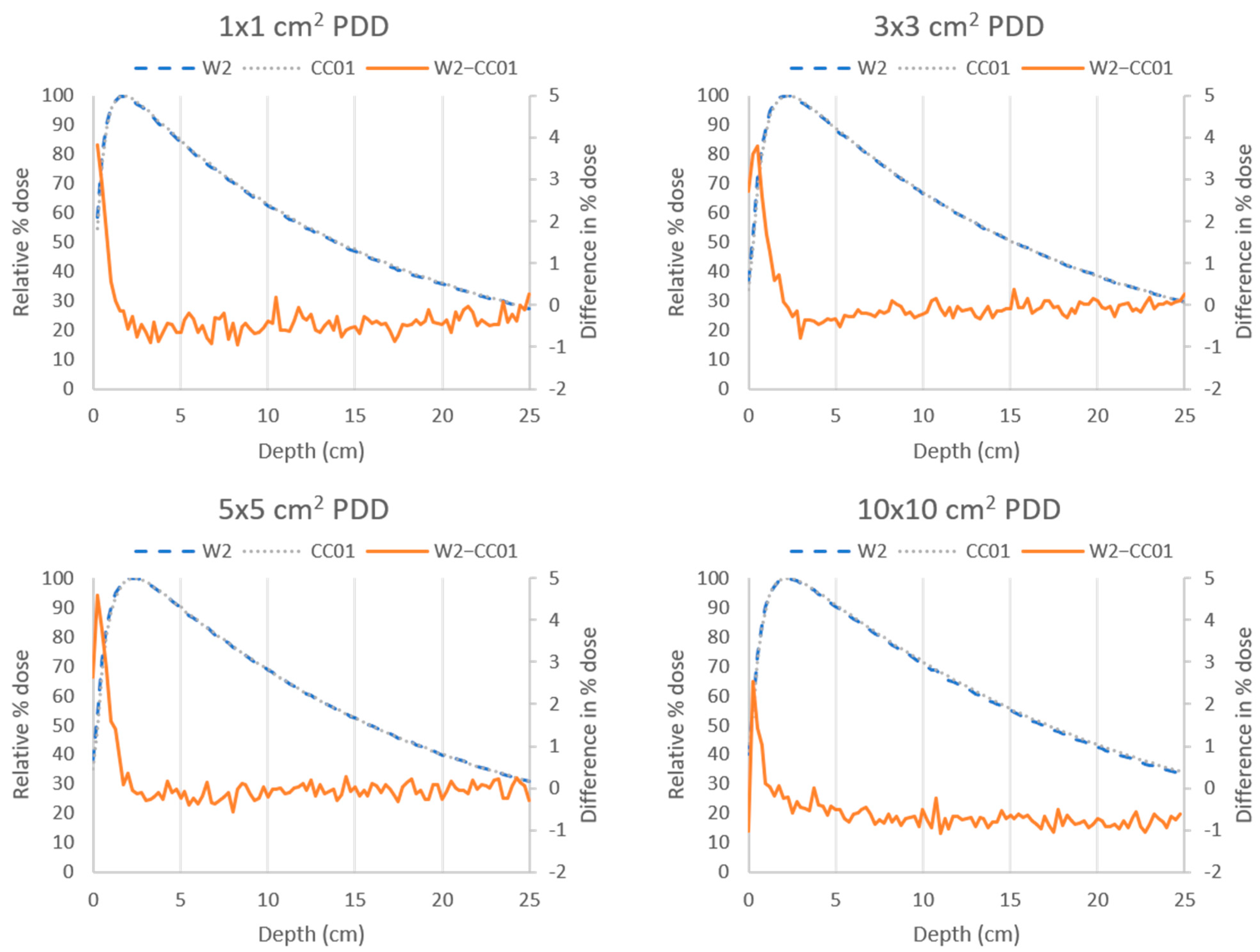
3.2. PDD Measurements
4. Discussion
5. Conclusions
Author Contributions
Funding
Institutional Review Board Statement
Informed Consent Statement
Data Availability Statement
Acknowledgments
Conflicts of Interest
References
- Xiao, Y.; Kry, S.F.; Popple, R.; Yorke, E.; Papanikolaou, N.; Stathakis, S.; Xia, P.; Huq, S.; Bayouth, J.; Galvin, J.; et al. Flattening filter-free accelerators: A report from the AAPM Therapy Emerging Technology Assessment Work Group. J. Appl. Clin. Med. Phys. 2015, 16, 12. [Google Scholar] [CrossRef] [PubMed]
- Kry, S.F.; Popple, R.; Molineu, A.; Followill, D.S. Ion recombination correction factors (Pion) for Varian TrueBeam high-dose-rate therapy beams. J. Appl. Clin. Med. Phys. 2012, 13, 318–325. [Google Scholar] [CrossRef] [PubMed]
- Lang, S.; Hrbacek, J.; Leong, A.; Klöck, S. Ion-recombination correction for different ionization chambers in high dose rate flattening-filter-free photon beams. Phys. Med. Biol. 2012, 57, 2819. [Google Scholar] [CrossRef] [PubMed]
- Jacqmin, D.J.; Miller, J.R.; Barraclough, B.A.; Labby, Z.E. Commissioning an Exradin W2 plastic scintillation detector for clinical use in small radiation fields. J. Appl. Clin. Med. Phys. 2022, 23, 23. [Google Scholar] [CrossRef]
- Brown, T.A.D.; Ayers, R.G.; Popple, R.A. Commissioning a multileaf collimator virtual cone for the stereotactic radiosurgery of trigeminal neuralgia. J. Appl. Clin. Med. Phys. 2022, 23, e13562. [Google Scholar] [CrossRef]
- Okamura, K.; Akino, Y.; Inoue, S.; Isohashi, F.; Seo, Y.; Tamari, K.; Hirata, T.; Hayashi, K.; Fumimoto, Y.; Ogawa, K. Evaluation of calibration methods of Exradin W2 plastic scintillation detector for CyberKnife small-field dosimetry. Radiat. Meas. 2022, 156, 106821. [Google Scholar] [CrossRef]
- Lam, S.E.; Bradley, D.A.; Khandaker, M.U. Small-field radiotherapy photon beam output evaluation: Detectors reviewed. Radiat. Phys. Chem. 2021, 178, 108950. [Google Scholar] [CrossRef]
- Beddar, A.S. Plastic scintillation dosimetry and its application to radiotherapy. Radiat. Meas. 2006, 41 (Suppl. S1), S124–S133. [Google Scholar] [CrossRef]
- Beddar, A.S.; Mackie, T.R.; Attix, F.H. Cerenkov light generated in optical fibres and other light pipes irradiated by electron beams. Phys. Med. Biol. 1992, 37, 925–935. [Google Scholar] [CrossRef]
- Haider, J.A.; Skarsgard, L.D.; Lam, G.K.Y.; Clift, M.A.; Sutton, R.A.; Webb, D.V. Water equivalence of plastic organic scintillators in megavoltageradiotherapy bremsstrahlung beams. Phys. Med. Biol. 2000, 45, 1885. [Google Scholar] [CrossRef]
- Beddar, A.S.; Mackie, T.R.; Attix, F.H. Water-equivalent plastic scintillation detectors for high-energy beam dosimetry: I. Physical characteristics and theoretical considerations. Phys. Med. Biol. 1992, 37, 1883. [Google Scholar] [CrossRef] [PubMed]
- Beddar, A.S. Water equivalent plastic scintillation detectors in radiation therapy. Radiat. Prot. Dosim. 2006, 120, 1–6. [Google Scholar] [CrossRef] [PubMed]
- Beddar, A.S.; Mackie, T.R.; Attix, F.H. Water-equivalent plastic scintillation detectors for high-energy beam dosimetry: II. Properties and measurements. Phys. Med. Biol. 1992, 37, 1901. [Google Scholar] [CrossRef]
- Nascimento, L.F.; Veronese, I.; Loi, G.; Mones, E.; Vanhavere, F.; Verellen, D. Radioluminescence results from an Al2O3:C fiber prototype: 6 MV medical beam. Sens. Actuators A Phys. 2018, 274, 1–9. [Google Scholar] [CrossRef]
- Beierholm, A.R.; Behrens, C.F.; Andersen, C.E. Dosimetric characterization of the Exradin W1 plastic scintillator detector through comparison with an in-house developed scintillator system. Radiat. Meas. 2014, 69, 50–56. [Google Scholar] [CrossRef]
- Underwood, T.S.A.; Rowland, B.C.; Ferrand, R.; Vieillevigne, L. Application of the Exradin W1 scintillator to determine Ediode 60017 and microDiamond 60019 correction factors for relative dosimetry within small MV and FFF fields. Phys. Med. Biol. 2015, 60, 6669–6683. [Google Scholar] [CrossRef]
- Archambault, L.; Therriault-Proulx, F.; Beddar, S.; Beaulieu, L. A mathematical formalism for hyperspectral, multipoint plastic scintillation detectors. Phys. Med. Biol. 2012, 57, 7133–7145. [Google Scholar] [CrossRef]
- Archambault, L.; Beddar, A.S.; Gingras, L.; Roy, R.; Beaulieu, L. Measurement accuracy and Cerenkov removal for high performance, high spatial resolution scintillation dosimetry. Med. Phys. 2006, 33, 128–135. [Google Scholar] [CrossRef]
- Guillot, M.; Gingras, L.; Archambault, L.; Beddar, S.; Beaulieu, L. Spectral method for the correction of the Cerenkov light effect in plastic scintillation detectors: A comparison study of calibration procedures and validation in Cerenkov light-dominated situations. Med. Phys. 2011, 38, 2140–2150. [Google Scholar] [CrossRef]
- Galavis, P.E.; Hu, L.; Holmes, S.; Das, I.J. Characterization of the plastic scintillation detector Exradin W2 for small field dosimetry. Med. Phys. 2019, 46, 2468–2476. [Google Scholar] [CrossRef]
- Wegener, S.; Herzog, B.; Sauer, O.A. Detector response in the buildup region of small MV fields. Med. Phys. 2020, 47, 1327–1339. [Google Scholar] [CrossRef] [PubMed]
- Almond, P.R.; Biggs, P.J.; Coursey, B.M.; Hanson, W.F.; Huq, M.S.; Nath, R.; Rogers, D.W. AAPM’s TG-51 protocol for clinical reference dosimetry of high-energy photon and electron beams. Med. Phys. 1999, 26, 1847–1870. [Google Scholar] [CrossRef]
- CNMC. Radiation Physics|Thimble Ionization Chambers. Available online: http://www.teambest.com/CNMC_docs/radPhysics/thimble/CNMC_Wellhofer_compact.pdf (accessed on 25 July 2022).
- IBA. Detectors for Relative and Absolute Dosimetry. Available online: https://www.iba-dosimetry.com/fileadmin/user_upload/products/02_radiation_therapy/_Detectors/Detectors-RD-_-AD_Rev.3_0718_E.pdf (accessed on 25 July 2022).
- Standard Imaging. Exradin W2 Scintillator. 2019, pp. 1–2. Available online: https://static.standardimaging.com/literature/ExradinW2_DS_1418-02.pdf (accessed on 25 July 2022).
- Mcewen, M.; DeWerd, L.; Ibbott, G.; Followill, D.; Rogers, D.W.; Seltzer, S.; Seuntjens, J. Addendum to the AAPM’s TG-51 protocol for clinical reference dosimetry of high-energy photon beams. Med. Phys. 2014, 41, 041501. [Google Scholar] [CrossRef] [PubMed]
- Gibbons, J.P. Khan’s the Physics of Radiation Therapy, 6th ed.; Lippincott Williams & Wilkins: Riverwoods, IL, USA, 2019. [Google Scholar]
- Hyun, M.A.; Miller, J.R.; Micka, J.A.; Dewerd, L.A. Ion recombination and polarity corrections for small-volume ionization chambers in high-dose-rate, flattening-filter-free pulsed photon beams. Med. Phys. 2017, 44, 618–627. [Google Scholar] [CrossRef] [PubMed]


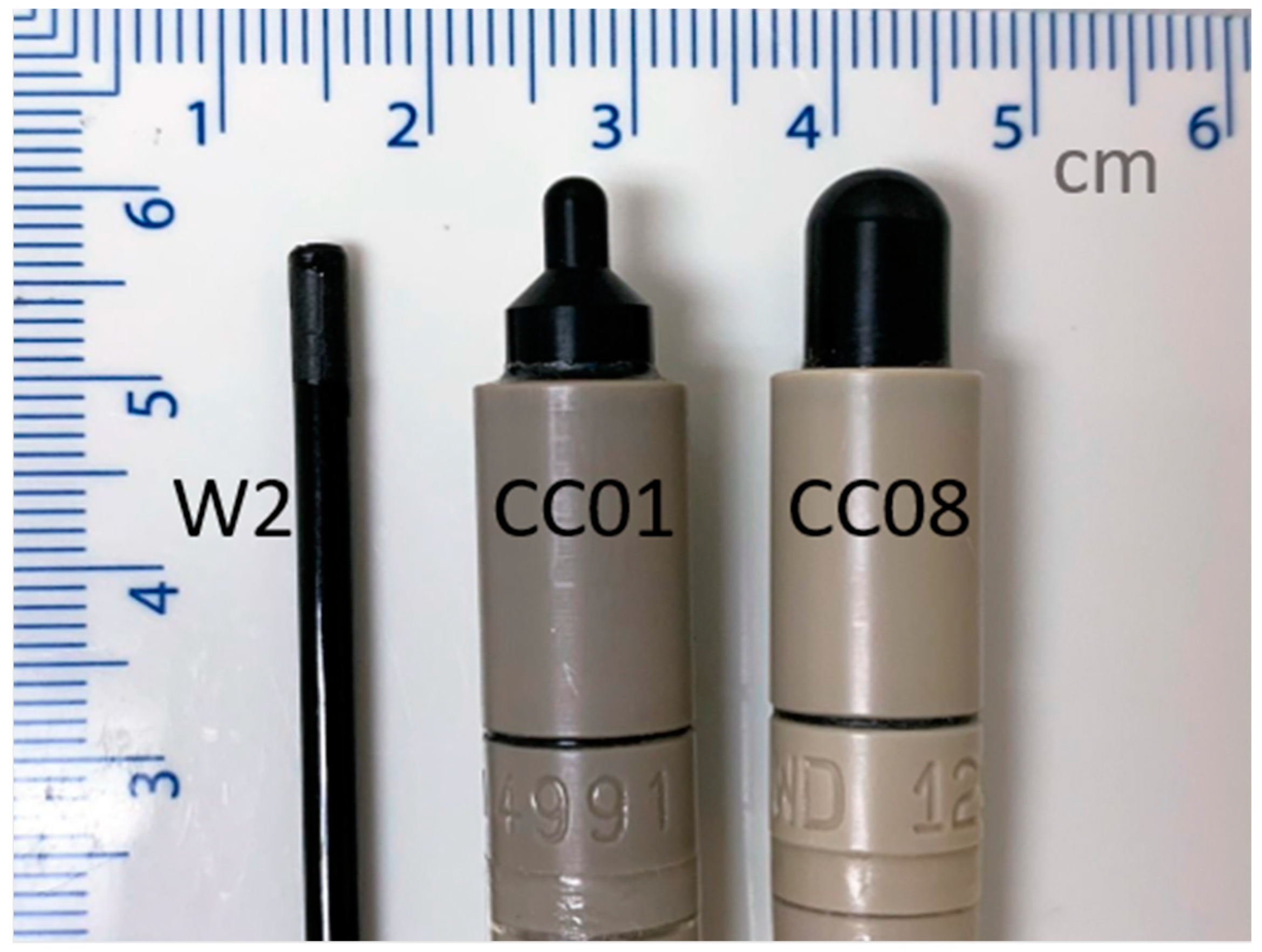
| Detector | Dimensions: Length, Inner Diameter (mm) | Active Volume (cm3) | Detector Material |
|---|---|---|---|
| CC01 [23] | 3.6, 2.0 | 0.01 | 0.35 mm diameter steel electrode in air |
| CC08 [24] | 4.0, 6.0 | 0.08 | 1 mm diameter C552 electrode in air |
| Exradin W2 [25] | 1.0, 1.0 | 0.0008 | Solid polystyrene with polyimide stem |
Publisher’s Note: MDPI stays neutral with regard to jurisdictional claims in published maps and institutional affiliations. |
© 2022 by the authors. Licensee MDPI, Basel, Switzerland. This article is an open access article distributed under the terms and conditions of the Creative Commons Attribution (CC BY) license (https://creativecommons.org/licenses/by/4.0/).
Share and Cite
Thrower, S.; Prajapati, S.; Holmes, S.; Schüler, E.; Beddar, S. Characterization of the Plastic Scintillator Detector System Exradin W2 in a High Dose Rate Flattening-Filter-Free Photon Beam. Sensors 2022, 22, 6785. https://0-doi-org.brum.beds.ac.uk/10.3390/s22186785
Thrower S, Prajapati S, Holmes S, Schüler E, Beddar S. Characterization of the Plastic Scintillator Detector System Exradin W2 in a High Dose Rate Flattening-Filter-Free Photon Beam. Sensors. 2022; 22(18):6785. https://0-doi-org.brum.beds.ac.uk/10.3390/s22186785
Chicago/Turabian StyleThrower, Sara, Surendra Prajapati, Shannon Holmes, Emil Schüler, and Sam Beddar. 2022. "Characterization of the Plastic Scintillator Detector System Exradin W2 in a High Dose Rate Flattening-Filter-Free Photon Beam" Sensors 22, no. 18: 6785. https://0-doi-org.brum.beds.ac.uk/10.3390/s22186785