Article

4-Hydroxyderricin Isolated from the Sap of Angelica keiskei Koidzumi: Evaluation of Its Inhibitory Activity towards Dipeptidyl Peptidase-IV

1
Pharmaceutical Biology Research Group, School of Pharmacy, Bandung Institute of Technology, Jl. Ganesha 10, Bandung 40132, Indonesia
2
Department of Pharmaceutical Biology, Indonesian School of Pharmacy (Sekolah Tinggi Farmasi Indonesia), Jl. Soekarno-Hatta No 354, Bandung 40266, Indonesia
3
Department of Pharmacology and Toxicology, School of Pharmacy, Bandung Institute of Technology, Jl. Ganesha 10, Bandung 40132, Indonesia
4
Department of Pharmacology and Clinical Pharmacy, Faculty of Pharmacy, Universitas Padjadjaran, Jl. Raya Bandung-Sumedang km. 21, West Java 45363, Indonesia
*
Author to whom correspondence should be addressed.
Submission received: 29 September 2019 / Revised: 8 November 2019 / Accepted: 11 November 2019 / Published: 15 November 2019

Abstract

:
Angelica keiskei sap is used as a blood-sugar reducer in Indonesia, however its molecular mechanism has not yet been explored. 4-hydroxyderricin (4-HD) is one of the major components extracted from A. keiskei sap. The aim of this work was to isolate 4-HD from the sap of A. keiskei planted in Lombok, Indonesia, and to study in silico and in vitro mechanisms against dipeptidyl peptidase-IV (DPP-IV). The dried sap was submitted to liquid–liquid extraction using solvents with different polarity. Further purification processing was conducted using gradient elution column chromatography. The isolated compound was a yellowish powder, m/z 339.2215 [M + H]+, which was confirmed as 4-HD. Sitagliptin, a DPP-IV inhibitor, was employed as the positive control for both the in silico and in vitro studies. It was indicated that 4-HD interacts with Glu206 and Phe357, important amino acid residues in the DPP-IV binding pocket. These interactions are similar to that of sitagliptin. The affinity of 4-HD (inhibition constant (Ki) = 3.99 μM) to DPP-IV is lower than that of sitagliptin (inhibition constant (Ki) = 0.17 μM). Furthermore, in vitro study showed that 4-HD inhibits DPP-IV (IC50 = 81.44 μM) weaker than for sitagliptin (IC50 = 0.87 μM). We concluded that 4- HD might have potential in inhibiting DPP-IV. However, by considering the polar interaction of sitagliptin with DPP-IV, a further structure modification of 4-HD, e.g., by introducing a polar moiety such as a hydroxyl group, might be needed to obtain stronger activity as a DPP-IV inhibitor.

1. Introduction

Diabetes mellitus (DM) is considered as a serious health problem due to its increasing prevalence in most developing countries [1], therefore new antidiabetic drugs and therapies are always of interest to be explored [2]. DM is a chronic condition when the beta-cells in the pancreas’ Langerhans islets are not able to produce sufficient insulin and eventually elevates the blood sugar level [3]. More than one million Indonesians were diagnosed with DM in 2018 [4], whereas 451 million (age 18–99 years) have been reported to suffer the same problem globally [1,3]. Type 2 DM (T2DM) is characterized by insulin insensitivity, inhibition of insulin production, and eventually, pancreatic beta-cell failure. This leads to a decrease in glucose transport into the liver, muscle cells, and fat cells [5].
Treating T2DM by regulating incretin, a group of metabolic hormones such as glucagon-like peptide-1 (GLP-1), maintains glucose homeostasis by stimulating the biosynthesis insulin or insulin release. Subsequently, it inhibits the release of glucagon. This condition slows down gastric emptying, and reduces appetite [6,7]. GLP-1 is released from the intestine in response to carbohydrate intake from food. Although GIP activity is impaired in those with T2DM, GLP-1 insulinotropic effects are preserved, and thus GLP-1 represents a potentially beneficial therapeutic option. DPP-IV inhibitors prevent the breakdown of endogenous GLP-1 as well as glucose-dependent insulinotropic peptide (GIP) [8]. Therefore, DPP-IV inhibitors are believed to prolong the half-life of incretins (GLP-1 and GIP) and are associated with increased insulin release and reduced glucagon release [9]
However, similarly to GIP, GLP-1 is rapidly inactivated by DPP-IV in vivo [10]. DPP-IV inhibitors represent an innovative approach for the treatment of T2DM. These inhibitors, e.g., sitagliptin, are attached in the DPP-IV binding pocket via hydrogen bonding with Glu205, Glu206, and Tyr662 [9] and prevent the breaking down of GLP-1 [11]. As a result, GLP-1 remains active for a longer time, thus it increases the insulin production and holds back the glucagon production, both of which help to reduce the blood glucose level. A controlled blood glucose may reduce the risk of potentially life-threatening complications, i.e., heart disease, kidney problems, nerve damage, and visual impairment [12]. Unlike many other T2DM therapies, DPP-IV inhibitors do not cause weight gain. This is an advantage as body weight can be a challenge to manage [13,14].
Chalcones have been found to be potent as inhibitors of DPP-IV, which is an effective target on glucose homeostasis [15,16,17]. The root and the stem of Angelica keiskei (ashitaba) of the Apiaceae family, contain yellow sap [18], in which two major chalcones (4-hydroxyderricin or 4-HD and xanthoangelol) [19,20] are produced. According to Luo [21], the ashitaba extract and xanthoangelol (IC50 = 11.54 µM) indicated excellent activity in inhibiting alpha-glucosidase; however, a moderate inhibition was shown by 4-HD (IC50 = 33.76 µM) [21]. Ashitaba extract could lower cholesterol, glucose, and increase insulin levels in C57BL/6 strain mice and prevent adiposity through modulating lipid metabolism [16,17,19,20,22]. Our work aimed to explore the inhibitory activity of 4-HD against DPP-IV through in silico and in vitro studies.

2. Materials and Methods

2.1. Materials

2.1.1. Plant Material

The yellow sap of Angelica keiskei was collected from Mount Rinjani, Lombok, Indonesia. Taxonomic determination (No. 401/[email protected]/PL/2019) was conducted by a certified botanist at the Herbarium Bandungense, Bandung Institute of Technology (ITB). The yellow sap (1 L) was freeze-dried.

2.1.2. Chemicals

Human DPP-IV (Sigma-Aldrich, Cat. No. D4943, Saint Louis, MI, USA), Gly-Pro-p-Nitroanilide Hydrochloride (GPPN) (Sigma-Aldrich, Cat. No. G0513), Tris-HCl Buffer (Sigma-Aldrich, Cat. No. T3253), and other chemicals were purchased from Merck (Saint Louis, MI, USA). Sitagliptin standard was purchased from PT. Kimia Farma Tbk, Indonesia.

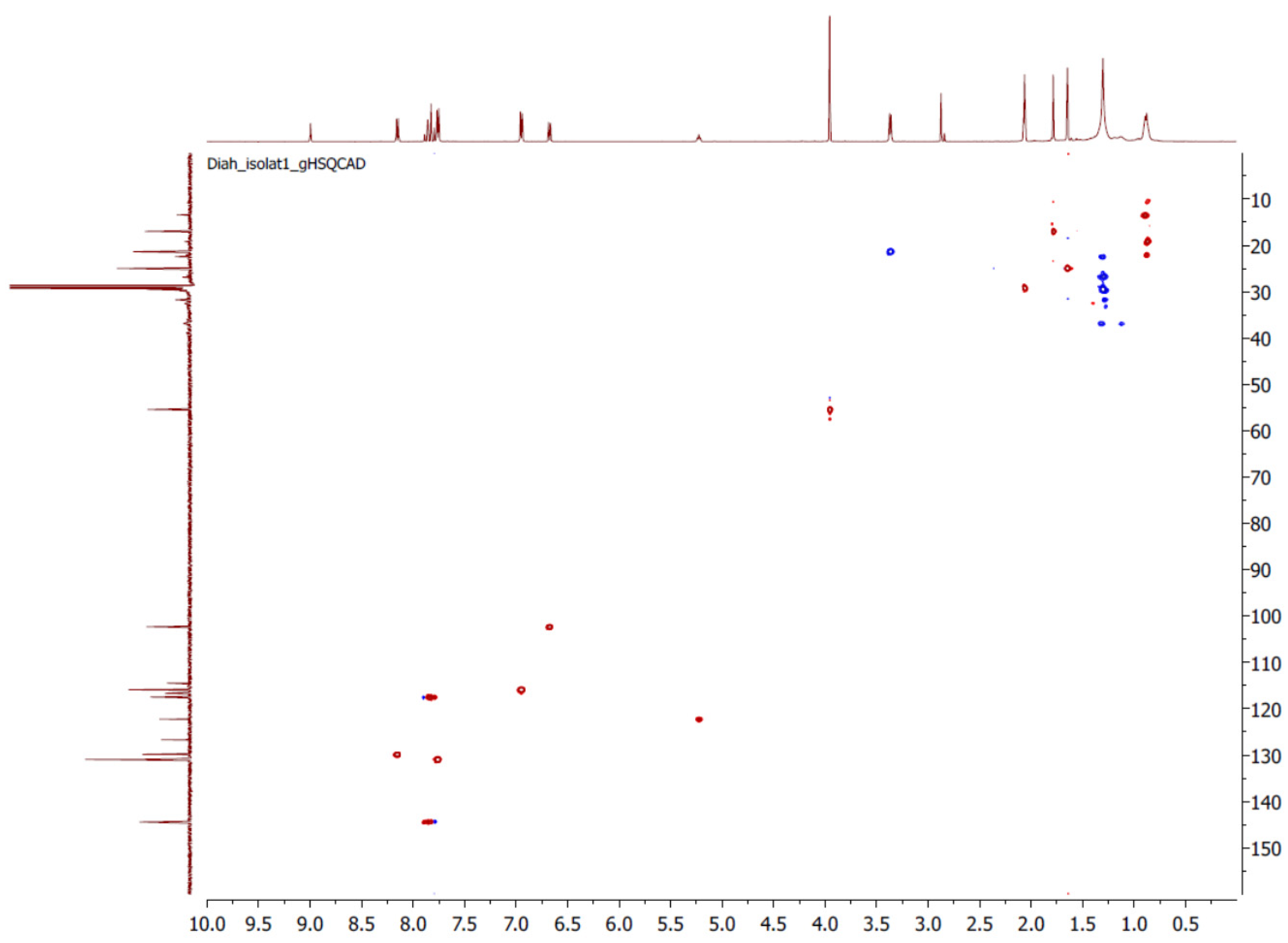
2.2. Extraction and Isolation

The yellow sap powder (10 g) was extracted with ethanol (1:1 w/v), followed by the evaporation of the solvent. The viscous extract was fractionated by liquid–liquid extraction three times by using three solvents, i.e., n-hexane (yielded 0.43 g), ethyl acetate (yielded 5.86 g), and a combination of ethanol-water (7:3 v/v) (yielded 2.90 g). The resulting fractions were evaporated to a certain volume for the isolation process. The isolation was conducted on the ethyl acetate fraction (2 g) by using column chromatography (2.0 × 45 cm) packed with 32.33 g of kieselgel 60 (Merck), and was gradient eluted using a combination of n-hexane-acetone (100:0 → 0:100 v/v) to give F1–F6 fractions. Furthermore, the F3 (300 mg) fraction was passed onto a 1.2 × 35 cm column packed with 6 g of kieselgel 60 (Merck). A combination of n-hexane-acetone (100:0 → 0:100 v/v) was used as the eluent, yielding 75.5 mg of isolated compound 1. This isolated compound, which was obtained from twice partitioning on a column, was confirmed by its structure as 4-HD (Figure 1a).
Compound Characterization. (4-HD) Compound 1, which forms as a yellowish powder, with purity 84.91% at retention 5.36 min by Liquid Chromatography (AquityTM Ultra Performance LC, RP18 1.7 µm, 2.1 × 100 mm column, with mobile phase A, water–0.1% formic acid v/v and mobile phase B, methanol (70:30 → 40:60 v/v), flow rate 0.2 mL/minute), shows a maximum at 364 nm using UV spectrophotometry (Agilent 845x UV-Visible system). Mass spectra (XevoTM QTOF MS, ionization mode ES+) resulted [M + H]+ m/z = 339.2215. The 13C-NMR (Agilent, 125 MHz) and 1H-NMR (Agilent, 500 MHz) data shown in Table 1 was compared to that of Kim and co-workers [23]. The isolated compound was dissolved in 600 µL deuterated acetone (CD3)2CO in a 5 mm diameter NMR tube, and two dimensional (2D) NMR was performed with heteronuclear single quantum coherence (1H,13C gHSQC) and heteronuclear multiple bond correlation (gHMBC), and confirms C21H22O4 for 4- hydroxyderricin reference (Table 1). The 1H-NMR (500 MHz, (CD3)2CO):1.63 (3H,s, 4″-CH3); 3.36 (1H,D, J = 7.2 Hz,1″-H); 1.77 (3H, s, 5″-CH3); 3.95 (3H, s, 4′-OCH3); 6.67 (1H,d, J = 9.1 Hz, 5′-CH); 6.93 (2H, d, J = 8.6 Hz, 3,5-H); 7.73 (1H, d, J = 15.4 Hz, α); 7.80 (2H, d, J = 8.6 Hz, 2.6-H); 5.21 (1H, t, J = 7.3 Hz, 2″-H); 8.13 (1H,d, J = 9.1 Hz, 6′-H); 7.84 (1H,d, J = 15.4 Hz, β); 13.76 (1H, s, 2′-OH). The 13C- NMR (125 MHZ, (CD3)2CO)): 17.88 (4″-C); 22.27 (1″-C); 25.89 (5″-C; 56.32 (4′-C); 103.19 (5′-C); 116.79 (3,5-C); 117.60 (α-C); 118.38 (2,6-C); 123.16 (2″-C); 130.76 (6′C); 145.35 (β-C); 193.46 (C=O). The gHSQCAD spectra (Figure 2) exhibited the presence of two methyl groups, one methylene, one methoxy (δ 56.32). The absence correlation of seven signal carbon (δ 127.59, 131.84, 161.06, 163.38, 163.79, 164.14, 164.18) to proton spectra indicated the presence of three quaternary carbons. In addition the signals at δ 5.19, 5.21, 5.22 are recognizable for a prenyl group, and the signal at δ 193.46 is known as the carbonyl group in chalcone.

2.3. In Silico Study

2.3.1. Protein Preparation

The 3D X-ray crystallographic data of DPP-IV (PDB code: 1X70, resolution 2.1 Å in the complex with sitagliptin, crystallized by Kim [9] was downloaded from the online Protein Data Bank (www.rcsb.org). Water molecules, ligands, and other hetero atoms were removed from the protein molecule along with the unused chain, using Swiss-Pdb Viewer v4.0.4 (Swiss Institute of Bioinformatics, downloaded from www.expasy.org). The addition of hydrogen atoms and Kollman charges to the protein was performed using AutoDockVina (Molecular Graphics Laboratory, Scripps Research Institute, downloaded from http://autodock.scripps.edu). The binding modes of DPP-IV with sitagliptin were studied using Ligand Explorer Viewer v.4.1.0 (Research Col laboratory for Structural Bioinformatics, embedded on http://www.pdb.org/pdb/explore).

2.3.2. Ligand Preparation

All ligands were geometry-optimized using ChemBioDraw Ultra 12.0 free trial (downloaded from www.cambridgesoft.com) by employing MM2 and AM1 at default settings. The ligands were calculated for their log P, mass, and volume; and were saved in PDB file format for further processing.

2.3.3. Validation of the Docking Simulation

Sitagliptin was separated from DPP-IV, and validation was performed by re-docking the extracted-sitagliptin into its origin location for 50× using Linux script command. This step was continued by calculating the standard deviation (SD) of the binding energy and Ki. The re-docked sitagliptin was then superimposed with the extracted-sitagliptin and the root mean square deviation (RMSD) of the structures was measured.

2.3.4. Molecular Docking Simulation

Docking simulation of 4-HD was repeated 50× using Linux script command for AutoDockVina [24] embedded in MGL Tools v.1.5.6 at position x 41.1841; y 51.045; z 35.6287. These coordinates range within 7 Å distance centered to the ligand position, respectively, and were calculated by employing bind module in PLANTS 1.2. The default parameters of the automatic settings were used to set the genetic algorithm parameters. The docked conformation which had the highest docking score (Ei) was selected to analyze the binding mode [25]. Sitagliptin was used as the reference and the structure is represented in Figure 1b.

2.4. In Vitro Study

Dipeptidyl Peptidase-IV (DPP-IV) Assay

The assay was conducted according to the method proposed by ref. [26] with slight modification, in 96 microwell plates. Samples (@ 35 µL) in various concentrations (2.5; 1.25; 0.625; and 0.325 µg/mL) dissolved in 50 mM Tris HCl buffer (pH 7.5), were put into the wells and 15 µL of DPP-IV (0.05 units/mL) solution were added. The mixtures were incubated for 10 min in 37 °C. After incubation, a 50 µL of Gly-Pro-p-Nitroanilide Hydrochloride (0.2 mM in Tris HCl buffer pH 7.5) substrate was added and the mixtures were shaken and incubated for 30 min at 37 °C. Acetic acid 25% in water (v/v) (25 µL) was added to the mixture to stop the enzymatic reaction. Sitagliptin (0.2; 0.4; 0.8; 1.6; and 3.2 µg/mL in a 50 mM Tris HCl buffer pH 7.5) was used as the reference.
The substrate, a chromogenic Gly-Pro-p-Nitroanilide Hydrochloride was cleaved by DPP-IV, and released p-nitroaniline (pNA), a yellow colored product was recorded using a Multiscan (Nanoquant, Infinite M2000 Pro, TECAN) at 405 nm. The procedure was performed in three replications. One unit of enzyme activity was defined as the amount of enzyme that catalyzes the release of 1 μmoL of pNA from the substrate/min in the experimental conditions. All data was expressed as mean ± SD. The percentage of inhibition was calculated using the following formula:
%   Inhibition   =   A b s .   n e g a t i v e   c o n t r o l A b s .   i n h i b i t o r A b s .   n e g a t i v e   c o n t r o l   ×   100

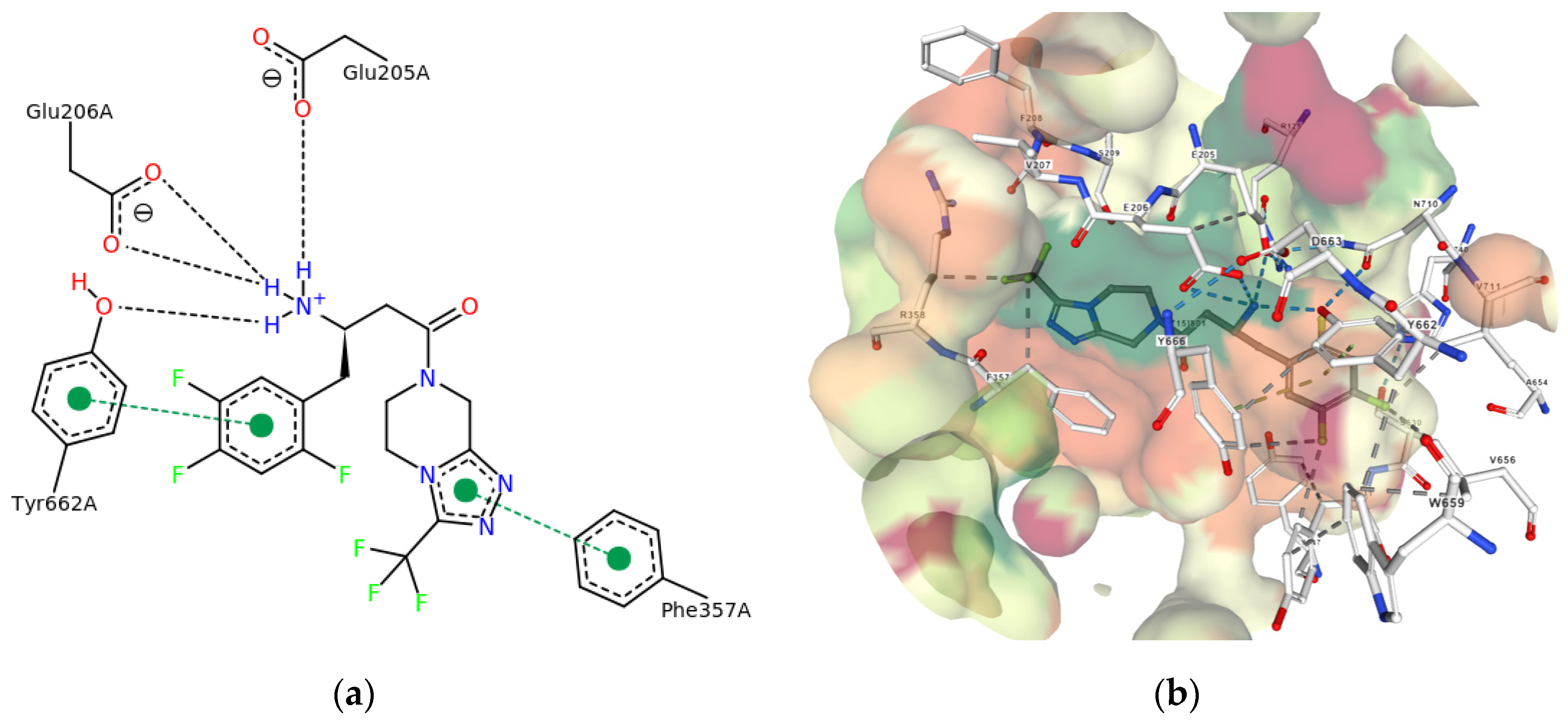
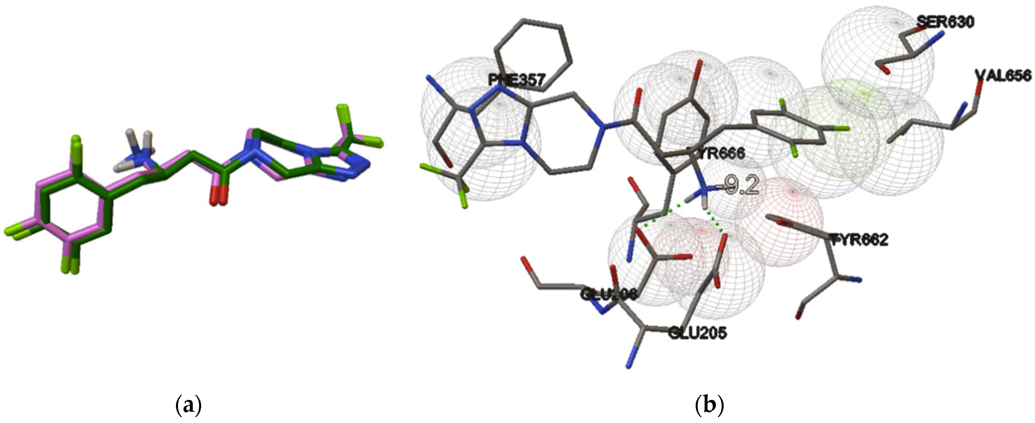
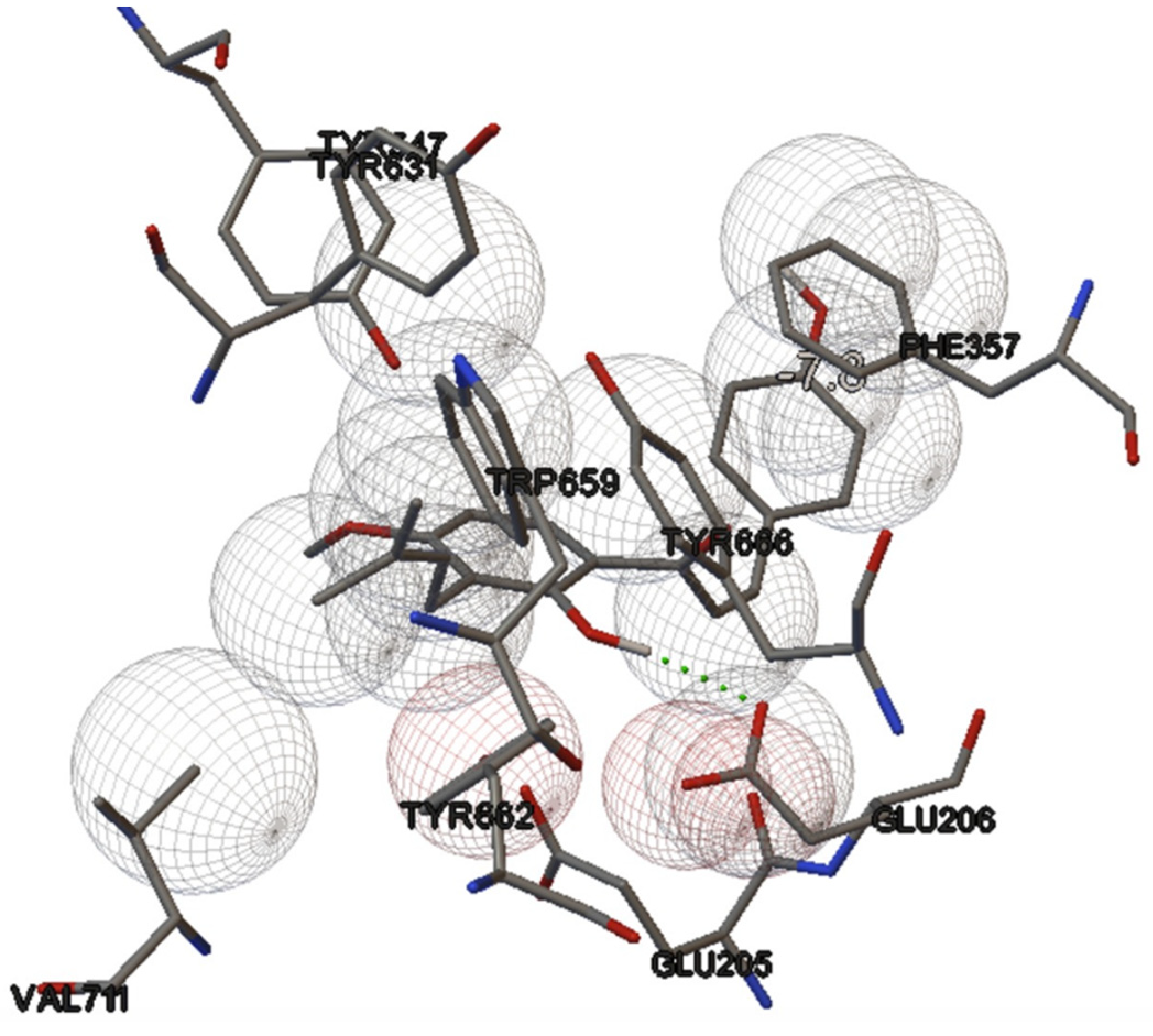
3. Results and Discussion

Our work showed that 4-HD, an alkylated-chalcone, was found to be abundant in the ethyl acetate fraction of A. keiskei sap. Comparing our work to that of Li and colleagues [27], who determined the distribution of 4-HD in different parts of A. keiskei planted in China, the stems were the best source of this particular chalcone (149.16 µg/g) [27]. In the biosynthesis of flavonoids, chalcones which possess an open C-ring, i.e., isomers of dihydroflavones, represent the key precursors. These compounds are also responsible for the plant’s color [15,28]. The presence of a double bond in conjugation with the carbonyl (C=O) functionality of chalcones is believed to be responsible for their biological activities, as removal of this functionality make them inactive [29,30]. Carbonyl is a relatively strong dipole which allows its oxygen to act as an H-bond acceptor (HBA). This pharmacophoric feature of chalcones as HBAs has been proven for antileishmanial activity [31].
Geometry optimization of both sitagliptin and 4-HD, which was performed using MM2 and AM1, resulted in minimization energy respectively −210.71 for sitagliptin and −90.14 for 4-HD. Both ligands showed similar volume (1010.5 Å for sitagliptin and 1012.5 Å for 4-HD). However, 4-HD is more hydrophobic (log P 4.275) than sitagliptin (log P 1.286). A crystal structure of DPP-IV was employed in this work (2.1 Å resolution, crystallized by Kim [9]). PoseView image in Figure 3a (in 2D) and Figure 3b (in 3D) showed that sitagliptin interacts with Glu205, Glu206, and Tyr662 via four HBs. This drug also interacts with Phe357 and Tyr662 via two aromatic or π–π stackings. The last is a lipophilic interaction. Sitagliptin was separated from its complex with DPP-IV, and validation was performed by re-docking the extracted-sitagliptin into its origin location (x 41.1841; y 51.045; z 35.6287) for 50× using Linux script command. The re-docked sitagliptin was then superimposed with the extracted-sitagliptin extracted from the enzyme. The molecular docking simulation was categorized as valid (RMSD value = 1.013) and is represented in Figure 4a.
Re-docking simulation confirmed that the binding mode of extracted-sitagliptin is similar to that of its origin. The extracted-sitagliptin builds two HBs with polar amino acid residues in the DPP-IV’s binding pocket: Glu205 and Glu206. Meanwhile, two aromatic stackings with Phe357 and Tyr662 (the docking score –9.24 kcal/mol; Ki = 0.172 μM) are observed in Figure 4b, whereas the origin sitagliptin builds two HBs with DPP-IV amino acid residues (1 × 70). The docking reveals that 4-HD (docking score −7.42 kcal/mol; inhibition constant 3.99 μM) interacts with DPP-IV via one HB with Glu206 (the hydroxyl group of 4-HD is an HBD) and an aromatic stacking with Phe357 (binding energy −7.8 kcal), as observed in Figure 5.
DPP-IV inhibitors, in the N-terminus of the substrate peptide, form salt bridges with Glu205 and Glu206 in the S2 subsite, two important amino acid residues which play a vital role in their inhibitory activity. The amino group of sitagliptin forms four HBs, e.g., one HB with the hydroxyl group of Tyr662 and three HBs with the carboxylate oxygens of two glutamate residues (Glu205 and Glu206) [9,32]. As predicted, our work reveals that 4-HD also interacts with Glu206, an important amino residue in the N-terminus. This interaction might play a role in its inhibitory activity.
In vitro assay indicated that 4-HD inhibits DPP-IV (IC50 = 81.44 µM). However, its inhibitory activity is weaker than that of sitagliptin (IC50 = 0.87 µM). Our in vitro result is in line with the in silico prediction; 4-HD could attach to the DPP-IV binding pocket and indicates similar interaction with that of sitagliptin, a known DPP-IV inhibitor, which was employed as the standard.
It can be summarized that 4-HD could inhibit DPP-IV activity via hydrogen bonding with Glu206, an important amino residue in the N-terminus, and with aromatic stacking with Phe357. 4-HD might also exert its antihyperglycemic activity via several mechanisms: (a) It could stimulate the glucose uptake in 3T3-L1 adipocytes, and this activity of 4-HD was stronger than that of xanthoangelol [18]; (b) It could stimulate the phosphorylation of 5′-AMPK and its downstream target acetyl-CoA carboxylase [17].

4. Conclusions

A chalcone compound, 4-hydroxyderricin (4-HD), was successfully isolated from the ethyl acetate fraction of the yellow sap of Angelica keiskei collected from Mount Rinjani, Lombok, Indonesia. In silico study revealed that 4-HD may interact with important amino acid residues in the DPP-IV binding pocket via one hydrogen bond (HB) with Glu206 and an aromatic stacking with Phe357. Sitagliptin, a known DPP-IV inhibitor, builds two HBs with Glu205 and Glu206, and two aromatic stackings with Phe357 and Tyr662. In vitro study indicated that 4-HD inhibits DPP-IV (IC50 = 81.44 µM) weaker than for sitagliptin (IC50 = 0.87 µM). We concluded that 4-HD might have potential in inhibiting DPP-IV. However, further structure modification of 4-HD, e.g., by introducing a polar moiety (–OH), is still needed to obtain a stronger activity as a DPP-IV inhibitor.

Author Contributions

S.S., J.L. and I.K.A. substantially contributed to the conception and design of the study. D.L.A. participated in the collection and assembly of data. S.S., J.L., I.K.A., and D.L.A. participated in processing analysis, and interpretation of reported data. D.L.A. and J.L. contributed to the drafting of the article. S.S., J.L., and I.K.A. contributed to the critical revision of the article on important intellectual content. All authors read and approved the final manuscript before submission.

Funding

We would like to thank the Indonesia Endowment Fund for Education (LPDP) for funding and supporting this research. Funding No. NPRJ-5723 /LPDP.3/2016.

Acknowledgments

We would like to thank to PT. Kimia Farma, Indonesia for kindly providing sitagliptin.

Conflicts of Interest

The authors declare no conflict of interest.

References

  1. Cho, N.H.; Shaw, J.E.; Karuranga, S.; Huang, Y.; da Rocha Fernandes, J.D.; Ohlrogge, A.W.; Malanda, B. IDF Diabetes Atlas: Global estimates of diabetes prevalence for 2017 and projections for 2045. Diabetes Res. Clin. Pract. 2018, 138, 271–281. [Google Scholar] [CrossRef]
  2. Dicker, D. DPP-4 Inhibitors: Impact on glycemic control and cardiovascular risk factors. Diabetes Care 2011, 34, 276–278. [Google Scholar] [CrossRef]
  3. International Diabetes Federation. IDF Diabetes Atlas, 7th ed.; IDF: Brussels, Belgium, 2015; ISBN 9782930229812. [Google Scholar]
  4. Anonymous. Indonesian Report on Basic Health Research; Ministry of Health (Indonesia): Jakarta, Indonesia, 2018; ISBN 978-602-8937-24-5.
  5. Deacon, C.F. Dipeptidyl peptidase-4 inhibitors in the treatment of type 2 diabetes: A comparative review. Diabetes Obes. Metab. 2011, 13, 7–18. [Google Scholar] [CrossRef]
  6. Nauck, M.A.; Kemmeries, G.; Holst, J.J.; Meier, J.J. Rapid tachyphylaxis of the glucagon-like peptide 1-induced deceleration of gastric emptying in humans. Diabetes 2011, 60, 1561–1565. [Google Scholar] [CrossRef]
  7. Richter, B.; Bandeira-echtler, E. Emerging Role of Dipeptidyl Peptidase-4 Inhibitors in the Management of Type 2 Diabetes. Vasc. Health Risk Manag. 2008, 4, 753–768. [Google Scholar] [CrossRef]
  8. Olokoba, A.B.; Obateru, O.A.; Olokoba, L.B. Type 2 Diabetes Mellitus: A Review of Current Trends. Oman Med. J. 2012, 27, 269–273. [Google Scholar] [CrossRef]
  9. Kim, D.; Wang, L.; Beconi, M.; Eiermann, G.J.; Fisher, M.H.; He, H.; Hickey, G.J.; Kowalchick, J.E.; Leiting, B.; Lyons, K.; et al. (2R)-4-Oxo-4-[3-(Trifluoromethyl)-5,6-dihydro[1,2,4]triazolo[4,3-a]pyrazin- 7(8H)-yl]-1-(2,4,5-trifluorophenyl)butan-2-amine: A Potent, Orally Active Dipeptidyl Peptidase IV Inhibitor for the Treatment of Type 2 Diabetes. J. Med. Chem. 2005, 141, 141–151. [Google Scholar] [CrossRef]
  10. Zilleßen, P.; Celner, J.; Kretschmann, A.; Pfeifer, A.; Racké, K.; Mayer, P. Metabolic role of dipeptidyl peptidase 4 (DPP4) in primary human (pre)adipocytes. Sci. Rep. 2016, 6, 23074. [Google Scholar] [CrossRef]
  11. Avogaro, A.; Fadini, G.P. The Effects of Dipeptidyl Peptidase-4 Inhibition on Microvascular Diabetes Complications. Diabetes Care 2014, 37, 2884–2894. [Google Scholar] [CrossRef]
  12. Fadini, G.P.; Avogaro, A. Cardiovascular effects of DPP-4 inhibition: Beyond GLP-1. Vascul. Pharmacol. 2011, 55, 10–16. [Google Scholar] [CrossRef]
  13. Kahn, S.E.; Hull, R.L.; Utzschneider, K.M. Mechanisms Linking Obesity to Insulin Resistance and Type 2 Diabetes. Nature 2006, 444, 840–846. [Google Scholar] [CrossRef]
  14. Hill, M.J.; Metcalfe, D.; McTernan, P.G. Obesity and Diabetes:Lipids, “Nowhere to run to”. Clin. Sci. 2009, 116, 113–123. [Google Scholar] [CrossRef]
  15. Wang, T.Y.; Li, Q.; Bi, K.S. Bioactive Flavonoids in Medicinal Plants: Structure, Activity and Biological Fate. Asian J. Pharm. Sci. 2017, 13, 12–23. [Google Scholar] [CrossRef]
  16. Enoki, T.; Ohnogi, H.; Nagamine, K.; Kudo, Y.; Sugiyama, K.; Tanabe, M.; Kobayashi, E.; Sagawa, H.; Kato, I. Antidiabetic Activities of Chalcones Isolated from a Japanese Herb, Angelica keiskei. J. Agric. Food Chem. 2007, 55, 6013–6017. [Google Scholar] [CrossRef]
  17. Zhang, T.; Yamashita, Y.; Yasuda, M.; Yamamoto, N.; Ashida, H. Ashitaba (Angelica keiskei) Extract Prevents Adiposity in High-fat Diet-fed C57BL/6 Mice. Food Funct. 2015, 6, 134–144. [Google Scholar] [CrossRef]
  18. Ohta, M.; Fujinami, A.; Kobayashi, N.; Amano, A.; Ishigami, A.; Tokuda, H.; Suzuki, N.; Ito, F.; Mori, T.; Sawada, M.; et al. Two chalcones, 4-Hydroxyderricin and Xanthoangelol, Stimulate GLUT4-dependent Glucose Uptake through the LKB1/AMP-activated Protein Kinase Signaling Pathway in 3T3-L1 Adipocytes. Nutr. Res. 2015, 35, 618–625. [Google Scholar] [CrossRef]
  19. Akihisa, T.; Tokuda, H.; Ukiya, M.; Iizuka, M.; Schneider, S.; Ogasawara, K.; Mukainaka, T.; Iwatsuki, K.; Suzuki, T.; Nishino, H. Chalcones, Coumarins, and Flavanones from the Exudate of Angelica keiskei and Their Chemopreventive Effects. Cancer Lett. 2003, 201, 133–137. [Google Scholar] [CrossRef]
  20. Sarker, S.D.; Nahar, L. Natural Medicine: The Genus Angelica. Curr. Med. Chem. 2004, 11, 1479–1500. [Google Scholar] [CrossRef]
  21. Luo, L.; Wang, R.; Wang, X.; Ma, Z.; Li, N. Compounds from Angelica keiskei with NQO1 Induction, DPPH Scavenging and α-Glucosidase Inhibitory Activities. Food Chem. 2012, 131, 992–998. [Google Scholar] [CrossRef]
  22. Caesar, L.K.; Cech, N.B. A Review of the Medicinal Uses and Pharmacology of Ashitaba. Planta Med. 2016, 82, 1236–1245. [Google Scholar] [CrossRef] [PubMed]
  23. Kim, J.H.; Son, Y.K.; Kim, G.H.; Hwang, K.H. Xanthoangelol and 4-Hydroxyderricin are the Major Active Principles of the Inhibitory Activities Against Monoamine Oxidases on Angelica keiskei K. Biomol. Ther. 2013, 21, 234–240. [Google Scholar] [CrossRef] [PubMed]
  24. Trott, O.; Olson, A.J. Software news and Update AutoDock Vina: Improving the Speed and Accuracy of Docking with a New Scoring Function, Efficient Optimization, and Multithreading. J. Comput. Chem. 2010, 31, 455–461. [Google Scholar] [PubMed]
  25. Levita, J.; Rositama, M.R.; Alias, N.; Khalida, N.; Saptarini, N.M.; Megantara, S. Discovering COX-2 Inhibitors from Flavonoids and Diterpenoids. J. Appl. Pharm. Sci. 2017, 7, 103–110. [Google Scholar]
  26. Riyanti, S.; Suganda, A.G.; Sukandar, E.Y. Dipeptidyl peptidase-IV Inhibitory Activity of some Indonesian Medicinal Plants. Asian J. Pharm. Clin. Res. 2016, 9, 375–377. [Google Scholar]
  27. Li, Y.; Luo, Y.; Liu, J.; Wu, J.; Zhong, Y.; Wang, Z. Determination of 4-Hydroxyderricin and Xanthoangelol in Different Parts of Angelica keiskei by HPLC-PDA. Food Sci. 2016, 37, 2014–2017. [Google Scholar]
  28. Kumar, A.; Premoli, M.; Aria, F.; Bonini, S.A.; Maccarinelli, G.; Gianoncelli, A.; Memo, M.; Mastinu, A. Cannabimimetic plants: Are they new cannabinoidergic modulators? Planta 2019, 249, 1681–1694. [Google Scholar] [CrossRef]
  29. Brashier, D.S.; Sharma, A.; Dahiya, N.; Singh, S.; Khadka, A. Lorcaserin: A novel antiobesity drug. J. Pharmacol. Pharmacother. 2014, 5, 175. [Google Scholar] [CrossRef]
  30. Zhou, B.; Xing, C. Diverse Molecular Targets for Chalcones with Varied Bioactivities. Med. Chem. 2015, 5, 388–404. [Google Scholar] [CrossRef]
  31. Desai, S.A.; Kumbhar, S.S.; Katti, V.S.; Choudhari, P.B.; Bhatia, M.S. 3D QSAR and pharmacophore modelling on chalcones as antileishmanial agents potential trypanothione reductase Inhibitors. J. Appl. Pharm. Sci. 2013, 3, 99–102. [Google Scholar]
  32. Arulmozhiraja, S.; Matsuo, N.; Ishitsubo, E.; Okazaki, S.; Shimano, H.; Tokiwa, H. Comparative Binding Analysis of Dipeptidyl Peptidase IV (DPP-4) with Antidiabetic Drugs -An Ab Initio Fragment Molecular Orbital Study. PLoS ONE 2016, 11, e0166275. [Google Scholar] [CrossRef] [PubMed] [Green Version]
Figure 1. 2D structure of 4-hydroxyderricin (4-HD) (a) and sitagliptin (b).
Scipharm 87 00030 g001
Figure 2. gHSQCAD spectrum of 4-hydroxyderricin (4-HD).
Scipharm 87 00030 g002
Figure 3. The binding mode of sitagliptin with DPP-IV: 2D (a) and 3D (b) (Downloaded from https://www.rcsb.org/3d-view/1X70).
Scipharm 87 00030 g003
Figure 4. Validation of docking method. Superimposed structures of sitagliptin (RMSD 1.013 Å) (a), re-docking simulation of sitagliptin into DPP-IV. Sitagliptin forms 2 HBs with Glu205 and Glu206 (showed with green dotted-lines) (b).
Scipharm 87 00030 g004
Figure 5. The binding mode of 4-HD with DPP-IV. 4-HD interacts with Glu206 via HB (shown with green dotted-lines).
Scipharm 87 00030 g005
Table 1. 1H and 13C NMR data of 4-hydroxyderricin from A. keiskei.
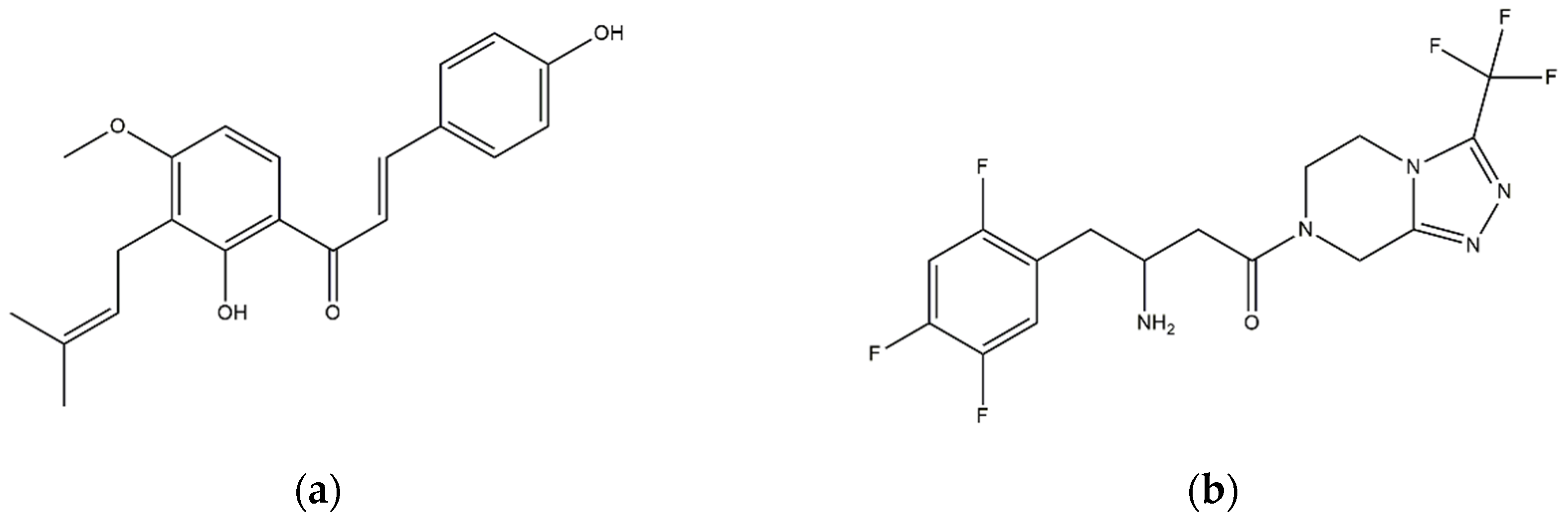
4-HD [23] (solvent CDCl3)
δ C (ppm)δ H (ppm)CH (HSQC)Position
-1.60(3H,s,4″-CH3)4″
-3.31(1H, d, J = 7.0 Hz,1″-H)1″
-1.71(3H, s, 5″-CH3)5″
-3.82(3H, s, 4-OCH3)4′
-6.41(1H, d, J = 9.0 Hz, 5′-H)5′
-6.79(2H, d, J = 8.5 Hz, 3,5-H)3,5
-7.36(1H, d, J = 15.4 Hz, α)α
-7.70(2H, d, J = 9.4 Hz, 2,6-H)2,6
-5.30(1H, t, J = 7.0 Hz, 2″-H)2″
-7.70(1H, d, J = 9.4 Hz, 6′-H)6′
-7.72(1H, d, J = 15.4 Hz, β)β
-13.38(1H, s, 2′-OH)2′
Compound 1 (4-HD) (solvent (CD3)2CO)
δ C (ppm)δ H (ppm)CH (HSQC)Position
17.881.63(3H,s, -CH3)4″
22.273.36(1H,d, J = 7.2 Hz, 1″-H)1″
25.891.77(3H,s, -5″-CH3)5″
56.323.95(3H,s, 4′-OCH3)4′
103.196.67(1H,d, J = 9.1 Hz, 5′-H)5′
116.796.93(2H,d, J = 8.6 Hz, 3,5-H)3,5
117.607.73(1H,d, J = 15.4 Hz, α)α
118.387.80(2H,d, J = 8.6 Hz, 2,6-H)2,6
123.165.21(1H,t, J = 7.3 Hz, 2″-H)2″
127.59---
130.768.13(1H,d, J = 9.1 Hz, 6′-H)6′
131.84--3″
145.357.84(1H,d, J = 15.4 Hz, β)β
-13.76(1H,s, 2′-OH)2′
161.06---
163.38---
163.79---
164.14---
164.18---
193.46-(C=O)-

