Article

LC-MS Phytochemical Screening, In Vitro Antioxidant, Antimicrobial and Anticancer Activity of Microalgae Nannochloropsis oculata Extract

by
Adil Farooq Wali
1,*,
Yusra Al Dhaheri
2,
Jayachithra Ramakrishna Pillai
1,
Ahlam Mushtaq
3,
Padma G. M. Rao
4,
Syed Arman Rabbani
4,
Aimen Firdous
5,
Mohamed Soliman Elshikh
6 and
Dunia A. Al Farraj
6
1
Department of Pharmaceutical Chemistry, RAK College of Pharmaceutical Sciences, RAK Medical and Health Sciences University, Ras Al Khaimah 11172, UAE
2
Department of Biology, College of Science, United Arab Emirates University, Al Ain 15551, UAE
3
Department of Biochemistry, Government Medical College (GMC), Karan Nagar, Srinagar 190010, India
4
Department of Clinical Pharmacy and Pharmacology, RAK College of Pharmaceutical Sciences, RAK Medical and Health Sciences University, Ras Al Khaimah 11172, UAE
5
Department of Processing Technology, Kerala University of Fisheries and Ocean Studies (KUFOS), Panangad, Kerala 682506, India
6
Department of Botany and Microbiology, College of Science, King Saud University, Riyadh 11451, Saudi Arabia
*
Author to whom correspondence should be addressed.
Submission received: 24 August 2020 / Revised: 16 September 2020 / Accepted: 18 September 2020 / Published: 8 October 2020

Abstract

:
Nowadays, marine microalgae are recognized to be a considerably novel and rich origin of bioactive moieties utilized in the sectors of nutraceuticals and pharmaceuticals. In the present study, Nannochloropsis oculata extract (AME) was associated with a wide variety of pharmacological studies such as in vitro antioxidant, antibacterial, and antifungal and anticancer activity (MDA-MB-231) in cancer cells through in vitro models. In the study, the chemical composition and structure of the bioactive compounds found in the AME extract were studied using the LC-MS technique. The results of the anticancer activity showed a decrease in the percentage of cell viability of the MDA-MB-231 cells in a concentration- and time-dependent manner (400 μg/mL at 24 h, 300 μg/mL at 48 h, and 200 μg/mL at 72 h). We have also observed morphological changes in the cells that could be associated with treatment with AME extract. Our observation of the AME extract-treated MDA-MB231 cells under light microscopy showed that when the concentration increased, the number of cells began to decrease. As far as LC-MS analysis is concerned, it showed the presence of the bioactive molecules was terpenoids along with carotenoids, polyphenolic and fatty acids. The result revealed that the AME extract exhibited noteworthy in vitro free radical scavenging potential, with an IC50 value of 52.10 ± 0.85 µg/L in DPPH assay, 122.84 ± 2.32 µg/mL in H2O2 assay and, 96.95 ± 1.68 µg/mL in ABTS assay. The activity was found to be highly significant against bacteria (Gram-positive and negative) and moderately significant against fungal strain with minimum inhibitory concentration (MIC) and minimum bactericidal concentration (MBC)/minimum fungicidal concentration (MFC) values between 15.63 and 500 µg/mL.

1. Introduction

Natural products are important components of medicines. The use of plants as medicine may date back to the ancient period. Nature has created a rich repository of herbal medicine to treat human disease [1]. The discovery of therapeutic potential in flora and fauna has driven the disclosure of bioactive compounds, with significant implications in the laboratory context, and recognition of dynamic concepts of clinical value [2]. Marine natural products were, moreover, the target of finding novel drugs of chemical and biological significance. Marine organisms are the source of major new drugs with numerous different biological activities [3]. Microalgae could be found over almost all ecosystems and habitats on the planet, from the Northern Hemisphere to the equator. They may be free-floating or growing attached to substrates. Microalgae may even form mattresses on silt surfaces [4]. There is a possibility of the well-founded opportunity for a larger range of microalgae species to be utilized for human nourishment. A wide variety of microphytes comprise very rich sources proteins for humans [5]. A few of the most widely accepted microalgae pigments are essential for human health, have anticancer properties, and function as antioxidants. Nutritious polyunsaturated fatty acids prosper in a few algal species, advertising imperative fatty acids required for human wellbeing. Despite these prospects, few microalgae species have been identified for use during the nutritional sciences industry [6]. Microalgae manufacture a variety of secondary metabolites with biological effects, including anticancer, antimicrobial, antifungal, antiviral, anti-inflammatory, and other substances, and are a possible future source of novel therapeutic drugs [7]. They produce various bioactive molecules as a reaction to ecological pressure. They are essential biological resources that have a large number of physical, biological, and molecular approaches to handle stress. They have a broad range of metabolites and natural active components such as fatty acids, polysaccharides, phycocyanins, carotenoids, terpenoids, enzymes, and phenolic compounds [8]. The analysis of macro-algae extracts has recently been studied for their significant effectiveness in the prevention of different diseases. Despite the enormous proportion of marine algae in this region, only a few studies have been performed regarding the phytochemical, antioxidant, and pharmacological activities of algae [9,10].
Cancer has become one of the world’s leading adverse health effects with significant social and economic implications. Presently, cancer incidence rates account for one in seven deaths in the world, with far more deaths than AIDS, tuberculosis, and malaria combined [11]. A major number of women across the world die of breast cancer [12]. Of all breast cancers, a subtype of cancer that lacks three receptors (progesterone, estrogen, and HER2) known as triple-negative breast cancer (TNBC) constitutes about 15% in the United States of America. TNBC is distinguished by its attacking nature, unique molecular structure, lack of active treatment, and specific metastatic trends [13]. The essentiality of the presence of receptors for cancer cells makes common medications such as hormone therapy and estrogen, progesterone, and HER-2 targeting drugs ineffective. Therefore, chemotherapy using the cytotoxic drug that inhibits cancer cells is mainly used in the treatment of TNBCs. TNBCs often exhibit metastasis to secondary sites such as the brain and lungs. Furthermore, they are extremely proliferative and consistently develop resistance against chemotherapeutic agents [14]. Natural compounds are an extremely valuable resource of biologically active moieties which play a major role in the quest for new drugs by acting as lead molecules for the discovery of new drug candidates [15]. Natural compounds, obtained from various sources, act as a reserve of biologically active compounds that have transformed and improved the range of cancer treatment [16]. At present, the percentage of drugs derived from natural sources, with anticancer potential, is quite high corresponding to approximately 60 percent [17]. Several research findings have reported microalgae species with anti-cancer properties [18].
The green algae genus Nannochloropsis (Order: Eustigmatales; Family: Eustigmatophyceae.) is characterized by unicellular lacking flagella, planktonic, with 3 to 4 × 1.5 μm cylindrical cells [19]. They seem to be mostly rich in phytochemicals, making them suitable to be utilized as a nutritive supplement and natural therapy for the management of several ailments due to their ability to accumulate elevated levels of PUFAs (polyunsaturated fatty acids) [20].
This research was aimed at performing the phytochemical screening and assessing the antioxidant, antimicrobial, and anticancer activity of Nannochloropsis oculata.

2. Material and Methods

2.1. Preparation of Material and Extraction

Nannochloropsis oculata was cultured in the laboratory by adapting Sharifah et al. [21] culture technique. An amount of 100 g of microalgae (Nannochloropsis oculata) was thoroughly washed with demineralized water. The algae are therefore transferred to an Erlenmeyer flask and extracted with absolute methanol using an ultrasonic homogenizer (BioLogics, Inc., Manassas, VA, USA) for 1 hr at RT, 15 kHz. The mixture was filtered and using a rotary evaporator (Buchi Rotavapor® R-210, Flawil, Switzerland) the solvent was removed. By making use of freeze-drying technique the mass was lyophilized. The Nannochloropsis oculata extract (AME) was then placed in a desiccator to store it for further evaluation.

2.2. Total Phenolics Content (TPC)

Folin–Ciocalteu test was employed to estimate the TPC of AME extract following the methodology of Aryal et al. [22]. The standard reference sample used for plotting curve was gallic acid (20–100 μg/mL) and the TPC of AME extract was reported w.r.t gallic acid as μg/mg of corresponding gallic acid i.e., GAE/mg.

2.3. Total Flavonoid Content (TFC)

To evaluate this parameter the technique of colorimetry was employed as reported by Shi et al. [23]. For plotting the curve, the reference standard used was quercetin (20–100 μg/mL). AME extract was reported as μg/mg of the corresponding quercetin i.e., QE/mg.

2.4. Tannins Content (TC)

The methodology outlined by Siddhuraju et al. [24] was employed to measure the tannins content of the AME extract.

2.5. In Vitro Antioxidant Activity

The free radical scavenging potential of AME extract was assessed by DPPH assay [25], H2O2 assay [26] and ABTS assay [27]. The AME extract was compared to the reference standard butylated hydroxyanisole (BHA) and was evaluated in triplicate.
The value of IC50 defines the extract concentration that inhibits 50% of the radicals. The anti-radical concentration for 50% of DPPH, H2O2 and ABTS free radical was quantified from the logarithmic regression equation.

2.6. Phytochemical Screening of AME Extract by Using LC-MS

The chemical screening of AME extract was performed on an LC-MS instrument connected to HPLC with an ESI interface. For chromatographic separation Acquity C18 column (100 × 4.6) × 5 µm was used. Isocratic elution condition was employed for mobile phase with A:B Methanol:2mM Ammonium acetate:formic acid (65:35:0.1) maintaining a flow rate of 0.6 mL/min. The linearity was achieved from 1 to 250 ppm. The LC-MS conditions were voltage 30 V, capillary voltage 3.5 V, desolvation gas 900 L/h, desolvation temperature 400 °C, cone gas 50 L/h, source temp 150 °C, collision energy 22 V. The ionization was carried out in positive ion mode. The spectral data were obtained using PDA detector. To verify the compounds, all the data, procurements, and evaluations were regulated using the database library.

2.7. In Vitro Cytotoxicity Assay

2.7.1. Cell Line and Cell Culture

The cell line utilized for this study was triple-negative human breast cancer cell line (MDA-MB-231). The cell lines were provided with Dulbecco’s Modified Eagle’s Medium (DMEM). The medium of culture that included Dulbecco phosphate-buffered saline (DPBS) was accompanied by bovine fetal serum (10%) along with streptomycin/penicillin (100 U/mL) at 37 °C 5%, 95% humidity and CO2. After the removal of confluent media, the washing of sub-cultured cells was done with phosphate saline buffer (1×), then Trypsin was added to MDA-MB-231 cells for detaching the cells from substrate surface and counting the cells (cells/mL) at the time of plates seeding for cell viability experiment

2.7.2. Treatment of AME Extract to MDA-MB-231

MDA-MB-231 cells were transferred into 96-well plates. The density at which seeding was performed was 5000 cells/well. This was followed by overnight incubation of seeded plates which were then tested with increasing concentrations of AME extract, dissolved in 70% ethanol (50, 100, 200, 400 and 600 μg/mL) or ethanol (control). These were then incubated for another 24, 48 and 72 h. Cells were then subjected to cell viability measurement.

2.7.3. Measurement of Cell Viability

The cell viability measurement, at the indicated times was done by removing the media from each well then 50 μL of fresh media was added first, followed by addition of CellTiter-Glo (Promega, Madison, WI, USA) reagent (50 μL). After mixing the plate for 2 min, another 10 min incubation period was done at room temperature. After that, luminescence was noted using the GloMax® Explorer Multimode Detection System. CellTiter-Glo measures the amount of ATP signaling the prevalence of metabolically active cells. Data was provided as proportional viability (%) by equating the data for treated cells to untreated cells (possessing the viability of 100 percent).

2.8. Antimicrobial Activity

To assess the antimicrobial activity, the agar well plate technique for determination of minimum inhibitory concentration (MIC), minimum bactericidal concentration (MBC), and minimum fungicidal concentration (MFC) values was used [28]. Four different American Type Culture Collection (ATCC) strain/s of microbes (bacteria and fungi) were used for the study, they are: Staphylococcus aureus ATCC 25923 (Gram-positive bacteria), Pseudomonas aeruginosa ATCC 27853, Escherichia coli ATCC 25922 (Gram-negative bacteria) and Candida albicans ATCC 24433 (Yeast). Three replicates were made against each of the tested microorganisms. The data have been presented as mean ± standard deviation.

2.9. Statistical Analysis

All findings are tabulated as Mean ± S.D. For paired or unpaired values, the Student t-test was performed. The p-value of <0.05 taken as significant.

3. Results

3.1. Estimation of TPC, TFC, and TC

The TPC, TFC, and TC of AME extract were measured for the assessment of chemical composition are presented in Table 1. The AME extract was recorded as having 437 ± 0.95 μg QE/g: R2 = 0.964 amount of flavonoid content, 53.92 ± 0.83 mg GAE/g with R2 = 0.989 amount of phenolic content, and 47.05 ± 0.43 mg GAE/g with R2 = 0.955 amount of tannins content, respectively.

3.2. In Vitro Antioxidant Activity

Table 2 represents the antioxidant potential of AME extract. Although the antioxidant ability of the AME extract is low with IC50 values for DPPH 52.10 ± 0.85 μg/mL, H2O2 122.84 ± 2.32 μg/mL and ABTS 96.95 ± 1.23 μg/mL at 250 μg/mL, respectively, in comparison to BHA. Activity with higher IC50 values represents less anti-oxidant activity.

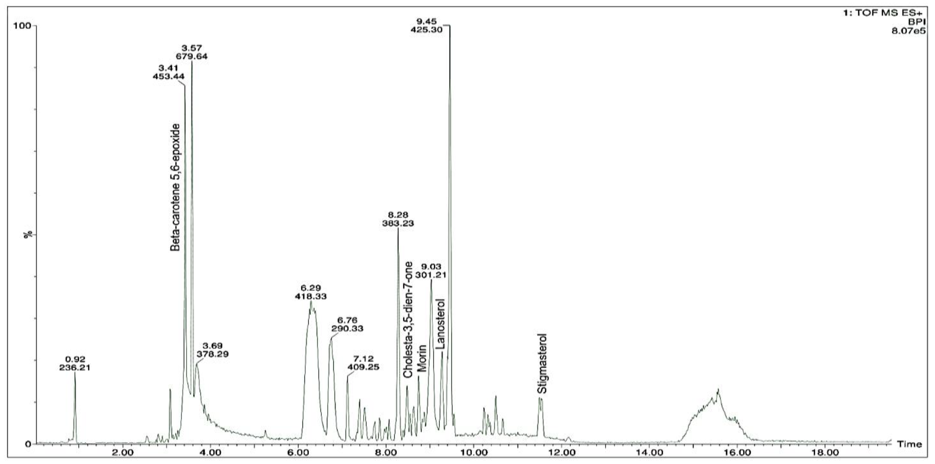
3.3. Phytochemical Profiling of AME Extract by LC-MS

A total of five bioactive molecules were identified and characterized by a correlation of the molecular (precursor) ions and the fragmentation patterns (i.e., product ions) acquired employing LC-MS analysis (Table 3). The LC-MS data were then compared to the reported literature, database, and computer repositories for reference compounds. The bioactive molecules identified in AME extract were terpenoid along with carotenoids, polyphenolic, and fatty acid compounds as shown in Figure 1. The compounds found in the AME extract were known for their pharmacological properties.

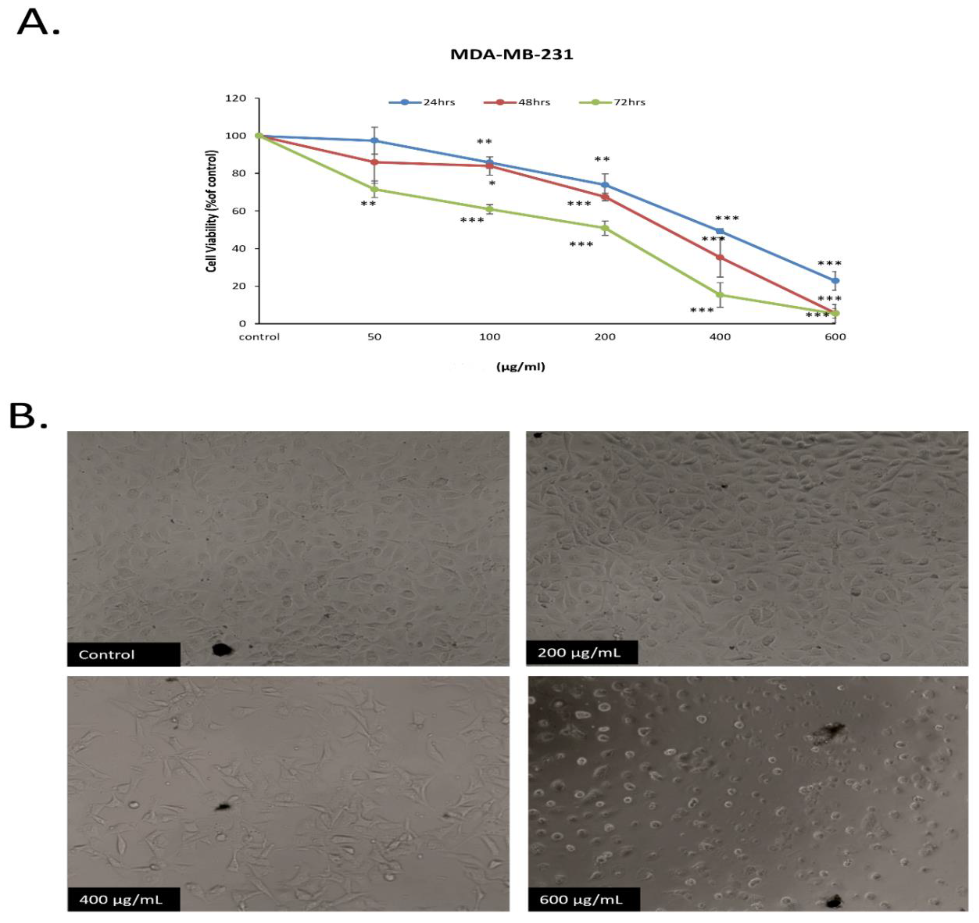
3.4. Anticancer Activity

This part of the study was intended to measure the potential anticancer activity of AME extract. For this, the AME extract was tested in vitro for the inhibition of proliferation of neoplastic cells using human breast cancer cell line. Experimentally, an estimation of time and concentration dependence of the activity of AME was assessed. This was achieved by the incubation of MDA-MB-231 cells to a series of different concentrations of AME (50–600 μg/mL) performed for different time periods (24, 48, and 72 h). Our findings demonstrate that AME extract inhibited the viability of the MDA-MB231 cells in a concentration and time-dependent manner (Figure 2A).
We also monitored cell morphological changes that might be associated with AME extract treatment. The study reflected that the MDA-MB231 cells, when observed under light microscope, show a decrease in the cell count with increase in concentration of AME (Figure 2B).

3.5. Antimicrobial Activity

The antimicrobial potential of the AME extract was studied using Muller Hinton agar as a medium against different pathogen strains. In this investigation, for determining the antimicrobial activity of the AME extract, a sequential dilution technique was employed. Findings showed that the AME extract exhibited poor inhibitory activity on the fungi evaluated, the MIC and MFC of AME extract values against Candida albicans was higher than 500 μg/mL. The MIC and MBC for Staphylococcus aureus was 15.62 and 31.25 μg/mL, likewise, for Escherichia coli the MIC values were recorded to be 125 and 250 μg/mL and for Pseudomonas aeruginosa 62.50 and 125.00 μg/mL respectively as shown in Table 4.

4. Discussion

Microalgae exhibit a wide range of molecules that promote their sustainability and metabolism in an excessively harsh and challenging environment [29]. Research and development of new and unique bioactive components responsible for the production of their synthesis pathway of secondary metabolites have produced a resurgence of interest in the pharmaceutical industry.
Our research is among the few to simultaneously evaluate microalgae extraction, phytochemical screening, and biological activity evaluation such as anti-cancer, antioxidant, and antimicrobial activity.
Polyphenolic compounds are abundant in plants and algae, which could facilitate public health and safety [30]. Polyphenols (e.g., flavonoids) have recently been recognized to be non-toxic antioxidants [31]. Many more findings suggest that adequate nutritional quality of organic phenolics is significantly related to increased life span, substantially reduced chances of acquiring certain chronic illnesses, such as chemopreventive agents [32]. Our observation corresponds to the observation of Haoujar et al., according to whom Nannochloropsis oculata has higher polyphenol content [33] but is lower than reported by Maadane et al. [34]. Polyphenolic constituents found in the microalgae are associated with antioxidant activity and therefore are highly reliant mostly on solvent and algae organisms used during extraction [35].
The significant relationship between polyphenolic constituents and the antioxidant activity of algae is thoroughly reported [36] and as per several studies performed on microalgae, it has been found that phenolics are amongst the antioxidants of chief importance [37]. In the present study, DPPH, H2O2, and ABTS assays were employed for the evaluation of the antioxidant capacity of AME extract. Typically, the DPPH•− scavenging model was used to determine the potential of hydrogen to transfer atoms since the transition of hydrogen atoms is a required mechanism in the DPPH•− scavenging system [38]. DPPH is a free radical having an absorption peak at 517 nm. The purple color of DPPH disappears easily whenever it confronts a proton radical scavenger. ABTS assay is perhaps the best technique for the evaluation of free radical scavenging ability of diverse bioactive molecules/extracts/fractions. The phenolic antioxidant compounds appear to give an electron to ABTS•+ free radical which incapacitates the positive electrical charge present on it and as a result, a stable neutral ABTS molecule is formed [39]. Hydrogen peroxide individually is not all that reactive, although it has a deleterious effect on cells since it leads to the formation of hydroxyl radicals. Therefore, the elimination of both H2O2 and O2 is extremely essential for the protection of antioxidants in cells or nutrition [40].
The results from this study are similar to the findings of the former studies by Millao et al. [41] and Custodio et al. [42] that reported maximum scavenging activity in methanol extract attributed to polyphenols, carotenoids, polyunsaturated fatty acids, etc. [43].
The findings of the study exhibited that the AME extract successfully prohibited the proliferation of both Gram-positive as well as Gram-negative bacteria in comparison to fungi. Our observations correspond with the findings of Kokou et al. [44]. Secondary metabolites such as terpenoids [45], fatty acids [46] and, polyphenolic [47] can damage the plasma membrane of both types of bacteria. Nannochloropsis oculata will serve as a possible future source of novel phytochemical constituents of importance for diagnostic and therapeutic applications [48]. The presence of different antimicrobial compounds in extraction liquid extracts from the organisms studied might just be the cause of the increase in antimicrobial efficacy.
Cancer can be regarded as a multifactorial disease characterized by the unrestrained production and growth of cells in the body [42,49]. Microalgae are rich reservoirs of a variety of biomolecules such as pigments, fats, and fatty acids, carbohydrates, vitamins, and so on [49]. Though microalgae possess a huge array of promising biomolecules, and a variety of compounds are recovered from them with the attribution of many pharmacological activities to them, only a few recent studies have reported the anticancer activity of microalgae [50].
Our findings on MDA-MB-231 cells showed that AME extract exhibits a significant effect on their proliferation which was affected by concentration as well as time. At a concentration of about 200 µg/mL, approximately 25% of cell-viability inhibition was reached after 24 h. After 48 h, more than 50% of cell-viability inhibition was reached at 400 µg/mL concentration. After 72 h, cell-viability inhibition increased dramatically compared with 24 h and 48 h. Thus, the cell-viability inhibition had the least average at 72 h, which means that AME extract shows its potent effect after 72 h. Interestingly, we also noted that MDA-MB231 cells treated with concentrations of 400 and 600 μg/mL experienced a fall in cell count when the concentration increased, besides undergoing structural alterations caused due to loss of architecture of epithelium, echinoid spikes, and cellular rounding visible after 48 h of treatment.
To learn whether apoptosis is related to the anti-proliferative effect and observed morphological changes, further analysis for the apoptosis process was done, using apoptosis assays, as one possible mechanism exerted by AME anti-proliferative effect is required.
Recently, in agreement with what has been found in this study, several reports have been made regarding the in vitro anticancer activity of some microalgae species using different human and mouse cell lines.
Sanjeewa et al. [51] assessed the anti-cancer effect of hexane fractions of Nannochloropsis oculata using various cell lines (A-549, HEP-3B, HCT-116, SW-480, and HL-60). Cells were incubated with different concentrations of hexane fractions for 24 h and the viability of the cells were measured using colorimetric 3-(4,5-dimethylthiazol-2-yl)-2,5-diphenyltetrazolium bromide (MTT) assay. The authors performed a cytotoxicity study and observed that there was a substantial dose-dependent decrease in the viability of cancer cell lines. Microalgae may, therefore, represent an interesting source of biologically active moieties that can be considered as promising anti-cancer agents for clinical prospects.

5. Conclusions

The outcomes of this study suggested there was substantial variance in the in vitro antioxidant, antimicrobial, and anticancer potential of Nannochloropsis oculata. Further research on the phytochemical compounds in the extract to recognize unique and novel constituents and to assess which compound/s is responsible for all these pharmacological activities and to evaluate their effects on in vivo models to enhance the system antioxidant function by defending against different oxidative stress-induced diseases.

Author Contributions

Conceptualization, A.F.W. and J.R.P. and A.M.; Data curation, A.F.W. and Y.A.D.; Formal analysis, A.F.W., Y.A.D., M.S.E., D.A.A.F., A.F. and S.A.R.; Investigation, A.F.W. and Y.A.D.; Methodology, A.F.W. and J.R.P.; Project administration, A.F.W. and P.G.M.R.; Resources A.F.W., Y.A.D., M.S.E. and D.A.A.F.; Software, A.F.W. and S.A.R.; Supervision J.R.P., P.G.M.R. and A.F.W.; Writing—original draft, A.F.W., A.F. and Y.A.D.; Writing—review and editing, A.F.W., A.M., Y.A.D., M.S.E. and D.A.A.F. All authors have read and agreed to the published version of the manuscript.

Funding

This research was funded by RAKMHSU-REC-123-2018-F-P and RSP-2020/190.

Acknowledgments

The authors wish to thank RAK Medical and Health Sciences University, Ras Al Khaimah, United Arab Emirates, for their research support for the RAKMHSU-REC-123-2018-F-P research project. The authors would like to extend their sincere appreciation to the Researchers Supporting Project Number (RSP-2020/190), King Saud University, Riyadh, Saudi Arabia.

Conflicts of Interest

The authors declare that there is no conflict of interest.

References

  1. Wang, H.; Khor, T.O.; Shu, L.; Su, Z.-Y.; Fuentes, F.; Lee, J.-H.; Kong, A.-N.T. Plants vs. Cancer: A Review on Natural Phytochemicals in Preventing and Treating Cancers and Their Druggability. Anti-Cancer Agents Med. Chem. 2012, 12, 1281–1305. [Google Scholar] [CrossRef] [PubMed]
  2. Thomford, N.; Senthebane, D.; Rowe, A.; Munro, D.; Seele, P.; Maroyi, A.; Dzobo, K. Natural Products for Drug Discovery in the 21st Century: Innovations for Novel Drug Discovery. Int. J. Mol. Sci. 2018, 19, 1578. [Google Scholar] [CrossRef] [PubMed] [Green Version]
  3. Montaser, R.; Luesch, H. Marine Natural Products: A New Wave of Drugs? Future Med. Chem. 2011, 3, 1475–1489. [Google Scholar] [CrossRef] [PubMed] [Green Version]
  4. Omar, W.M. Perspectives on the Use of Algae as Biological Indicators for Monitoring and Protecting Aquatic Environments, with Special Reference to Malaysian Freshwater Ecosystems. Trop. Life Sci. Res. 2010, 21, 51–67. [Google Scholar] [PubMed]
  5. Barkia, I.; Saari, N.; Manning, S.R. Microalgae for High-Value Products towards Human Health and Nutrition. Mar. Drugs 2019, 17, 304. [Google Scholar] [CrossRef] [Green Version]
  6. Kent, M.; Welladsen, H.M.; Mangott, A.; Li, Y. Nutritional Evaluation of Australian Microalgae as Potential Human Health Supplements. PLoS ONE 2015, 10, e0118985. [Google Scholar] [CrossRef]
  7. Petersen, L.-E.; Kellermann, M.Y.; Schupp, P.J. Secondary Metabolites of Marine Microbes: From Natural Products Chemistry to Chemical Ecology. In YOUMARES 9—The Oceans: Our Research, Our Future; Springer: Berlin/Heidelberg, Germany, 2019; Volume 1, pp. 159–180. [Google Scholar]
  8. Nethravathy, M.U.; Jitendra, G.M.; Sandeep, N.M.; Ajam, Y.S. Recent Advances in Microalgal Bioactives for Food, Feed, and Healthcare Products: Commercial Potential, Market Space, and Sustainability. Compr. Rev. Food Sci. Food Saf. 2019, 18, 1882–1897. [Google Scholar]
  9. Barbosa-Filho, J.M.; Alencar, A.A.; Nunes, X.P.; Tomaz, A.C.D.A.; Sena-Filho, J.G.; Athayde-Filho, P.F.; Silva, M.S.; Souza, M.F.V.D.; Da-Cunha, E.V.L. Sources of Alpha-, Beta-, Gamma-, Delta- and Epsilon-Carotenes: A Twentieth Century Review. Rev. Bras. Farmacogn. 2008, 18, 135–154. [Google Scholar] [CrossRef] [Green Version]
  10. Matta, C.B.B.D.; Souza, É.T.D.; Queiroz, A.C.D.; Lira, D.P.D.; Araújo, M.V.D.; Cavalcante-Silva, L.H.A.; Miranda, G.E.C.D.; Araújo-Júnior, J.X.D.; Barbosa-Filho, J.M.; Santos, B.V.D.O.; et al. Antinociceptive and Anti-Inflammatory Activity from Algae of the Genus Caulerpa. Mar. Drugs 2011, 9, 307–318. [Google Scholar] [CrossRef] [Green Version]
  11. Alves, C.; Silva, J.; Pinteus, S.; Gaspar, H.; Alpoim, M.C.; Botana, L.M.; Pedrosa, R. From Marine Origin to Therapeutics: The Antitumor Potential of Marine Algae-Derived Compounds. Front. Pharmacol. 2018, 9, 777. [Google Scholar] [CrossRef] [Green Version]
  12. Azamjah, N.; Soltan-Zadeh, Y.; Zayeri, F. Global Trend of Breast Cancer Mortality Rate: A 25-Year Study. Asian Pac. J. Cancer Prev. 2019, 20, 2015–2020. [Google Scholar] [CrossRef] [PubMed]
  13. Mehanna, J.; Haddad, F.G.; Eid, R.; Lambertini, M.; Kourie, H.R. Triple-Negative Breast Cancer: Current Perspective on the Evolving Therapeutic Landscape. Int. J. Women’s Health 2019, 11, 431–437. [Google Scholar] [CrossRef] [PubMed] [Green Version]
  14. Varghese, E.; Samuel, S.; Abotaleb, M.; Cheema, S.; Mamtani, R.; Büsselberg, D. The “Yin and Yang” of Natural Compounds in Anticancer Therapy of Triple-Negative Breast Cancers. Cancers 2018, 10, 346. [Google Scholar] [CrossRef] [PubMed] [Green Version]
  15. Calcabrini, C.; Catanzaro, E.; Bishayee, A.; Turrini, E.; Fimognari, C. Marine Sponge Natural Products with Anticancer Potential: An Updated Review. Mar. Drugs 2017, 15, 310. [Google Scholar] [CrossRef] [Green Version]
  16. Wali, A.F.; Majid, S.; Rasool, S.; Shehada, S.B.; Abdulkareem, S.K.; Firdous, A.; Beigh, S.; Shakeel, S.; Mushtaq, S.; Akbar, I.; et al. Natural Products against Cancer: Review on Phytochemicals from Marine Sources in Preventing Cancer. Saudi Pharm. J. 2019, 27, 767–777. [Google Scholar] [CrossRef]
  17. Ratovitski, E. Anticancer Natural Compounds as Epigenetic Modulators of Gene Expression. Curr. Genom. 2017, 18, 175–205. [Google Scholar] [CrossRef]
  18. Andrade, K.M.; Lauritano, C.; Romano, G.; Ianora, A. Marine Microalgae with Anti-Cancer Properties. Mar. Drugs 2018, 16, 165. [Google Scholar] [CrossRef] [Green Version]
  19. Borowitzka, M.A. Biology of Microalgae. In Microalgae in Health and Disease Prevention; Academic Press: Cambridge, MA, USA, 2018; pp. 23–72. [Google Scholar]
  20. Matos, J.; Cardoso, C.; Bandarra, N.M.; Afonso, C. Microalgae as Healthy Ingredients for Functional Food: A Review. Food Funct. 2017, 8, 2672–2685. [Google Scholar] [CrossRef]
  21. Sharifah, E.N.; Eguchi, M. The phytoplankton Nannochloropsis oculata enhances the ability of Roseobacter clade bacteria to inhibit the growth of fish pathogen Vibrio anguillarum. PLoS ONE 2011, 6, e26756. [Google Scholar] [CrossRef] [Green Version]
  22. Aryal, S.; Baniya, M.K.; Danekhu, K.; Kunwar, P.; Gurung, R.; Koirala, N. Total Phenolic Content, Flavonoid Content and Antioxidant Potential of Wild Vegetables from Western Nepal. Plants 2019, 8, 96. [Google Scholar] [CrossRef] [Green Version]
  23. Shi, P.; Du, W.; Wang, Y.; Teng, X.; Chen, X.; Ye, L. Total Phenolic, Flavonoid Content, and Antioxidant Activity of Bulbs, Leaves, and Flowers Made from Eleutherine Bulbosa (Mill.) Urb. Food Sci. Nutr. 2018, 7, 148–154. [Google Scholar] [CrossRef] [PubMed] [Green Version]
  24. Siddhuraju, P.; Manian, S. The Antioxidant Activity and Free Radical-Scavenging Capacity of Dietary Phenolic Extracts from Horse Gram (Macrotyloma Uniflorum (Lam.) Verdc.) Seeds. Food Chem. 2007, 105, 950–958. [Google Scholar] [CrossRef]
  25. Akbar, M.; Mubashir, H.M.; Adil, F.W.; Mudasir, A.M.; Nida, S.S. Antioxidant potential of methanolic root extract of Mentha arvensis L. From Kashmir region. J. Appl. Pharm. 2014, 4, 50–57. [Google Scholar]
  26. Ramakrishna, J.; Wali, A.F.; Bakhit, F.E.M.; Elawad, A.O.; Ayaabdin, A. In Vitro Antioxidant Activity and Quantitative Elemental Analysis of Adansonia Digitata L. Fruit Using Inductively Coupled Plasma Optical Emission Spectroscopy. Ann. Phytomed. 2019, 8, 127–133. [Google Scholar] [CrossRef]
  27. Lee, K.J.; Oh, Y.C.; Cho, W.K.; Ma, J.Y. Antioxidant and Anti-Inflammatory Activity Determination of One Hundred Kinds of Pure Chemical Compounds Using Offline and Online Screening HPLC Assay. Evid.-Based Complement. Altern. Med. 2015, 2015, 1–13. [Google Scholar] [CrossRef] [Green Version]
  28. National Committee for Clinical Laboratory Standards (NCCLS). Introduces New Editions of Antimicrobial Susceptibility Testing Standards. Clin. Microbiol. Newsl. 1997, 19, 40. [Google Scholar] [CrossRef]
  29. Khan, M.I.; Shin, J.H.; Kim, J.D. The Promising Future of Microalgae: Current Status, Challenges, and Optimization of a Sustainable and Renewable Industry for Biofuels, Feed, and Other Products. Microb. Cell Fact. 2018, 17, 36. [Google Scholar] [CrossRef]
  30. Yan, S.; Asmah, R. Comparison of Total Phenolic Contents and Antioxidant Activities of Turmeric Leaf, Pandan Leaf and Torch Ginger Flower. Int. Food Res. J. 2010, 17, 417–423. [Google Scholar]
  31. Panche, A.N.; Diwan, A.D.; Chandra, S.R. Flavonoids: An Overview. J. Nutr. Sci. 2016, 5, e47. [Google Scholar] [CrossRef] [Green Version]
  32. Farasat, M.; Khavari-Nejad, R.-A.; Nabavi, S.M.B.; Namjooyan, F. Antioxidant Properties of Two Edible Green Seaweeds from Northern Coasts of the Persian Gulf. Jundishapur J. Nat. Pharm. Prod. 2013, 8, 47–52. [Google Scholar] [CrossRef] [Green Version]
  33. Haoujar, I.; Cacciola, F.; Abrini, J.; Mangraviti, D.; Giuffrida, D.; Majdoub, Y.O.E.; Kounnoun, A.; Miceli, N.; Taviano, M.F.; Mondello, L.; et al. The Contribution of Carotenoids, Phenolic Compounds, and Flavonoids to the Antioxidative Properties of Marine Microalgae Isolated from Mediterranean Morocco. Molecules 2019, 24, 4037. [Google Scholar] [CrossRef] [PubMed] [Green Version]
  34. Maadane, A.; Merghoub, N.; Ainane, T.; Arroussi, H.E.; Benhima, R.; Amzazi, S.; Bakri, Y.; Wahby, I. Antioxidant Activity of Some Moroccan Marine Microalgae: Pufa Profiles, Carotenoids and Phenolic Content. J. Biotechnol. 2015, 215, 13–19. [Google Scholar] [CrossRef] [PubMed]
  35. Ebrahimzadeh, M.A.; Khalili, M.; Dehpour, A.A. Antioxidant Activity of Ethyl Acetate and Methanolic Extracts of Two Marine Algae, Nannochloropsis Oculata and Gracilaria Gracilis-An in Vitro Assay. Braz. J. Pharm. 2018, 54, e17280. [Google Scholar] [CrossRef] [Green Version]
  36. Horincar, V.B.; Parfene, P.; Bahrim, G. Evaluation of Bioactive Compounds in Extracts Obtained from Three Romanian Marine Algae Species. Rom. Biotechnol. Lett. 2011, 16, 71–78. [Google Scholar]
  37. Zakaria, N.A.; Ibrahim, D.; Sulaiman, S.F.; Supardy, A. Assessment of Antioxidant Activity, Total Phenolic Content and In vitro Toxicity of Malaysian Red Seaweed, Acanthophora Spicifera. J. Chem. Pharm. 2011, 3, 182–191. [Google Scholar]
  38. Li, X. 2-Phenyl-4,4,5,5-Tetramethylimidazoline-1-Oxyl 3-Oxide (PTIO•) Radical Scavenging: A New and Simple Antioxidant Assay in Vitro. J. Agric. Food Chem. 2017, 65, 6288–6297. [Google Scholar] [CrossRef]
  39. Wang, G.; Li, X.C.; Zeng, H.P. Synthesis, Antioxidation Activity of (E)-9-p-Tolyl-3-[2-(8-hydroxy-quinol-2-yl) vinyl]-carbazole and (E)-9-(p-Anisyl)-3-[2-(8-hydroxy-quinol-2-yl) vinyl]-carbazole and Their Induction Proliferation of Mesenchymal Stem Cells. Acta Chim. Sin. 2009, 67, 974–982. [Google Scholar]
  40. Gülçin, I.; Küfrevioǧlu, Ö.; Oktay, M.; Büyükokuroǧlu, M.E. Antioxidant, Antimicrobial, Antiulcer and Analgesic Activities of Nettle (Urtica Dioica L). J. Ethnopharmacol. 2004, 90, 205–215. [Google Scholar] [CrossRef]
  41. Millao, S.; Uquiche, E. Antioxidant Activity of Supercritical Extracts from Nannochloropsis Gaditana: Correlation with Its Content of Carotenoids and Tocopherols. Supercrit. Fluids 2016, 111, 143–150. [Google Scholar] [CrossRef]
  42. Custódio, L.; Soares, F.; Pereira, H.; Rodrigues, M.J.; Barreira, L.; Rauter, A.P.; Alberício, F.; Varela, J. Botryococcus Braunii and Nannochloropsis Oculata Extracts Inhibit Cholinesterases and Protect Human Dopaminergic SH-SY5Y Cells from H2O2-Induced Cytotoxicity. J. Appl. Phycol. 2014, 27, 839–848. [Google Scholar] [CrossRef]
  43. Kumar, S.S.; Saramma, A.V. In Vitro Antioxidant Activity and Total Phenolic Content of Nannochloropsis salina. Int. J. Pharmacogn. 2018, 10, 160–164. [Google Scholar]
  44. Kokou, F.; Makridis, P.; Kentouri, M.; Divanach, P. Antibacterial Activity in Microalgae Cultures. Aquac. Res. 2011, 43, 1520–1527. [Google Scholar] [CrossRef]
  45. Guimarães, A.C.; Meireles, L.M.; Lemos, M.F.; Guimarães, M.C.C.; Endringer, D.C.; Fronza, M.; Scherer, R. Antibacterial Activity of Terpenes and Terpenoids Present in Essential Oils. Molecules 2019, 24, 2471. [Google Scholar] [CrossRef] [Green Version]
  46. Karimi, E.; Jaafar, H.Z.; Ghasemzadeh, A.; Ebrahimi, M. Fatty Acid Composition, Antioxidant and Antibacterial Properties of the Microwave Aqueous Extract of Three Varieties of Labisia Pumila Benth. Biol. Res. 2015, 48, 9. [Google Scholar] [CrossRef] [Green Version]
  47. Bouarab-Chibane, L.; Forquet, V.; Lantéri, P.; Clément, Y.; Léonard-Akkari, L.; Oulahal, N.; Degraeve, P.; Bordes, C. Antibacterial Properties of Polyphenols: Characterization and QSAR (Quantitative Structure–Activity Relationship) Models. Front. Microbiol. 2019, 10, 829. [Google Scholar] [CrossRef]
  48. Zanella, L.; Vianello, F. Microalgae of the Genus Nannochloropsis: Chemical Composition and Functional Implications for Human Nutrition. J. Funct. Foods 2020, 68, 103919. [Google Scholar] [CrossRef]
  49. Fouad, Y.A.; Aanei, C. Revisiting the Hallmarks of Cancer. Am. J. Cancer Res. 2017, 7, 1016–1036. [Google Scholar]
  50. Dyshlovoy, S.; Honecker, F. Marine Compounds and Cancer: Where Do We Stand? Mar. Drugs 2015, 13, 5657–5665. [Google Scholar] [CrossRef]
  51. Sanjeewa, K.K.A.; Fernando, I.P.S.; Samarakoon, K.W.; Lakmal, H.H.C.; Kim, E.; Kwon, O.N.; Dilshara, M.G.; Lee, J.; Jeon, Y. Anti-inflammatory and anti-cancer activities of sterol rich fraction of cultured marine microalga Nannochloropsis oculata. Algae 2016, 31, 277–287. [Google Scholar] [CrossRef] [Green Version]
Figure 1. LC-MS chromatographic representation of AME extract in positive mode.
Separations 07 00054 g001
Figure 2. Impact of AME extract on morphological changes and viability of human breast cancer cells MDA-MB-231. (A) Cell viability in percentage subsequent to incubation of exponentially proliferating cells with specified concentration level of AME extract and control for 24, 48 and 72 h. (B) After 48 h of incubation, the photomicrograph shows the effect of different concentrations of AME extract on MDA-MB-231 cells. The data display the mean of the test as measured in triplicate. (* p = 0.05, ** p = 0.005 and *** p = 0.0005).
Separations 07 00054 g002
Table 1. Total phenolics content, total flavonoid content and tannins content of AME extract.
ExtractTotal Phenolics
μg QE/g
R2Total Flavonoid
mg GAE/g
R2Tannins
mg GAE/g
R2
AME437 ± 0.950.96453.92 ± 0.830.98947.05 ± 0.430.955
Tests were performed in triplicates and represented as mean ± SD.
Table 2. In vitro antioxidant activity of AME extract.
DPPH
IC50 µg/mL
H2O2
IC50 µg/mL
GAE/g ABTS
IC50 µg/mL
AME52.10 ± 0.85122.84 ± 2.3296.95 ± 1.68
BHA35.91 ± 0.3864.37 ± 1.7645.27 ± 1.53
Values expressed as mean ± standard deviation (n = 3).
Table 3. The major components found in the AME extract based on LC-MS analysis.
Peak No.Compound NameRetention Time
(min)
Molecular Formula
(gmol−1)
Theoretical (m/z)Measured (m/z)Percentage Content
1.Stigmasterol11.49C29H48O412.69413.352.76
2.Unidentified10.50--609.39-
3.Lanosterol9.43C30H50O425.30426.713.98
4.Morin8.85C15H10O7301.29302.231.84
5.Cholesta-3,5-dien-7-one8.27C27H42O382.62383.232.42
6.Unidentified6.75--290.33-
7.Beta-carotene 5,6-epoxide3.41C40H56O552.90454.348.76
8.Unidentified3.07--249.21-
Table 4. Antimicrobial activity of Nannochloropsis oculata extract.
Pathogens StrainsZoIMICMBC/MFC
µg/mL
Staphylococcus aureus10 ± 1.2315.6231.25
Escherichia coli7 ± 1.58125250
Pseudomonas aeruginosa9 ± 1.9662.50125
Candida albicans5 ± 0.82500-
ZoI, zone of inhibition (mm); [-], no activity; MIC, minimum inhibition concentration (mg/mL); MBC, minimum bactericidal concentration (µg/mL); MFC, minimum fungicidal concentration (µg/mL).

