Article

Determination of Lidocaine in Postmortem Whole Blood Samples after Unsuccessful Cardiopulmonary Resuscitation

by
Amvrosios Orfanidis
1,
Nikolaos Raikos
1,
Evdokia Brousa
1,
Eleni Zangelidou
2 and
Orthodoxia Mastrogianni
1,2,*
1
Laboratory of Forensic Medicine and Toxicology, School of Medicine, Aristotle University, 54124 Thessaloniki, Greece
2
Laboratory of Toxicology, Forensic Service of Ministry of Justice, 56334 Thessaloniki, Greece
*
Author to whom correspondence should be addressed.
Submission received: 9 July 2021 / Revised: 30 July 2021 / Accepted: 7 August 2021 / Published: 9 August 2021
(This article belongs to the Section Forensics/Toxins)

Abstract

:
Forensic toxicologists often detect lidocaine in the biological fluids of the deceased, due to cardiopulmonary resuscitation (CPR) attempts prior to death. Here, we describe the development of a rapid, sensitive and robust method for the detection of lidocaine in postmortem whole blood using liquid−liquid extraction (LLE) followed by GC/MS analysis. The method showed a dynamic linear range of 100 to 6000 ng/mL with a linearity expressed by the regression coefficient (R2) and a value of 0.9947. The quantitation limit (LOQ) was found to be 0.03 ng/mL and the detection limit (LOD) 0.01 ng/mL. Recovery accuracy and repeatability were satisfactory. Finally, the method was applied to 23 real whole blood samples from cases where CPR was attempted. Blood concentrations ranged from 0.21–0.96 μg/mL.

1. Introduction

Lidocaine is a drug commonly used as a local anesthetic and also as a first class antiarrhythmic reagent [1], when given by intravenous injection. Its application as a first line emergency drug is no longer recommended and it has been replaced by amiodarone [2].
In forensic toxicology, lidocaine is frequently detected in the biological fluids of the deceased; circumstances suggest that CPR (cardiopulmonary resuscitation) was attempted prior to death and lidocaine-containing lubricant gel during bladder catheterization was used. Absorption of lidocaine into the blood [3,4] and tissues [5,6,7,8] may be observed whilst it can be further metabolized to monoethylglycinexylidide (MEGX), an active metabolite which has 83% of the antiarrhythmic activity and 129% of the convulsant activity of lidocaine [9] (Figure 1). MEGX also contributes significantly to the toxic effects of the parent drug. Metabolism occurs via N-dealkylation, hydroxylation, amide hydrolysis and glucuronide formation mainly in the liver, whilst only 3% of the parent drug is eliminated unchanged in urine. As stated by Benowitz et al. [10]: “Pharmacokinetic studies in man show wide variability in drug disposition between patients, even when cardiac and hepatic status is considered, making specific dosing recommendations a problem”, thus the development of a method for the determination of lidocaine in the biological fluids of the deceased is crucial for the accurate interpretation of the toxicological results.
Previous studies have shown that postmortem lidocaine concentrations, after unsuccessful CPR, in whole blood and urine lie between the subtherapeutic and therapeutic range [11]. In literature, there are analytical methods for the determination of lidocaine in blood, urine and tissues with GC/MS, most of these studies all have one common feature, they require evaporation and the majority of them use big amounts of organic solvent. Some of them use solid phase extraction (SPE) [12,13] and other use liquid−liquid extraction (LLE) [6,9,14]. To our best knowledge, almost all of the methods have either an evaporation step or a complex sample preparation procedure, where additional time is needed and the cost of the analysis also increases. Therefore, the aim of this study is to develop and validate a new simple, rapid, reliable and accurate method for the determination of lidocaine in human blood by GC/MS, in order to ensure minimum volume of extraction solvent, ease of extraction, low cost analysis and also to avoid instrumentation, such as LC MS/MS or TOF/MS, which are not available in every forensic toxicology laboratory, require highly trained personnel, and the running and maintenance costs of which are extremely high. Full validation was carried out, including selectivity, lower limit of quantification (LLOQ), limit of detection (LOD), precision, accuracy, carry over effect and short term stability, using internationally accepted guidelines for forensic toxicology methods. Method validation was performed according to the guidelines of Scientific Working Group for Forensic Toxicology (SWGTOX) Standard Practices for Method Validation in Forensic Toxicology [15]. A simple liquid−liquid extraction method using butyl acetate as the extracting solvent was used for extracting lidocaine from postmortem blood and the method was successfully applied to real human blood samples, in an effort to try discriminate if the lidocaine was used as a coating to the endotracheal tube in order to facilitate intubation by improving sliding properties or as an antiarrhythmic reagent intravenously. For forensic toxicologists is crucial to know the site of administration and also the dose of the local anesthetic delivered, since they both constitute independent risk factors for systemic toxicity.

2. Materials and Methods

2.1. Chemicals and Reagents

Methanol and butyl acetate HPLC grade were obtained from Sigma-Aldrich (St. Louis, MO, USA). For pH alkalization, a saturated solution (pH = 12) of potassium carbonate (K2CO3) from Panreac (Barcelona, Spain) was used. Standards of lidocaine and cocaine-D3 were purchased from Lipomed (Arlesheim, Switzerland). Standard stock solutions (100 μg/mL in methanol) were stored in a freezer at −20 °C.

2.2. Apparatus and Conditions

GC/MS analysis was performed with a gas chromatograph Agilent Technologies 7890A with an MS 5975C inrtXL, EI/CI MSD with Triple-Axis detector. The mass spectrometer was operated with electron energy of 70 eV in the electron impact (EI) mode. An Agilent J&W HP-5MS fused-silica capillary column (30 x 0.25 mm i.d., 0.25 film thickness) was used for GC separation. GC oven temperature program was the following; initial temperature: 120 °C for 1 min, final temperature: 300 °C with a rate of 15 °C/min, total run time: 33 min. The acquisition mode was in full scan from 40 to 500 amu. Ion m/z 86 was selected for the quantification of lidocaine and m/z 85 for cocaine-D3 (Figure 2).

2.3. Calibrator and Control Preparation

Calibration curves were prepared at concentrations ranging from 100 to 6000 ng/mL. A minimum of 6 calibrators were used to construct the calibration curve. Controls (QC) were prepared at concentrations of 250 ng/mL (LQC), 1250 ng/mL (MQC) and 5000 ng/mL (HQC) in order to verify the accuracy of the method. These stocks solutions were prepared in methanol. For calibration standards and QCs, 50 μL of the working standards were added to 1 mL of whole blood, to yield the calibrator concentrations as given in Table 1. All solutions and QC samples were stored at −20 °C.

2.4. Sample Pretreatment and Extraction

Calibrators, controls and real samples were prepared by the following procedure:
Our laboratory identified 23 lidocaine-positive fatalities from cases that had received cardiopulmonary resuscitation (CPR) prior to death. For the calibration curves and the quality controls, postmortem whole blood samples free of drugs were used. Blood samples were stored at −20 °C in tubes containing 1.00% w/v sodium fluoride/potassium oxalate prior to analysis.
To 1 mL of whole blood, 50 μL of internal standard (5 μg/mL) was added. Subsequently, liquid−liquid extraction was applied by adding 250 μL of butyl acetate and additionally 500 μL of K2CO3 to adjust the pH to 12. Samples were then vortexed for 10 min and centrifuged at 4 °C at 12,000 rpm for 10 min. Finally, 1 μL of the supernatant was subjected to GC/MS analysis.

2.5. Method Validation

Validation of the method was performed with respect to international guidelines. Evaluated parameters were as follows: recovery, matrix effect, carry over, linearity, sensitivity, selectivity, stability, accuracy, repeatability, limit of detection and limit of quantification.

2.5.1. Recovery, Matrix effect and Carry over

The percentage recovery of the analytes was assessed by the ratio of responses of spiked blood prior to and after extraction at three concentration levels (LQC, MQC, HQC). In addition, in order to determine whether endogenous matrix substances could affect the signal of the study compound, matrix effect was calculated. This was calculated from the ratio of the signal of the compound at 250, 1250 and 5000 ng/mL in a drug-free blood extract to the signal of the respective methanolic solution of the compounds at the same concentration (250 ng/mL (LQC), 1250 ng/mL (MQC), 5000 (HQC) ng/mL)). In addition, for the carry over study, a drug free sample was injected into the system immediately after the analysis of a high concentration (6000 ng/mL) sample with lidocaine.

2.5.2. Linearity, Sensitivity and Selectivity

Calibration curves were constructed with six concentration levels as described above by three replicate analyses of each standard and expressed by the coefficient of regression (R2). Regression lines were assessed based on peak area ratios of lidocaine to that of the internal standard. In addition, to determine the sensitivity of the method, a blank blood sample was analyzed in order to study the selectivity of the method. The LOD was evaluated for the analyte as the concentration where a signal-to-noise ratio greater than 3:1 was fulfilled. The limit of quantification (LOQ) had to fulfill the requirement of a signal-to-noise ratio of greater than 10:1.

2.5.3. Stability

The stability study of the substances in blood extracts (control samples) was performed in a median concentration (2000 ng/mL). Short-term stability was evaluated in samples that remained in the autosampler for up to 48 h as well as samples kept in the freezer for 72 h.

2.5.4. Accuracy and Repeatability

For the assessment of intraday and day-to-day repeatability, five identical QC samples including lidocaine in low, medium and high concentrations were analyzed. Intraday accuracy was estimated at three different concentrations (LQC, MQC, HQC) and expressed as a percentage of the actual value.

3. Results

3.1. Recovery, Matrix Effect, Carry Over

The percentage recovery of the analytes was calculated from the ratio of the substances’ responses when a drug-free blood sample was fortified before or after the extraction process at three concentration levels (LQC, MQC, HQC) ranging from 103% to 112%. These results suggest that the extraction efficiency of the analytes was satisfactory. The results of the recovery and the matrix effect are given in Table 1.
In addition, the results showed that the matrix did not significantly affect the signal intensity. The differentiation due to the presence of the substrate was found to be less than 14% in low, medium and high control samples. This effect was less than 1% in high concentration blood extracts (6000 ng/mL).

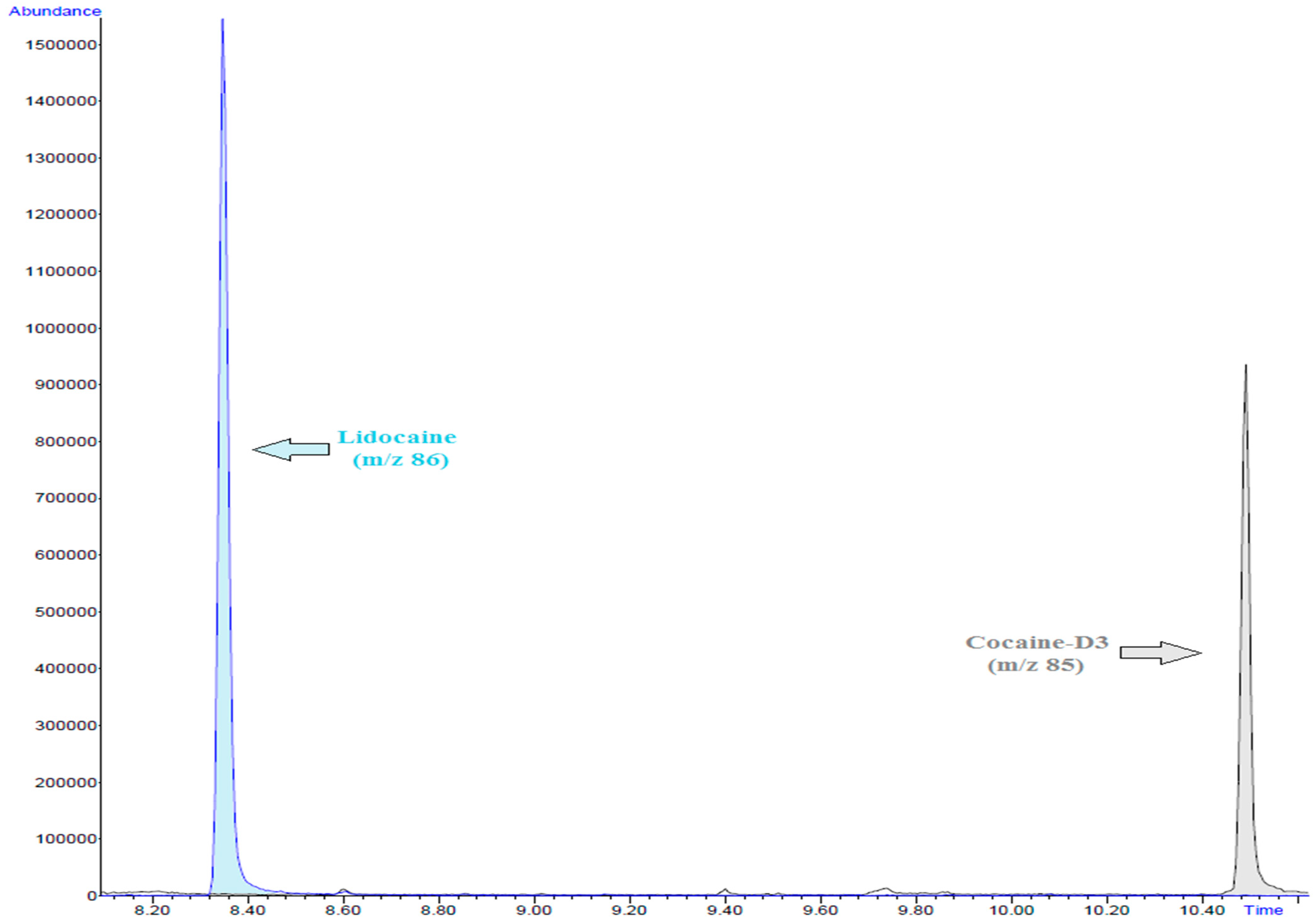
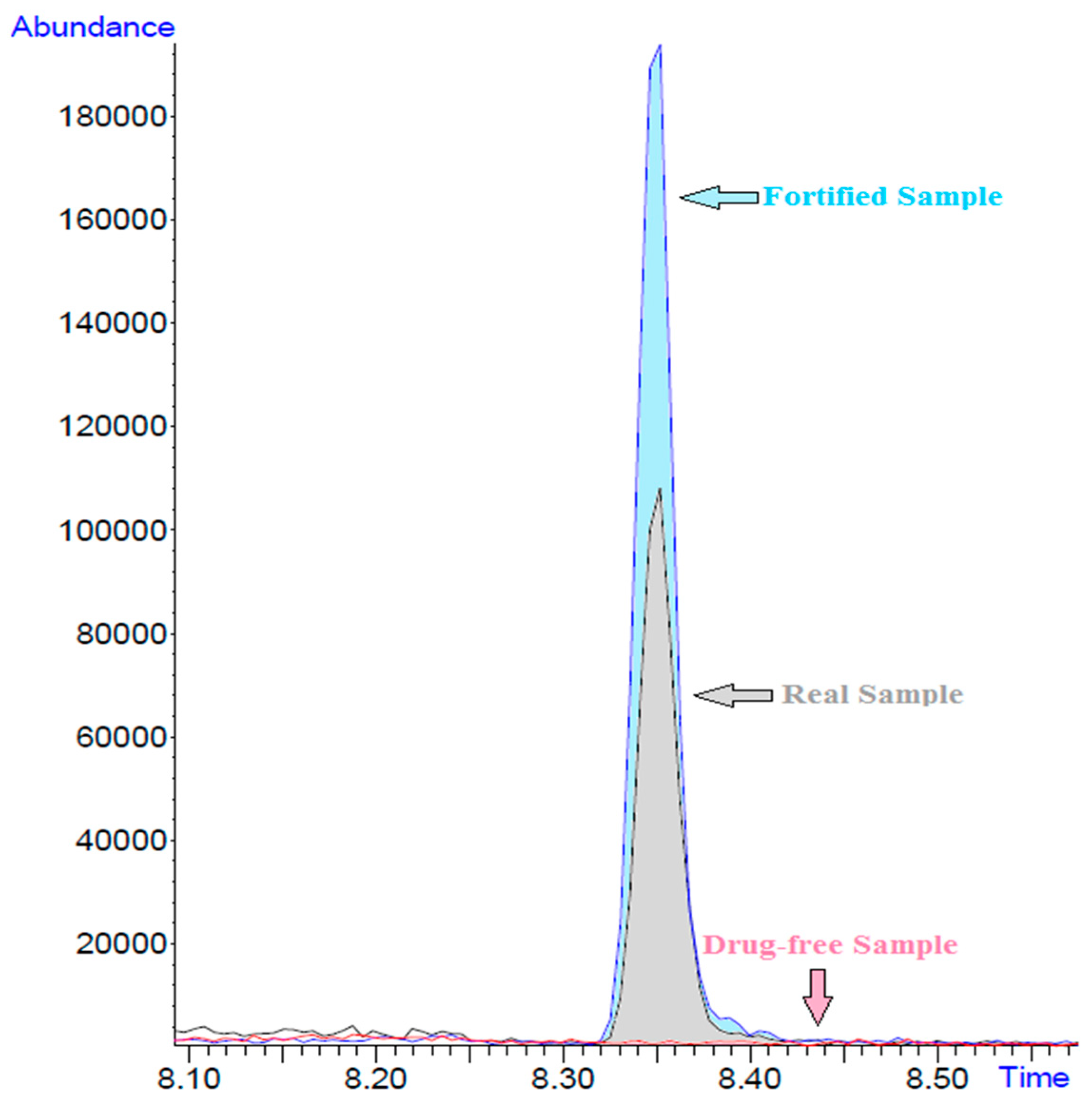
3.2. Linearity, Sensitivity and Selectivity

Calibration curves were constructed at six different concentration levels, as described above, and were analyzed in triplicate. Regression lines were evaluated based on the peak area ratios of lidocaine to those of the internal standard. The method showed a dynamic linear range of 100 to 6000 ng/mL with a linearity expressed by the regression coefficient (R2) and a value of 0.9947. The quantitation limit (LOQ) was found to be 0.03 ng/mL and the detection limit (LOD) 0.01 ng/mL.
The selectivity of the method was satisfactory. In addition, there was no interference from endogenous substances nor substances coeluting at the same time. Analysis of the drug-free blood samples from thirty incidents also showed that there is no interference within the retention time span of lidocaine.

3.3. Stability

Concerning short-term stability, working solutions and extracted blood samples at a concentration of 2000 ng/mL were found stable for at least 48 h at autosampler temperature (4 °C) and at −20 °C for 72 h. Short-term stability was assessed as sufficient.

3.4. Accuracy and Repeatability

The accuracy was calculated from the calibration curve at three different concentrations (MQC, LQC, HQC), expressed as a percentage of the ratio of the found concentration to the theoretical value. The results showed that the accuracy was within acceptable limits and ranged between 99% and 105%.
Finally, the method showed good repeatability. In the intraday, expressed as RSD%, it was much below 15%, with the maximum value being 10.4%. Repeatability from day to day was again below 15%. The highest RSD was 5.9%, which showed very satisfactory repeatability. All the above data are in detail in Table 1.

3.5. Application in real samples

After the validation of the method, it was applied to the analysis of twenty-three real cases (postmortem samples) which belonged to patients receiving CPR prior to death. Age, gender and cause of death of each case are presented in Table 2. The concentrations found in each case are included at Table 3 (S.D: 0.25, median: 0.32 μg/mL, mean: 0.28 μg/mL). Representative chromatograms of real, fortified and drug free samples are presented in Figure 3.

4. Discussion

The aim of the present study was to develop a method for the detection of lidocaine in postmortem blood samples, because it may be evidence that reanimation was attempted before death, thus helping the forensic toxicologists to interpret the results with accuracy.
Previously, Wunder et al. [11] have reported that the lidocaine concentration in postmortem cardiac whole blood samples, where antemortem CPR had been performed, ranged between 0.07–1.07 μg/mL and that these levels were at the same order of magnitude as determined in antemortem samples after one hour of controlled exposure. Our results were in accordance with the previously described studies as the concentrations were found to be in the range of 0.21–0.96 μg/mL. To our knowledge, this is the first study where such a large number of postmortem samples were analyzed in an attempt to identify the site and route of administration of lidocaine. To reach this goal, a rapid method was fully validated according to international guidelines. All samples were collected from peripheral vessels (femoral vein) in cases where the heart never resumed beating in spite of resuscitation attempts. Detection of lidocaine in all of these samples led to the assumption that artificial circulation was achieved due to cardiac massage and so tracheal lidocaine could be absorbed and distributed, even though normal circulation was never restored. The low concentrations of lidocaine found in all samples may imply that lidocaine was used for reanimation and not as an antiarrhythmic drug.
Another parameter that should be further investigated is that lidocaine has potential for postmortem redistribution (PMR) [16,17,18]. A large number of studies have been published in recent years suggesting that lidocaine may exhibit PMR. The time intervals between time of death and autopsy were unknown to us and thus this factor did not constitute a part of this study.

5. Conclusions

Our aim was to develop and validate a rapid, sensitive and reliable method for the detection of lidocaine and to finally apply it to postmortem whole blood samples from patients receiving CPR prior to death. Lidocaine was successfully detected and quantified in all blood samples. Finally, the results from this work are in accordance with previous published studies that have demonstrated that lidocaine is absorbed in the trachea from an endotracheal tube coated with a lidocaine-containing gel [11].

Author Contributions

Conceptualization, E.Z., O.M.; methodology, A.O., O.M.; validation, A.O., E.B., O.M.; investigation, E.Z.; writing—original draft preparation, A.O., O.M.; writing—review and editing, N.R., O.M.; visualization, A.O.; supervision, O.M. All authors have read and agreed to the published version of the manuscript.

Funding

This research received no external funding.

Conflicts of Interest

The authors declare no conflict of interest.

References

  1. Schwartz, M.L.; Meyer, M.B.; Covino, B.G.; Narang, R.M.; Sethi, V.; Schwartz, A.J.; Kamp, P. Antiarrhythmic Effectiveness of Intramus-cular Lidocaine: Influence of Different Injection Sites. J. Clin. Pharmacol. 1974, 14, 77–83. [Google Scholar] [CrossRef] [PubMed]
  2. Paul, D.; Dan, C.; Schwartz, B.; Cooper, R.; Gelaznikas, R.; Barr, A. Amiodarone as Compared with Lidocaine for Shock-Resistant Ventricular Fibrillation. N. Engl. J. Med. 2002, 21, 884–890. [Google Scholar]
  3. Moriya, F.; Hashimoto, Y. Postmortem diffusion of tracheal lidocaine into heart blood following intubation for cardiopul-monary resuscitation. J. Forensic Sci. 1997, 42, 296–299. [Google Scholar] [CrossRef] [PubMed]
  4. Moriya, F.; Hashimoto, Y. Absorption of Intubation-Related Lidocaine from the Trachea during Prolonged Cardiopulmonary Resuscitation. J. Forensic Sci. 1998, 43, 718–722. [Google Scholar] [CrossRef] [PubMed]
  5. Moriya, F.; Hashimoto, Y. Determining the state of the deceased during cardiopulmonary resuscitation from tissue distri-bution patterns of intubation-related lidocaine. J. Forensic Sci. 2000, 45, 846–849. [Google Scholar] [CrossRef] [PubMed]
  6. Moriya, F.; Hashimoto, Y. Tissue distribution of lidocaine in critical care patients after intubation. Forensic Sci. Int. 2003, 137, 183–187. [Google Scholar] [CrossRef]
  7. Moriya, F.; Hashimoto, Y. Tissue Distribution of Intubation-Related Lidocaine in Brain-Dead Patients. Am. J. Forensic Med. Pathol. 2004, 25, 351–354. [Google Scholar] [CrossRef] [PubMed]
  8. Moriya, F.; Hashimoto, Y.; Nakanishi, A. Distributions of ethanol and intubation-related lidocaine in the brain of a trauma patient who was brain dead for about 5 days. Leg. Med. 2003, 5, S118–S122. [Google Scholar] [CrossRef]
  9. Moriya, F.; Hashimoto, Y. Concentrations of monoethylglycinexylidide in body fluids of deceased patients after use of li-docaine for endotracheal intubation. Leg. Med. 2000, 2, 31–35. [Google Scholar] [CrossRef]
  10. Benowitz, N.L.; Meister, W. Clinical pharmacokinetics of lignocaine. Clin. Pharmacokinet. 1978, 3, 177–201. [Google Scholar] [CrossRef] [PubMed]
  11. Wunder, C.; Meier, J.; Reyher, C.; Könitz, V.; Paulke, A.; Zacharowski, K.; Toennes, S.W. Use of lidocaine in endotracheal intubation. Blood and urine concentrations in patients and deceased after unsuccessful resuscitation. Forensic Sci. Int. 2014, 244, 259–262. [Google Scholar] [CrossRef] [PubMed]
  12. Barbaro, A. Teatino About an unusual case: GC-MS detection of lidocaine. Forensic Sci. Int. 2004, 146, S93–S94. [Google Scholar]
  13. Centini, F.; Fiore, C.; Riezzo, I.; Rossi, G.; Fineschi, V. Suicide due to oral ingestion of lidocaine: A case report and review of the literature. Forensic Sci. Int. 2007, 171, 57–62. [Google Scholar] [CrossRef] [PubMed]
  14. Zhang, N.; Sun, J.; Wei, Z.; He, W.; Jin, G.; Zhang, X.; Gao, P.; Wang, L.; Yun, K. Comparison of the Concentrations of Lido-caine in Different Body Fluids/Tissues after Subarachnoid Space and Intravenous Administration of a Lethal Dose of Lido-caine. J. Forensic. Sci. Med. 2015, 1, 48–53. [Google Scholar]
  15. Scientific Working Group for Forensic Toxicology (SWGTOX) Standard Practices for Method Validation in Forensic Toxicology. J. Anal. Toxicol. 2013, 37, 452–474. [CrossRef] [PubMed]
  16. Wei, Z.; Yu, J.; Zhang, C.; Jia, J.; Cao, J.; Wang, Y.; Gao, C.; Wang, Y.; Yun, K. Postmortem Redistribution of Lidocaine after Epidural Anesthesia Accidental Death. J. Forensic Res. 2017, 8, 2. [Google Scholar] [CrossRef]
  17. Zhang, C.; Liang, B.; Wang, C.; Wei, Z.; Yun, K. Postmortem redistribution of lidocaine after epidural injection in beagle dogs. Int. J. Clin. Exp. Med. 2015, 8, 18500–18505. [Google Scholar] [PubMed]
  18. Han, E.; Kim, E.; Hong, H.; Jeong, S.; Kim, J.; In, S.; Chung, H.; Lee, S. Evaluation of postmortem redistribution phenomena for commonly encountered drugs. Forensic Sci. Int. 2012, 219, 265–271. [Google Scholar] [CrossRef] [PubMed]
Figure 1. (a) Chemical structure of lidocaine; (b) Chemical structure of MEGX.
Separations 08 00117 g001
Figure 2. Extracted ion chromatograms for lidocaine and cocaine-D3.
Separations 08 00117 g002
Figure 3. Overlayed extracted ion chromatograms of a real, fortified and drug-free sample.
Separations 08 00117 g003
Table 1. Validation parameters evaluated in three different concentrations (LQC, MQC, HQC) of fortified blood sample with lidocaine.
Validation Parameters0.25 μg/mL
(LQC)
1.25 μg/mL
(MQC)
5.00 μg/mL
(HQC)
Recovery %106112103
Matrix Effect %9.712.68.3
Accuracy%10110599
Intraday repeatability (RSD%)7.110.48.3
Day-to-day repeatability (RSD%)5.92.34.2
Table 2. Case histories.
CaseMale (M)/Female (F)AgeWeight (Kg)Cause of Death
1M1770Hemorrhagic shock—traffic accident
2M2780Hemorrhagic shock—gunshot injury
3M2375Hemorrhagic shock—traffic accident
4M4278Respiratory infection—coronary atherosclerosis
5M8485Hypertrophic cardiomyopathy—pulmonary edema
6F4662Myocardial infarction—coronary atherosclerosis
7M4875Myocardial infarction—coronary atherosclerosis
8M5180Aortic dissection
9F39110Cardiac arrest—epilepsy
10M5970Pericarditis—hypertrophic cardiomyopathy
11M3880Myocardial infarction—coronary atherosclerosis
12M4675Respiratory infection—coronary atherosclerosis
13F2758Hemorrhagic shock—traffic accident
14F4960Myocardial infarction—coronary atherosclerosis
15M65110Hemorrhagic shock—gunshot injury
16M6572Myocardial infarction—right coronary artery thrombosis
17M5685Hypertensive heart disease
18M6880Drowning—ischemic heart disease
19M4184Myocardial infarction—coronary atherosclerosis
20M8078Ischemic heart disease—gastric cancer
21F4962Aortic dissection
22M2695Craniocerebral gunshot injury
23M8560Hypertrophic cardiomyopathy
Table 3. Concentration of lidocaine found in the 23 real samples.
Case NoLidocaine (μg/mL)Case NoLidocaine (μg/mL)
10.27140.89
20.96150.38
30.90160.25
40.21170.36
50.36180.27
60.32190.22
70.29200.32
80.58210.37
90.88220.27
100.72230.25
110.30
120.40
130.23
Publisher’s Note: MDPI stays neutral with regard to jurisdictional claims in published maps and institutional affiliations.

