1. Introduction
Traditional medicines composed of plants and their extracts used to cure various infections and ailments are being modified and refined to modern formulations in order to play a significant role in the treatment of various diseases such as diabetes, ischemic heart diseases, atherosclerosis and the initiation of carcinogenesis or liver diseases [
1,
2]. The physiological response imparted by the phytochemicals induces the desired therapeutic action [
3,
4]. The most essential of these bioactive constituents are phenolic compounds, alkaloids, saponins, tannins and flavonoids.
Cucurbits form an important and large fruit crop set, cultivated extensively in subtropical and tropical countries, and contains a terpenoid substance known as Cucurbitacin [
5]. The whole fruit of
Cucumis melo L. (
C. melo or muskmelon) is indicated for chronic eczema and to treat light burns and scrapes [
6].
Dose-dependent cytotoxic activities exhibited by an aqueous fruit extract of
C. melo in human prostate carcinoma PC 3 cell lines is evidence of its anti-cancer property [
7]. High superoxide dismutase activity (SOD) is responsible for the in vitro and in vivo antioxidant and anti-inflammatory properties of their extract [
8]. The in vitro evaluation of the antioxidant properties of
C. melo leaves and fruit extracts reported concentration-dependent scavenging activity. 1,1-diphenyl-2-picrylhydrazyl (DPPH) was employed as the free radical in the evaluation of the reducing substances [
9]. Previous investigations suggested that
C. melo is a potential source of natural antioxidants and can act as a therapeutic agent in preventing oxidative stress-related disorders [
10,
11,
12]. Advanced techniques using supercritical and subcritical CO
2 are also recommended to improve the bioavailability of the active ingredients [
13]. In the present study, the antioxidant activity of a methanolic extract of
C. melo fruit (F1 hybrid) was evaluated using standard procedures.
2. Materials and Methods
2.1. Chemicals
The chemicals and drugs were of pharmacopoeia/AnalaR or HPLC grade as required by the nature of the experiment and extraction. They were purchased from the licensed distributors of the Central Drug House (New Delhi, India), Himedia Labs (Mumbai, India) and Sigma-Aldrich chemicals (Bangalore, India).
2.2. Procurement of the Research Raw Materials
C. melo (family: Cucurbitaceae) fruit (
Figure 1) were purchased from Vadanerkunam (Tindivanam T.K., Villupuram District, Tamilnadu, India) and identified and authenticated by experts of the Department of Botany, St. Berchmans College, Changanacherry, Kottayam-686101, India.
2.3. Fruit Extract Preparation
The fruit of
C. melo were cut into pieces with a stainless-steel knife. The mean dimensions were 1.8 cm diameter × 3.5 to 4.0 cm cylinders [
14] and weighed approximately 100 g (including the seeds). These were dried in an oven at 60 °C. The dried fruit were then comminuted to a coarse powder and then passed through a No.10 sieve.
2.4. Preparation of MECM
The coarse powder was extracted with methanol (absolute) by a hot continuous percolation process. A total of 100 g of the dry powder was packed in a filter paper thimble each time and extracted in a Soxhlet extractor (five cycles/day for three consecutive days). The marc was then further extracted until it was colourless with 95% methanol. The process was repeated with fresh powder and the entire alcoholic extractives were mixed. Excess solvent was recovered by distillation under a reduced pressure and evaporated under a vacuum to form a soft extract, which was then weighed, re-heated (ensuring it was free of alcohol) and again weighed. The yield was calculated as the percentage weight per weight (
w/
w) of powder.
2.5. Phytochemical Screening
The following chemical tests were performed on the methanol extracts of
C. melo (MECM) [
15].
2.5.1. Tests for Carbohydrates
MECM (1 g) was boiled with 10 mL of distilled water for 10 min. It was cooled and filtered. The filtrate was used to conduct the following tests:
Molisch’s test: Filtrate (2 mL) was added with two drops of alcoholic solution of α-naphthol. The mixture was shaken well and 1 mL of concentrated sulphuric acid was added slowly along the sides of the test tube. It was allowed to stand for a few seconds. The colour of the mixture was noted.
Iodine test: Filtrate (2 mL) was added with a few drops of dilute iodine solution and observed for the formation of a blue, orange or red colour.
Fehling’s test: Filtrate (1 mL) was boiled in a water bath with 2 mL of a mixed Fehling’s solution (1 mL each of Fehling’s solutions A and B). The change in the colour of the solution was noted.
Benedict’s test: Filtrate (0.5 mL) was added with 0.5 mL of Benedict’s reagent and heated. The mixture was kept on a boiling water bath for 2 min. The change in the colour of the solution was noted.
Barfoed’s test: Filtrate (1 mL) was added with 1 mL of Barfoed’s reagent. It was heated in a boiling water bath for 2 min. The change in the colour of the solution was noted.
2.5.2. Tests for Glycosides
MECM was hydrolysed (50 mg) by heating with concentrated hydrochloric acid for 2 h on a water bath. It was filtered and, in the hydrolysate obtained, the following tests were conducted:
Borntrager’s test: Filtered hydrolysate (2 mL) was added with 3 mL chloroform and shaken. The chloroform layer was separated and a 10% ammonia solution was added to it. The change in the colour of the solution was noted.
Legal’s test: MECM (50 mg) was dissolved in pyridine. A sodium nitroprusside solution was added and made alkaline by using a 10% sodium hydroxide solution. The change in the colour of the solution was noted.
2.5.3. Tests for Alkaloids
MECM (2 g) was shaken with an ammonia solution for 20 min and then filtered. The mixture was extracted with petroleum ether and the ethereal extract was acidified with dilute HCl and extracted with 10 mL of water. The aqueous extract was again made ammoniacal and re-extracted with petroleum ether. The ethereal extract was evaporated to dryness and the residue was stirred with 10 mL of dilute hydrochloric acid and filtered. The filtrate was tested carefully with various reagents for the detection of alkaloids as follows:
Mayer’s test: Filtrate (2 mL) was taken in a test tube. Two drops of Mayer’s reagent were added along the sides and the change in the colour was noted.
Dragendorff’s test: Filtrate (2 mL) was added with 1 mL of Dragendorff’s reagent and the colour change and formation of any precipitate were noted.
Hager’s test: Filtrate (2 mL) was added with 1 mL of Hager’s reagent and the change in the colour of the solution was noted.
Wagner’s test: Filtrate (2 mL) was added with 1 mL of Wagner’s reagent and the change in the colour of the solution was noted.
2.5.4. Tests for Amino acids
A total of 500 mg of MECM was shaken vigorously with 10 mL distilled water and filtered through a Whatman No.1 filter paper. The filtrate was used to tests for proteins and amino acids as follows:
Biuret test: Filtrate (2 mL) was treated with one drop of a 2% copper sulphate solution. A total of 1 mL of (95%) ethanol was added to the solution followed by an excess of potassium hydroxide pellets and the change in the colour was observed.
Ninhydrin test: Ninhydrin solution of 1% was added (2 drops) to the aqueous filtrate (2 mL). The mixture was heated and the change in the colour was observed.
Million’s test: A few drops of Million’s reagent were added to the filtrate (2 mL) and the colour change was noted.
Xanthoprotein test: The filtrate (3 mL) was added with 1 mL of concentrated nitric acid. The solution was cooled and made alkaline with 10% sodium hydroxide. The change in the colour of the solution was noted.
2.5.5. Tests for Sterols/Steroids
Liebermann’s sterol test: MECM (500 mg) was mixed with 2 mL of glacial acetic acid and one drop of concentrated sulphuric acid was added. The change in the colour of the solution was noted.
Liebermann–Burchard test: MECM (500 mg) was mixed with 5 mL chloroform and a few mL of acetic anhydride was added. The mixture was added with one drop of concentrated sulphuric acid. This was then shaken well and the change in the colour was observed.
Salkowski’s test: MECM (500 mg) was shaken vigorously with 3 mL of chloroform and concentrated sulphuric acid was added through the sides of the test tube. The colour change of the solution was observed.
2.5.6. Tests for Fixed Oils and Fats
Grease spot test: MECM (a small quantity) was pressed between two filter papers.
Saponification test: MECM (a small quantity) was added with a few drops of a 0.5 N alcoholic potassium hydroxide solution along with a drop of phenolphthalein and the mixture was heated on a water bath for 2 h. The solution was observed for the formation of soap or any colour change indicating the neutralisation of potassium hydroxide on saponification.
2.5.7. Tests for Phenolic Compounds and Tannins
Ferric chloride test: MECM (50 mg) was boiled with 5 mL distilled water and filtered. A few drops of a neutral, freshly prepared 5% ferric chloride solution were added to the filtrate and the colour change was noted (brownish green or blue-black colouration indicated the presence of tannins and phenolic compounds).
Test for phlobatannins: MECM (1 g) was boiled with 1% HCl in a boiling tube and observed for the deposition of a red precipitate.
Lead acetate test: MECM (50 mg) was boiled with distilled water and filtered. The filtrate was added with 3 mL of a 10% lead acetate solution. The formation of any precipitate was observed.
Gelatine test: MECM (50 mg) was boiled with distilled water and 2 mL of a 1% solution of gelatine was added containing 10% sodium chloride. The solution was observed for the formation of a precipitate.
2.5.8. Tests for Terpenes/Terpenoids
2.5.9. Tests for Saponins
Foam test: MECM (50 mg) was dissolved in distilled water and was diluted to 20 mL. The suspension was shaken vigorously in a graduated cylinder for 15 min. It was observed for the formation of foam.
MECM (1 g) was boiled with 20 mL distilled water for 5 min and then cooled and filtered. The filtrate (10 mL) was shaken vigorously after adding 5 mL of distilled water. The frothing solution was mixed with a few drops of arachis oil and again shaken vigorously and observed for the formation of an emulsion.
2.5.10. Tests for Gums and Mucilage
Precipitation test: MECM (100 mg) was shaken vigorously with 10 mL of distilled water and 25 mL of absolute alcohol was then added with constant stirring.
Ruthenium red test: MECM (50 mg) was allowed to swell in water and then a few drops of Ruthenium red were added.
2.5.11. Tests for Flavones and Flavonoids
Aqueous sodium hydroxide: An MECM solution of 2 mL was added with an aqueous solution of sodium hydroxide and mixed well. The change in the colour was observed.
Concentrated sulphuric acid: An MECM solution (50 mg dissolved in 10 mL of water) was added with a concentrated sulphuric acid solution.
Shinoda test: MECM (50 mg) was dissolved in alcohol. A few magnesium turnings and concentrated hydrochloric acid (dropwise) were added and the change in the colour was noted.
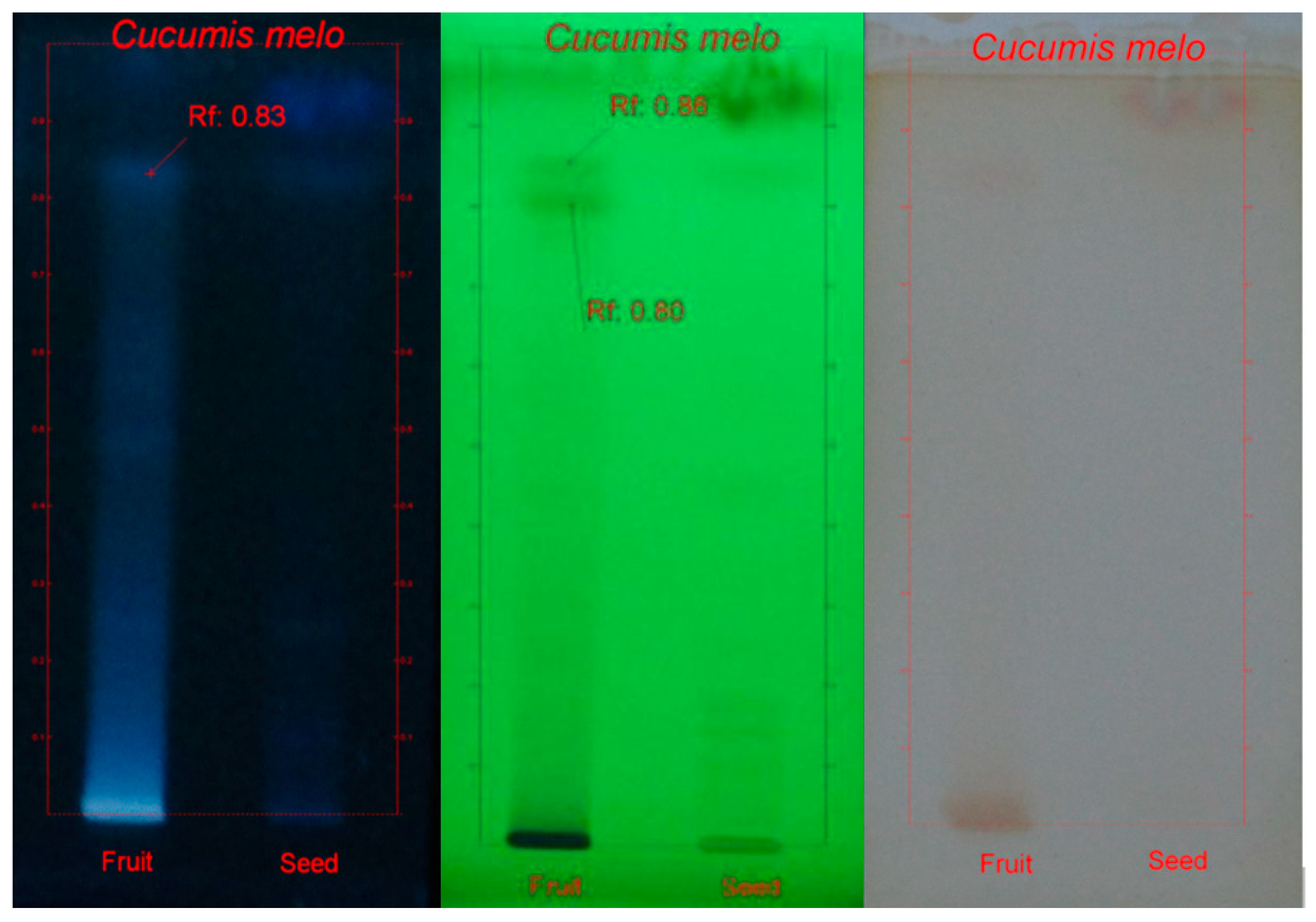
2.6. HPTLC Analysis
HPTLC was used for the detection of the phenolic compounds. HPTLC is a highly efficient, reliable and cost-efficient separation technique that is ideally applicable for the analysis of herbal drugs as well as botanicals. A Camag HPTLC instrument with a Linomat V automatic spotter fitted with a 100 μL syringe connected to a nitrogen cylinder and a Scanner-III were used. The developing chamber used was a twin trough and the viewing cabinet was equipped with dual-wavelength UV lamps (Camag, Muttenz, Switzerland). The precoated HPTLC plates used were aluminium backed with silica gel 60 F254 and the thickness was 0.2 mm. The HPTLC plates were cleaned by predevelopment with methanol before the analysis and activated at 110 °C for 5 min for the removal of solvents. Specific mobile phases were used for the separation of a particular group of phytochemicals.
Sample Application
A total of 10 μL of the sample was spotted using Linomat V on a precoated TLC plate as a narrow bandwidth (8 mm) 10 mm from the bottom and 15 mm from left and right edges of the plates. The samples were applied under a continuous dry stream of nitrogen gas.
THF (toluene, formic acid and water with a ratio of 16:8:2:1) was used as mobile phase for the development of the plates spotted with the samples for the detection of the phenolic compounds. The method of development was linear ascending and it was carried out in a 10 × 10 cm twin trough glass chamber that was equilibrated with the mobile phase. It was saturated for 20 min at 25 ± 2 °C with a relative humidity of 60 ± 5%. The volume of the mobile phase used for the development was 10 mL (5 mL in a trough containing the plate and 5 mL in another trough). It was allowed to migrate up to a distance of 85 mm from the point of the sample application. The TLC plate was dried after the development and the chromatogram was viewed in a UV chamber at 254 nm and 366 nm to visualise the phenolic compounds. Fast blue salt B was used to detect the phenolic compounds.
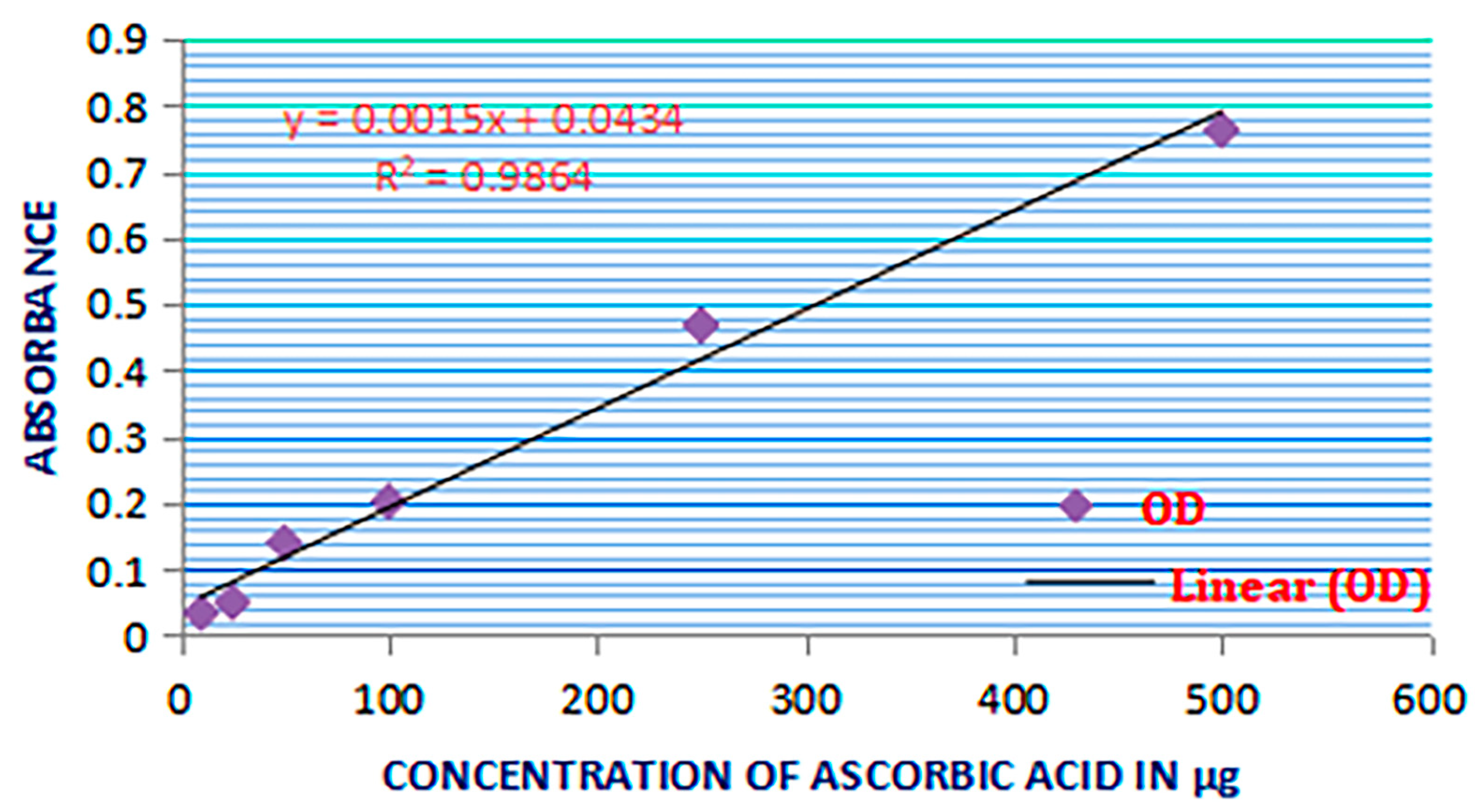
2.7. Total Antioxidant Activity Estimation
Test solutions containing 50, 100 and 200 µg/mL of MECM in methanol were prepared and 0.3 mL of the solution was mixed with a reagent solution (3 mL) containing 0.6 mL H2SO4, 28 mM sodium phosphate and 4 mM ammonium molybdate. The tubes containing the reaction solutions were incubated at 95 °C for 90 min and then cooled to room temperature. The absorbance of the resulting solution was measured at 695 nm against a blank. The solution of methanol (0.3 mL) was the control
The antioxidant activity was expressed as the number of grams equivalent to ascorbic acid. The antioxidant activity values were portrayed in standard graphs plotted with an OD of the standard against the various concentrations of ascorbic acid (10, 25, 50, 100, 250 and 500 µg/mL) treated similarly.
2.8. DPPH Radical Scavenging Assay
The free radical scavenging property of the extracts was estimated by a DPPH radical scavenging assay [
16,
17,
18,
19]. The hydrogen atom-donating capability of the plant extractives was determined by the decolourisation of the methanol solution of the DPPH (2,2-diphenyl-1-picrylhydrasyl) that produces a violet/purple colour in the methanol solution and fades to shades of yellow in the presence of antioxidants. A solution of 0.1 mM DPPH in methanol was prepared and 2.4 mL of this solution was mixed with 1.6 mL of the methanol extract at different concentrations (12.5–150 μg/mL). The reaction mixture was vortexed thoroughly and left in the dark at RT for 30 min. The absorbance of the mixture was measured spectrophotometrically at 517 nm. The percentage DPPH radical scavenging activity was calculated by the following equation:
where A
0 was the absorbance of the control and A
1 was the absorbance of the extractives/standard. The % of inhibition was then plotted against the concentration and, from the graph, the IC
50 was calculated. The experiment was repeated three times at each concentration [
20,
21,
22,
23,
24].
2.9. Hydroxyl Radical Scavenging Activity
The assay principle was based on the competition estimation between the MECM and 2-deoxyribose for the hydroxyl radical that was formed by the Fenton’s reaction [
24,
25]. The underlying mechanism is the oxidation of ferrous ion to ferric ion by hydrogen peroxide and hydroxyl free radicals and hydroxide ions are also formed. The Fe
3+ form is reduced back to the Fe
2+ form by reacting with another molecule of H
2O
2, forming a superoxide radical and an H
+. This reaction is essential in biological systems because transition metals such as iron and copper can donate or accept free electrons through various reactions taking place within the cells and can generate free radicals. The hydroxyl ion formed in the Fenton’s reaction can react with barbituric acid and convert it into alloxan [
26].
2.10. Nitric Oxide Generation and the Assay of Nitric Oxide Scavenging
Nitric oxide (NO) was generated from sodium nitroprusside and measured by the Greiss reaction as described previously. Sodium nitroprusside in an aqueous solution at a physiological pH spontaneously generates NO, which interacts with oxygen to produce nitrite ions that can be estimated by the Greiss reagent [
27,
28]. Scavengers of NO compete with oxygen, leading to a reduced production of NO. Sodium nitroprusside (5 mM) in phosphate-buffered saline was mixed with different concentrations of various plant extracts dissolved in suitable solvent systems and incubated at 258 °C for 150 min and reacted with the Greiss reagent (1% sulphanilamide, 2% H
3PO
4 and 0.1% naphthylethylenediamine dihydrochloride). The absorbance of the chromophore formed during the diazotisation of the nitrite with sulphanilamide and the subsequent coupling was noted.
2.11. Evaluation of the Reducing Power Activity
Concentrations of 50, 100 and 200 µg/mL of MECM and standard ascorbic acid of 50, 100 and 200 µg/mL each were mixed with 2.5 mL of phosphate buffer (pH 6.6). To the resultant solution, 1% potassium ferricyanide (2.5 mL) was added and boiled for 20 min at 50 °C. To that mixture, TCA (2.5 mL) was added and centrifuged for 10 min at 2000 rpm. The supernatant was collected and distilled water (1 mL) and 0.1% ferric chloride (250 µL) were added. The absorbance of the solution was then measured at 700 nm. The reducing power activity was indicated by the increase in the optical density [
29,
30].
3. Results
3.1. Qualitative Screening Tests of the Methanolic Extract of Muskmelon
The qualitative analysis of the methanolic extract of
C. melo fruit showed the presence of various phytochemical constituents such as carbohydrates, alkaloids, sterols, phenolic compounds, terpenes and flavonoids (
Table 1). The Rf value obtained from HPTLC analysis of the MECM for phenolic compounds were 0.80, 0.83 and 0.86 (
Figure 2).
3.2. Estimation of the Total Antioxidant Activity
The antioxidant activity of MECM in concentrations of 50, 100 and 200 µg were equated to the antioxidant activity of 3.3 ± 0.1732, 6.867 ± 0.5457 and 13.63 ± 0.8295 µg of ascorbic acid, respectively (
Figure 3).
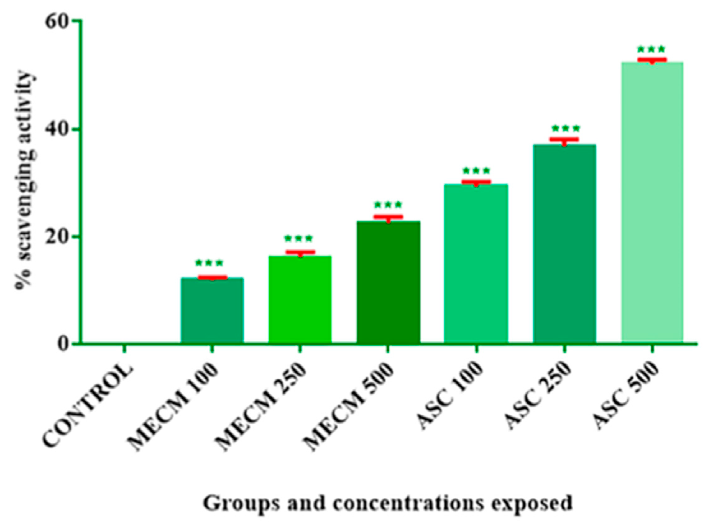
3.3. DPPH Scavenging Activity
DPPH, 2,2-Diphenyl-1-picrylhydrasyl, is a dark-coloured compound composed of stable free radical molecules. The purple colour of DPPH decays in the presence of antioxidants. The change in absorbance at 517 nm in the presence of antioxidants can be equated with the antioxidant potential of the compound.
The standard employed was ascorbic acid. Both the extract and the ascorbic acid were tested in concentrations of 100, 250 and 500 µg. It was observed that the calculated percentage scavenging activity of ascorbic acid at these concentrations was 29.24 ± 0.8712, 36.76 ± 1.3 and 52.06 ± 0.7963, respectively (mean ± SEM). The percentage inhibition for MECM calculated at similar concentrations was 11.79 ± 0.5469, 16.50 ± 1.065 and 22.45 ± 1.131; when compared with the control, these values were significant (
p < 0.001). The results are summarised in
Table 2 and
Figure 4.
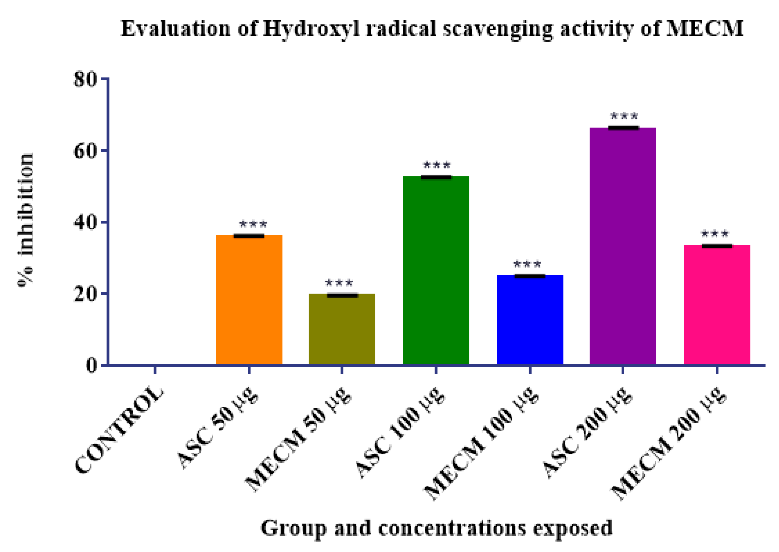
The ability of MECM to scavenge hydroxyl radicals generated by Fenton’s reaction was tested in this study. The test depends upon the formation of the coloured product by the reaction of hydroxyl radicals with thiobarbituric acid and the measurement of its optical density by a colorimetric assay. A reduction in the OD correlated with the ability of MECM to scavenge the hydroxyl radicals from the reaction mixture.
The standard employed was ascorbic acid. Both the extracts and the ascorbic acid were tested in concentrations of 50, 100 and 200 µg. It was observed that the calculated percentage inhibition by ascorbic acid at these concentrations was 36.09 ± 0.296, 52.4 ± 0.387 and 65.98 ± 0.589, respectively (mean ± SEM). The percentage inhibition for MECM calculated at similar concentrations was 19.56 ± 0.194, 24.92 ± 0.194 and 33.3 ± 0.194; when compared with the control, these values were significant (
p < 0.001). The results are summarised in
Table 3 and
Figure 5.
The IC
50 calculated for the ascorbic acid was 108.95 µg whereas that of MECM was 385 µg. A comparison of the inhibitory activities of ascorbic acid and MECM is shown in
Figure 4.
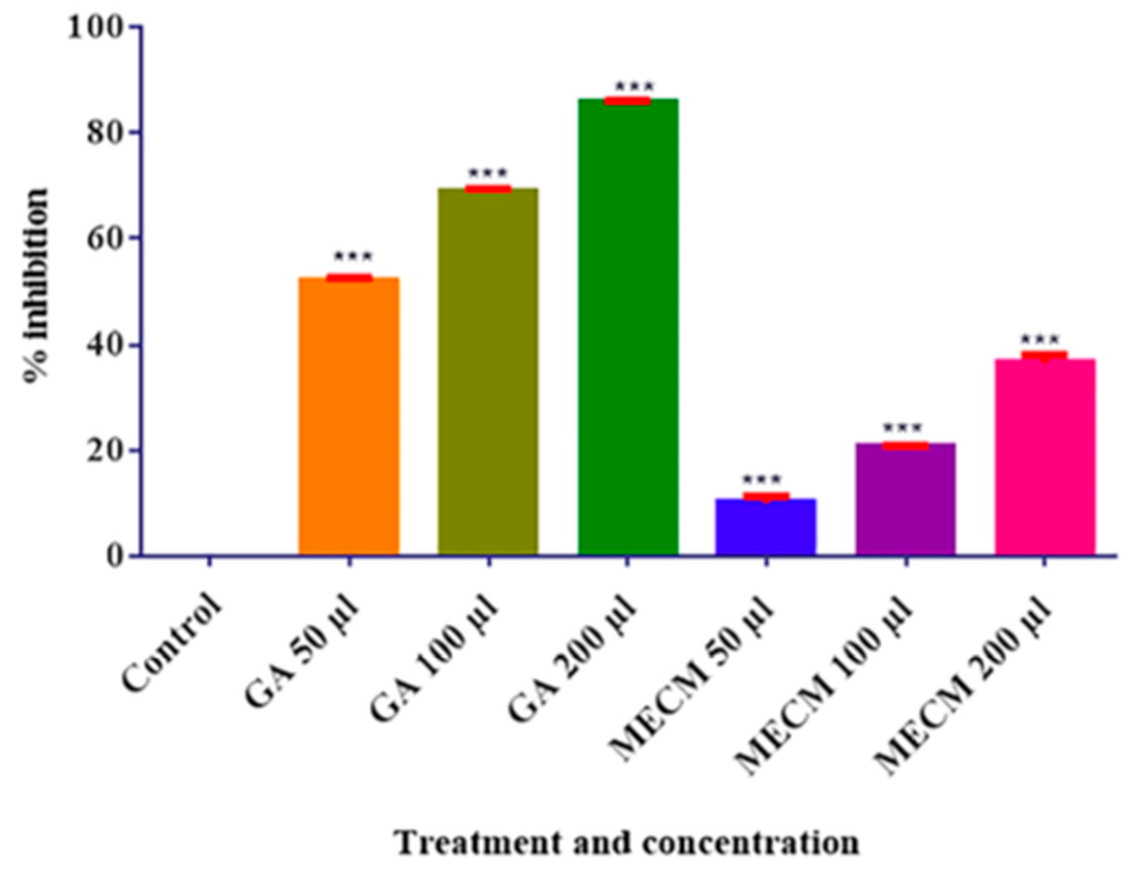
3.4. Nitric Oxide Scavenging Activity
We evaluated the nitric oxide scavenging activity of MECM. The standard used for the comparison was gallic acid. The increased concentration of nitric oxide generated was indicated by the increase in the optical density. Thus, the decrease in the optical density evaluated the nitric oxide scavenging activity (
Table 4).
The observed mean OD of the control group was 0.201 ± 0.007, which corresponded with a zero % inhibition. As the exposed concentrations of gallic acid were increased from 50, 100 and 250 µg/mL, there was a significant reduction in the OD (p < 0.05). Hence, the inhibitory effect on the nitric oxide generation had to be considered inhibited in a corresponding gradation. The percentage inhibition calculated (with reference to the control OD) for the optical density values of 0.099, 0.064 and 0.029 was 51.8 ± 0.744, 68.83 ± 0.562 and 85.7 ± 0.342% for the gallic acid standard. These values were statistically significant at 0.05 levels.
The OD values of MECM for concentrations of 50, 100 and 200 µg/mL were 0.184 ± 0.004, 0.163 ± 0.003 and 0.130 ± 0.002, respectively. These values corresponded with the percentage inhibition of 10.1 ± 1.39, 20.13 ± 0.281 and 36.5 ± 1.55. When compared with the inhibition produced by gallic acid at the same concentrations, these values were significantly low (p < 0.05). Moreover, although the decrease in the OD was significant at 100 and 200 µg/mL concentrations, the effect of 50 µg was found to be insignificant at p < 0.05.
The IC
50 for gallic acid was calculated to be 30.74 µg and that for MECM was 275.29 µg (
Figure 6).
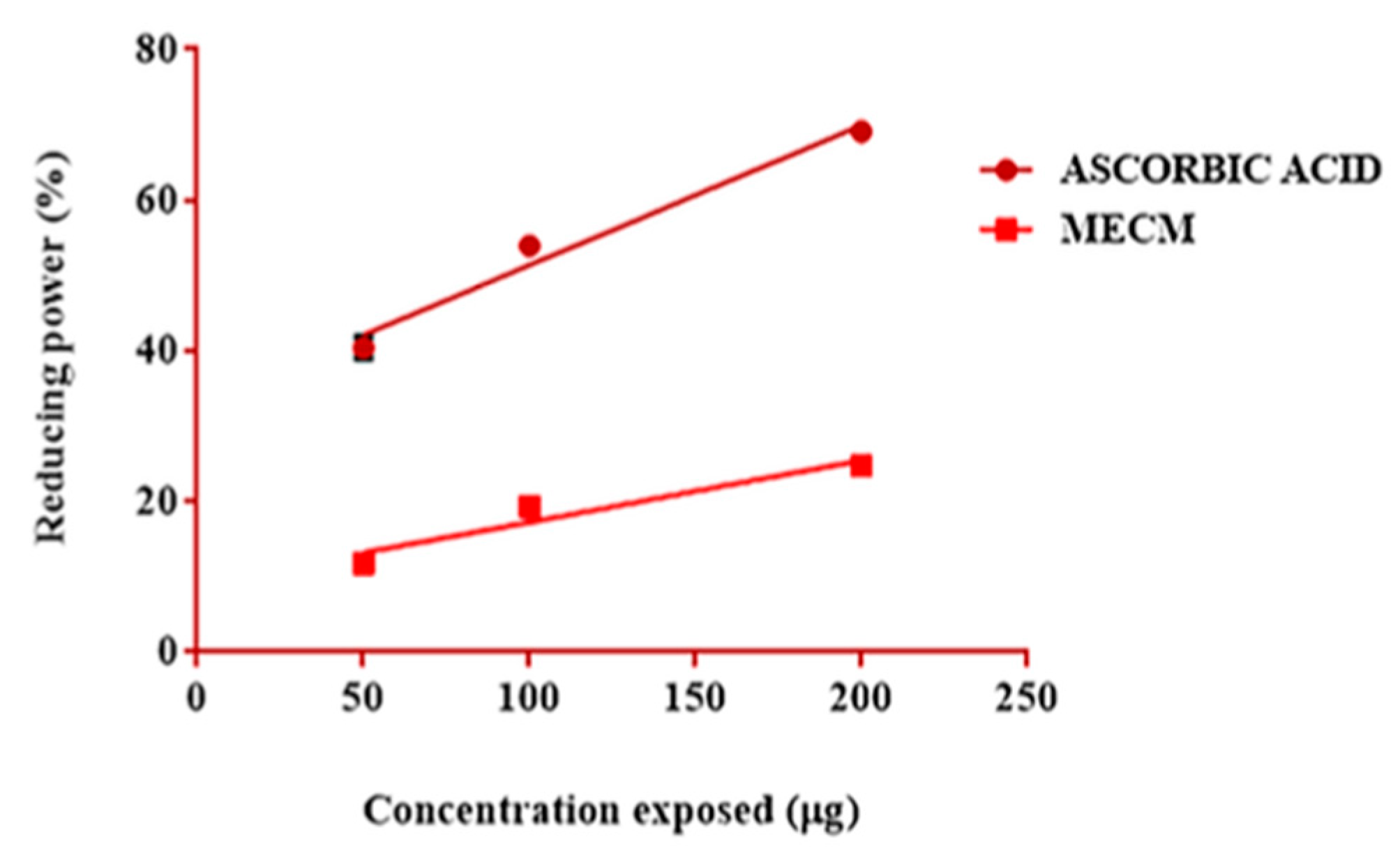
3.5. Evaluation of the Reducing Power Activity
The evaluation of the reducing power activity was conducted by a simple colorimetric method. The change in the absorbance of a reaction system containing trichloroacetic acid, ferric chloride and potassium ferricyanide was extrapolated to calculate the reducing power activity of a compound.
The percentage increase in the absorbance with respect to the control was taken as the percentage reducing power. The result of the study is tabulated in
Table 5.
The reducing power of ascorbic acid at concentrations of 50, 100 and 200 µg was found to be (mean ± SEM) 40.42 ± 1.35, 53.98 ± 0.2405 and 69.14 ± 0.2309 whereas that of MECM in similar concentrations was 11.8 ± 0.6132, 19.37 ± 0.8192 and 24.78 ± 0.8110 (
Figure 7).
4. Discussion
Oxidative stress plays an important role in the development and pathophysiology of many diseases [
1]. The
C. melo extract showed good activities in hydroxyl, nitric oxide radical and DPPH radical scavenging activity [
11]. Moreover, the antioxidant potency of MECM was evaluated by estimating the total antioxidant activity and reducing power activity [
12]. Our finding indicates that MECM is a good source of antioxidant constituents, which may be due to the presence of a series of components acting synergistically by different mechanisms. Natural antioxidants are considered better than synthetic compounds due to minimum adverse effects and
C. melo fruit could be a dietary supplement that could be recommended to prevent oxidative stress.
The phytochemical analysis revealed the presence of a series of phytochemicals in MECM and the HPTLC analysis revealed the presence of polyphenolic compounds. The study mainly aimed to evaluate the antioxidant activity of MECM. Hence, by comparing the antioxidant activity with the standard ascorbic acid in its pure form, we demonstrated that MECM had an antioxidant activity. Ascorbic acid was used in its pure form and is a standard and potent antioxidant. Specifically, the study proved that MECM had an activity compared with the control, with a significance level of a p-value less than 0.05.
4.1. DPPH Radical Scavenging Activity of MECM
This method is one of the most popular procedures to test the antioxidant potential of a plant extract. DPPH is a relatively stable radical that acts as an antioxidant or free radical scavenger by donating hydrogen ions to the compounds in the oxidised state [
31]. The estimation of the free radical scavenging activity in food and plant-based drugs is extensively performed utilising this assay and the fruit extract had an excellent activity [
32,
33].
4.2. Hydroxyl Radical Scavenging Potential of MECM
The neutral form of a hydroxide ion (OH
−) is a hydroxyl radical and is highly reactive. Therefore, it exists only for a short period. Several biological membranes including DNA can be damaged by hydroxyl radicals. To neutralise this radical, no endogenous enzymatic scavenging pathways are present inside the body and most often it is neutralised by several endogenous molecules such as glutathione, melatonin and antioxidants supplemented through diet [
34]. MECM showed a statistically significant hydroxyl radical scavenging potential and the fruit could act as an excellent dietary source to produce this activity.
4.3. Nitric Oxide Scavenging Potential of MECM
Nitric oxide (NO), which is an important bioactive molecule, possesses several physiological functions [
35]. It assists in maintaining vascular homeostasis, fights against pathological microorganisms and it is known to have an anti-cancer activity. NO can also act on the blood vessels, producing vasodilatation, altering the vascular endothelial permeability and acting as a neurotransmitter. When combined with a superoxide radical, it generates a peroxynitrite anion and becomes highly reactive and causes severe damage to intracellular components. The substances that scavenge nitric oxide could have a cytoprotecting property [
35].
The methanolic extract of the F1 Hybrid variety of
C. melo was tested for its ability to scavenge nitric oxide using gallic acid as a standard. In the in vitro study, the significant capacity of MECM to reduce the formation of NO in the reaction system was established. It can act as a potent scavenger of free radicals, thus inferring its cytoprotective effect [
36,
37,
38].
5. Conclusions
Cucumis melo Linn. (C. melo) is an important horticultural crop cultivated worldwide. In the present study, we evaluated the antioxidant activity of a methanolic extract of Cucumis melo Linn (MECM) quantitatively and qualitatively using standard protocols. The qualitative analysis showed that MECM contained carbohydrates, alkaloids, sterols, phenolic compounds, terpenes and flavonoids. The total antioxidant activity of the concentrations of 50, 100 and 200 µg was 3.3 ± 0.1732, 6.867 ± 0.5457 and 13.63 ± 0.8295 µg of ascorbic acid, respectively. Significant nitric oxide and DPPH scavenging activities as well as a reducing power activity were also observed.
Author Contributions
Conceptualization, S.P.I., R.S.R.; methodology, R.S.R., S.P.I.; investigation, R.S.R.; data curation, P.P.N., M.E.-M.; writing—original draft preparation, R.S.R.; writing—review and editing, R.S.R., P.P.N., M.E.-M., M.S.K., G.S.A.; visualization, R.S.R.; supervision, S.P.I.; funding acquisition, R.S.R., P.P.N., M.E.-M., M.S.K. All authors have read and agreed to the published version of the manuscript.
Funding
This research received no external funding.
Institutional Review Board Statement
Not applicable.
Informed Consent Statement
Not applicable.
Acknowledgments
The authors extend their appreciation to the Deanship of Scientific Research at King Khalid University, Saudi Arabia for funding this work through the Research Group Program under Grant No: RGP 2/191/42.
Conflicts of Interest
There is no conflict of interest.
References
- Adwas, A.A.; Elsayed, A.S.I.; Azab, A.E. Oxidative stress and antioxidant mechanisms in human body. J. Appl. Biotechnol. Bioeng. 2019, 6, 43–47. [Google Scholar] [CrossRef]
- Kasole, R.; Martin, H.D.; Kimiywe, J. Traditional medicine and its role in the management of diabetes mellitus: “patients’ and herbalists’ perspectives. Evid.-Based Complement. Altern. Med. 2019, 2019, 2835691. [Google Scholar] [CrossRef] [PubMed]
- Contardi, M.; Lenzuni, M.; Fiorentini, F.; Summa, M.; Bertorelli, R.; Suarato, G.; Athanassiou, A. Hydroxycinnamic acids and derivatives formulations for skin damages and disorders: A review. Pharmaceutics 2021, 13, 999. [Google Scholar] [CrossRef] [PubMed]
- Nuzzo, D.; Contardi, M.; Kossyvaki, D.; Picone, P.; Cristaldi, L.; Galizzi, G.; Bosco, G.; Scoglio, S.; Athanassiou, A.; Di Carlo, M. Heat-resistant Aphanizomenon flos-aquae (AFA) Extract (Klamin®) as a functional ingredient in food strategy for prevention of oxidative stress. Oxid. Med. Cell. Longev. 2019, 2019, 9481390. [Google Scholar] [CrossRef] [PubMed] [Green Version]
- Mujeeb, F.; Bajpai, P.; Pathak, N. Phytochemical evaluation, antimicrobial activity, and determination of bioactive components from leaves of Aegle marmelos. Biomed Res. Int. 2014, 2014, 497606. [Google Scholar] [CrossRef] [PubMed] [Green Version]
- Tungmunnithum, D.; Thongboonyou, A.; Pholboon, A.; Yangsabai, A. Flavonoids and other phenolic compounds from medicinal plants for pharmaceutical and medical aspects: An overview. Medicines 2018, 5, 93. [Google Scholar] [CrossRef]
- Larayetan, R.; Ololade, Z.S.; Ogunmola, O.O.; Ladokun, A. Phytochemical constituents, antioxidant, cytotoxicity, antimicrobial, antitrypanosomal, and antimalarial potentials of the crude extracts of Callistemon citrinus. Evid.-Based Complement. Altern. Med. 2019, 2019, 5410923. [Google Scholar] [CrossRef] [Green Version]
- Rajasree, R.S.; Sibi, P.I.; Francis, F.; William, H. Phytochemicals of Cucurbitaceae family—A review. Int. J. Pharmacogn. Phytochem. Res. 2016, 8, 113–123. [Google Scholar]
- Marwat, S.K.; Rehman, F.U. Medicinal folk recipes used as traditional phytotherapies in district Dera Ismail Khan, KPK, Pakistan. Pak. J. Bot. 2011, 43, 1453–1462. [Google Scholar]
- Ittiyavirah, S.P.; George, A.; Santhosh, A.M.; Kurian, S.T.; Pappachan, P.; Jacob, G. Studies of cytotoxic potential of Cucumismelo. Linn fruit aqueous extract in prostate cancer cell lines PC-3 using MTT and neutral red assay. Iran. J. Pharmacol. Ther. 2013, 12, 24–30. [Google Scholar]
- Mallek-Ayadi, S.; Bahloul, N.; Kechaou, N. Characterization, phenolic compounds and functional properties of Cucumis melo L. peels. Food Chem. 2017, 221, 1691–1697. [Google Scholar] [CrossRef]
- Ismail, H.I.; Chan, K.W.; Mariod, A.A.; Ismail, M. Phenolic content and antioxidant activity of cantaloupe (Cucumis melo) methanolic extracts. Food Chem. 2010, 119, 643–647. [Google Scholar] [CrossRef]
- Baldino, L.; Scognamiglio, M.; Reverchon, E. Supercritical fluid technologies applied to the extraction of compounds of industrial interest from Cannabis sativa L. and to their pharmaceutical formulations: A review. J. Supercrit. Fluids 2020, 165, 104960. [Google Scholar] [CrossRef]
- Portela, S.I.; Cantwell, M.I. Cutting blade sharpness affects appearance and other quality attributes of fresh-cut cantaloupe melon. J. Food Sci. 2001, 66, 1265–1270. [Google Scholar] [CrossRef]
- Gokhale, S.B.; Kokate, C.K.; Purohit, A.P. A Textbook of Pharmacognosy, 28th ed.; Niraliprakashan: New Delhi, India, 2007; pp. 1–20. [Google Scholar]
- Becket, A.H.; Stenlake, J.B. Practical Pharmaceutical Chemistry, 2nd ed.; C.B.S. Publishers and Distributors: New Delhi, India, 1986; pp. 333–337. [Google Scholar]
- Vouldoukis, I.; Lacan, D.; Kamate, C.; Coste, P.; Calenda, A.; Mazier, D.; Conti, M.; Dugas, B. Antioxidant and anti-inflammatory properties of a Cucumis melo LC. extract rich in superoxide dismutase activity. J. Ethnopharmacol. 2004, 94, 67–75. [Google Scholar] [CrossRef]
- Reddy, G.M.; Muralikrishna, A.; Padmavathi, V.; Padmaja, A.; Tilak, T.K.; Rao, C.A. Synthesis and antioxidant activity of styryl sulfonyl methyl 1, 3, 4-oxadiazoles, pyrazolyl/isoxazolyl-1, 3, 4-oxadiazoles. Chem. Pharm. Bull. 2013, 61, 1291–1297. [Google Scholar] [CrossRef] [PubMed] [Green Version]
- Chandran, P.R.; Jothi, T.A. Multisource five level inverter using an improved PWM scheme. Int. J. Sci. Res. 2013, 2, 279–282. [Google Scholar]
- Nishikimi, M.; Rao, N.A.; Yagi, K. The occurrence of superoxide anion in the reaction of reduced phenazine methosulfate and molecular oxygen. Biochem. Biophys. Res. Commun. 1972, 46, 849–854. [Google Scholar] [CrossRef]
- McCord, J.M.; Fridovich, I. The utility of superoxide dismutase in studying free radical reactions: I. Radicals generated by the interaction of sulfite, dimethyl sulfoxide, and oxygen. J. Biol. Chem. 1969, 244, 6056–6063. [Google Scholar] [CrossRef]
- Umamaheswari, M.; Chatterjee, T.K. In vitro antioxidant activities of the fractions of Coccinia grandis L. leaf extract. Afr. J. Tradit. Complement. Altern. Med. 2008, 5, 61–73. [Google Scholar] [CrossRef] [Green Version]
- Blois, M.S. Antioxidant determinations by the use of a stable free radical. Nature 1958, 181, 1199–1200. [Google Scholar] [CrossRef]
- Brand-Williams, W.; Cuvelier, M.E.; Berset, C.L. Use of a free radical method to evaluate antioxidant activity. LWT-Food Sci Technol. 1995, 28, 25–30. [Google Scholar] [CrossRef]
- Elizebeth, K.; Rao, M.W.A. Oxygen radical scavenging activity of Curcumin. Int. J. Pharm. 1991, 58, 237–240. [Google Scholar]
- Enrol, D.; Mechmet, U.; Ferda, C.; Dimitra, D.; Gulhan, V.U.; Mosschos, P.; Atalay, S. Antimicrobial and antioxidant activities of essential oils and methanol extract of Saliva cryptantha (Montbret et AucherexBenth) and Saliva multicaulis (Vahl). J. Food Chem. 2013, 84, 519–525. [Google Scholar]
- Kumaran, A.; Karunakaran, R.J. Nitric oxide radical scavenging active components from Phyllanthus emblica L. Plant Foods Hum. Nutr. 2006, 61, 1. [Google Scholar] [CrossRef] [PubMed]
- Yen, G.C.; Duh, P.D.; Tsai, C.L. Relationship between antioxidant activity and maturity of peanut hulls. J. Agric. Food Chem. 1993, 41, 67–70. [Google Scholar] [CrossRef]
- Lu, Y.; Foo, L.Y. Antioxidant activities of polyphenols from sage (Salvia officinalis). Food Chem. 2001, 75, 197–202. [Google Scholar] [CrossRef]
- Da Porto, C.; Calligaris, S.; Celotti, E.; Nicoli, M.C. Antiradical properties of commercial cognacs assessed by the DPPH test. J. Agric. Food Chem. 2000, 48, 4241–4245. [Google Scholar] [CrossRef]
- Soare, J.R.; Dinis, T.C.; Cunha, A.P.; Almeida, L. Antioxidant activities of some extracts of Thymus Zygis. Free Radic. Res. 1997, 26, 469–478. [Google Scholar] [CrossRef]
- Badarinath, A.V.; Rao, K.M.; Chetty, C.M.; Ramkanth, S.T.; Rajan, T.V.; Gnanaprakash, K. A review on in-vitro antioxidant methods: Comparisons, correlations and considerations. Int. J. Pharmtech. Res. 2010, 2, 1276–1285. [Google Scholar]
- Pham-Huy, L.A.; He, H.; Pham-Huy, C. Free radicals, antioxidants in disease and health. Int. J. Biomed. Sci. 2008, 4, 89–96. [Google Scholar]
- Garratt, D.C. The Quantitative Analysis of Drugs, Japan; Chapman and Hall: Tokyo, Japan, 1964; Volume 3, pp. 456–458. [Google Scholar]
- Marcocci, L.; Maguire, J.J.; Droylefaix, M.T.; Packer, L. The nitric oxide-scavenging properties of Ginkgo biloba extract EGb 761. Biochem. Biophys. Res. Commun. 1994, 201, 748–755. [Google Scholar] [CrossRef] [PubMed]
- Tamir, S.; Tannenbaum, S.R. The role of nitric oxide (NO) in the carcinogenic process. Biochim. Biophys. Acta Rev. Cancer 1996, 1288, 31–36. [Google Scholar] [CrossRef]
- Sreejayan, N.; Rao, M.N.; Priyadarsini, K.I.; Devasagayam, T.P. Inhibition of radiation-induced lipid peroxidation by curcumin. Int. J. Pharm. 1997, 151, 127–130. [Google Scholar] [CrossRef]
- Kala, M.; Shaikh, M.V.; Nivsarkar, M. Equilibrium between anti-oxidants and reactive oxygen species: A requisite for oocyte development and maturation. Reprod. Med. Biol. 2017, 16, 28–35. [Google Scholar] [CrossRef]
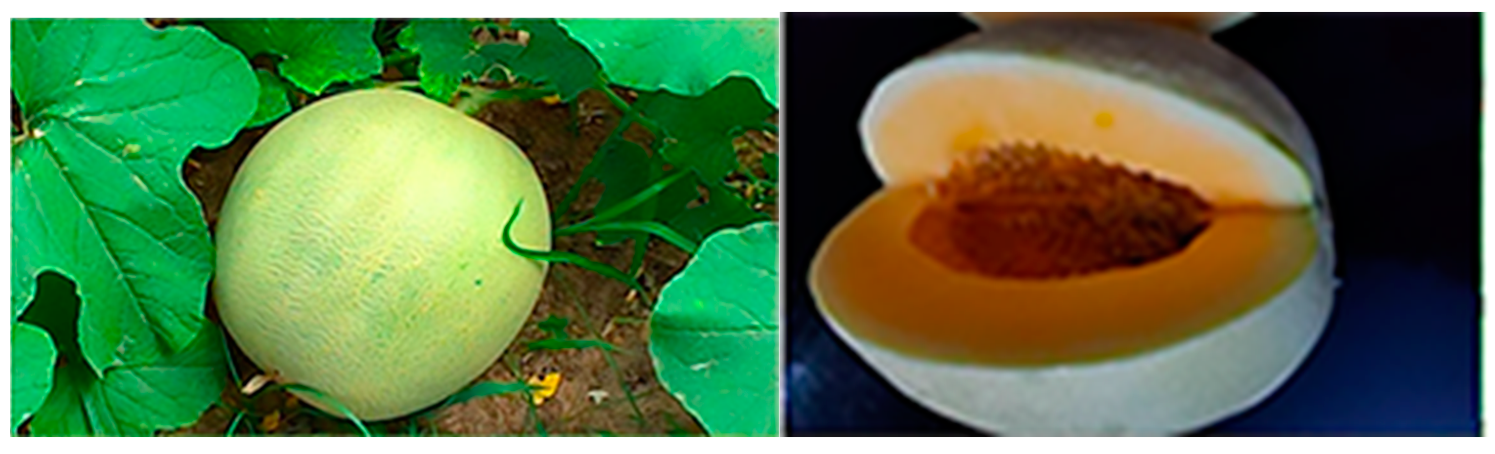
Figure 1.
Muskmelon is a member of the Cucurbitaceae family, genus Cucumis, which comprises about 118 genera and 825 species. The fruit has been cultivated in China since 2000 BC. Diverse fruit forms have evolved around the world and are widely spread in subtropical and tropical regions. China and the USA are the highest producers of muskmelons.
Figure 2.
HPTLC of the phenolic compounds: UV 366 nm, UV 254 nm and derivatised under visible light using a fast blue salt B reagent.
Figure 2.
HPTLC of the phenolic compounds: UV 366 nm, UV 254 nm and derivatised under visible light using a fast blue salt B reagent.
Figure 3.
Evaluation of the total antioxidant activity: a standard curve of ascorbic acid.
Figure 3.
Evaluation of the total antioxidant activity: a standard curve of ascorbic acid.
Figure 4.
The DPPH scavenging activity of MECM using ascorbic acid as a standard. Compared with ascorbic acid in its pure form, MECM showed a significant DPPH scavenging activity (*** Significant, p < 0.001).
Figure 4.
The DPPH scavenging activity of MECM using ascorbic acid as a standard. Compared with ascorbic acid in its pure form, MECM showed a significant DPPH scavenging activity (*** Significant, p < 0.001).
Figure 5.
Comparison of the hydroxyl radical scavenging activity of MECM and ascorbic acid. Compared with ascorbic acid in its pure form, MECM showed a significant hydroxyl radical scavenging activity (*** Significant, p < 0.001).
Figure 5.
Comparison of the hydroxyl radical scavenging activity of MECM and ascorbic acid. Compared with ascorbic acid in its pure form, MECM showed a significant hydroxyl radical scavenging activity (*** Significant, p < 0.001).
Figure 6.
Evaluation of the nitric oxide scavenging activity of MECM: a comparison with gallic acid (*** Significant, p < 0.001).
Figure 6.
Evaluation of the nitric oxide scavenging activity of MECM: a comparison with gallic acid (*** Significant, p < 0.001).
Figure 7.
A comparison of the reducing power of MECM with that of ascorbic acid at concentrations of 50,100 and 200 micrograms. The x-axis shows the concentrations exposed and the y-axis shows the percentage reducing activity. Compared with ascorbic acid in its pure form, MECM showed a significant reducing power.
Table 1.
Qualitative chemical tests of the methanolic extract of muskmelons.
Table 1.
Qualitative chemical tests of the methanolic extract of muskmelons.
| Sl No | Phytochemicals | Test/Reagent Used | Result |
|---|
| 01 | Carbohydrates | Molisch’s test | + |
| Iodine test | − |
| Fehling’s test | − |
| Benedict’s test | − |
| | | Barfoed’s test | − |
| | | Million’s test | − |
| 02 | Proteins | Biuret test | − |
| | | Xanthoproteic test | − |
| | | Ninhydrin test | − |
| | | Grease spot test | − |
| 03 | Fats/oil | Saponification test | − |
| | | Bontrager’s test | − |
| 04 | Alkaloids | Hager’s test | + |
| | | Dragendorff’s test | + |
| | | Liebermann’s sterol test | + |
| 05 | Sterols/steroids | Liebermann–Burchard test | + |
| | | Salkowski’s test | + |
| | | Ferric chloride test | + |
| 06 | Phenolics | Lead acetate test | + |
| | | Gelatine test | − |
| 07 | Terpenes/terpenoids | Tin and thionyl chloride test | + |
| 08 | Saponin glycosides | Form test | − |
| Haemolysis test | − |
| 09 | Gums/mucilage | Precipitation test | − |
| Ruthenium red test | − |
| | | Alkali (aqueous NaOH) | + |
| 10 | Flavones/flavonoids | Conc. H2SO4 | + |
| | | Shinoda test | + |
Table 2.
DPPH scavenging activity.
Table 2.
DPPH scavenging activity.
| Sl No. | Concentrations Exposed (µg/mL) | % Scavenging |
|---|
| | | Control | MECM | Ascorbic Acid |
|---|
| 1 | 100 | 0.00 | 11.79 ± 0.5469 *** | 29.24 ± 0.8712 *** |
| 2 | 250 | 16.50 ± 1.065 *** | 36.76 ± 1.3 *** |
| 3 | 500 | 22.45 ± 1.131 *** | 52.06 ± 0.7963 *** |
Table 3.
Evaluation of the hydroxyl radical scavenging activity of MECM.
Table 3.
Evaluation of the hydroxyl radical scavenging activity of MECM.
| Group Treated | Concentration Exposed (µg) | % Inhibition (Mean ± SEM) | Groups Compared and Significance |
|---|
| Control (A) | -- | 00 | -- |
Standard (B)
ascorbic acid | 50 (B1) | 36.09 ± 0.296 | A and B1 *** |
| 100 (B2) | 52.4 ± 0.387 | A and B2 *** |
| 200 (B3) | 65.98 ± 0.589 | A and B3 *** |
Test (C)
MECM | 50 (C1) | 19.56 ± 0.194 | A and C1 *** |
| 100 (C2) | 24.92 ± 0.194 | A and C2 *** |
| 200 (C3) | 33.3 ± 0.194 | A and C3 *** |
Table 4.
Nitric oxide scavenging activity of MECM.
Table 4.
Nitric oxide scavenging activity of MECM.
| Sl No | Group | % Inhibition (Mean ± SEM) | Level of Significance and Groups Compared |
|---|
50 µg
(A) | 100 µg
(B) | 200 µg
(C) |
|---|
| 1 | Control (I) | 00.00 | 00.00 | 00.00 | --- |
| 2 | Std (II)
(Gallic acid) | 51.8 ± 0.744 | 68.8 ± 0.562 | 85.7 ± 0.342 | I(A) and II(A), II(B), II(C) *** |
| 3 | MECM (III) | 10.1 ± 1.39 | 20.13 ± 0.281 | 36.5 ± 1.55 | I(A) and III(A), III(B), III(C)
*** |
Table 5.
Evaluation of the reducing power of MECM.
Table 5.
Evaluation of the reducing power of MECM.
| Sl NO | Concentration (µg /mL) | Reducing Power as % (Mean ± SEM) | Statistics |
|---|
| Control (A) | MECM (B) | Ascorbic Acid (C) |
|---|
| 1 | 50 (i) | 0.0 | 11.8 ± 0.6132 | 40.42 ± 1.35. | Bi and Ci ***
Bi and A ***
Ci and A *** |
| 2 | 100 (ii) | 19.37 ± 0.8192 | 53.98 ± 0.2405 | Bii and Cii **
Bii and A ***
Cii and A *** |
| 3 | 200 (iii) | 24.78 ± 0.8110 | 69.14 ± 0.2309 | Biii and Ciii ***
Biii and A ***
Ciii and A *** |
| Publisher’s Note: MDPI stays neutral with regard to jurisdictional claims in published maps and institutional affiliations. |
© 2021 by the authors. Licensee MDPI, Basel, Switzerland. This article is an open access article distributed under the terms and conditions of the Creative Commons Attribution (CC BY) license (https://creativecommons.org/licenses/by/4.0/).


 ,
, 


