Review

Electromyography: A Simple and Accessible Tool to Assess Physical Performance and Health during Hypoxia Training. A Systematic Review

by
Diego Fernández-Lázaro
1,2,*,
Juan Mielgo-Ayuso
3,
David P. Adams
4,
Jerónimo J. González-Bernal
5,
Ana Fernández Araque
6,
Alicia Cano García
1 and
Cesar I. Fernández-Lázaro
1,7
1
Department of Cellular Biology, Histology and Pharmacology, Faculty of Health Sciences, University of Valladolid, Campus of Soria, 42003 Soria, Spain
2
Neurobiology Research Group, Faculty of Medicine, University of Valladolid, 47005 Valladolid, Spain
3
Department of Biochemistry, Molecular Biology and Physiology, Faculty of Health Sciences, University of Valladolid, Campus of Soria, 42003 Soria, Spain
4
Dual Enrollment Program, Point University-Savannah Campus, Savannah, GA 31419, USA
5
Department of Health Sciences, Cavidito Research Group, University of Burgos, Paseo comendadores s/n 09001 Burgos, Spain
6
Department of Nursing, Faculty of Health Sciences, University of Valladolid, Campus of Soria, 42003 Soria, Spain
7
Department of Preventive Medicine and Public Health, School of Medicine, University of Navarra, IdiSNA, 31008 Pamplona, Spain
*
Author to whom correspondence should be addressed.
Sustainability 2020, 12(21), 9137; https://0-doi-org.brum.beds.ac.uk/10.3390/su12219137
Submission received: 30 August 2020 / Revised: 16 October 2020 / Accepted: 27 October 2020 / Published: 3 November 2020
(This article belongs to the Special Issue Physical Performance and Health Care for a Sustainable Lifestyle)

Abstract

:
Hypoxia causes reduced partial pressure of oxygen in arterial blood and induces adaptations in skeletal muscle that may affect individuals’ physical performance and muscular health. These muscular changes are detectable and quantifiable by electromyography (EMG), an instrument that assesses electrical activity during active contraction at rest. EMG is a relatively simple and accessible technique for all patients, one that can show the degree of the sensory and motor functions because it provides information about the status of the peripheral nerves and muscles. The main goal of this review is to evaluate the scientific evidence of EMG as an instrument for monitoring different responses of skeletal muscles subjected to external stimuli such as hypoxia and physical activity. A structured search was conducted following the Preferred Reporting Items for Systematic Review and Meta-Analyses (PRISMA) guidelines in Medline/PubMed, Scielo, Google Scholar, Web of Science, and Cochrane Library Plus. The search included articles published in the last 25 years until May 2020 and was restricted to English- and Spanish-language publications. As such, investigators identified nine articles that met the search criteria. The results determined that EMG was able to detect muscle fatigue from changes in the frequency spectrum. When a muscle was fatigued, high frequency components decreased and low frequency components increased. In other studies, EMG determined muscle activation increased during exercise by recruiting motor units and by increasing the intensity of muscle contractions. Finally, it was also possible to calculate the mean quadriceps quadratic activity used to obtain an image of muscle activation. In conclusion, EMG offers a suitable tool for monitoring the different skeletal muscle responses and has sufficient sensitivity to detect hypoxia-induced muscle changes produced by hypoxic stimuli. Moreover, EMG enhances an extension of physical examination and tests motor-system integrity.

1. Introduction

Hypoxia is defined as the reduction of oxygen (O2) content or pressure at the cellular level. Hypoxemic hypoxia is one of the main approaches to improving muscular performance. It has two subtypes: hypobaric hypoxia, characterized by an atmospheric pressure lower than 760 mm Hg and a fraction of inspired O2 (FiO2) of 20.9%; and normobaric hypoxia, with a barometric pressure of 760 mm Hg and a FiO2 of less than 20.9%. In terms of exposure time, hypoxia is classified as acute, chronic, or intermittent. Intermittent hypoxia characterizes a state in which subjects are exposed to alternating cycles of hypoxia and normoxia. These artificial hypoxic conditions (hypobaric or normobaric) can be simulated by devices such as mixed gas masks, chambers, tents or rooms, and reduced-oxygen breathing devices [1]. Two common techniques that simulate altitude training in elite athletes are intermittent hypoxic exposure (IHE) and intermittent hypoxic training (IHT). IHE is implemented through lengths of passive stays in a hypoxic environment or through breathing in an environment with reduced oxygen concentration, whereas IHT consists of training sessions under hypoxic conditions [2].
Monitoring arterial saturation of oxygen (SaO2) is essential to controlling artificial hypoxia situations. SaO2 is extremely useful in the control of the intensity of the hypoxic stimulus. Thus, when SaO2 is lower than 90%, the adaptive physiological response is met in different systems such as the respiratory, cardiovascular, endocrine, metabolic, hematological, and immune systems as well as in skeletal muscle (Table 1) [1]. In this context, functional and molecular adaptations in skeletal muscle tissue under hypoxic conditions are achieved [3].
Electromyography (EMG) is an indirect diagnostic device to evaluate the electrical activity produced in muscles by a nerve impulse that generates a potential for action in the membrane of myocytes in situations of rest or activation phase [4,5]. In general, EMG can be classified according to the test protocol conducted on the individual being evaluated: resting EMG (determines basal muscular electrical activity), voluntary EMG (assesses muscular response after the action), and EMG with evoked potentials (evaluates motor units) [6].
EMG is a type of recording equipment composed of several elements: electrodes, amplifiers, and a registration system. It has two types of electrodes: internal or needle electrodes (deep EMG or integrated EMG (iEMG), and surface electrodes (kinesiological or surface EMG (sEMG)). The EMG signal is emitted by the electrodes and is collected in the amplifier, which detects potential difference and eliminates interference. Next, the signal is quantified by means of a recording system that expresses the signal entity compared to a previously obtained reference value. There are two primary methods of signal amplification: monopolar and bipolar configurations. Moreover, there are different types of recording systems that collect information, such as graphic recording, oscilloscopic recording, permanent records on paper, or permanent records by photographic means [4,6,7,8].
EMG has multiple applications, not least as a diagnostic tool. In some cases, patients are diagnosed after the onset of the first symptoms that follow a lengthy preclinical phase. Delayed diagnosis may cause health problems and notably reduce the quality of life. In this context, EMG provides a relatively simple and accessible diagnosis, one that yields information about the state of the peripheral nerves and muscles. EMG may also be useful in the early diagnosis of neuromuscular disease, distinguishing the specific type(s) of pathology and its effect on the axons or the nerve myelin. In other words, EMG may act as a record of muscle electrical activity; therefore, it constitutes an extension of physical examination that can evaluate the integrity of the motor system [1,3,6,8].
Another EMG application may involve monitoring real-time muscle status and identifying thresholds at which transition occurs between molecular systems during physical activity [8]. Inadequate application of intensities during exercise results in modifications of the molecular systems that alter health, physical performance, quality of life, and social and individual sustainability [3]. Furthermore, EMG may be considered a useful tool in clinical practice [4]. The purpose of this study is to systematically review EMG’s application as point-of-care testing for the evaluation and monitoring of different responses of skeletal muscles subjected to an external stimulus such as hypoxia.

2. Material and Methods

2.1. Search Strategy

This systematic review was conducted following the Preferred Reporting Items for Systematic Reviews and Meta-Analyses (PRISMA) guidelines [9]. To select the studies, the PICOS question model [10] was used as follows: P (population)—“physically active and healthy men,” I (intervention)—“hypoxia training recorded by EMG,” C (comparison)—“some conditions with/without hypoxia,” O (outcomes)—”hypoxia-induced modifications at the muscular level: fatigue, muscular activation and quadriceps mean quadratic activity,” S (study design)—“randomized design without cross-placebo.”
A structured search was conducted in the following databases: Medline (PubMed), Scielo, Google Scholar (GS), Web of Science (WOS), and Cochrane Library Plus (LP). We sought articles published over the last 25 years prior to May 2020. Search terms were a mix of medical subject headings (MeSH) and key words related to EMG, muscle, exercise, and hypoxia. They were the following: (“electromyography” OR “electromyogram”) AND (“muscle” OR “muscle fatigue” OR “muscle strength”) AND (“hypoxia”) AND (“exercise” OR “physical activity” OR “sports”). We used the snowball strategy to identify other potential manuscripts. All titles and abstracts of the identified studies were cross-referenced to identify duplicates and any missing studies. Titles and abstracts were then screened for a subsequent full-text review. The search for published studies was independently performed by two authors and disagreements were resolved by third-party evaluation.

2.2. Selection of Articles: Inclusion and Exclusion Criteria

We did not include any filter related to participants’ physical activity, age, or race/ethnicity. The only filter applied to participants’ characteristics was the exclusion of female participants. Female athletes have different intensity and physiological responses to exercise than male athletes, which directly influences EMG results [11,12,13]. Therefore, inclusion criteria included articles (i) depicting a well-designed study that included the use of EMG during physical activity under hypoxic conditions in humans; (ii) with the same study design but without hypoxia conditions; (iii) and specific information related to EMG and hypoxia. Alternatively, exclusion criteria included: (i) publications without results in human and non-related to hypoxia training; (ii) female participants; (iii) animal studies; (iv) studies written in languages other than English or Spanish; (v) articles published more than 25 years ago; and (vi) editorials, systematic reviews, letters to the editor, commentaries, and systematic reviews.

2.3. Data Extraction

Once the inclusion/exclusion criteria were applied to each selected study, data on the study source (including authors and year of publication), study design and sample size, characteristics of participants (gender and level of physical activity), type of hypoxia (dose and timing), type of electromyography, muscles tested, physical activity developed, outcomes, and main conclusions were extracted independently by two authors using a spreadsheet. Subsequently, disagreements were resolved by third-party evaluation.

2.4. Quality Assessment

The methodological quality evaluation of the selected articles was assessed using the McMaster’s Critical Review Form [14]. The aim of this evaluation was to exclude studies with poor methodology. The methodological quality of the selected studies was assessed by the same two authors, and any disagreements were resolved by third-party evaluation.

3. Results

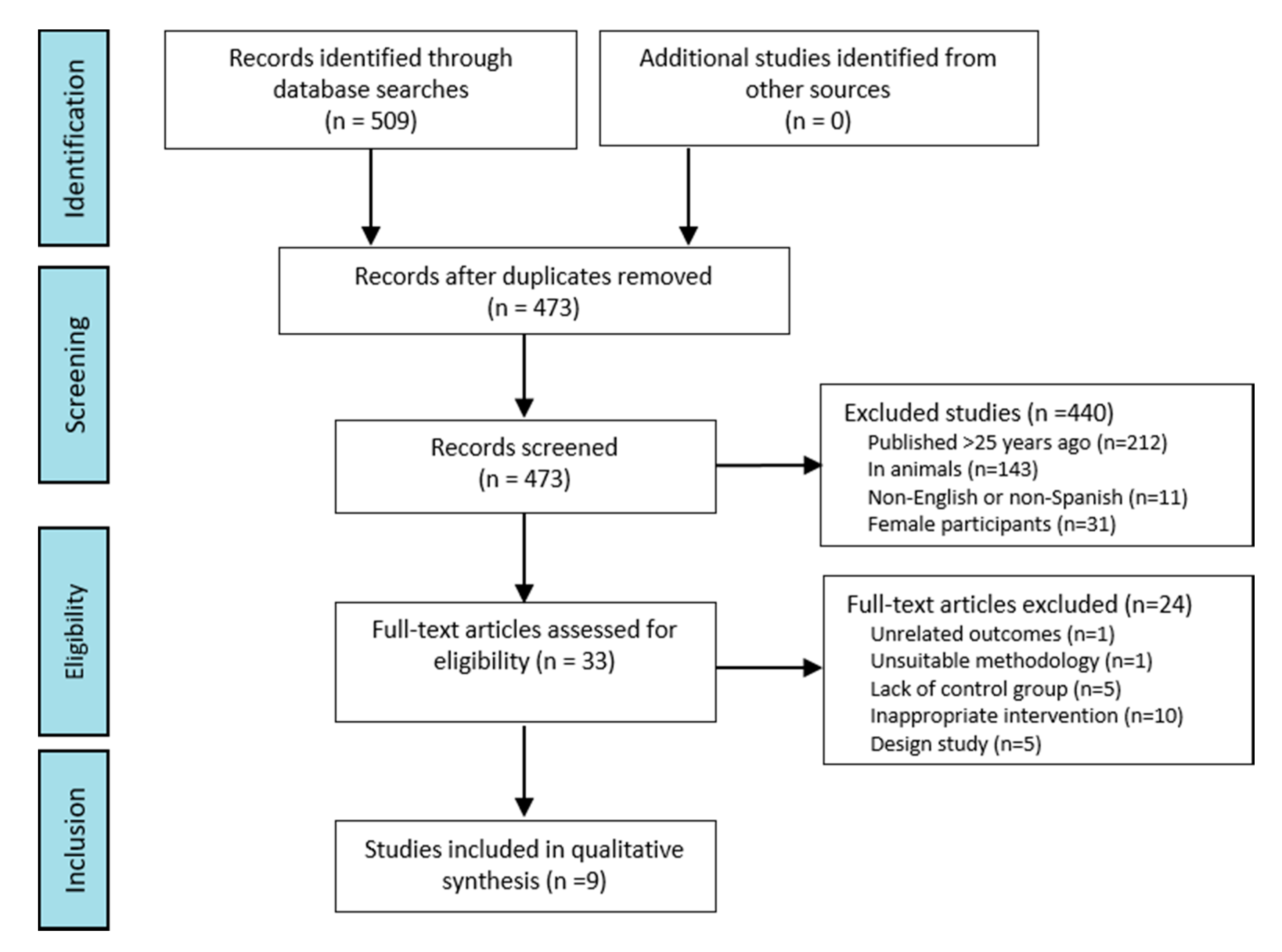
3.1. Selection of Studies

We identified an initial total of 509 records. Among those, we removed 36 duplicates, 212 manuscripts published more than 25 years ago, 143 articles conducted on animals, 11 additional non-Spanish or non-English studies, and 31 records that included female participants. We also excluded 24 articles after full-text review. Reasons for exclusions after full-text review were unrelated outcomes (n = 1), unsuitable methodology (n = 1), lack of control group (n = 5), inappropriate intervention (n = 10), and study design (n = 5). The remaining nine [15,16,17,18,19,20,21,22,23] studies met our inclusion criteria and were included in the present systematic review (Figure 1).

3.2. Results of the Quality Assessment

Then we conducted the quality assessment of the articles. The score of the selected articles ranged from 11 to 15 points. Two studies were assessed as “excellent,” six as “very good,” and one as “good.” No studies were excluded because of poor quality. Details about the results of the quality assessment are shown in Table 2.

3.3. Descriptive Information of the Selected Articles Included in the Systematic Review

The characteristics of the studies included in the systematic review appear in Table 3.

3.4. Performance Measures

Table 4 and Table 5 summarize the studies included in the present review. Table 4 displays information about the authors, publication year, study design, population, type of hypoxic stimuli, type of EMG used, muscles on which EMG was used, and type of physical activity performed in the study. Table 5 summarizes the main results and conclusions of the selected studies.

4. Discussion

The purpose of this systematic review was to summarize all the scientific evidence on EMG assessment of skeletal muscle changes produced by two stimuli applied simultaneously: hypoxia and exercise. Skeletal muscles show a remarkable plasticity that allows them to adapt to different external stimuli that influence the usual levels of contractile activity. Due to such a quality, skeletal muscle can efficiently perform its function in different circumstances [1]. Nonetheless, the response to hypoxia and/or exercise is not the same for every individual. How muscle function responds to hypoxia depends on the type of hypoxic interventions (e.g., acute vs. chronic, intermittent vs. continuous), and the response to physical activity depends on the type of muscle contraction (e.g., sustained vs. intermittent) developed during exercise [24]. One way to identify these responses is through indirect assessment of muscle activity by detecting the electrical activity generated by the passage of nerve impulses that create a potential for action in the muscle cell membrane. This allows EMG to be an objective, quantifiable, and precise assessment tool to study the intrinsic changes in skeletal muscle [4].

4.1. Study Design and Participants

The selected studies for this review had a crossover clinical trial design in which treatments were administered to each patient in randomly selected successive periods, allowing each subject to be their own control. The advantage of using crossover clinical studies is that the effect of the treatment can be estimated with greater precision, and therefore it is not necessary to recruit as many participants [25]. In this sense, the sample size of the analyzed studies was between eight and 14 participants. Disadvantages of cross-group trials include difficulties in avoiding dragging effects (the influence of the first phase of treatment “dragging” to the second phase of treatment); withdrawal of patients from the study, which complicates interpretation and analysis; and difficulties with late-phase adverse events from the intervention [25].

4.2. Additional Stimuli

Two studies [21,23] added temperature to hypoxia as an extra factor for muscle contraction, heat [21] and cold [23], which modified the EMG-measured response. The thermal rise induces lower cardiac output that directly affects blood flow to the muscle and alters contraction events [26]. The response to hypothermia alters the mechanics of the contraction by decreasing the maximum force and the rate of propagation of the potential action of motor end-plate [27]. Assessment of knowing the muscular response to hypoxia with heat or cold is necessary because various types of exercises are performed at moderate altitudes of between 2000 and 3000 m (11–13% FiO2) and extreme temperatures. For example, mountaineers, alpine skiers, or soldiers who frequently enter high-altitude regions must often deal with low or high temperatures. The results of muscular alterations, determined by EMG, would provide guidelines for acclimatization prior to such activities.

4.3. Hypoxia

The nine studies included in this review [15,16,17,18,19,20,21,22,23] were performed in hypoxic training that required subjects to be subjected to hypoxia while doing physical activity. Most of the studies [15,16,17,19,20,21,22,23] used normobaric hypoxia (NH), except the study conducted by Fulco et al. [18] that used hypobaric hypoxia (HH). This influences muscle responses and, consequently, EMG data. From one type of hypoxia to another, the response of the muscles is conditioned and therefore influences data recording. HH produces a more intense stimulus than NH; in turn, it produces more intense physiological responses. Nonetheless, the two types of hypoxia are effective in acclimatizing to similar physiological responses. It has been shown, however, that some differences exist between the two hypoxia types, e.g., increased NH ventilation and an alveolar gas imbalance. It has also been suggested that oxidative stress increases during HH. Other physiological responses such as heart rate, O2 intake, SaO2, or hormonal responses that produce both hypoxias are similar [28].

4.4. Electromyography

A total of three [19,20,22] studies used only sEMG. sEMG has some advantages such as being pain-free, having electrodes that are easy to place, and being able to reproduce good-quality signals of muscular activity obtained from dynamic actions. Moreover, sEMG provides useful information for the study of actions that involve movement. The signal collection area, however, is limited and noise can be registered when electrical activity is detected in adjacent muscles (cross talk).
Alternatively, there was only one study [15] that used only iEMG to minimize the aforementioned sEMG limitations. iEMG can register a specific area of deep muscles, isolate areas of long muscles, and/or register small muscles. In this case, it is used to isolate some areas of the long muscles since the muscles used for the analysis are the external vastus, the internal vastus, and the femoral rectum, e.g., the quadriceps. Discomfort can increase tension and/or spasticity, causing cramps that may contribute to lower reproducibility of findings [29].
Both sEMG and iEMG provide real-time information, regardless of muscle of activity or inactivity, and three studies used sEMG and iEMG [16,17,18]. In other words, these tools furnish information about activity levels and relationships or interactions with the rest of the muscles involved in the action under study. The best approach may be to use both sEMG and iEMG to obtain optimal results concerning the quality of movement through assessment of the gesture, gait, and agonist and antagonist muscles. It may be useful as an assessment tool in muscular or neuromuscular disorders, fatigue, or sports performance, improving quality of care and quality of life.

4.5. Muscles

Eight articles selected for the present systematic review studied the muscles of the lower limbs [15,16,17,18,19,20,21,22], and one study [23] evaluated those of the forearm. Taylor et al. [15] and Osawa et al. [19] examined a single muscle, the quadriceps, while the rest of the studies [16,17,18,20,21,22] examined different muscles in the same subjects. Most of the studies [16,17,18,20] observed the quadriceps and the femoral biceps [18,20] or the quadriceps and femoral biceps attached to the gluteus maximus [16,17]. The anterior tibialis muscle was studied with the soleus [21] or with the femoral rectum, the vastus externalis, the femoral biceps, and the calves [22]. The selection of muscles was performed according to the different physical activities practiced in each of the studies, analyzing the muscles that most predominated in the exercise.
The most-studied muscles are in the lower limbs. First, the physical activity for which the studies were conducted focused on exercises that strengthened the lower limb. Another reason to study lower-limb muscles is their large size and number of fibers. The more fibers the muscle has, the more information is provided from it. Moreover, they are muscles that are easily located. Alternatively, upper-limb muscles are more difficult to locate due to their small size and the large number of adjacent muscles in the forearm. Only one study [23] focused on the upper limbs.

4.6. Physical Activity

To conduct an EMG analysis, one must examine the activation of different muscles during a specific dynamic action. As such, it is necessary to synchronize the electromyographic data collection with other measurements that provide cinematic data [4]. Alternatively, analyses can be complemented with force analysis systems, i.e., kinetics, such as podometry and the force platform.
Some studies used the cycle ergometer, in which physical activity was developed as variants of cycling [15,19,20,21]. Therefore, the worked muscles were primarily the quadriceps, the anterior tibialis, the gluteus maximus, and the hamstrings. Two studies [16,17] designed a squat exercise plan, an activity that worked several muscles at once. As for the lower extremity, the quadriceps, the gluteus maximus, the hamstrings, and the calves were the muscles analyzed in most of the studies [15,16,17,18,19,20,21]. In the rest of the studies, investigators used different strategies to make participants exercise. Fulco et al. [18], for example, chose to use exercises of submaximum extension of the knee until exhaustion to work in the extension of the quadriceps and when returning, the hamstrings. Girard et al. [22] used a protocol of sprints designed to work the hamstrings, quadriceps, anterior tibialis, calves, and soleus. Finally, Lloyd et al. [23] proposed exercises of voluntary isometric contraction to work the common extensor of the fingers, the radial flexor of the carpus, and the superficial common flexor of the fingers.

4.7. Electromyographic Evaluation of Muscle Activity by Electromyography

4.7.1. Muscular Activity

EMG [15,16,18,19,20,21,22,23] was able to record muscle activity after the application of hypoxia training stimuli. In this sense, we analyzed the joint and comparative use of iEMG and sEMG [16,17,18] as tools for monitoring the activity of different muscles. The analysis provided differences in the recording of the recruitment of muscle motor units [17] by sEMG during the process of muscle activity that were not observed by iEMG. Taylor et al. [15] were able to detect patterns of change in the determination of muscle activity with a trend towards an increased myoelectric output response under moderate hypoxia (HM) conditions in three quadriceps muscles by iEMG. The basis for iEMG non-significance may have been related to iEMG, sample size, and the level of hypoxia induced. Thus, these results may establish sEMG as a better diagnostic device to evaluate myoelectric activity.

4.7.2. Muscle Fatigue

Physical fatigue or muscular fatigue involves the decrease in the physical capacity of the individual. This may occur because of a static, dynamic, or repetitive muscular tension, due to an excessive tension of the whole organism, or due to excessive effort by the psychomotor system that directly affects skeletal muscle [30]. Fatigue can be evaluated by EMG by monitoring the signal emitted. Changes in the frequency spectrum cause low frequency components to increase and high frequency components to decrease when muscle fatigue occurs [29]. In a fatigued state, the muscle contraction cannot be maintained and induces a decrease in force that causes a decrease in the excitation of motor units. This is reflected in a reduction in frequency amplitude [31]. Exhaustion time was measured by iEMG [15], sEMG [21], or both [18], a lower result with hypoxic stimulus compared to the control group in normoxia. Estimation of the neuromuscular fatigue threshold from the EMG amplitude may be useful in evaluation and diagnosis in sports to personalize athletes’ training to improve their performance [4].

4.7.3. Muscle Activation

Optimal muscle activation is established by the connection between the central nervous system and skeletal muscles. The muscle activation in EMG is demonstrated by the amplitude of the surface electromyogram increases during incremental exercise until exhaustion [19]. A greater amplitude reflects a combination of gradual recruitment of motor units and an increase in the final frequency to enhance muscle-contraction intensity. Muscle activation also increases during repeated static contraction and dynamic submaximal muscle contractions at an exercise intensity determined by the recruitment of additional motor units [29,31]. The determination of muscle activation by sEMG [16,17,19,20] or iEMG [16,17] in hypoxia training showed an increase in relation to stimulus load [16,19,20], although there were no significant differences in the recruitment of motor units by iEMG between the hypoxic and normoxic groups [17].
The mean quadratic muscle activity is the value of the power of the electromyographic signal [4]. Using the root mean square (RMS), investigators can measure the electrical power of the electromyography signal (the square root of the area between the square of the signal and the computed time in a time interval divided by that time). The RMS does not need rectification; it is obtained at various times according to the activity being studied and provides more information than the integrated signal [31]. RMS, therefore, is an algorithm used to obtain an image of muscle activation that is easier to understand, and its formula represents the signal power [5]. In a study conducted by Girard et al. [22], the RMS of the quadriceps was higher in hypoxia than in normoxia, which determines that muscle activation is higher in hypoxia.

5. Application of Electromyography for a Sustainable Lifestyle

5.1. Quality of Health and Quality of Life

The application of EMG in rehabilitation medicine may prove especially useful. EMG allows clinicians to study muscular physiology and diagnose neuromuscular diseases (Figure 2). Furthermore, EMG can be used to detect spasms, muscular hyperactivity, muscular imbalances, and the resting and occlusal positions of the jaw [6]. Moreover, it allows for the analysis of muscular movement (i.e., muscle walking), to determine the activation time of the muscle, the beginning and end of the articular position, and the degree of muscular activation that marks the muscular effort that is necessary to generate the action of gesture and intermuscular coordination [5]. These parameters are important when assessing pathologies with movement disorders from neurological origin, post-surgical care after a joint prosthesis, and in case of instability and/or injuries of the ligaments [32,33,34,35]. Additionally, EMG allows for the assessment of motor unit potentials, identification of neuromuscular alterations, [36] and differentiation of neuropathic and/or myopathy processes [5] (Figure 3).

5.2. Physical Performance

EMG can analyze dynamic situations that may be of special interest in assessments of sports performance. Potential uses of EMG in physical performance include improvements in the effectiveness of a gesture, co-activation, fatigue, myofeedback techniques, diagnosis, and treatment of muscle injuries.
The effectiveness of a gesture implies the correct use of muscular work in terms of effort economy, profitability, and injury prevention. During training, improvements can be made in muscular activity. In particular, the execution of a task can be improved in terms of muscular activation and/or in terms of muscular fatigue based on the analysis of the frequencies of the electromyographic traces obtained. In other words, EMG improves the effectiveness of an action by saving effort and improving the prevention of muscular injuries [5].
Co-activation also involves simultaneous activity in agonist and antagonist muscles [37]. This is very important when assessing the quality of movement because alterations in coercion are related to immaturity of the neuromuscular system [5]. Furthermore, it is possible to evaluate fatigue with EMG by studying its evolution and potentially providing a solution with an appropriate treatment [38]. This is of special interest for sports and occupational medicine because it could improve sports performance and productivity [5]. EMG facilitates myofeedback techniques that are widely used in physiotherapy with the aim of postural re-education and proprioception work [39]. EMG gives the opportunity to assess muscle activity during a diagnostic and/or therapeutic process in sports medicine. Moreover, it analyzes muscular activation, which allows for the comparison of healthy and pathological states. Lastly, EMG provides information about muscular coordination and the relationship between agonist and antagonist muscles [39].

6. Perspectives

Muscular alterations as a consequence of hypoxic situations contribute not only to a decrease in life expectancy but also to a lower quality of life and health status. Our perspective, based on the results of this systematic review, is that EMG is a suitable tool for monitoring the different skeletal muscle responses and has sufficient sensitivity to detect the muscle changes produced by hypoxic stimuli. Therefore, sEMG maybe provide a practical point-of-care diagnostic test for medical diagnoses as well as a tool to improve sports performance. iEMG studies the physiology and pathology of denervation, re-innervation, and various myopathies. It also analyzes deep musculature such as muscular behavior, temporal activity patterns, fatigue, and muscular activation. sEMG is suitable for providing information about global muscle behavior, temporal activity patterns, muscle fatigue, and the activation level of the superficial musculature.

Author Contributions

Conceptualization, D.F.-L. and C.I.F.-L.; methodology, J.M.-A., D.P.A., J.J.G.-B. and C.I.F.-L.; writing—original draft preparation, D.F.-L. and C.I.F.-L.; writing—review and editing, D.P.A.; visualization, D.P.A., A.F.A. and A.C.G.; supervision, C.I.F.-L.; All authors have read and agreed to the published version of the manuscript.

Funding

This research received no external funding.

Acknowledgments

The authors want to thank the Department of Cellular Biology, Histology and Pharmacology, Faculty of Health Sciences, University of Valladolid and Neurobiology Research Group, Faculty of Medicine, University of Valladolid for their collaboration on infrastructure and computer support.

Conflicts of Interest

The authors declare no conflict of interest.

References

  1. Fernandez-Lazaro, D.; Diaz, J.; Caballero, A.; Cordova, A. Strength and endurance training in hypoxia: Effect on muscular hypertrophy. Biomedica 2019, 39, 212–220. [Google Scholar]
  2. Millet, G.P.; Roels, B.; Schmitt, L.; Woorons, X.; Richalet, J.P. Combining hypoxic methods for peak performance. Sports Med. 2010, 40, 1–25. [Google Scholar] [CrossRef] [PubMed]
  3. Cordova Martinez, A.; Pascual Fernandez, J.; Fernandez Lazaro, D.; Alvarez Mon, M. Muscular and heart adaptations of execise in hypoxia. Is training in slow hypoxy healthy? Med. Clin. 2017, 148, 469–474. [Google Scholar]
  4. Córdova Martínez, A.; Nuin, I.; Fernández-Lázaro, D.; Latasa Zudaire, I.A.; Rodríguez Falces, J. Electromyographic activity (EMG) during pedaling, its usefulness in the diagnosis of fatigue in cyclists. Arch. Med. Dep. 2017, 34, 217–223. [Google Scholar]
  5. Massó, N.; Rey, F.; Romero, D.; Gual, G.; Costa, L.; Germán, A. Applications of surface electromyography in sport. Apunt. Med. Esport. 2010, 45, 127–136. [Google Scholar] [CrossRef]
  6. Caballero, K.; Duque, L.M.; Ceballos, S.; Ramírez, J.C.; Peláez, A. Basic concepts for electromyographic analysis. Ces. Odontol. 2002, 15, 41–50. [Google Scholar]
  7. Tankisi, H.; Burke, D.; Cui, L.; de Carvalho, M.; Kuwabara, S.; Nandedkar, S.D.; Rutkove, S.B.; Stålberg, E.; Van Putten, M.J.; Fuglsang-Frederiksen, A. Standards of instrumentation of EMG. Clin. Neurophysiol. 2020, 131, 243–258. [Google Scholar] [CrossRef]
  8. Rojas Martínez, M.; Mañanas Villanueva, M.Á. Multichannel surface electromyography as a non-invasive tool in neuromuscular rehabilitation. In Proceedings of the 4th CEA Bioengineering Symposium 2012: Brain Computer Interface and Rehabilitation Technologies, Valladolid, Spain, 29 May 2012; University of Valladolid: Valladolid, Spain, 2012; pp. 73–79. Available online: https://upcommons.upc.edu/handle/2117/17970 (accessed on 2 September 2020).
  9. Liberati, A.; Altman, D.G.; Tetzlaff, J.; Mulrow, C.; Gøtzsche, P.C.; Ioannidis, J.P.; Clarke, M.; Devereaux, P.J.; Kleijnen, J.; Moher, D. The PRISMA statement for reporting systematic reviews and meta-analyses of studies that evaluate health care interventions: Explanation and elaboration. PLoS Med. 2009, 6, e1000100. [Google Scholar] [CrossRef]
  10. O’Connor, D.; Green, S.; Higgins, J.P. Defining the review question and developing criteria for including studies. In Cochrane Handbook for Systematic Reviews of Interventions, 1st ed.; Higgins, J.P., Green, S., Eds.; Wiley Online Library: Chichester, UK, 2008; p. 83. [Google Scholar]
  11. Gallo Flórez, R. Physiological changes in sports women. Educ. Fis. Sports 1995, 17, 1–7. [Google Scholar]
  12. Córdova Martínez, A. Sports Physiology, 1st ed.; Síntesis: Madrid, Spain, 2013. [Google Scholar]
  13. París, C.L. Influence of sex in the practice of sports. Biol. Sportswom. Arbor. 2000, 165, 249–263. [Google Scholar]
  14. Law, M.; Stewart, D.; Letts, L.; Pollock, N.; Bosch, J.; Westmorland, M. Guidelines for Critical Review of Qualitative Studies; McMaster University Occupational Therapy Evidence-Based Practice Research Group: Hamilton, ON, Canada, 1998; pp. 1–9. [Google Scholar]
  15. Taylor, A.; Bronks, R. Effect of acute normobaric hypoxia on quadriceps integrated electromyogram and blood metabolites during incremental exercise to exhaustion. Eur. J. Appl. Physiol. Occup. Physiol. 1996, 73, 121–129. [Google Scholar] [CrossRef]
  16. Scott, B.R.; Slattery, K.M.; Sculley, D.V.; Lockhart, C.; Dascombe, B.J. Acute physiological responses to moderate-load resistance exercise in hypoxia. J. Strength Cond. Res. 2017, 31, 1973–1981. [Google Scholar] [CrossRef] [PubMed]
  17. Scott, B.R.; Slattery, K.M.; Sculley, D.V.; Smith, S.M.; Peiffer, J.; Dascombe, B.J. Acute physiological and perceptual responses to high-load resistance exercise in hypoxia. Clin. Physiol. Funct. Imaging 2018, 38, 595–602. [Google Scholar] [CrossRef]
  18. Fulco, C.S.; Lewis, S.F.; Frykman, P.N.; Boushel, R.; Smith, S.; Harman, E.A.; Cymerman, A.; Pandolf, K.B. Muscle fatigue and exhaustion during dynamic leg exercise in normoxia and hypobaric hypoxia. Eur. J. Appl. Physiol. 1996, 81, 1891–1900. [Google Scholar] [CrossRef] [Green Version]
  19. Osawa, T.; Kime, R.; Hamaoka, T.; Katsumura, T.; Yamamoto, M. Attenuation of muscle deoxygenation precedes EMG threshold in normoxia and hypoxia. Med. Sci. Sports Exerc. 2011, 43, 1406–1413. [Google Scholar] [CrossRef]
  20. Torres-Peralta, R.; Losa-Reyna, J.; Gonzalez-Izal, M.; Pérez-Suárez, I.; Calle-Herrero, J.; Izquierdo, M.; Calbet, J.A.L. Muscle activation during exercise in severe acute hypoxia: Role of absolute and relative intensity. High Alt. Med. Biol. 2014, 15, 472–482. [Google Scholar] [CrossRef] [Green Version]
  21. Girard, O.; Racinais, S. Combining heat stress and moderate hypoxia reduces cycling time to exhaustion without modifying neuromuscular fatigue characteristics. Eur. J. Appl. Physiol. 2014, 114, 1521–1532. [Google Scholar] [CrossRef] [Green Version]
  22. Girard, O.; Brocherie, F.; Morin, J.-B.; Millet, G.P. Neuro-mechanical determinants of repeated treadmill sprints-Usefulness of an “hypoxic to normoxic recovery” approach. Front. Physiol. 2015, 6, 260. [Google Scholar] [CrossRef] [PubMed] [Green Version]
  23. Lloyd, A.; Hodder, S.; Havenith, G. The interactive effect of cooling and hypoxia on forearm fatigue development. Eur. J. Appl. Physiol. 2015, 115, 2007–2018. [Google Scholar] [CrossRef] [Green Version]
  24. Lockhart, C.; Scott, B.R.; Thoseby, B.; Dascombe, B.J. Acute effects of interset rest duration on physiological and perceptual responses to resistance exercise in hypoxia. J. Strength Cond. Res. 2020, 34, 2241. [Google Scholar] [CrossRef] [PubMed]
  25. Hulley, S.B.; Cummings, S.R.; Browner, W.S.; Grady, D.G.; Newman, T.B. Designing Clinical Research: An Epidemiologic Approach, 2nd ed.; Lippincott, Williams & Wilkins: Philadelphia, PA, USA, 2014. [Google Scholar]
  26. Mitchell, J.B.; Rogers, M.M.; Basset, J.T.; Hubing, K.A. Fatigue during high-intensity endurance exercise: The interaction between metabolic factors and thermal stress. J. Strength Cond. Res. 2014, 28, 1906–1914. [Google Scholar] [CrossRef] [PubMed]
  27. Cè, E.; Rampichini, S.; Agnello, L.; Limonta, E.; Veicsteinas, A.; Esposito, F. Combined effects of fatigue and temperature manipulation on skeletal muscle electrical and mechanical characteristics during isometric contraction. J. Electromyogr. Kinesiol. 2012, 22, 348–355. [Google Scholar] [CrossRef]
  28. Urdampilleta, A.; León-Guereño, P.; Calleja-González, J.; Roche, E.; Mielgo-Ayuso, J. Inclusion of resistance routines in a hypoxia training program does not interfere with prevention of acute mountain sickness. Phys. Sportsmed. 2020, 1–7. Available online: https://0-www-tandfonline-com.brum.beds.ac.uk/doi/abs/10.1080/00913847.2020.1786344 (accessed on 20 August 2020). [CrossRef]
  29. Fernández, J.M.; Acevedo, R.C.; Tabernig, C.B. Influencia de la fatiga muscular en la señal electromiográfica de músculos estimulados eléctricamente. Rev. EIA Esc. Ing. Antioq. 2007, 7, 111–119. [Google Scholar]
  30. Boyas, S.; Guével, A. Neuromuscular fatigue in healthy muscle: Underlying factors and adaptation mechanisms. Ann. Phys. Rehabil. Med. 2011, 54, 88–108. [Google Scholar] [CrossRef] [Green Version]
  31. Aparicio, M.V. Kinesiological electromyography. Rehabilitation 2005, 39, 255–264. [Google Scholar]
  32. Benedetti, M. Muscle activation intervals and EMG envelope in clinical gait analysis. IEEE Eng. Med. Biol. Mag. 2001, 20, 33–44. [Google Scholar] [CrossRef]
  33. Benedetti, M.G.; Catani, F.; Bilotta, T.; Marcacci, M.; Mariani, E.; Giannini, S. Muscle activation pattern and gait biomechanics after total knee replacement. Clin. Biomech. 2003, 18, 871–876. [Google Scholar] [CrossRef]
  34. Benoit, D.L.; Lamontagne, M.; Cerulli, G.; Liti, A. The clinical significance of electromyography normalisation techniques in subjects with anterior cruciate ligament injury during treadmill walking. Gait Posture 2003, 18, 56–63. [Google Scholar] [CrossRef]
  35. Frigo, C.; Crenna, P. Multichannel SEMG in clinical gait analysis: A review and state-of-the-art. Clin. Biomech. 2009, 24, 236–245. [Google Scholar] [CrossRef]
  36. Meekins, G.D.; So, Y.; Quan, D. American Association of Neuromuscular & Electrodiagnostic Medicine evidenced-based review: Use of surface electromyography in the diagnosis and study of neuromuscular disorders. Muscle Nerve 2008, 38, 1219–1224. [Google Scholar]
  37. Balestra, G.; Frassinelli, S.; Knaflitz, M.; Molinari, F. Time-frequency analysis of surface myoelectric signals during athletic movement. IEEE Eng. Med. Biol. Mag. 2001, 20, 106–115. [Google Scholar] [CrossRef]
  38. Nordander, C.; Hansson, G.-Å.; Rylander, L.; Asterland, P.; BystrÖm, J.U.; Ohlsson, K.; Balogh, I.; Skerfving, S. Muscular rest and gap frequency as EMG measures of physical exposure: The impact of work tasks and individual related. Ergonomics 2000, 43, 1904–1919. [Google Scholar] [CrossRef] [PubMed]
  39. So, R.; Ng, J.; Lam, R.; Lo, C.; Ng, G. EMG wavelet analysis of quadriceps muscle during repeated knee extension movement. Med. Sci. Sports Exerc. 2009, 41, 788–796. [Google Scholar] [CrossRef]
Figure 1. Preferred Reporting Items for Systematic Reviews and Meta-Analyses (PRISMA) flow diagram study selection process for the systematic review.
Sustainability 12 09137 g001
Figure 2. Application of electromyography in sports rehabilitation.
Sustainability 12 09137 g002
Figure 3. Main pathologies diagnosed by electromyography.
Sustainability 12 09137 g003
Table 1. Changes produced by hypoxia in different body systems.
Body SystemAdaptive Physiological Response by Hypoxia
Respiratory
  • Increases breathing rate and inspired volume
  • Facilitates the elimination of CO2
  • Alveolar vasoconstriction
  • Peripheral vasodilatation
Cardiovascular
  • Increases heart rate and cardiac output
  • Decreases maximum heart rate
  • Reduces maximum oxygen consumption (VO2 max)
  • Increases the number and diameter of blood capillaries
  • Decreases muscular and peripheral vascular resistance
  • Increases the Borg effect (the difference in pH between arterial and venous blood increases)
  • Increases 2,3-Diphosphoglycerate (2.3-DPG) and oxygen release to tissues
  • Decreases hemoglobin affinity for oxygen
Endocrine
  • Increases adrenaline, noradrenaline, cortisol, growth hormone, thyroid stimulating hormone, T3 and T4 hormones, and testosterone
  • Decreases aldosterone and insulin
Metabolic
  • Use of carbohydrates
  • Reference by glycolytic pathways
  • Increases activity of the glycolytic pathway
  • Increases expression of glucose transporters at membrane level
  • Aids in the control of postprandial blood glucose
Hematological
  • Stimulates erythropoietin (EPO) secretion
  • Increases iron demand
  • Expands erythrocyte volume
  • Increases volume of red blood cells and blood viscosity
Immune
  • Acute response: increased cardiac output, ventilation, bronchodilation, natural killer (NK) cells, and pro-inflammatory cytokines such as IL-6.
  • Maintained response: Elevation of IL-6 and increased monocyte levels
Muscle
  • Increases oxidative activity, mitochondrial activity, and myoglobin content
  • Changes aerobic metabolism
  • Increases volume, strength, and cross-sectional area of muscle fibers
Table 2. Quality assessment of the studies included in the systematic review.
Author/sItemsT1%MQ
12345678910111213141516
Taylor et al. [15]11110111111011101381.25VG
Scott et al. [16]11111111111111101593.75E
Scott et al. [17]11111100111111101381.25VG
Fulco et al. [18]11110111111011101381.25VG
Osawa et al. [19]11110100111011101168.75G
Torres et al. [20]11110111111011101381.25VG
Girard et al. [21]11110111111011101381.25VG
Girard et al. [22]11111111111011111593.75E
Lloyd et al. [23]11111111111011101487.50VG
T29999497799929991
Abbreviations: (1) Criterion was met; (0) Criterion was not met; (T1) Total items fulfilled by study; (T2) Number of studies that fulfilled the item; (%) Percentage of methodological quality assessment; (MQ) Methodological quality; (P) poor ≤ 8 points; (A) acceptable 9–10 points; (G) good 11–12 points; (VG) very good 13–14 points; (E) excellent ≥ 15 points.
Table 3. Descriptive synthesis of the studies included in the systematic review.
CharacteristicsTypeStudy Reference
Additional stimuliHeat[21]
Cold[23]
Hypoxia trainingNormobaric hypoxia[15,16,17,19,20,21,22,23]
Hypobaric hypoxia[18]
ElectromyographyIntegrated electromyography
(iEMG)
[15]
Surface electromyography
(sEMG)
[19,20,22]
Both integrated and surface electromyography
(iEMG & sEMG)
[16,17,18]
MusclesLower limb[15,16,17,18,19,20,21,22]
Forearm[23]
Physical activationCycling[15,19,20,21]
Squat exercise plan[16,17]
Exercises of submaximum extension of the knee[18]
Sprints[22]
Exercises of voluntary isometric contraction[23]
Muscular activityIntegrated electromyography
(iEMG)
[16,17,18]
Surface electromyography
(sEMG)
[16,17,18]
Muscle fatigue (exhaustion time)Integrated electromyography
(iEMG)
[15,18]
Surface electromyography
(sEMG)
[18,21]
Muscle activationIntegrated electromyography
(iEMG)
[16,17,19,20]
Surface electromyography
(sEMG)
[16,17]
Table 4. Summary of the results of the studies included in the systematic review.
Author/s–YearStudy DesignPopulationHypoxiaEMGMusclePhysical Activity
Taylor et al. (1996) [15]One normoxia test and one HE test was performed in random order. A unique blind experiment design was used. The tests were separated by 24 h.14 healthy trained men
20.9 ± 2.7 years
HE
(FiO2 13.5%)
iEMG.Vastus lateralis, vastus internalis, and femoral rectumThe tests were carried out on a cycle ergometer. Each test began at 60 W and continued with exercise increments of 30 W ∙ 4 min−1.
Scott et al. (2017)
[16]
The subjects went to the lab 3 times, each separated by at least 1 week. A blind and counterbalanced crossover design was used. They visited the lab 2 more times to complete an exercise protocol.14 healthy untrained men
21–29 years
HM (FiO2 16%)iEMG and sEMGGluteus major, femoral biceps, vastus lateralis, and vastus internalisTwo sets of warm-up squats (10 repetitions at 40 and 50% of 1RM) before doing the first of 3 sets of 10 repetitions at 60% of 1RM, with a 60 s break between sets. They rested 8 min before doing the same warm-up and exercise protocol for dead weight.
Scott et al. (2018)
[17]
The subjects went to the lab 4 times, each separated by at least 1 week. A randomized single-blind crossover design was used. They went 3 times more to complete an exercise protocol.12
healthy trained men
25.3 ± 4.3 years
HM (FiO2 16%)
and high hypoxia (FiO2 13%)
iEMG and
sEMG
Middle gluteus, femoral biceps, external and internal vastusTwo sets of squats (10 repetitions at 50% of 1RM and 7 repetitions at 65% of 1RM) separated by 90 s. They rested 180 s before the first 5 sets of 5 repetitions at 80% of 1RM, with a 180 s break. Then they rested 180 s before starting the same dead-weight protocol.
Fulco et al. (1996)
[18]
Each subject was evaluated in 4 days, each separated by 2 to 5 days. The order of exercise days in normoxia or hypoxia was randomized.8 healthy untrained men
19.0 ± 1.0 years
HH (FiO2 13% and 464 Torr)iEMG
and sEMG
Vastus lateralis, vastus internalis, femoral rectum, and femoral bicepsOn two of the trial days, the maximum rate of knee extension work was determined on one leg, and on the other two days a submaximal knee extension exercise was performed on one leg until exhaustion.
Osawa et al. (2011) [19]In random order, with an interval of 48 h between each exercise session9 healthy and physically
23.0 ± 2.0 years
HN (FiO2 12%)sEMGVastus lateralis The exercises were of incremental cycling on a ramp. A cycle ergometer was used and the frequency of pedaling was maintained at 60 rpm. It started at 10 W for 4 min and increased to a ramp speed of 20 W/min.
Torres et al. (2014)
[20]
Subjects came to the laboratory on 2 different test days, at least 1 week apart. On each test day, the exercises were performed in random order.11 healthy and physically active men
21.0 ± 2.0 years
HN (FiO2 10.8%)sEMGFemoral rectum, vastus internalis, vastus externalis, and femoral bicepsTwo series of exercises with an incremental cycle ergometer, with a rest of 90 min. The load was at 60 W (hypoxia) or 80 W (normoxia), and after 2 min the intensity increased by 20–30 W (hypoxia) or 30–40 W (normoxia) every 2 min until exhaustion. They were asked to maintain a pedal speed of 80 rpm. Burnout was defined as the inability to maintain a pedal speed greater than 50 rpm despite verbal stimulation for 5 s.
Girard et al. (2014)
[21]
Trials were randomized, separated by at least 5–7 days and conducted at the same time of day11 healthy and physically active menModerate hypoxia (FiO2 15%) with heat 35 °C and 40% humidity and room temperature 22 °CsEMGAnterior soleus and tibialis10-min warm-up on an ergometer at 75 W (pedal speed 70–80 rpm); 5-min rest; up to the exhaustion limit with a fixed workload, 66% of the output power associated with VO2 (pedal speed 80–90 rpm); 5-min recovery, including 90 s of pedaling at 50 W, 60–70 rpm, with a rest of 3 min. It was over when the pedal cadence dropped from 60 rpm to more than 5 s.
Girard et al. (2015) [22]During 3 experimental sessions (random crossover counterbalanced in double blind mode), at least 3–4 days apart. The protocols were executed in double-blind mode.13 healthy recreational team and racket sport players (31.2 ± 4.8 years)Moderate HN (FiO2 16%) and Severe HN (FiO2 13%) sEMGFemoral rectum, vastus lateralis, femoral biceps, anterior tibialis, internal calves, and external calves The first 8, 5 s of sprints with 25 s of rest. Followed by 6 min of rest and 4, 5 s of sprints with 25 s of rest. Before 10 min of race to 10 km∙h−1, with 15 min of specific muscle warm-up [3× (high knee, high heels, full kicks, jumping 10 s with 30 s walking), 3× (accelerations of 3 steps in a sense of subjective effort in 7, 8, and 9), 2× (sprints of 3 s with a sense of subjective effort in 8 and 9). A total of 3 sprints of 5 s, with 2 min rest. And 5 min of cooling was left before the repeated sprint protocol.
Lloyd et al. (2015) [23]They were blinded and exposed once to the 4 conditions. The order was random and the exposures were separated by at least 4 days. 8 healthy untrained men (21.9 ± 0.8 years)HN (FiO2 16%) with cold 5 °C and room temperature 22 °CsEMGExtensor and flexor (radial, common, fingers)After 15 min of rest, they performed intermittent dynamic exercises of the forearm at a maximum voluntary isometric contraction of 15% during 8 work sessions of 5 consecutive minutes. Each test was separated with a 110 s break.
EMG: electromyography; sEMG: surface electromyography; iEMG: integrated electromyography; FiO2: inspired oxygen fraction; HE: stagnation hypoxia; HH: hypobaric hypoxia; HI: intermittent hypoxia; HM: moderate hypoxia; HN: normobaric hypoxia; RM: maximum resistance; RMS: root mean square; rpm: revolution per minute; VO2 max: maximum oxygen consumption; MVC: controlled voluntary movement; W: watts.
Table 5. Summary of results and conclusions of the studies included in this systematic review.
Author/s–YearResultsConclusions
Taylor et al. (1996) [15]HE increased quadriceps iEMG during ergometry, although these responses were not significant.iEMG showed that HE reduced the time to exhaustion.
Scott et al. (2017)
[16]
iEMG was significantly higher at HM, p ≤ 0.032.HM with moderate load increased muscle activation. HM can increase muscle development.
Scott et al. (2018)
[17]
For squatting backwards, MH gave a higher iEMG result than in normoxia and high hypoxia, but the differences were not significant. But for dead weight, significant differences were observed, with high hypoxia being the one with the greatest results.No significant differences were observed in iEMG during motor unit recruitment in MH, high hypoxia, or normoxia.
Fulco et al. (1996)
[18]
The activity of the iEMG during maximum voluntary contraction decreased significantly when the duration of dynamic exercise was increased in both normoxia and HH.Exhaustion was more related to the reduced speed of shortening than to the failure to generate force.
Osawa et al. (2011) [19]EMG activity was not significantly higher in hypoxia at the same absolute exercise intensity and peak VO2.Muscle deoxygenation was related to changes in muscle activity in both normoxia and hypoxia.
Torres et al. (2014)
[20]
RMS increased with exercise intensity in the femoral rectum, vastus lateralis, vastus internalis, and femoral biceps, with greater effect in hypoxia, and the same relative intensity was greater in normoxia than in hypoxia.Muscle activation during exercise increases almost linearly with exercise intensity following a specific muscle pattern, which is adjusted depending on FiO2 and the relative intensity of the exercise.
Girard et al. (2014)
[21]
M-wave amplitude and mean quadratic activity were reduced in warm conditions compared to temperate conditions, while maximum EMG activity did not change.The altitude had no effect on any measured parameter, but hypoxia combined with heat reduced the time to exhaustion.
Girard et al. (2015) [22]Mean quadriceps root quadratic activity in severe HN was superior. During the first sprint of the subsequent normoxic set the electromyogram had no significant difference.The sprint and neural alterations were influenced by hypoxia. However, hypoxia had no residual effect during a subsequent set performed in normoxia.
Lloyd et al. (2015) [23]Electromyographic activity in relation to the force produced during MVC increased for cold and hypoxia. When stressors were combined, the effect was additive.Both cold and hypoxia significantly reduced the production of brief CVS force. This appeared to be of mechanical origin, not a failure to recruit muscle fibers. In addition, the reduction
EMG: electromyography; sEMG: surface electromyography; iEMG: integrated electromyography; FiO2: inspired oxygen fraction; HE: stagnation hypoxia; HH: hypobaric hypoxia; HI: intermittent hypoxia; HM: moderate hypoxia; HN: normobaric hypoxia; RMS: root mean square; MVC: controlled voluntary movement.
Publisher’s Note: MDPI stays neutral with regard to jurisdictional claims in published maps and institutional affiliations.

