Article

Polyphenol-Based Microencapsulated Extracts as Novel Green Insecticides for Sustainable Management of Polyphagous Brown Marmorated Stink Bug (Halyomorpha halys Stål, 1855)

by
Ivana Pajač Živković
1,*,
Slaven Jurić
2,
Marko Vinceković
2,
Marija Andrijana Galešić
1,
Marijan Marijan
2,
Kristina Vlahovićek-Kahlina
2,
Katarina M. Mikac
3 and
Darija Lemic
1
1
Department for Agricultural Zoology, Faculty of Agriculture, University of Zagreb, Svetošimunska 25, 10000 Zagreb, Croatia
2
Department for Chemistry, Faculty of Agriculture, University of Zagreb, Svetošimunska 25, 10000 Zagreb, Croatia
3
Centre for Sustainable Ecosystem Solutions, Faculty of Science, Medicine and Health, School of Earth, Atmospheric and Life Sciences, University of Wollongong, Wollongong, NSW 2522, Australia
*
Author to whom correspondence should be addressed.
Sustainability 2020, 12(23), 10079; https://0-doi-org.brum.beds.ac.uk/10.3390/su122310079
Submission received: 2 November 2020 / Revised: 17 November 2020 / Accepted: 1 December 2020 / Published: 3 December 2020
(This article belongs to the Special Issue Sustainable Fruit Growing: From Orchard to Table)

Abstract

:
The brown marmorated stink bug (Halyomorpha halys Stål, 1855) is an invasive polyphagous species that threatens fruit growing both in the United States and Europe. Many pesticide active ingredients have been studied in H. halys management, but for sustainable fruit growing, which implies the reduction of chemical harm to the environment, new safe insecticides should be implemented into the practice. For this purpose, novel green insecticide based on natural polyphenols of species Stevia rebaudiana (Bertoni) Bertoni and Aronia melanocarpa (Michx.) Elliott 1821 was developed. Stevia leaves (SLE) and Aronia pomace (APE) aqueous extracts were prepared using the ultrasound-assisted extraction method. Optimal extraction conditions for bioactive compounds (total polyphenols, flavonoids, anthocyanins, and flavan-3-ols, respectively) and antioxidant activity were determined using response surface methodology. Bioactive compounds rich SLE and APE were encapsulated in calcium alginate microparticles by the ionic gelation method. Physicochemical characteristics (morphology, size, encapsulation efficiency, loading capacity, and swelling) of microparticles showed very good properties with especially high encapsulation efficiency. Fitting to simple Korsmeyer–Peppa’s empirical model revealed that the underlying release mechanism of polyphenols is Fickian diffusion. SLE loaded microparticles showed very good pesticidal efficiency against Halyomorpha halys, especially on younger larval stages after both contact and digestive treatment. Microparticles loaded with APE did not achieve satisfactory digestive efficiency, but a certain toxic impact has been observed at contact application on all H. halys growth stages. Microparticles loaded with SLS exhibited prolonged insecticidal action against H. halys and could be a potential candidate as a green insecticide whose application could increase fruit growing safety.

1. Introduction

An insect that has caught a lot of attention from many scientists in the last several years is the brown marmorated stink bug (Halyomorpha halys Stål, 1855; Hemiptera: Pentatomidae). Native to Eastern Asia, it was introduced to North America [1] and most recently Chile [2]. On the European continent, it was first recorded in Switzerland in 2007 and soon after in Germany, France, Italy, Greece, and Hungary. More recently, it had spread to the rest of Europe, including Croatia [1,3,4]. Pathways of movement of H. halys are human-mediated and adults have been found as stowaways in cargo, packing crates, aircraft, machinery, vehicles, and personal luggage [5,6].
Halyomorpha halys is polyphagous and feeds on a wide range of plant species (>170 plant species), including economically important plants and crops [4,7]. In its native and newly invaded range, H. halys can cause 100% crop loss in fruit and corn production [1,8,9]. Damage on plants caused by H. halys is made by inserting their feeding stylets into fruits, seeds, or pods, which leads to scarring, pitting, faded sunken areas, and deformation [5,9,10]. Halyomorpha halys can also transmit different pathogenic bacteria and yeasts to the plants they infest [11]. Moreover, this insect is a human nuisance pest, as adults are known to overwinter inside protected environments (houses) and disturb people in their daily activities [6]. Although H. halys does not attack humans, adults release chemical defense compounds that are classified as a clinically significant indoor allergen, which can induce allergic sensitization like rhinitis or conjunctivitis in humans [12].
In the last decade, effectiveness of many pesticide active ingredients in the management of H. halys has been studied [7,13,14,15,16,17]. The best effect has been established for several pyrethroids, neonicotinoids, carbamates, some organophosphates, and organochlorines [16]. Till today, no resistance to insecticides has been detected in the management of H. halys [17,18]. However, chemical substances often used in the suppression of H. halys can harm beneficial arthropods and could cause increasing in pest outbreaks. Moreover, it is the unclear genetic structure of H. halys and possible resistance response to this extensive chemical control.
Natural sources of polyphenolic compounds are used in a wide range of industrial applications, as well as traditional medicine and a healthy diet. Polyphenols encompass several classes of structurally diverse natural products biogenetically arising from the shikimate-phenylpropanoids-flavonoids pathways. These compounds are necessary for plant growth, pigmentation, reproduction, resistance to pathogens, and other functions. These adaptive characteristics are a result of natural selection during evolution. Plants can respond this way to diverse enemies (e.g., pests) and stressors, thus making them more resistant [19,20,21].
Stevia (Stevia rebaudiana (Bertoni) Bertoni) [22] and Aronia (Aronia melanocarpa (Michx.) Elliott 1821) [23] are mass cultivated plants with relatively high total polyphenolic compound contents. As such, they represent a convenient and economical source of polyphenolic compounds and could be used in the form of simple extract to control insects. It is well documented that polyphenols are used as a repellent to reduce insect infestation through their deterrent properties or anti-feeding effects [24]. A large range of insects belonging to different orders appears to have a sensitivity to polyphenols including, Hemiptera (Homoptera) [25], Lepidoptera [26], Orthoptera [27], and Diptera [28]. While chemical composition and potential beneficial effects on human health of A. melanocarpa are well-known [29], the influence of S. rebaudiana extract on various insects is not well documented in the literature.
Polyphenols are very sensitive to heat and light, so it is very important to preserve their effectiveness during storage and application. Encapsulation in biopolymeric matrices via the ionic gelation method has been recognized as an effective method in preserving functionality, stability, and bioavailability of polyphenols allowing their controlled release [30]. Furthermore, this method is sustainable, economical, and uses nontoxic biodegradable natural materials, like sodium alginate [31,32,33,34,35]. To suppress the initial repellent properties of polyphenolic compounds, the encapsulation method represents a convenient way of targeted delivery to invasive pests.
Here, we test the potential of natural extracts on the invasive H. halls. Our objectives were to (1) optimize the extraction procedure of polyphenols from stevia leaves and black chokeberry pomace with only water as a solvent, (2) formulate microparticles loaded with extracts rich in bioactive compounds, (3) evaluate contact and digestive toxicity of the encapsulated natural extracts on H. halys.

2. Materials and Methods

All chemicals used for the experimental procedures were of analytical grade.

2.1. Preparation of Stevia Leaves Extract (SLE)

Optimization of the extraction procedure was performed using DesignExpert 7.0 program (Response surface methodology design, Box–Behnken design) and was used to determine optimal conditions for stevia leaves extraction in terms of total polyphenolic compounds, total flavonoids, and antioxidant activity. Commercially available dry stevia leaves were powdered using FOSS homogenizer 2094 (Hillerød, Denmark) to a mesh size <450 μm, and were weighed out and mixed with 100 mL of distilled water. The extraction of polyphenols from stevia leaves was performed using an ultrasound-assisted extraction (UAE) technique (Hielscher UP200St-G-Ultrasonic generator, Sonotrode S26d14). Optimization of the extraction procedure was based on the following parameters: (i) concentration: 2–6 g/L; (ii) amplitude: 25%–75%; and (iii) time: 3–9 min (Table A1).

2.2. Preparation of Aronia Pomace Extract (APE)

Aronia (Aronia melanocarpa, cv. ‘Nero’) pomace was obtained from field-collected samples. After the processing of the Aronia sample to produce juice, the dried “spent” pomace was used for further extraction. The extraction procedure was optimized using the DesignExpert 7.0 program (Response surface methodology design, Box–Behnken design) to maximize the yield of polyphenolics and anthocyanins and obtain the highest antioxidant activity for the plant extract. Herein, based on our pre-trials and the literature, the temperature of extraction was taken into consideration since anthocyanins are susceptible to thermal degradation at temperatures above 60 °C and are more stable below this threshold [36]. APE was milled into the powder using FOSS homogenizer 2094 (Hillerød, Denmark) to a mesh size <450 μm and was subjected to UAE in distilled water (100 mL). The extraction of polyphenols from Aronia pomace was performed using an ultrasound-assisted extraction (UAE) technique (Hielscher UP200St-G-Ultrasonic generator, Sonotrode S26d14). Optimization of the extraction procedure was based on the following parameters: (i) concentration: 10–30 g/L; (ii) amplitude: 25%–75%; and (iii) time: 1–3 min (Table A3).

2.3. Determination of Total Polyphenolic Content (TPC)

The modified Folin Ciocalteu’s method [37] was used for the determination of TPC. A mixture of 0.1 mL extract (SLE or APE) with 7.9 mL distilled water and 0.5 mL Folin Ciocalteu reagent (diluted with distilled water in 1:2 ratio) and 1.5 mL 20% Na2CO3 was left for 2 h to react. The intense blue color was developed and the optical absorbance was measured at 765 nm using a UV−vis spectrophotometer (UV-1700, Shimadzu, Japan) [38]. The calibration curve was plotted using standard gallic acid and the data are expressed as mg gallic acid equivalents (GAE) per L of extract.

2.4. Determination of Total Flavonoids (TF)

The total flavonoids (TF) were determined as reported by Ivanova et al. [39]. One mL of extract was added in a 10 mL volumetric flask containing 4 mL of distilled water. The volume of 300 μL of NaNO2 (0.5 g/L) solution was added to the suspension and after 5 min, 300 μL of AlCl3 (1 g/L), respectively. After 6 min, 2 mL of NaOH (1 mol/L) was added to the mixture. The final volume was set to 10 mL with the addition of distilled water. The optical absorbance was measured at 360 nm against the blank (distilled water) using a UV−vis spectrophotometer (UV-1700, Shimadzu, Japan). The calibration curve was plotted using the quercetin standard and the data are expressed as mg quercetin equivalents (QE) per L of extract.

2.5. Radical Scavenging Assays (ABTS and DPPH)

The antioxidant activity (AA) of the extracts was determined via 2,2-diphenyl-1-picrylhydrazyl (DPPH) and 2,2-azino-bis (3-ethylbenzothiazoline-6-sulfonic acid) (ABTS) methods, according to the well-known procedures [40,41], respectively. Briefly, for the DPPH method, a volume of 3.9 mL of methanolic DPPH solution was added to 100 μL of a sample. The free radical-scavenging capacity of the sample was determined by measuring the absorbance decrease at 517 nm after 30 min of incubation against the blank sample. For the ABTS method, an amount of 40 μL of the extract was added to 4 mL of the ABTS radical solution, and the absorbance readings were taken after exactly 6 min against the appropriate reagent blank instead of the sample. Measurements were performed using a UV−vis spectrophotometer (UV-1700, Shimadzu, Japan). For both methods, a water-soluble vitamin E analog, Trolox (100–1000 μM) was used to plot the calibration curve, and the data obtained are expressed as mmol Trolox equivalents (TE) per L of extract.

2.6. Determination of Total Anthocyanins (TA)

TA was determined using a modified method with 1% (v/v) hydrochloric acid in 70% EtOH solution [42]. Juice samples were diluted, added to the extraction solution and absorbance was measured at 525 nm. Results were calculated as per the equation:
T o t a l   a n t h o c y a n i n s = A 525 × M r m a l v i d i n   3 g l u c o s i d e × 1000 ε × 5
Measurements were performed using a UV−vis spectrophotometer (UV-1700, Shimadzu, Japan). Results are expressed as mg malvidin 3-glucoside equivalents (M3GE) per L of APE.

2.7. Encapsulation of Bioactive Compounds Using Ionic Gelation Method

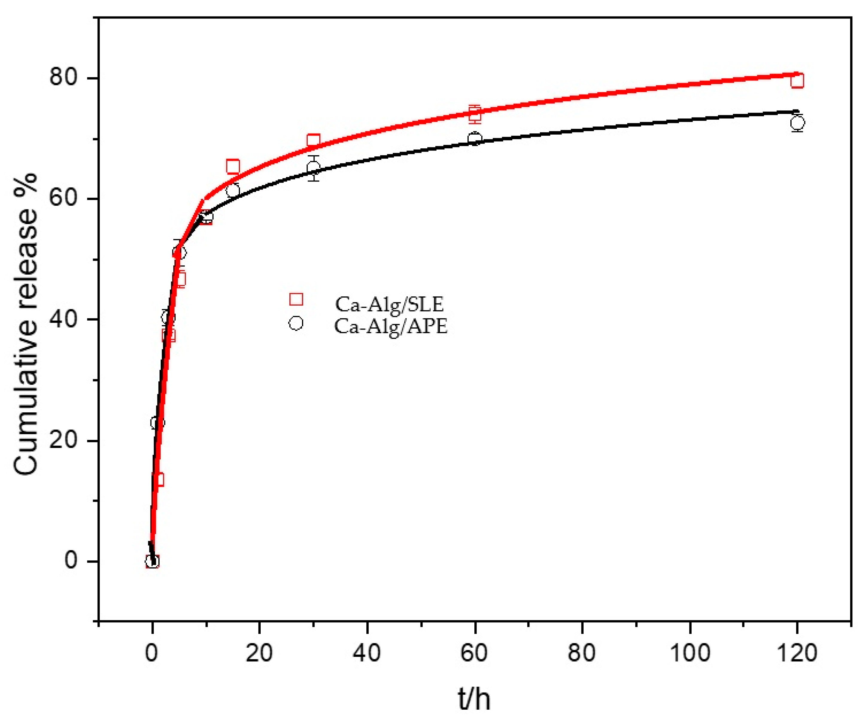
Obtained extracts (SLE or APE) were loaded into biopolymeric microparticles. Sodium alginate (1.5% w/v) and CaCl2 (2% w/v) solutions were prepared by dissolving the latter, separately in the extracts obtained (SLE or APE). Encapsulation was performed with Büchi—Encapsulator B-390 (Switzerland) via ionic gelation method by dropwise addition of sodium alginate (carrier) enriched with bioactive compounds into the calcium-containing extract solution (cross-linking solution). Conditions of the encapsulations were: Nozzle size 300 μm, frequency of 600 Hz (amplitude 3), and the pressure of 0.4 bar. Microparticles (MPs) were stored overnight in the extract containing Ca2+ ions to harden. Two types of microparticles were obtained: (1) MPs containing SLE (Ca-Alg/SLE) and (2) MPs containing APE (Ca-Alg/APE). MPs were air-dried for 24 h until a constant mass was achieved and stored in a sealed container.

2.8. Physicochemical Characterization of Microparticles and Total Polyphenols Release Kinetics

The size of MPs (μm) was determined using optical microscopy (OM) (Leica MZ16a stereomicroscope, Leica Microsystems Ltd., Switzerland). For the determination of encapsulation efficiency, loading capacity, and swelling degree of dry MPs, detailed methods are described in our previous publications [31,33]. The loading capacity of TPC in MPs was determined by dissolving 10 mg of dry microparticles in 5 mL of a mixture of 0.2 M NaHCO3 and 0.06 M Na3C6H5O7 × 2H2O at pH 8 [31]. Results are presented as mg GAE g−1 of dry MPs. Release kinetics were observed as a cumulative release (%) of TPC from prepared MPs. To observe the release profile of TPC from MPs loaded with SLE or APE for up to 120 h, 5 g of dry microparticles Ca-Alg/SLE, or 6 g of dry microparticles Ca-Alg/APE was put into 100 mL distilled water [41].

2.9. Preparation of Viscous Solution for the Application of Microparticles

MPs were added to the sodium alginate (0.2% w/v) solution (5 g/100 mL SLE or 6 g/100 mL APE). The suspension was stirred for 5 min and used as a dipping medium for soybean leaves and pods [43]. Dipping was performed with submersion of leaves/pods and transfer to the Petri dishes.
A laboratory trial was set up in autumn 2019 with adults and third and fourth larval stages of H. halys collected in a soybean field in the vicinity of Šašinovec (middle Croatia, 45°50′13.9″ N 16°11′38.9″ E). Collected insects were kept in entomological cages to recover overnight before testing, without additional feeding and previous contact with insecticides. From the same soybean field, leaves and pods have been collected for a digestive experiment.
In two experiments, the contact and digestive efficacy of two encapsulated extracts (SLE or APE) were evaluated. Investigated ingredients and doses are shown in Table 1. Each type of prepared microparticles was evaluated for contact and digestive action.
For all treatments, ten adult or larvae (depending on variant) of H. halys were placed in a Petri dish ( r = 90   mm ). Contact action was evaluated by applying encapsulated ingredients on the bugs in the Petri dishes ( r = 90   mm ) by spraying 0.2% sodium alginate solution containing MPs using a laboratory sprayer in a volume of 3 mL per Petri dish. One Petri dish represented one replicate. Digestive action was evaluated by placing H. halys into Petri dishes in which treated soybean leaves and pods were placed. The untreated control for all experiments included a treatment in which bugs were placed into Petri dishes treated with water or, in case of digestive action, they were fed with soybean leaves and pods treated with water. Each application and the investigated action of tested ingredients occurred in four replicates. Each replicate had ten individuals of a specific life stage of H. halys. In total 16 different variants were tested on 640 H. hyalis individuals.

2.10. Efficacy Assessment and Data Analyses

The number of dead H. halys in each Petri dish was determined every 24 h for three days. Based on the number of dead H. halys found in the treatment and the untreated control, the efficacy of the ingredients was determined according to Abbott’s formula. Statistical data analysis (one-way ANOVA, Kruskal Wallis test) was performed using ARM 2019® GDM software (Gylling Data Management, 2019) [44].

3. Results and Discussion

3.1. Optimal Extraction Parameters for SLE and APE

Optimization of ultrasound-assisted extraction was performed to achieve an economical method of extraction using only water as a solvent. For SLE, optimal values were found to be at maximum investigated values concentration: 6 g/L, amplitude: 75%, and time: 9 min. (Table 2 and Table A2). In this case, increased temperatures did not significantly decrease polyphenols extraction (Figure A1). APE optimal values were found to be at 10.23 g/L, 74.13% amplitude, with 2 min and 55 s of extraction time (Table 3 and Table A4). For APE, in our preliminary trials (data not shown), we observed degradation of some polyphenolic compounds (anthocyanins) at higher temperatures, so the maximum temperature threshold was set to ~55 °C (Figure A2) and this was also reported in the literature [36]. The end time of the extraction procedure was significantly lower compared to the optimal SLE. Thus, we observe higher values in terms of total polyphenols in SLE (approx. 2.7×), respectively to the APE, but with significantly higher input in energy and time. Lower TPC in APE may also be ascribed to the fact that “spent” pomace was used in the extraction procedure, where most of the compounds were released from the cells in the process of the juice production. Results for TF/TA and AA were in high correlation (above 0.94). Predicted values were following experimental values, with a relative error for TPC of 0.0694%–0.589%. In the case of TA, a higher relative error value (7.9%) can be observed, which may be ascribed to the relative instability of these compounds during and after the processing (Table 3).

3.2. Physicochemical Characteristics of Microparticles Loaded with SLE or APE

Morphological characteristics of microparticles are important parameters that should be considered in the formulation and subsequent application of it to pest control, as spherical beads with uniform size distributions are preferable in the delivery of active functional ingredients [45]. The size and shape analysis of microparticles Ca-Alg/SLE and Ca-Alg/APE was undertaken after the encapsulation procedure. In general, both prepared wet microparticles showed similar narrow size distributions (Table 4). There were visible dry extract particles entrapped into the Ca-alginate matrix. The color of prepared formulations was either slightly green (Ca-Alg/SLE) or red (Ca-Alg/APE). This occurred because of the natural colors present in the stevia leaves (chlorophylls) and Aronia pomace (anthocyanins). The prepared MPs were almost spherical, but after drying to constant mass (approximately four weeks on air at room temperature), their sphericity was lost (Figure 1). Stabilization was achieved since we have not observed the degradation of the bioactive compounds for six months when samples were kept under room temperature in the dark chamber (data not shown). The surface of dried MPs is not smooth and rounded anymore. During the drying process, Ca-Alg/SLE and Ca-Alg/APE MPs significantly decreased in size. The decrease in size was ~45%, resulting from the high-water content in microparticles. There is a loss of irregularity on the surface and shape, which could be explained as a consequence of the water and gel network collapsing during the drying process [46].
Encapsulation efficiency (EE%) of TPC in Ca-alginate microparticles tends to depend on the physical properties of particles (i.e., structure), and the type of encapsulation process. To overcome polyphenolic losses by diffusion and to maximize encapsulation efficiency during the encapsulation process, extracts were also added to the cross-linking solution. Because of the significant diffusional migration of polyphenolic compounds in and out of the microparticles during the ionic gelation process, the same concentration of solutions (extracts) was prepared both for the carrier (sodium-alginate) and cross-linking-solution (CaCl2). This was undertaken to overcome polyphenolic losses by diffusion and to increase the EE% [45]. High EE% values (over 110%) were then observed (Table 4). Furthermore, others have shown that the drying process also has a significant impact on quality parameters and the content of bioactive compounds encapsulated in microparticles [47].
Swelling of microparticles occurred by water absorption, which loosens the networks of gel structure, resulting in the accelerated release of polyphenol from prepared microparticles [48]. When particles are exposed to continuous water absorption, gel particles erode through excessive swelling. For Ca-alginate microparticles, alginate influenced the density of the gel particle structure, while Ca2+ influenced the cross-linking strength [49]. The swelling degree of Ca-Alg/SLE microparticles was almost 50% higher than the swelling degree of Ca-Alg/APE microparticles (Table 4). This could be explained by the variety of present polyphenolic compounds influencing the gel structure firmness. The penetration of water into the denser network with high cross-linking density is difficult, that is, swelling is limited by cross-links and the swelling can be used as a measure of the extent of cross-linking [50]. Accordingly, loading with SLE indicated a lower cross-linking degree of Ca-Alg/SLE in comparison with Ca-Alg/APE.
Physicochemical characterization revealed good morphological properties, loading capacity, and swelling degree, especially EE% of microparticles loaded with either SLE or APE.

3.3. In Vitro Release of Total Polyphenols from Microparticles Loaded with SLE and APE

Microparticles loaded with SLE or APE prepared by the ionic gelation method through extrusion dripping are reservoirs of bioactive components surrounded by a wall that can control the release. The release profiles for SLE and APE are presented in Figure 2. Both release profiles are characterized by rapid initial release followed by a slower release (obeying the power law equation). A plateau was reached after about 20 min, which leads to quick initial efficiency. SLE release fraction is higher than APE which is following a higher degree of swelling. Moreover, the loading capacity of polyphenols in Ca-Alg/SLE microparticles was relatively higher than in the Ca-Alg/APE microparticles, so the higher fraction of release in the latter could also be explained by this result.
To explain and understand the kinetics and type of mechanism involved in the release of total polyphenolic compounds from microparticles, the KorsmeyerPeppas model was applied [51]. According to this model, different controlling mechanisms may be distinguished by a simple empirical equation:
f   =   k t n
where f is cumulative release at time t, k is a kinetic constant characteristic for a particular system considering structural and geometrical aspects, n is the release exponent representing the release mechanism.
The values of the release constants k, and exponents n obtained by fitting the release curves are listed in Table 5. Lower n values than 0.43 indicate that the release process of polyphenols is controlled by diffusion through microparticles. Differences in microparticle composition (i.e., type of extract) do not affect the controlling release mechanism. The values of kinetic constant (Table 5) indicated that the release rate of APE is slower than of SLE. Slower APE diffusion through the alginate matrix could be ascribed to the denser Ca-Alg/APE structure in comparison Ca-Alg/SLE.
Generally, it has been shown that of primary importance in the release of loaded compounds are (i) the calcium alginate network structure, (ii) steric reasons due to the existence of physical entanglements of cross-linked calcium alginate, and (iii) specific interactions between loaded compounds and calcium ions [52]. Polyphenols structure and size of molecules also play a role in possible interactions with alginate residues, all of which are factors that affect release kinetics. This may also explain the possible soldering of specific polyphenols [53].
The underlying mechanism of polyphenols release is diffusion through the alginate gel matrix. This has also been reported for other low-molecular-weight compounds [54]. It means that the rate of polyphenols diffusion is much less than that of polymer swelling and relaxation. Equilibrium of absorption in the surface exposure of the polymeric system takes place rapidly, leading to conditions of time-dependent links. The kinetics of this phenomenon are characterized by diffusivity. The values of kinetic constant (Table 5) exhibited that the release rate of APE is slower than of SLE. Slower APE diffusion through the alginate matrix could be ascribed to the interaction’s denser Ca-Alg/APE structure in comparison to Ca-Alg/SLE. From these results, it could be concluded that Ca-alginate microparticles act as a barrier for polyphenol release [46,55,56].

3.4. The Efficiency of Plant-Based Microparticles on H. halys

Results in Table 6 show that stevia-based microparticles have certain efficiency against H. halys, especially on younger larval stages (third stage) and that the main efficiency is achieved after contact (73%) but also very good efficiency after digestive (60%) treatment. Older H. halys larvae (fourth stage) and adults showed some efficiency only when microparticles have been applied for contact action. Application based on microparticles loaded with APE did not reach the level expected, however, some efficiency has been observed at contact application on both investigated H. halys growth stages.
It is interesting that in all variants, except the third larval stage in the SLE microparticles variant, significant efficiency occurred on the first day after application and did not increase over time. Considering polyphenolic compound release (Figure 2), it is obvious that SLE and APE both had quick initial insecticidal activity, with prolonged SLE insecticidal action in the following days, and without the further insecticidal activity of APE compounds. In the untreated control, all tested individuals survived.
The insecticidal effect of non-nutritive sweetener Truvia and other sweeteners like mannose has already been proven for some insects. Baudier et al. [57] investigated the effect of Erythritol, non-nutritive sugar alcohol, on species Drosophila melanogaster and noted its toxic effect on its movement and life span. Erythritol is the mortality-causing agent within Truvia. The same authors have also emphasized that consumption of Erythritol is safe for humans, even when consumed at high levels, suggesting its use as a novel, human-safe insecticide, which could be applied in urban pest control. Although mannose is toxic to a very important representative of Hymenoptera species (e.g., honeybee), the same effect was not observed for some Dipteran species (e.g., D. melanogaster and Ceratitis capitata). Erythritol toxicity needs to be inspected in other insect species [56]. Ahmad et al. [58] investigated ethanolic and methanolic extracts and dichloromethane extract of S. rebaudiana, Ginkgo biloba, and Parthenium hysterophorous against insect species Anopheles stephensi. It was observed that S. rebaudiana methanolic extract caused high larvicidal activity. S. rebaudiana can also be used as a repellent plant as its repulsive effect was observed at coffee plantations on coffee berry borer, Hypothenemus hampei [59]. Results from our study also demonstrated a potentially good insecticidal effect of S. rebaudiana against the third larval stage of invasive H. halys. This nuisance pest causes discomfort in humans and causes some health risks, so its suppression needs to be carefully planned to avoid additional side effects on human health. Based on this study SLE loaded microparticles could be promising candidates for urban control of this pest. The insecticidal effect of A. melanocarpa on insect species is not known in scientific research, therefore, these results provide the first insights into the effect of APE on insects.
Further tests using SLE- or APE-loaded microparticles, other insects, and other methods of application are needed to determine the full capacity of these potential “green” insecticides. For field applications, phototoxicity, photodegradation, and dispersal ease (among many others) of polyphenolic compounds are necessary to determine the effectiveness of the compounds as a potential bio-pesticide. Additionally, determining the exact mode of action may better facilitate the reduction of the lethal concentration to be comparable to some of the other (bio) insecticides on the market.

4. Conclusions

Ultrasound-assisted extraction presents an economical and effective extraction method for bioactive compounds from stevia leaves and black chokeberry pomace using only water as a solvent. Furthermore, to stabilize natural extracts and make application easier, we have prepared Ca-alginate microparticles loaded with SLE or APE. In the end, a sufficient number of polyphenolic compounds to achieve the desired insecticidal effect was obtained. SLE microparticles showed pesticidal efficiency against H. halys, especially on younger larval stages. Very good efficiency was achieved after contact, while lower but still satisfactory efficiency was achieved after digestive treatment. Microparticles loaded with APE did not achieve satisfactory efficiency, probably due to the significantly lower total polyphenolic content, but a certain toxic impact has been observed at contact application on all H. halys growth stages. These results provide the first insights into the effect of microencapsulated SLE and APE in insect pest suppression and could be a promising tool in the sustainable management of this invasive species, especially in urban areas and home applications.

Author Contributions

Conceptualization, I.P.Ž., S.J., M.V. and D.L.; Formal analysis, I.P.Ž., S.J., M.V., M.A.G., M.M., K.V.-K. and D.L.; Funding acquisition, M.V.; Investigation, I.P.Ž., S.J., M.V. and D.L.; Methodology, S.J., M.V., M.M. and K.V.-K.; Project administration, I.P.Ž., M.V. and D.L.; Resources, I.P.Ž., S.J., M.V. and D.L.; Software, D.L.; Supervision, K.M.M.; Validation, I.P.Ž., S.J., M.V. and D.L.; Writing–original draft, I.P.Ž., S.J., M.V. and D.L.; Writing–review & editing, M.V., K.V.-K. and K.M.M. All authors have read and agreed to the published version of the manuscript.

Funding

This research was funded by the Croatian Science Foundation (Project: UIP-2014-501 09-6462) and HORIZON2020.CloudingFacturing.

Acknowledgments

Authors thank Matej Orešković for technical assistance.

Conflicts of Interest

The authors declare no conflict of interest.

Appendix A. (Stevia Leaves Extract)

Table A1. Response surface methodology design (Box–Behnken design) for stevia (Stevia rebaudiana (Bertoni) Bertoni) leaves extract preparation (random generation).
StdRunAmplitude (%)Concentration (g/L)Time (min)
1017543
625063
1237549
1345046
1655046
565023
775029
387526
1192549
9102543
1112526
17125046
2132566
14145046
15155046
4167566
8175069
Table A2. Sequential Model Sum of Squares [Type I].
ResponseSourceSum of SquaresdfMean SquareF Valuep-Value Prob > F
TPCQuadratic vs. 2FI1575.523525.175.520.0291
ABTS2FI vs. Linear0.3030.104.170.0372
DPPHLinear vs. Mean5.0331.6810.050.0011
Flavon.Linear vs. Mean1456.943485.6577.31<0.0001
Figure A1. The temperature profile of ultrasound-assisted extraction at optimal variables.
Sustainability 12 10079 g0a1

Appendix B. (Black Chokeberry Pomace Extract)

Table A3. Response surface methodology design (Box–Behnken design) for Aronia (Aronia melanocarpa (Michx.) Elliott) pomace extract preparation (random generation).
StdRunAmplitude (%)Concentration (g/L)Time (min)
8175203
17250202
7325203
11450103
16550202
12650303
2775102
13850202
9950101
101050301
151150202
61275201
141350202
31425302
11525102
41675302
51725201
Table A4. Sequential Model Sum of Squares [Type I].
ResponseSourceSum of SquaresdfMean SquareF Valuep-Value Prob > F
TPC2FI vs. Linear18.0936.0312.750.0009
ABTS2FI vs. Linear3.264 × 10-431.088 × 10-46.330.0111
DPPH2FI vs. Linear2.463 × 10-438.210 × 10-45.460.0175
Anthocy.2FI vs. Linear0.1130.03665.83<0.0001
Figure A2. The temperature profile of ultrasound-assisted extraction at optimal variables.
Sustainability 12 10079 g0a2

References

  1. Bariselli, M.; Bugiani, R.; Maistrello, L. Distribution and damage caused by Halyomorpha halys in Italy. EPPO Bull. 2016, 46, 332–334. [Google Scholar] [CrossRef]
  2. Leskey, T.C.; Nielsen, A.L. Impact of the Invasive Brown Marmorated Stink Bug in North America and Europe: History, Biology, Ecology, and Management. Annu. Rev. Entomol. 2018, 63, 599–618. [Google Scholar] [CrossRef] [Green Version]
  3. Rot, M.; Devetak, M.; Carlevaris, B.; Žežlina, J.; Žežlina, I. First record of Brown Marmorated Stink Bug (Halyomorpha hayis (Stål, 1855)) in Slovenia. Acta Entomol. Slov. 2018, 26, 5–12. [Google Scholar]
  4. Vetek, G.; Koranyi, D. Severe damage to vegetables by the invasive brown marmorated stink bug, Halyomorpha halys (Hemiptera: Pentatomidae), in Hungary. Period. Biol. 2017, 119, 131–135. [Google Scholar] [CrossRef]
  5. Gariepy, T.D.; Bruin, A.; Haye, T.; Milonas, P.; Vétek, G. Occurrence and genetic diversity of new populations of Halyomorpha halys in Europe. J. Pest Sci. 2015, 88, 451–460. [Google Scholar] [CrossRef]
  6. Haye, T.; Gariepy, T.; Hoelmer, K.; Rossi, J.-P.; Streito, J.-C.; Tassus, X.; Desneux, N. Range expansion of the invasive brown marmorated stinkbug, Halyomorpha halys: An increasing threat to field, fruit and vegetable crops worldwide. J. Pest Sci. 2015, 88, 665–673. [Google Scholar] [CrossRef]
  7. Lee, D.-H.; Short, B.D.; Joseph, S.V.; Bergh, J.C.; Leskey, T.C. Review of the Biology, Ecology, and Management of Halyomorpha halys (Hemiptera: Pentatomidae) in China, Japan, and the Republic of Korea. Environ. Entomol. 2013, 42, 627–641. [Google Scholar] [CrossRef]
  8. Nielsen, A.L.; Hamilton, G.C.; Matadha, D. Developmental Rate Estimation and Life Table Analysis for Halyomorpha halys (Hemiptera: Pentatomidae). Environ. Entomol. 2008, 37, 348–355. [Google Scholar] [CrossRef]
  9. Candian, V.; Pansa, M.G.; Briano, R.; Peano, C.; Tedeschi, R.; Tavella, L. Exclusion nets: A promising tool to prevent Halyomorpha halys from damaging nectarines and apples in NW Italy. Bull. Insectol. 2018, 71, 21–30. [Google Scholar]
  10. Maistrello, L.; Vaccari, G.; Caruso, S.; Costi, E.; Bortolini, S.; Macavei, L.; Foca, G.; Ulrici, A.; Bortolotti, P.P.; Nannini, R.; et al. Monitoring of the invasive Halyomorpha halys, a new key pest of fruit orchards in northern Italy. J. Pest Sci. 2017, 90, 1231–1244. [Google Scholar] [CrossRef]
  11. Rice, K.B.; Bergh, C.J.; Bergmann, E.J.; Biddinger, D.J.; Dieckhoff, C.; Dively, G.; Fraser, H.; Gariepy, T.; Hamilton, G.; Haye, T.; et al. Biology, Ecology, and Management of Brown Marmorated Stink Bug (Hemiptera: Pentatomidae). J. Integr. Pest Manag. 2014, 5, 1–13. [Google Scholar] [CrossRef]
  12. Mertz, T.L.; Jacobs, S.B.; Craig, T.J.; Ishmael, F.T. The brown marmorated stinkbug as a new aeroallergen. J. Allergy Clin. Immunol. 2012, 130, 999–1001.e1. [Google Scholar] [CrossRef]
  13. Nielsen, A.L.; Hamilton, G.C. Seasonal Occurrence and Impact of Halyomorpha halys (Hemiptera: Pentatomidae) in Tree Fruit. J. Econ. Entomol. 2009, 102, 1133–1140. [Google Scholar] [CrossRef] [PubMed]
  14. Nielsen, A.L.; Hamilton, G.C.; Shearer, P.W. Seasonal Phenology and Monitoring of the Non-Native Halyomorpha halys (Hemiptera: Pentatomidae) in Soybean. Environ. Entomol. 2011, 40, 231–238. [Google Scholar] [CrossRef] [Green Version]
  15. Lee, D.-H.; Cullum, J.P.; Anderson, J.L.; Daugherty, J.L.; Beckett, L.M.; Leskey, T.C. Characterization of Overwintering Sites of the Invasive Brown Marmorated Stink Bug in Natural Landscapes Using Human Surveyors and Detector Canines. PLoS ONE 2014, 9, e91575. [Google Scholar] [CrossRef]
  16. Leskey, T.C.; Short, B.D.; Butler, B.R.; Wright, S.E. Impact of the Invasive Brown Marmorated Stink Bug, Halyomorpha halys (Stål), in Mid-Atlantic Tree Fruit Orchards in the United States: Case Studies of Commercial Management. Psyche A J. Entomol. 2012, 2012, 1–14. [Google Scholar] [CrossRef] [Green Version]
  17. Lee, D.-H.; Leskey, T.C. Flight behavior of foraging and overwintering brown marmorated stink bug, Halyomorpha halys (Hemiptera: Pentatomidae). Bull. Entomol. Res. 2015, 105, 566–573. [Google Scholar] [CrossRef] [Green Version]
  18. Zobel, E.S.; Hooks, C.R.R.; Dively, G.P. Seasonal Abundance, Host Suitability, and Feeding Injury of the Brown Marmorated Stink Bug, Halyomorpha halys (Heteroptera: Penatomidae), in Selected Vegetables. J. Econ. Entomol. 2016, 109, 1289–1302. [Google Scholar] [CrossRef]
  19. Lattanzio, V.; Lattanzio, V.M.T.; Cardinali, A. Role of Polyphenols in the Resistance Mechanisms of Plants against Fungal Pathogens and Insects. In Phytochemistry, Advances in Research; Imperato, F., Ed.; Research Signpost: Trivandrum, Kerala, India, 2006; pp. 23–67. [Google Scholar]
  20. Vinceković, M.; Maslov Bandić, L.; Jurić, S.; Jalšenjak, N.; Čaić, A.; Živičnjak, I.; Đermić, E.; Karoglan, M.; Osrečak, M.; Topolovec-Pintarić, S. The enhancement of bioactive potential in Vitis vinifera leaves by application of microspheres loaded with biological and chemical agents. J. Plant Nutr. 2019, 42, 543–558. [Google Scholar] [CrossRef]
  21. Jurić, S.; Sopko Stracenski, K.; Król-Kilińska, Ż.; Žutić, I.; Fabek Uher, S.; Đermić, E.; Topolovec-Pintarić, S.; Vinceković, M. The enhancement of plant secondary metabolites content in Lactuca sativa L. by encapsulated bioactive agents. Sci. Rep. 2020, 10, 3737. [Google Scholar] [CrossRef] [Green Version]
  22. Myint, K.; Wu, K.; Xia, Y.; Fan, Y.; Shen, J.; Zhang, P.; Gu, J. Polyphenols from Stevia rebaudiana (Bertoni) leaves and their functional properties. J. Food Sci. 2020, 85, 240–248. [Google Scholar] [CrossRef] [Green Version]
  23. Lee, J.E.; Kim, G.-S.; Park, S.; Kim, Y.-H.; Kim, M.-B.; Lee, W.S.; Jeong, S.W.; Lee, S.J.; Jin, J.S.; Shin, S.C. Determination of chokeberry (Aronia melanocarpa) polyphenol components using liquid chromatography–tandem mass spectrometry: Overall contribution to antioxidant activity. Food Chem. 2014, 146, 1–5. [Google Scholar] [CrossRef]
  24. Simmonds, M.S.J.; Blaney, W.M.; Delle Monache, F.; Marini Bettolo, G.B. Insect antifeedant activity associated with compounds isolated from species ofLonchocarpus andTephrosia. J. Chem. Ecol. 1990, 16, 365–380. [Google Scholar] [CrossRef]
  25. Dreyer, D.L.; Jones, K.C. Feeding deterrency of flavonoids and related phenolics towards Schizaphis graminum and Myzus persicae: Aphid feeding deterrents in wheat. Phytochemistry 1981, 20, 2489–2493. [Google Scholar] [CrossRef]
  26. De Paula, J.R.; Vieira, I.J.C.; Das, M.F.; Da Silva, M.F.; Fo, E.R.; Fernandes, J.B.; Vieira, P.C.; Pinheiro, A.; Vilela, E.F. Sesquiterpenes, triterpenoids, limonoids and flavonoids of Cedrela odorata graft and speculations on the induced resistance against Hypsipyla grandella. Phytochemistry 1997, 44, 1449–1454. [Google Scholar] [CrossRef]
  27. Dini, J.; Owen-Smith, N. Condensed tannin in Eragrostis chloromelas leaves deters feeding by a generalist grasshopper. Afr. J. Range Forage Sci. 1995, 12, 49–52. [Google Scholar] [CrossRef]
  28. Capasso, R.; Evidente, A.; Tremblay, E.; Sala, A.; Santoro, C.; Cristinzio, G.; Scognamiglio, F. Direct and mediated effects on Bactrocera oleae (Gmelin) (Diptera; Tephritidae) of natural polyphenols and some of related synthetic compounds: Structure-activity relationships. J. Chem. Ecol. 1994, 20, 1189–1199. [Google Scholar] [CrossRef]
  29. Kulling, S.; Rawel, H. Chokeberry (Aronia melanocarpa)—A Review on the Characteristic Components and Potential Health Effects. Planta Med. 2008, 74, 1625–1634. [Google Scholar] [CrossRef] [Green Version]
  30. Belščak-Cvitanović, A.; Jurić, S.; Đorđević, V.; Barišić, L.; Komes, D.; Ježek, D.; Bugarski, B.; Nedović, V. Chemometric evaluation of binary mixtures of alginate and polysaccharide biopolymers as carriers for microencapsulation of green tea polyphenols. Int. J. Food Prop. 2017, 20, 1971–1986. [Google Scholar] [CrossRef] [Green Version]
  31. Vinceković, M.; Jalšenjak, N.; Topolovec-Pintarić, S.; Đermić, E.; Bujan, M.; Jurić, S. Encapsulation of Biological and Chemical Agents for Plant Nutrition and Protection: Chitosan/Alginate Microcapsules Loaded with Copper Cations and Trichoderma viride. J. Agric. Food Chem. 2016, 64, 8073–8083. [Google Scholar] [CrossRef]
  32. Vinceković, M.; Topolovec-Pintarić, S.; Jurić, S.; Viskić, M.; Bujan, M.; Đermić, E.; Žutić, J.; Fabek Uher, S. Release of Trichoderma viride Spores from Microcapsules Simultaneously Loaded with Chemical and Biological Agents. Agric. Conspec. Sci. 2017, 82, 395–401. [Google Scholar]
  33. Vinceković, M.; Jurić, S.; Đermić, E.; Topolovec-Pintarić, S. Kinetics and Mechanisms of Chemical and Biological Agents Release from Biopolymeric Microcapsules. J. Agric. Food Chem. 2017, 65, 9608–9617. [Google Scholar] [CrossRef]
  34. Jurić, S.; Šegota, S.; Vinceković, M. Influence of surface morphology and structure of alginate microparticles on the bioactive agents release behavior. Carbohydr. Polym. 2019, 218, 234–242. [Google Scholar] [CrossRef]
  35. Jurić, S.; Đermić, E.; Topolovec-Pintarić, S.; Bedek, M.; Vinceković, M. Physicochemical properties and release characteristics of calcium alginate microspheres loaded with Trichoderma viride spores. J. Integr. Agric. 2019, 18, 2534–2548. [Google Scholar] [CrossRef]
  36. Loypimai, P.; Moongngarm, A.; Chottanom, P. Thermal and pH degradation kinetics of anthocyanins in natural food colorant prepared from black rice bran. J. Food Sci. Technol. 2016, 53, 461–470. [Google Scholar] [CrossRef] [Green Version]
  37. Singleton, V.L.; Orthofer, R.; Lamuela-Raventós, R.M. Analysis of total phenols and other oxidation substrates and antioxidants by means of folin-ciocalteu reagent. Methods Enzymol. 1999, 299, 152–178. [Google Scholar] [CrossRef]
  38. Ough, C.S.; Amerine, M.A. Nitrogen compounds. In Methods Analysis of Musts and Wines; Wiley-Interscience: New York, NY, USA, 1988; pp. 172–195. [Google Scholar]
  39. Ivanova, V.; Stefova, M.; Chinnici, F. Determination of the polyphenol contents in Macedonian grapes and wines by standardized spectrophotometric methods. J. Serbian Chem. Soc. 2010, 75, 45–59. [Google Scholar] [CrossRef]
  40. Brand-Williams, W.; Cuvelier, M.E.; Berset, C. Use of a free radical method to evaluate antioxidant activity. LWT Food Sci. Technol. 1995, 28, 25–30. [Google Scholar] [CrossRef]
  41. Re, R.; Pellegrini, N.; Proteggente, A.; Pannala, A.; Yang, M.; Rice-Evans, C. Antioxidant activity applying an improved ABTS radical cation decolorization assay. Free Radic. Biol. Med. 1999, 26, 1231–1237. [Google Scholar] [CrossRef]
  42. Fuleki, T.; Francis, F.J. Quantitative Methods for Anthocyanins: 1. Extraction and Determination of Total Anthocyanins in Cranberries. J. Food Sci. 1968, 33, 266–274. [Google Scholar] [CrossRef]
  43. IRAC. Susceptibility Test Method 033. Available online: https://irac-online.org/content/uploads/Method_033_Lygus_30March16.pdf (accessed on 15 September 2019).
  44. Gylling Data Management Inc. ARM 2019® GDM Software, Revision 2019.4, 5 August 2019 (B = 25105); Gylling Data Management Inc.: Brookings, South Dakota, 2019. [Google Scholar]
  45. Arriola, N.D.A.; Chater, P.I.; Wilcox, M.; Lucini, L.; Rocchetti, G.; Dalmina, M.; Pearson, J.P.; Dias de Mello, R.; Amboni, C. Encapsulation of stevia rebaudiana Bertoni aqueous crude extracts by ionic gelation—Effects of alginate blends and gelling solutions on the polyphenolic profile. Food Chem. 2019, 275, 123–134. [Google Scholar] [CrossRef] [Green Version]
  46. Dai, Y.; Li, P.; Zhang, J.; Wang, A.; Wei, Q. Swelling characteristics and drug delivery properties of nifedipine-loaded pH sensitive alginate–chitosan hydrogel beads. J. Biomed. Mater. Res. Part B Appl. Biomater. 2008, 86B, 493–500. [Google Scholar] [CrossRef]
  47. Katsube, T.; Tsurunaga, Y.; Sugiyama, M.; Furuno, T.; Yamasaki, Y. Effect of air-drying temperature on antioxidant capacity and stability of polyphenolic compounds in mulberry (Morus alba L.) leaves. Food Chem. 2009, 113, 964–969. [Google Scholar] [CrossRef]
  48. Sriamornsak, P.; Thirawong, N.; Korkerd, K. Swelling, erosion and release behavior of alginate-based matrix tablets. Eur. J. Pharm. Biopharm. 2007, 66, 435–450. [Google Scholar] [CrossRef]
  49. Draget, K.I.; Gaserod, O.; Aune, I.; Andersen, P.O.; Storbakken, B.; Stokke, B.T.; Smidsrod, O. Effects of molecular weight and elastic segment flexibility on syneresis in Ca-alginate gels. Food Hydrocoll. 2001, 15, 485–490. [Google Scholar] [CrossRef]
  50. Roger, S.; Talbot, D.; Bee, A. Preparation and effect of Ca2+ on water solubility, particle release and swelling properties of magnetic alginate films. J. Magn. Magn. Mater. 2006, 305, 221–227. [Google Scholar] [CrossRef]
  51. Korsmeyer, R.W.; Gurny, R.; Doelker, E.; Buri, P.; Peppas, N.A. Mechanisms of solute release from porous hydrophilic polymers. Int. J. Pharm. 1983, 15, 25–35. [Google Scholar] [CrossRef]
  52. Rousseau, I.; Le Cerf, D.; Picton, L.; Argillier, J.F.; Muller, G. Entrapment and release of sodium polystyrene sulfonate (SPS) from calcium alginate gel beads. Eur. Polym. J. 2004, 40, 2709–2715. [Google Scholar] [CrossRef]
  53. Martinsen, A.; Skjåk-Braek, G.; Smidsrød, O. Alginate as immobilization material: I. Correlation between chemical and physical properties of alginate gel beads. Biotechnol. Bioeng. 1989, 33, 79–89. [Google Scholar] [CrossRef]
  54. Stojanovic, R.; Belscak-Cvitanovic, A.; Manojlovic, V.; Komes, D.; Nedovic, V.; Bugarski, B. Encapsulation of thyme (Thymus serpyllum L.) aqueous extract in calcium alginate beads. J. Sci. Food Agric. 2012, 92, 685–696. [Google Scholar] [CrossRef]
  55. Kim, E.S.; Lee, J.-S.; Lee, H.G. Calcium-alginate microparticles for sustained release of catechin prepared via an emulsion gelation technique. Food Sci. Biotechnol. 2016, 25, 1337–1343. [Google Scholar] [CrossRef]
  56. Ćujić, N.; Trifković, K.; Bugarski, B.; Ibrić, S.; Pljevljakušić, D.; Šavikin, K. Chokeberry (Aronia melanocarpa L.) extract loaded in alginate and alginate/inulin system. Ind. Crops Prod. 2016, 86, 120–131. [Google Scholar] [CrossRef]
  57. Baudier, K.M.; Kaschock-Marenda, S.D.; Patel, N.; Diangelus, K.L.; O’Donnell, S.; Marenda, D.R. Erythritol, a Non-Nutritive Sugar Alcohol Sweetener and the Main Component of Truvia®, Is a Palatable Ingested Insecticide. PLoS ONE 2014, 9, e98949. [Google Scholar] [CrossRef]
  58. Ahmad, N.; Fazal, H.; Abbasi, B.H.; Iqbal, M. In vitro larvicidal potential against Anopheles stephensi and antioxidative enzyme activities of Ginkgo biloba, Stevia rebaudiana and Parthenium hysterophorous. Asian Pac. J. Trop. Med. 2011, 4, 169–175. [Google Scholar] [CrossRef] [Green Version]
  59. Castro, A.M.; Tapias, J.; Ortiz, A.; Benavides, P.; Góngora, C.E. Identification of attractant and repellent plants to coffee berry borer, Hypothenemus hampei. Entomol. Exp. Appl. 2017, 164, 120–130. [Google Scholar] [CrossRef]
Figure 1. Dry Ca-Alg/SLE microparticle under the light microscope (left) and Ca-Alg/APE microparticles under the phase-contrast light microscope (right). Scale bars are indicated.
Sustainability 12 10079 g001
Figure 2. The release profile of total polyphenols from dry microparticles loaded with SLE or APE as the cumulative release (%) over time (min).
Sustainability 12 10079 g002
Table 1. Treatment labels, insect stages, investigated actions, and applied dosage.
TreatmentStage of InsectAction InvestigatedA Dose of a.i. Applied per Repetition (mg)
Ca-ALG/SLEadultscontact150
digestive
3rd larval stagecontact150
digestive
4th larval stagecontact150
digestive
Ca-ALG/APEadultscontact180
digestive
4th larval stagecontact180
digestive
Untreatedadultscontactwater
digestive
3rd larval stagecontact
digestive
4th larval stagecontact
digestive
Table 2. Optimal variable setup with predicted-optimal vs. actual-experimental responses for SLE.
VariablesAmplitude (%)Concentration (g/L)Time (min)Desirability
Optimal values75690.984
ResponsesTPC (mg GAE/L)TF (mg QE/L)ABTS (mmol TE/L)DPPH (mmol TE/L)
Predicted (Opt.)465.16249.0612.3693.104
Actual (Exp.)467.92649.6392.4163.036
Relative error %0.5891.1641.9572.229
Table 3. Optimal variable setup with predicted-optimal vs. actual-experimental responses for APE.
VariablesAmplitude (%)Concentration (g/L)Time (min)Desirability
Optimal values70.1811.522 min 55 s1.000
ResponsesTPC (mg GAE/L)Anthocyanins (mg M3GE/L)ABTS (mmol TE/L)DPPH (mmol TE/L)
Predicted (Opt.)174.352912.49340.91700.8018
Actual (Exp.)174.231911.50500.90550.7672
Relative error %0.06947.91121.25714.3113
Table 4. Physicochemical characteristics of microparticle formulations, Ca-Alg/SLE, and Ca-Alg/APE.
SampleSize of Dry Microparticles (μm)Encapsulation Efficiency (%)—TPCLoading Capacity (mg TPC/g Dry Microparticles)Swelling Degree (%) of Dry Microparticles
Ca-Alg/SLE *220.121 ± 48.235112.229 ± 1.3152.586 ± 0.178123.225 ± 1.385
Ca-Alg/APE **218.657 ± 62.147111.021 ± 4.5211.017 ± 0.06860.111 ± 18.018
* SLE—stevia leaves extract; ** APE—Aronia pomace extract.
Table 5. Variation of the release constant (k/h), exponent (n), and correlation coefficient (R2) of polyphenols released from microparticles loaded with SLE and APE.
Dry Microparticlesk/hnR2
Ca-Alg/SLE50.5600.09440.9989
Ca-Alg/APE45.1270.10440.9981
Table 6. Mortality (%) of encapsulated natural extracts in the experiment.
TreatmentA Dose of a.i. Applied per Repetition (mg)Growth StageInvestigated ActionDays After the Treatment
123
Ca-Alg/SLE150adultscontact20 ab+20 bc20 c
digestive0 c0 d0 c
1503rd larval stagecontact25 a75 a75 a
digestive0 c60 a60 ab
1504th larval stagecontact0 c30 b47.5 b
digestive0 c5 cd5 c
Ca-Alg/APE180adultscontact10 bc10 cd10 c
digestive0 c0 d0 c
1804th larval stagecontact5 c5 cd5 c
digestive0 c0 d0 c
Bartlett’s X25.859.1412.55
P(Bartlett’s X2)0.016 *0.027 *0.006 *
P(KruskalWallis X2)0.0060.0010.001
+ Mean values of the same column followed by the same letter are not significantly different (* p ≥ 0.05; LSD test).
Publisher’s Note: MDPI stays neutral with regard to jurisdictional claims in published maps and institutional affiliations.

