Review

Heavy Metal Contamination of Natural Foods Is a Serious Health Issue: A Review

1
Department of Biochemistry, Government College University, Faisalabad 38000, Pakistan
2
Laboratory of Human Pathologies Biology, Department of Biology, Faculty of Sciences, Mohammed V University in Rabat, Rabat 10056, Morocco
3
Laboratory of Histology, Embryology and Cytogenetic, Faculty of Medicine and Pharmacy, Mohammed V University in Rabat, Rabat 10056, Morocco
4
Department of Clinical Neurophysiology, Hospital of Specialities, Rabat Institute, Ibn Sina University Hospital, Rabat 10056, Morocco
5
Laboratory of Biodiversity, Ecology, and Genome, Faculty of Sciences, Mohammed V University in Rabat, Rabat 10056, Morocco
6
Physiology and Physiopathology Team, Department of Biology, Mohammed V University in Rabat, Rabat 10056, Morocco
7
Department of Eastern Medicine, Government College University, Faisalabad 38000, Pakistan
8
Department of Scientific Research, Kurgan Federal State Budgetary Educational Institution of Higher Education, Kurgan State Agricultural Academy by T.S. Maltsev, 641300 Kurgan, Russia
9
Department of Scientific Research, South Ural State Agrarian University, 13 Gagarin St., 457100 Troitsk, Russia
10
V. M. Gorbatov Federal Research Center for Food Systems, 26 Talalikhina St., 109316 Moscow, Russia
11
Department of Scientific Research, K. G. Razumovsky Moscow State University of Technologies and Management (The First Cossack University), 73 Zemlyanoy Val, 109004 Moscow, Russia
12
Faculty of Biotechnology and Food Engineering, Ural State Agrarian University, 42 Karl Liebknecht Str., 620075 Yekaterinburg, Russia
13
Department of Biotechnology, Sri Shakthi Institute of Engineering and Technology, Coimbatore 641062, India
14
Department of Crop Science, College of Sanghuh Life Science, Konkuk University, Seoul 05029, Korea
*
Author to whom correspondence should be addressed.
Sustainability 2022, 14(1), 161; https://0-doi-org.brum.beds.ac.uk/10.3390/su14010161
Submission received: 2 November 2021 / Revised: 10 December 2021 / Accepted: 16 December 2021 / Published: 24 December 2021
(This article belongs to the Special Issue Chemical Pollution, Prevention, and Environmental Sustainability)

Abstract

:
Heavy metals play an important role in the homeostasis of living cells. However, these elements induce several adverse environmental effects and toxicities, and therefore seriously affect living cells and organisms. In recent years, some heavy metal pollutants have been reported to cause harmful effects on crop quality, and thus affect both food security and human health. For example, chromium, cadmium, copper, lead, and mercury were detected in natural foods. Evidence suggests that these elements are environmental contaminants in natural foods. Consequently, this review highlights the risks of heavy metal contamination of the soil and food crops, and their impact on human health. The data were retrieved from different databases such as Science Direct, PubMed, Google scholar, and the Directory of Open Access Journals. Results show that vegetable and fruit crops grown in polluted soil accumulate higher levels of heavy metals than crops grown in unpolluted soil. Moreover, heavy metals in water, air, and soil can reduce the benefits of eating fruits and vegetables. A healthy diet requires a rational consumption of foods. Physical, chemical, and biological processes have been developed to reduce heavy metal concentration and bioavailability to reduce heavy metal aggregation in the ecosystem. However, mechanisms by which these heavy metals exhibit their action on human health are not well elucidated. In addition, the positive and negative effects of heavy metals are not very well established, suggesting the need for further investigation.

1. Introduction

Heavy metal contamination is a global phenomenon disrupting the ecosystem and creating serious risks for human health. The main challenges include increased urbanization, property transition, and industrial development, particularly in highly populated and emerging countries [1,2]. Heavy metals are both anthropogenic and natural in origin, and some of them such as chromium (Cr), vanadium, and nickel (Ni) are essential for human health in minor quantities. Indeed, these metals play an important role in cellular activity. However, some heavy metals have harmful effects on the environment as well as on living organisms [3]. Human exposure to heavy metals is triggered by inhalation, oral ingestion, and skin application [4]. Further, the balance between positive effects and side effects of heavy metals depends on their concentrations in living cells. Therefore, the levels of metal ions must be maintained within an appropriate range to prevent nutritional deficiencies, whereas higher concentrations can cause health concerns.
Cobalt (Co), copper (Cu), nickel, iron (Fe), manganese (Mn), zinc (Zn), molybdenum (Mo), and selenium (Se) are vital for living organisms, but can cause serious health complications if consumed above the safe limit [5,6,7,8,9,10]. Other metals such as arsenic (As), cadmium (Cd), chromium, and lead (Pb) are not essential for human health and can cause serious health complications even at low concentrations [11,12,13,14,15,16,17,18,19,20]. Under specific industrial and ecological conditions, metals constitute an important class of toxic substances. The ubiquitous exposure to heavy metals is of great interest. Toxic metals induce environmental pollution due to their extensive use in everyday life [21]. Toxic metals are dispersed in the environment to a large extent. Heavy metal distribution in the form of particulate matter or vapor by wind depends on their physical state. Meta components are transferred from the atmosphere into the soil or water surface, resulting in environmental pollution. Industrial effluents are the major source of metallic pollution in the hydrosphere. Toxic heavy metals such as nickel, lead, copper, chromium, cadmium, and arsenic occurring in wastewater represent environmental and health risk [22,23].
These metals are listed among the top twenty toxic chemicals by the United States Environmental Protection Agency [24,25]. Heavy metals enter the environment via traditional routes and anthropogenic mechanisms. They can be traced to a variety of sources, including soil erosion, natural earth crust weathering, mining, liquid waste, municipal sewage, and insecticides [26]. Soil and air pollution contributes to heavy metal accumulation in vegetables [27]. In fact, natural food contamination occurs through contact with contaminated soils or via air pollution [2]. Heavy metals accumulate in the human skeleton and fatty tissues, leading to depletion of key nutrients and resulting in central nervous system deficits, in addition to cardiac, gastric, hematological, hepatocellular, renal, neurodevelopmental, reproductive, and immune disorders, as well as intrauterine retardation [3]. This review assesses heavy metal contamination in plant foods and underscores the need to prevent the consumption of contaminated plant foods.

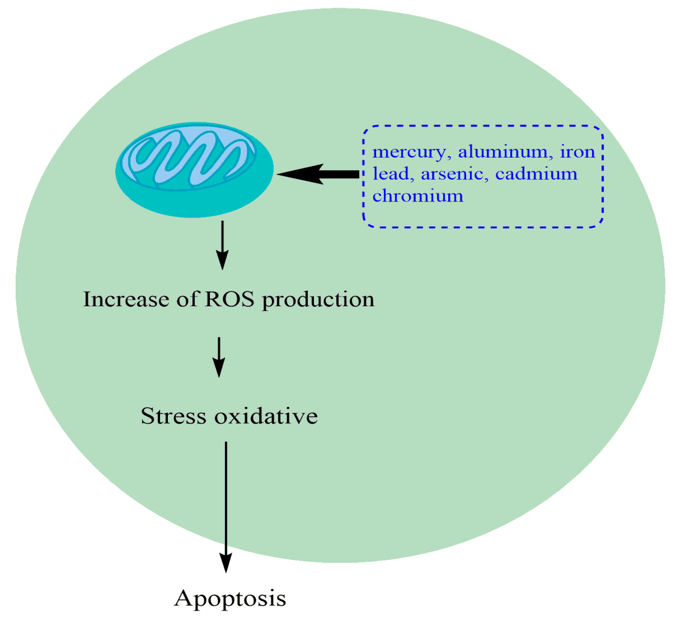
2. Different Heavy Metals and Their Toxicity
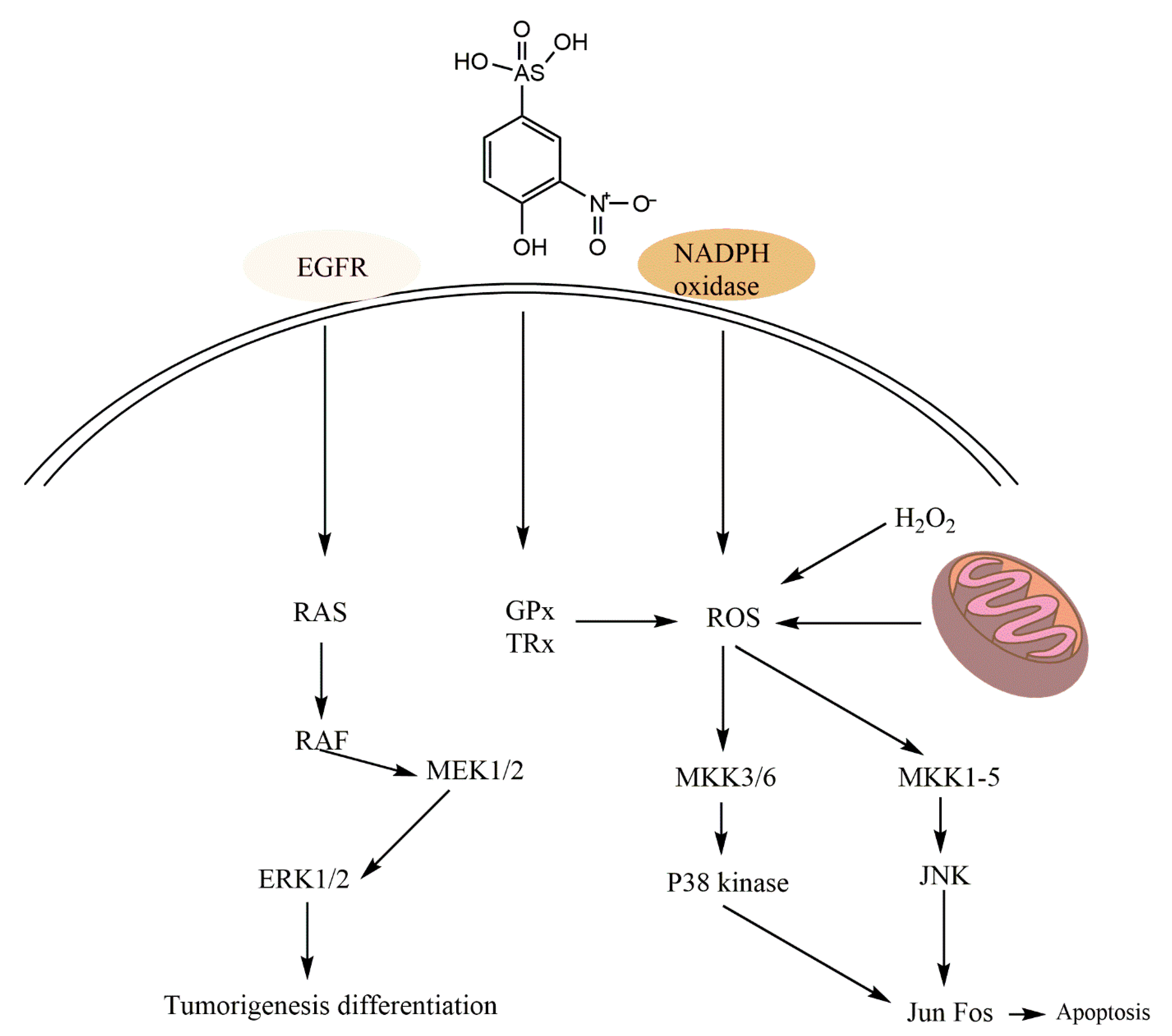
Current investigations demonstrate the toxic effects of heavy metals. Table 1 highlights the toxic effects and the underlying mechanisms of toxicity due to five major heavy metals, namely arsenic, chromium, cadmium, lead, and mercury (Table 1 and Figure 1). The results focus on preclinical and clinical outcomes involving acute and chronic metal exposures, as well as the associated adverse effects on target organs.
The most common natural sources of heavy metals in soil and agriculture include atmosphere, irrigation with sewage, pesticides, phosphate fertilizers, livestock manure, and sewage sludge. Other sources of anthropogenic contaminants derived from plants pose a major risk to human health following dietary intake of root crops contaminated with polluted soil or via direct atmospheric deposition of heavy metals on plant surfaces. In addition, particulate vehicular emissions may adversely affect global food safety [24].

2.1. Arsenic

Exposure to the highly toxic arsenic can be either occupational or through polluted food and water [44]. It is found in water, food, and environment as a contaminant in several forms: inorganic (As3+ and As5+), organic, metalloid (As0), and arsine (AsH3). The toxicity of As compounds occurs in the following order: organic arsenicals < As0 < inorganic species (As5+ < As3+) < arsine [28,45]. Metallic arsenic is an ecological toxin naturally present in all soils [46]. The major human exposure to arsenic in the ecosystem is polluted water. Arsenic enters the human food chain following exposure to polluted crops and feed [47]. Arsenic is not crucial but is generally toxic to plants. The roots are usually exposed first to arsenic resulting in inhibition of root growth [47]. It is widely present in the form of oxides of iron, sodium, and calcium, and is highly toxic and carcinogenic [48]. The inorganic arsenic compounds, namely arsenite and arsenate, are considered to be fatal to living beings and very harmful to the environment. Arsenic is the 20th most abundant element on earth. Humans encounter arsenic in natural and industrial sources. Improper disposal of arsenic-based chemicals can pollute drinking water [49]. Accidental consumption of arsenic by children or in suicide attempts can cause acute poisoning [50]. Interaction between arsenic and sulphydryl groups in cells impairs cellular enzymes and interferes with mitosis.

2.1.1. Mechanism of Arsenic Toxicity

Compared with arsenic, the arsenic metabolites formed enzymatically in biological systems can cause considerable side effects. During arsenic biotransformation, harmful inorganic arsenic compounds are methylated by bacteria, fungi, and humans to produce monomethylarsonic acid (MMA) and dimethylarsinic acid (DMA). During this biotransformation, inorganic arsenic species are enzymatically transformed into methylated arsenic compounds, which are the final metabolites and biomarkers associated with persistent arsenic levels. Both MMA and DMA products are formed via biomethylation and excreted in the urine, indicating chronic exposure to arsenic [51]. Previous studies suggested that arsenic is detoxified via methylation; however, other recent studies have shown conflicting results, demonstrating the increased toxicity of other methylated metabolites containing trivalent forms of arsenic compared with arsenite alone [52] (Figure 2). Further genotoxicity assays have shown that arsenic compounds inhibit DNA repair and induce chromosomal alterations, sister chromatid exchanges, and micronuclei formation in cultured human and rodent cells and exposed human cells [53]. Some in vitro studies investigating the carcinogenic mechanisms of arsenic have shown that this molecule and its compounds are cytotoxic and induce morphological changes in Syrian hamster embryo cells (SHE) as well as mouse C3H10T1/2 and BALB/3T3 cells [54,55]. An in vitro study of DNA damage using the comet test showed that arsenic trioxide (As2O3) induces DNA damage in human lymphocytes and mouse leukocytes [56]. Moreover, it has been shown that arsenic compounds induce gene amplification, inhibit mitosis, block DNA repair, and activate the expression of oxidative stress protein heme oxygenase and the c-fos gene in mammalian cells [57]. Trouba et al. [58] and Zhao et al. [59] reported that chronic exposure to high levels of arsenic increases the sensitivity of cells to mitogenic stimulation and altered mitogenic signaling contributes to its carcinogenicity. In this sense, arsenic can act as a carcinogen by causing DNA hypomethylation, which in turn increases the gene expression. Likewise, arsenic represents a potent stimulator of transactivation of the protein kinase regulated by extracellular Erk1 and AP-1, and an efficient inducer of the expression of c-fos and c-jun genes [60]. In contrast, other recent clinical trials have shown that As2O3 may be used to treat acute promyelocytic leukemia (APL) [61,62]. As2O3 is a tumor-specific agent that selectively induces apoptosis in APL cells [63,64]. Numerous cell culture and human studies of cancer chemotherapy in APL have shown that As2O3 treatment inhibits cell cycle and apoptosis of malignant cells. Other in vitro studies have also reported that arsenic modulates protein and gene expression, DNA synthesis, mitosis and/or apoptotic mechanisms, and genotoxicity in many cells including T cells, monocytes, microvascular endothelial cells, melanocytes, and keratinocytes [65], dendritic cells, dermal fibroblasts, colon cancer cells [61], lung cancer cells [66], human leukemia cells [67], Jurkat-T lymphocytes, and human hepatic carcinoma cells [68].

2.1.2. Systemic Effects of Dietary Arsenic
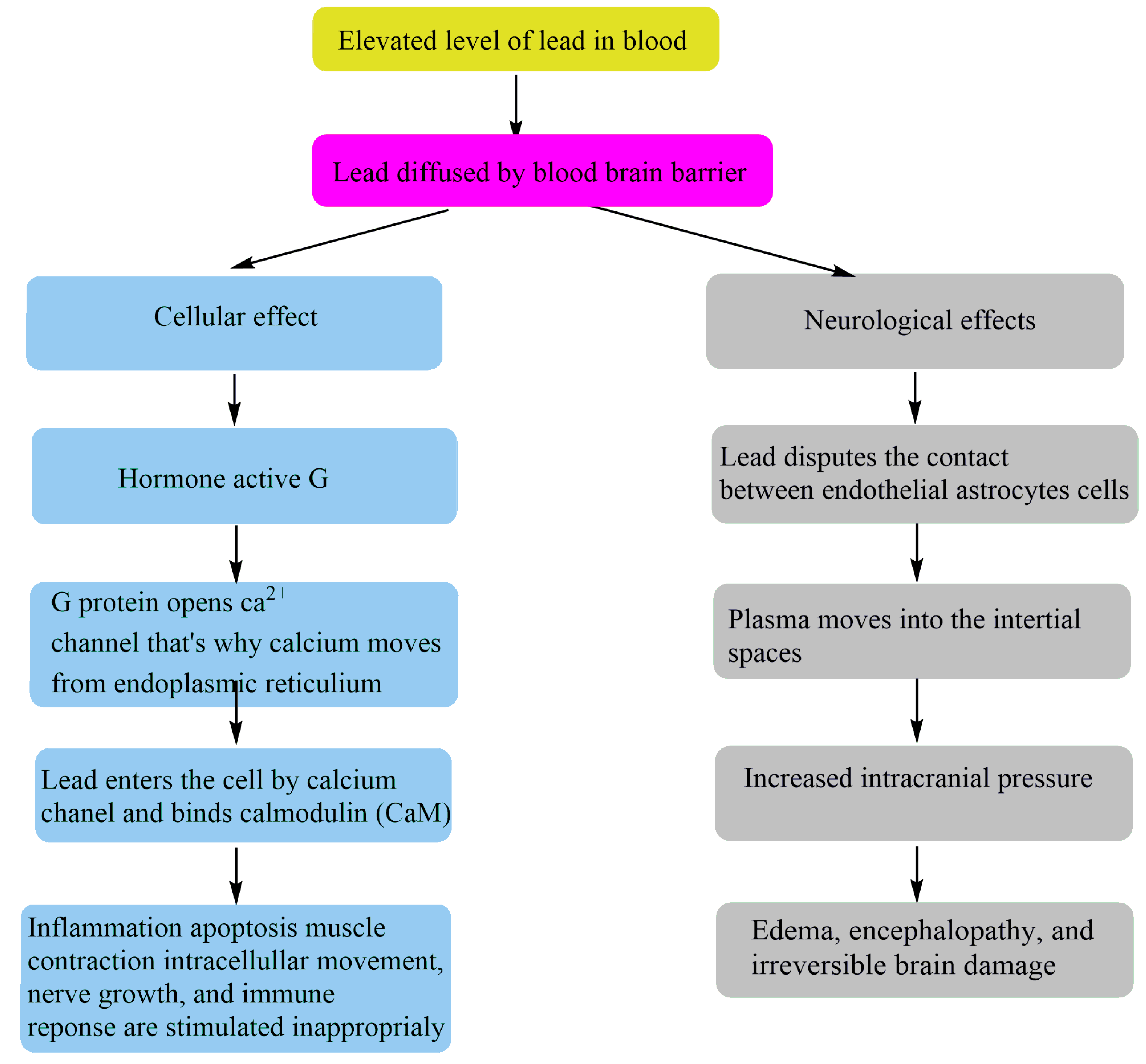
Exposure to soluble inorganic arsenic can trigger instant death, via vomiting, damage to the nervous system, and hemodynamic instability. This lethal effect can be triggered by the ingestion of a large amount of arsenic [69]. Long-term neurological effects, strokes, and cancers in sites other than the skin, kidneys, bladder, and lungs are triggered by exposure to the lowest levels of arsenic [46]. It has a weak effect on diabetes and reproduction, and a major impact on heart attacks, high blood pressure, and other circulatory diseases. Arsenic can also contaminate food and water bases such as shellfish and other seafood, as well as fruits and vegetables. Arsenic toxicity occurs due to exposure to contaminated wine or due to malicious intent. Long-term exposure to arsenic can cause skin changes such as redness and swelling in addition to sensory and motor nerve abnormalities. Further, the function of liver and kidneys may be affected [70]. Arsenic has been classified as a carcinogen by the Environmental Protection Agency (EPA).
The mechanism of heavy metal carcinogenicity is not well understood. Some heavy metals are carcinogenic primarily due to their binding to regulatory proteins involved in apoptosis, DNA repair and synthesis, and cell cycle regulation [71]. Some toxicity studies have shown that certain transcription factors (TFs) such as p53, nuclear factor-kappa B (NF-κB), and activator protein 1 (AP-1) are targets for arsenic and cadmium. Therefore, the inability to control the expression of protective genes can lead to uncontrolled cell growth and division [72]. Studies evaluated mutations in RAS proteins or increased activation following exposure to carcinogenic heavy metals. Ngalame et al. [73] reported the overexpression of RAS in human prostate epithelial cells after exposure to arsenic. In another investigation, cadmium exposure increased the levels of transcription factors jun and fos, as well as ERK 1/2 in vitro. In cultured cells, chromium (VI) also induced overexpression of c-jun. The kinase cascade was not deactivated, and the mutated RAS protein lost its inactivation. In addition, the gene expression of activated ERK 1/2 or intensified jun and fos was continued. Thus, a continuously activated proliferation was induced via irreversible activation of signaling pathway, thereby inducing increased tumor formation [74]. Ding et al. [75] showed that arsenic inhibits DNA repair by inhibiting poly ADP-ribose polymerase 1 (PARP-1), a DNA repair enzyme. Resistance to apoptosis due to heavy metal exposure disrupts basic cellular defense.
Oxidative stress in the reproductive organs led to comparable inactivation of the antioxidant enzymes superoxide dismutase (SOD), catalase (CAT), and glutathione peroxidase (GPx), as well as a simultaneous increase in lipid peroxidation following arsenic intoxication [76].

2.2. Lead (Pb)

Numerous studies have established the increased levels of lead toxicity [77,78]. Excessive exposure to lead can cause serious health issues in humans in addition to severe environmental contamination. The origins of lead toxicity can be traced to food, soil, air, industrial emissions, drinking water, e-waste, industrial process, herbal products, smoking, traditional medicines, cosmetics, and domestic sources [79,80,81]. Similarly, other sources of lead contamination include paints and gasoline in the faucets, pewter pitchers, toys, storage batteries, lead bullets, glazed ceramics, and plumbing pipes [82,83]. Annually, vehicles consume approximately 100 to 200,000 tons of lead in the United States. Lead is a very harmful metal that interferes with different physiological pathways in plants and unlike other metals such as copper, zinc and manganese, it has no biological role [84,85]. A high lead concentration in plants increases the synthesis of ROS by adversely affecting the photosynthetic and chlorophyll metabolism [86]. Studies suggest that lead reduces the quality of tea and biomass by altering its chemical composition [87]. It was found that exposure to even a low concentration of lead in plants causes extremely high ion instability, leading to large metabolic variation via potent inhibition of plant growth and photosynthetic capacity.

2.2.1. Lead Toxicity Mechanism

Lead interferes with a variety of metabolic processes, including calcium metabolism and protein reactions. Lead replaces calcium in the body and interacts with biological components, interfering with their normal function [88]. In living cells, lead poisoning is mostly mediated via ionic mechanisms and oxidative stress. Oxidative stress in living cells is attributed to the differences between the levels of antioxidants and free radicals to detoxify the reactive intermediates or restore the destructive effects. Figure 1 represents the toxicity of heavy metals in cells and the equilibrium between subsequent defense and ROS production induced by antioxidants. Antioxidants, such as glutathione, exist in the cell to protect against free radicals such as H2O2. However, higher levels of lead exposure increase ROS levels while decreasing the levels of antioxidants [89].
Since glutathione occurs in oxidized (GSSG) and decreased (GSH) states, the reduced form of glutathione generates equivalents (H++ e−) from the thiol classes of cysteine to ROS to ensure their stability. Reduced glutathione rapidly binds to an additional glutathione molecule after donating the electron and results in glutathione disulfide (GSSG) formation in the presence of the enzyme glutathione peroxidase. Under normal conditions, the oxidized form (GSSG) represents 10%, while the reduced form of GSH represents 90% of the overall content [90]. The GSSG concentration increases the concentration of GSH under oxidative stress. Another biomarker for oxidative stress is lipid peroxidation, which occurs when free radicals withdraw electrons from lipid molecules in the cell membrane [91,92]. The higher concentration of ROS at the cellular level may alter the structural integrity of cells, nucleic acids, proteins, lipids, and membranes, resulting in stress [93]. The ionic mechanism of lead toxicity usually occurs as the lead ions outnumber other monovalent cations such as Na+ and bivalent cations such as Fe2+, Ca2+, and Mg2+, which ultimately disrupts the metabolic rate of the cells. The ionic mechanism of lead poisoning alters the intra and intercellular signaling mechanisms, cell adhesion, apoptosis, ionic transportation, protein folding, mutation, release of neurotransmitters, and enzyme regulation. Furthermore, lead contamination alters the expression of protein kinase C, which regulates memory storage and neuronal function [92]. Lead remains a harmful environmental pollutant with high toxic effects on humans. Lead exposure is considered to be the most common cause of lead poisoning disease, with symptoms primarily affecting the gastrointestinal (GI) tract and central nervous system (CNS) in both children and adults [28]. Exposure to lead can induce cardiovascular, urinary, respiratory, and neurological diseases via immune modulation, and inflammatory and oxidative mechanisms, which can cause an imbalance in the oxidant-antioxidant system and subsequently lead to inflammatory responses in various organs, especially the renal system. Exposure to lead can alter the physiological functions of the body, leading to many diseases [94].

2.2.2. Dietary Effects of Lead on Kidney

Exposure to lead can damage kidneys and also cause kidney failure, based on several studies. Large doses of lead can induce serious kidney damage such as rupture of proximal tubular function, resulting in glycosuria, aminoaciduria, and hyperphosphaturia (Fanconi-like syndrome), which are reversible. Nevertheless, repetitive or continuous exposure can induce toxic stress on the kidney, resulting in chronic and irreversible nephropathy (interstitial nephritis) due to lead toxicity (Figure 3). However, no concomitant developmental studies involving undernutrition are available [95]. The impact of chronic lead intoxication before conception on the biochemical parameters and renal collapse was investigated in rats. In this study, the male Wistar rats were treated for 8 weeks with lead acetate, which led to a decrease in animal body weight [96]. This result is consistent with other studies reporting diminished growth due to insufficient food intake following loss of appetite caused by lead exposure. The anorectic impact exerted by this toxic metal supports its role in catecholaminergic, glutamatergic, and serotoninergic neurotransmission. In addition to the anorectic impact in the treated rats, the water consumption was decreased significantly (p < 0.05) compared with the control group.
This reduction in water intake is related to the dose, which explains the decreased weight following lead exposure. An important factor in urinary chemistry is to elucidate kidney function [97]. Urinary volume is clearly enhanced in the lead group. Improved urine output may be due to the diuretic impact of lead. In this study, no specific gravity or variation in pH was noticed. The study by Ghorbe et al. [95] revealed a decline in urinary pH mainly from day 45 of the investigation. A number of studies reported that the crystals adversely affected the pathogenesis of hypercalciuria but not renal failure. The Ca/Ox ratio and the level of supersaturation affect crystalluria, which promotes nucleation, precipitation of calcium oxalate, successive crystal growth, and amplified urinary calcium concentration. In fact, crystals are formed due to the presence of a protein in the urine. However, lead exposure caused inflammation of the urinary tract in rats. In addition, the level of calcium and phosphorus ions in the urine increased, as well as the levels of calcium and lead acetate. The increased levels of these ions suggest the possibility of calcium phosphate crystal formation.
Renal failure results from renal lesions, reduced permeability of the kidney barrier in the lower urinary tract, or disturbed metabolism following lead contamination. Therefore, the reduction in urine and the increase in serum creatinine are attributed to lead toxicity. Rats exposed to lead showed a decrease in urea in the urine, although serum urea levels increased following chronic renal failure. Ghorbe et al. [95] have shown that oral administration of lead acetate increases blood urea and serum creatinine levels. The serum glucose level is reduced due to the inhibition of glucose uptake and transport by lead. Acute lead intoxication is attributed to the formation of intranuclear insertion bodies [97]. These bodies represent the assembly of both intracellular and extracellular materials, due to metabolic disturbances and altered nuclear permeability. Blood lead concentration increases up to 50 μg/dL due to its high absorption, which attenuates glomerular filtration rate and results in glomerular sclerosis. Renal damage is caused by an increase in the concentration of calcium, phosphorus, uremia, and creatinemia, as well as a decrease in creatinuria and glycemia. A study of nephrotoxicity induced by lead in male rats concluded that exposure to lead acetate for about 8 weeks caused several disorders and nephropathies.

2.3. Nickel (Ni)

Nickel is present in very low concentrations in the environment. It is the main component of steel and other metal products. It is largely found in jewelry as well as in some food products, such as chocolate and fats. Nickel absorption is increased by consuming vegetables that are cultivated on polluted soils. The absorption of nickel from plants is greater than from vegetables [98].

2.3.1. Systemic Effects of Nickel

Contact with nickel can trigger various secondary effects, such as allergy, cardiovascular and renal diseases, pulmonary fibrosis, and lung and nasal cancer. Although the molecular mechanisms of nickel-induced toxicity are unclear, mitochondrial dysfunction and oxidative stress are believed to play a primary and crucial role [99]. In addition, exposure to highly polluted nickel environment can cause a variety of pathological effects [100,101]. Long-term exposure to nickel in the body can cause lung fibrosis, renal and cardiovascular illness, and respiratory cancer in humans [102,103]. Animal studies, however, have revealed that several nickel compounds, such as nickel subsulfide, nickel chloride, nickel oxide, and nickel sulfate, exhibit carcinogenic potential [104]. Lung cancers such as adenocarcinomas, squamous cell carcinomas, and fibrosarcomas were found in mice exposed to nickel oxide (7 mg Ni/m 3; 6 h/day; 5 days/week) [105]. Indeed, treatment with a single dose of nickel (NiSO4; 0.05 mg Ni/kg, body weight) resulted in homonymous hemianopsia (intraocular effect) for 2 h in a human study. [106]. Furthermore Al-Rikaby et al. [107] reported that exposure to nickel nitrite causes pronounced adverse effects in the blood and organs such as heart, liver, and kidney, suggesting that nickel nitrate is a hazardous pollutant [107].

2.3.2. Chronic Bronchitis

Nickel exposure can lead to chronic bronchitis and lung and nasal cancers. Chronic bronchitis is a type of chronic obstructive pulmonary disease (COPD) involving inflammation in bronchial tubes filled with mucus, resulting in breathing difficulty as well as cough. The chronic inflammation is attributed to prolonged exposure to nickel in the air. Nickel levels are higher among people who work in nickel-processing plants.

2.4. Cadmium (Cd)

Cadmium is the most important pollutant in the environment. Excessive and toxic levels of cadmium cause serious problems not only in humans but also in animals [108]. This heavy metal can affect the cardiovascular system, liver, pancreas, kidneys, lungs, and testes. Liver and kidneys are more sensitive to the toxic effects of cadmium. Serious and most chronic effects of cadmium are observed in the liver. Cadmium concentrations in the blood below 10 μg/L suggest renal dysfunction [109]. The risks of liver dysfunction are not widely described, although a single dose of cadmium can trigger toxicity. However, the relationship between cadmium and liver dysfunction was only investigated in a few studies. Many scientists found no apparent abnormalities in aspartate aminotransferase and alanine aminotransferase levels related to liver function. We concluded that blood cadmium concentrations higher than normal increase the risk of liver dysfunction. Serum liver enzymes may also be elevated due to exposure to this heavy metal [110].

2.5. Chromium (Cr)

Chromium, a trivalent element, is an important mineral required for optimal health [111]. Chromium picolinate is used for the treatment of refractory type 2 diabetes mellitus, as it is absorbed comparatively better than trivalent chromium. It also enhances insulin sensitivity to maintain glucose homeostasis in both animals and humans [112]. However, some studies showed that trivalent chromium exhibits toxicity [113,114]. The second form of chromium, hexavalent chromium, has been considered toxic in humans following chronic exposure orally or via inhalation, with high levels causing damage to the kidneys, liver, and immune, blood and gastrointestinal systems. Ulceration and sensitivity of the skin, as well as contact dermatitis, can be caused by dermal exposure to this compound [115].

Effects of Chromium on Heart

Studies investigating the effect of chromium picolinate on the cardiovascular system suggest some relationship with carbohydrate metabolism [116]. The standard correlation between persistent diabetes (type 2) and several cardiovascular diseases (irregular vascular functions, elevated blood pressure, and ischemic heart disease) suggest cardiovascular effects of the supplements. Treatment of hypertensive rats with chromium picolinate alleviates the blood pressure induced by sucrose and also improves the vasodilator response elicited by acetylcholine and nitroprusside [117]. In humans, the levels of lipids, proteins, and carbohydrates are conserved by chromium. Binding of chromium to insulin receptors improves the function of insulin in the cell wall. Many studies adopt different methods to measure growth, for example, carcass characteristics, immune functions, reproduction, and tissue deposition [112]. These methods utilize chromium nicotinate, chromium picolinate, and chromium propionate. In kidneys, liver, pancreas, muscles, and blood, the concentration of chromium is improved by nanoparticles of chromium-loaded chitosan provided as a nutritional supplement. The biological availability of inorganic chromium is less than that of organic chromium [118].

2.6. Iron (Fe)

Iron is needed by the body and many biological systems. It is present in trace amounts. Iron also plays an important role in sustaining aerobic life on earth. Iron toxicity is most evident in cats, dogs, and several other animals. Excessive iron consumption has been linked to accidental death in children aged below 6 years.

Normal Percentages of Iron

The percentage of iron in myoglobin is 5–10%. In hemoglobin, the percentage is nearly 70% in mammals. Iron changes to ferrous form (Fe+2) upon binding to normal hemoglobin and myoglobin. The remaining 25% of iron in ferric form (Fe+3) is transferred to organs such as spleen, liver, and bone marrow, where it is converted to ferritin and hemosiderin. Iron is vital for many iron-containing enzymes such as catalase, peroxidase, and cytochrome-c ferric iron [119].

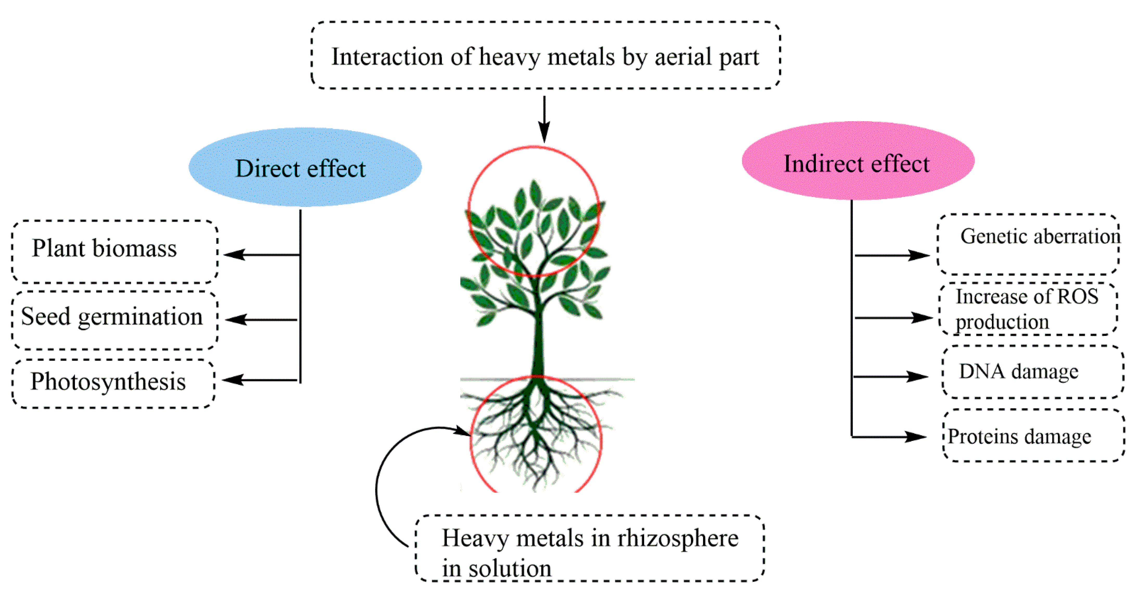
3. Heavy Metal Contaminants in Fruit and Vegetable Crops

Heavy metals, for example, cadmium, copper, chromium, lead, and mercury, are major environmental pollutants. The presence of heavy metals in water, atmosphere, and soil can cause serious problems in all living organisms (Figure 4). Additions of heavy metals to the soil have antagonistic effects on diet, crop growth rate, and environmental health.
The bioaccumulation of heavy metals in food can be extremely dangerous for human health. These metals enter the human body via inhalation and ingestion. This contamination is determined by soil mobility and bioavailability [120]. Here, we discuss the heavy metal contamination in fruit and vegetable crops.

3.1. Crops Contaminated with Heavy Metals

3.1.1. Rice

Rice is a popular food in Asia. The presence of heavy metals and contaminants in rice-based diets increases several risk factors for the human body. Recent studies evaluated the dietary exposure to heavy metals in rice and found elevated concentrations of various heavy metals such as arsenic, lead, and chromium in 71 irrigated and rain-fed rice (Table 1). During the irrigated season and due to the use of contaminated irrigation water, the concentration of heavy metals in the rice grains is generally high: arsenic, 0.153 ± 0.112 and 0.140 ± 0.080 mg/kg; cadmium, 0.073 ± 0.069 and 0.038 ± 0.032 mg/kg; lead, 0.264 ± 0.125 and 0.147 ± 0.077 mg/kg; and chromium, 1.208 ± 0.913 and 0.986 ± 0.796 mg/kg in irrigated and rain-fed rice, respectively [121]. Groundwater contaminated with arsenic is the biggest factor contributing to the high levels of arsenic in the rice samples. Contamination of industrial effluents is related to the higher levels of cadmium, lead, and chromium. In Bangladeshi adults, based on daily rice consumption of 400 g per 60 kg body weight, the estimated daily intake of arsenic, cadmium, lead, and chromium is 18.6–214 µg, 2.6–119 µg, 25.0–241 µg, and 59.0–1846 µg, respectively. Rice alone may contribute up to 46%, 57%, 50%, and 60% of the maximum tolerable daily intake (MTDI) for arsenic, cadmium, lead, and chromium, respectively, which in fact is an important factor in the dietary intake of these elements when compared with other food materials and drinking water. Therefore, for individuals consuming rice or rice products, e.g., in South Asia, the accumulation of heavy metals in rice grains is a big distress.

3.1.2. Wheat

Wheat-based diets are associated with several risk factors due to the presence of heavy metals and contaminants (Table 1). Wheat (Triticum aestivum) crop is the main and integral part of the diet. It contains proteins, carbohydrates, and some inorganic micronutrients [122]. When the accumulation of heavy metals is below controlled limits, wheat grain ingestion is safe [123]. However, when accumulation exceeds the safe limits, it leads to toxic effects and a range of diseases in humans [124]. The concentration ranges of cadmium, lead, arsenic, nickel, copper, zinc, chromium, and manganese are 0.011–0.039, 0.166–0.209, 0.005–1.113, 0.015–2.060, 0.089–4.625, 0.111–3.169, 0.013–1.018, and 0.100–4.467 mg/kg of wheat, respectively [124].

3.2. Vegetables Contaminated with Heavy Metals

3.2.1. Potato

Potato (Solanum tuberosum) is one of the primary food crops in the world. It contains highly digestible carbohydrates and protein, and therefore represents an excellent source of vital nutrients [125]. Heavy metals play both positive and negative roles in human life. Heavy metals, for example, mercury, lead, copper, and cadmium, are extremely toxic to the health of the ecosystem. Heavy metals such as iron, zinc, copper, and manganese are essential to human health. Unnecessary fertilization and pollution of irrigation water are sources of heavy metal pollution in areas of potato cultivation (Table 1). Extremely high intake of important heavy metals can result in toxic effects [126]. The concentrations of iron, copper, zinc, manganese, lead, nickel, and cadmium in potato were 48.87–72.64, 3.07–5.43, 13.80–18.88, 6.93–13.06, 0.51–0.77, 2.02–3.55, and 0.08–0.32 mg/kg, respectively. The accumulation of heavy metals in potato was in the order of iron > zinc > manganese > copper > nickel > lead > cadmium [127].

3.2.2. Tomato

Tomato (Solanum lycopersicum) is an edible food crop. It is used as a staple in several diets around the world. Heavy metals are stored in both edible and non-edible parts of many fruits and vegetables. Food safety concerns make this one of the most serious environmental issues. Vegetables grown in heavy metal-polluted soil carry a higher level of heavy metals than those grown in unpolluted soil [84]. In tomatoes, the concentrations of lead, cadmium, copper, zinc, and chromium are 0.14–0.28, 0.004–0.06, 1.64–2.86, 6.46–8.42, and 0.02–0.08 mg/kg, respectively (Table 1) [128].

3.2.3. Lettuce

Lettuce (Lactuca sativa) is a leafy vegetable. It is used as a staple in several foods around the world. It provides a source of highly digestible carbohydrates as well as protein. Vegetables are a good source of vitamins, minerals, and fiber, and they are also good for human health. These products, appropriately, contain both vital and toxic metals with a wide range of concentrations [129]. In lettuce, the concentrations of lead, cadmium, copper, zinc, and chromium are 0.48–0.63, 0.02–0.12, 1.12–2.44, 8.09–13.6, and 0.09–0.17 mg/kg, respectively (Table 1) [128].

3.2.4. Cabbage

Cabbage (family Brassicaceae) is an edible food crop. It is a rich source of protein. Vegetables grown in heavy metal-contaminated soil have a higher concentration of heavy metals than those grown in uncontaminated soil [84]. In cabbage, the concentrations of lead, cadmium, copper, zinc, and chromium are 0.22–0.53, 0.006–0.08, 1.84–3.40, 6.86–14.3, and 0.03–0.13 mg/kg, respectively (Table 1) [128].

3.2.5. Carrot

Carrot is an edible vegetable crop consumed as a staple food in several diets around the world. It is a source of keratin. Carrots are a good base of vitamins, minerals, and fiber and are also good for health. Carrots contain both crucial and toxic metals over a wide range of concentrations [129]. In carrots, the concentrations of lead, cadmium, copper, zinc, and chromium are 0.12–0.23, 0.01–0.05, 1.14–2.33, 8.22–10.6, and 0.06–1.22 mg/kg, respectively (Table 1) [128].

3.3. Fruits Contaminated with Heavy Metals

3.3.1. Avocado Pear

Avocado pear is widely used for gastronomic and dietary purposes. Avocado fruits are primarily composed of cellulose, hemicellulose, and pectin, which contribute to their smoothness and rigidity. Because of the presence of vitamins and mineral salts, fresh fruits and vegetables are extremely important in the diet. They also contain water, calcium, sulfur, iron, and potash [130]. However, heavy metal contamination can reduce the benefits of fruit consumption. These heavy metals are mostly recalcitrant in the environment. They are non-biodegradable and thermostable, and hence, they can easily accumulate to lethal levels. In avocado pear, the concentrations of cadmium, copper, zinc. iron, lead, nickel, manganese, and cobalt are 0.15, 3.10, 8.87, 28.60, 1.69, 3.34, 1.31, and 1.62 mg/kg, respectively (Table 1) [131].

3.3.2. Orange

Orange is generally used for gastronomic and dietary purposes. The fruits contain pectic and cellulosic materials that contribute to their rigidity and texture. Fresh vegetables and fruits are of great importance in the diet due to the abundance of minerals and vitamins [130]. In orange, the concentrations of cadmium, copper, zinc, iron, lead, nickel, manganese, and cobalt are 0.10; 0.23; 7.22; 19.0; 5.80; 2.99; 1.09; and 1.67 mg/kg, respectively (Table 1) [131].

3.3.3. Pawpaw

Pawpaw is widely used for gastronomic and dietary purposes. The fruits are made up of essential cellulose and hemicellulose ingredients that contribute to their rigidity. Fresh vegetables and fruits are of great importance in the diet because of an abundance of minerals and vitamins [130]. In pawpaw, the concentrations of cadmium, copper, zinc, iron, lead, nickel, manganese, and cobalt are 0.22, 05.29, 07.31, 29.60, 05.57, 05.87, 01.03, and 3.56 mg/kg, respectively (Table 1) [131].

3.3.4. Pineapple

Pineapple is commonly used for gastronomic and dietary purposes. The fruits mainly contain pectic and cellulose substances responsible for their texture. Because of the presence of vitamins and mineral salts, fresh fruits and vegetables are extremely important in the diet [130]. In pineapple, the concentrations of cadmium, copper, zinc, iron, lead, nickel, manganese, and cobalt are 0.08, 0.64, 6.78, 25.70, 4.52, 1.16, 2.60, and 1.43 mg/kg, respectively (Table 1) [131]. We always use fruits within safe limits to secure our life. A comparison of the concentrations of heavy metal accumulation in many crops, fruits, and vegetables was made. Heavy metals present in crops, fruits, and vegetables do not pose a direct threat to human health. However, continued consumption of these crops, fruits, and vegetables, even with adequate contamination levels, can lead to accumulation in the body and have long-term lethal consequences. Therefore, it is essential to implement an annual monitoring program for heavy metals in food (Table 1).

4. Heavy Metal Toxicity Biomarkers

Toxicology requires necessary tools and knowledge to assess exposure to toxic agents, measure the extent of the possible toxic response, and determine the likelihood of adverse effects. Thus, the techniques and tools necessary to perform this type of analysis are called “biomarkers”. In recent years, the emphasis has been on environmental viability of individuals and populations exposed to chemical pollution, which opens up many possibilities for rapid and sensitive detection of chemical stress in organizations [117]. Mussali-Galante et al. [132] investigated three types of biomarkers most commonly used to analyze heavy metal pollution in the environment at all levels of biological organization.

4.1. Types of Exposure Biomarkers

4.1.1. Internal Dose Biomarkers

Because of their reliability, precision, and relevance to individual risk, they are the most widely used biomarkers [133]. They are used in combination with parameters of external exposure. Currently, several techniques are available for highly sensitive analysis of very low concentrations of chemicals or their metabolites in various cells, organs, or body fluids. These biomarkers explain the differences in absorption, metabolism, bioaccumulation, and excretion of the compound in question. They indicate the actual dose of the substance in a specific organism and tissues [120]. These are used as internal dosimeters to measure metal exposure, such as total inorganic arsenic or its metabolites in hair, blood, nails, and urine [121].

4.1.2. Biologically Active Dose Biomarkers

These biomarkers appear relatively early in the path of disease exposure. Some of them have been linked to an enhanced risk of developing diseases like cancer. The best-known examples are DNA adducts, which are formed by covalent bonding of all or part of a metal molecule with chemical fragments of DNA, which are products generated when an activated chemical species binds covalently to negatively charged fragments. In this regard, DNA adducts are among the most informative biomarkers that can be used to measure exposure to genotoxic agents [134].

4.2. Role of Biomarkers in the Molecular Detection of Adverse Effects

Biomarkers reflect initial changes that may lead to clinical disease [135]. These biomarkers are not evidence of disease caused by environmental pollution, but rather tools for understanding a process that can eventually lead to negative consequences [136]. They can detect changes in important genetic targets such as DNA, including DNA breaks and chromosomal and micronuclear aberrations. Arsenic has been implicated in numerous studies investigating the role of biomarkers at the biochemical level, providing a wealth of information involving oxidative damage to DNA and proteins, as well as alterations in a wide range of enzymes such as DNA repair and metal-binding proteins [137]. Single-stranded (SSB) and double-stranded (DSB) DNA breaks are used to assess the potential consequences of environmental metal pollution in order to determine the genotoxic damage caused.

4.3. Biomarkers of Susceptibility

Given the importance of metabolism in toxicological research, there is substantial interest in the role of genetic variations in toxic response, as well as variations in susceptibility and related markers [138]. Several enzymes linked to disease show significant differences in terms of activity levels within the population, such as N-acetyl-transferase, several cytochromes P-450 (CYP), and glutathione transferase (GST), among others [139]. In some studies, trace metals have been shown to regulate CYP expression, as well as heavy metals such as mercury and lead [140]. Each of these enzymes has a colossal role in activating or detoxifying chemical exposure. Similarly, the genetic loci of these and other metabolic enzymes have been identified, allowing the detection of polymorphisms and phenotypic differences in the population. Polymorphisms and differences in enzymatic activity may play a role in different metal responses [141]. Single nucleotide polymorphisms (SNPs) are the most common types of DNA sequence variation in human genome, which contribute to phenotypic diversity by influencing the risk of certain diseases and the variable response to the ecosystem [139]. Therefore, the study and detection of SNPs is important when studying responses to metal exposures.

5. Conclusions

Despite their beneficial role in homeostasis, several heavy metals in water, air, and soil can cause serious biological and cellular effects in humans and other organisms. Contamination with these metals can diminish the health benefits of consuming fruits, crops, and vegetables, suggesting that different heavy metals exhibit toxic effects mediated via different mechanisms. However, these mechanisms were not completely elucidated, suggesting the need for further investigation into their toxicity.
Reducing the risk associated with the use of food crops polluted with heavy metals is a priority that requires a well-planned approach. To improve food security, the sources of heavy metal contamination must be reduced by implementing and enforcing laws and regulations, as well as adopting good agricultural practices. Despite the scarcity of farmland for plant growth, care should be taken when planting near industrial and mining sites. Furthermore, an assessment based on the accumulation of various heavy metals in soil and food crops is required to minimize public health challenges caused by the consumption of polluted agricultural crops.
We should always use food items within safe limits. Biological remediation, such as phytoremediation, may be an environmentally friendly and cost-effective method to reduce the toxicity of heavy metals in lightly polluted soil. Using secure measures, environmental technologies such as nano-tools and farmer brotherhood consciousness could greatly improve opportunities for regional economy and livelihood. Environmental pollution is a major contributor to this threat, which must be addressed by reducing heavy metal pollution and implementing sustainable agricultural practices.
The study limitations relate to the correlation between positive and negative effects of different heavy metal compounds. Indeed, as we indicated, some heavy metals play an important role in homeostasis and their toxicities are related only to their concentrations. Therefore, further studies are needed to evaluate the toxicity of heavy metals at different concentrations and establish the correlation between positive and negative effects of different heavy metals. Moreover, the degree of toxicity of each heavy metal was not well established, suggesting the need to determine the toxicity of each compound at the field level.

Author Contributions

N.M., M.J., A.B. (Abdelhakim Bouyahya), M.T., N.E.O., R.G., A.B. (Abdelaali Balahbib), Z.M. and M.A. conceived the review idea and focus and drafted the article. S.M.A.S., I.N.M., M.D., M.R., B.V., S.A. and M.A.S. were involved in the reference collection and writing the article. All authors have read and agreed to the published version of the manuscript.

Funding

This paper was supported by the KU Research Professor Program of Konkuk University, Seoul, Korea.

Institutional Review Board Statement

Not applicable.

Informed Consent Statement

Not applicable.

Data Availability Statement

Data are contained within the article.

Conflicts of Interest

The authors declare no conflict of interest.

References

  1. Zhang, C.; Gan, C.; Ding, L.; Xiong, M.; Zhang, A.; Li, P. Maternal Inorganic Mercury Exposure and Renal Effects in the Wanshan Mercury Mining Area, Southwest China. Ecotoxicol. Environ. Saf. 2020, 189, 109987. [Google Scholar] [CrossRef] [PubMed]
  2. Afonne, O.J.; Ifediba, E.C. Heavy Metals Risks in Plant Foods–Need to Step up Precautionary Measures. Curr. Opin. Toxicol. 2020, 22, 1–6. [Google Scholar] [CrossRef]
  3. El-Kady, A.A.; Abdel-Wahhab, M.A. Occurrence of Trace Metals in Foodstuffs and Their Health Impact. Trends Food Sci. Technol. 2018, 75, 36–45. [Google Scholar] [CrossRef]
  4. Sanaei, F.; Amin, M.M.; Alavijeh, Z.P.; Esfahani, R.A.; Sadeghi, M.; Bandarrig, N.S.; Fatehizadeh, A.; Taheri, E.; Rezakazemi, M. Health Risk Assessment of Potentially Toxic Elements Intake via Food Crops Consumption: Monte Carlo Simulation-Based Probabilistic and Heavy Metal Pollution Index. Environ. Sci. Pollut. Res. 2021, 28, 1479–1490. [Google Scholar] [CrossRef] [PubMed]
  5. Giri, S.; Mahato, M.K.; Bhattacharjee, S.; Singh, A.K. Development of a New Noncarcinogenic Heavy Metal Pollution Index for Quality Ranking of Vegetable, Rice, and Milk. Ecol. Indic. 2020, 113, 106214. [Google Scholar] [CrossRef]
  6. Gudkov, S.V.; Burmistrov, D.E.; Serov, D.A.; Rebezov, M.B.; Semenova, A.A.; Lisitsyn, A.B. A Mini Review of Antibacterial Properties of ZnO Nanoparticles. Front. Phys. 2021, 9, 641481. [Google Scholar] [CrossRef]
  7. Gudkov, S.V.; Burmistrov, D.E.; Serov, D.A.; Rebezov, M.B.; Semenova, A.A.; Lisitsyn, A.B. Do Iron Oxide Nanoparticles Have Significant Antibacterial Properties? Antibiotics 2021, 10, 884. [Google Scholar] [CrossRef] [PubMed]
  8. Dutta, N.; Miraz, S.M.; Khan, M.U.; Karekar, S.C.; Usman, M.; Khan, S.M.; Amin, U.; Rebezov, M.; Shariati, M.A.; Thiruvengadam, M. Heterologous Expression and Biophysical Characterization of a Mesophilic Tannase Following Manganese Nanoparticle Immobilization. Colloids Surf. B Biointerfaces 2021, 207, 112011. [Google Scholar] [CrossRef] [PubMed]
  9. Rajakumar, G.; Mao, L.; Bao, T.; Wen, W.; Wang, S.; Gomathi, T.; Gnanasundaram, N.; Rebezov, M.; Shariati, M.A.; Chung, I.-M.; et al. Yttrium Oxide Nanoparticle Synthesis: An Overview of Methods of Preparation and Biomedical Applications. Appl. Sci. 2021, 11, 2172. [Google Scholar] [CrossRef]
  10. Ahmad, B.; Shireen, F.; Rauf, A.; Shariati, M.A.; Bashir, S.; Patel, S.; Khan, A.; Rebezov, M.; Khan, M.U.; Mubarak, M.S. Phyto-Fabrication, Purification, Characterisation, Optimisation, and Biological Competence of Nano-Silver. IET Nanobiotechnol. 2021, 15, 1–18. [Google Scholar] [CrossRef] [PubMed]
  11. Rebezov, M.B.; Assirzhanova, Z.B.; Dautova, A.; Derkho, M.A.; Meshcheryakova, G.V.; Gumenyuk, O.A. Control by the Accuracy of the Results of Studies for the Lead Content in Samples Applying the Microwave Laboratory System PLP-01M. IOP Conf. Ser. Mater. Sci. Eng. 2021, 1047, 012188. [Google Scholar] [CrossRef]
  12. Rebezov, M.B.; Shariati, M.A.; Shinkarev, I.K.; Tarasova, A.A.; Zubkova, E.S. Results of Comparative Research Methods for Arsenic Content in Meat Samples of Broiler Chickens. IOP Conf. Ser. Earth Environ. Sci. 2021, 677, 052053. [Google Scholar] [CrossRef]
  13. Rebezov, M.B.; Shariati, M.A.; Artyukhova, S.I.; Kolosovskaya, I.I.; Trofimova, E.I. Comparative Analysis of Methods of Photoelectric Colorimetry and Stripping Voltammetry in Assessing the Content of Arsenic in Sea Bass Samples. IOP Conf. Ser. Earth Environ. Sci. 2021, 677, 052057. [Google Scholar] [CrossRef]
  14. Rebezov, M.B.; Kudryavtseva, T.M.; Meshcheryakova, G.V.; Derkho, M.A.; Shakirova, S.S.; Gumenyuk, O.A. Control of the Stability of the Results of Studies of Cadmium Content Using the Method of Additions in Cow’s Milk Samples. IOP Conf. Ser. Earth Environ. Sci. 2021, 677, 052051. [Google Scholar] [CrossRef]
  15. Cherkasova, E.I.; Rebezov, M.B.; Shariati, M.A.; Kharybina, M.M.; Muradova, Z.V. Monitoring the Stability of the Results of Studies of Chilled River Fish for Cadmium Content Using the Method of Additions. IOP Conf. Ser. Earth Environ. Sci. 2021, 677, 052060. [Google Scholar] [CrossRef]
  16. Abuova, A.B.; Rebezov, M.B.; Mukhamedyarova, L.G.; Shakirova, S.S.; Khaimuldinova, A.K.; Yermakhanova, F.R. Results of Studies of Wheat Bread for Lead Content Using the Additive Method. IOP Conf. Ser. Earth Environ. Sci. 2021, 677, 052050. [Google Scholar] [CrossRef]
  17. Rebezov, M.B.; Tretyak, L.N.; Solodov, S.A.; Galaev, A.V.; Korneev, I.N. Evaluation of the Use of the PLP-01M Microwave Laboratory System Using Working Samples to Control the Accuracy of the Results of Examining Product Samples for Lead Content. IOP Conf. Ser. Mater. Sci. Eng. 2021, 1047, 012191. [Google Scholar] [CrossRef]
  18. Maksimiuk, N.N.; Rebezov, M.B.; Tretyak, L.N.; Varivoda, A.A.; Artyukhova, S.I.; Tolstoguzova, T.T. Application of the PLP-01M Microwave Laboratory System Using Control Samples to Assess the Accuracy of the Results of Studies of Cadmium Content. IOP Conf. Ser. Mater. Sci. Eng. 2021, 1047, 012186. [Google Scholar] [CrossRef]
  19. Tretyak, L.N.; Rebezov, M.B.; Korablev, A.V.; Mikhaylova, T.M.; Voskanyan, E.A. Control by the Accuracy of the Results of Studies for the Cadmium Content in Samples Applying the Microwave Laboratory System PLP-01M. IOP Conf. Ser. Mater. Sci. Eng. 2021, 1047, 012183. [Google Scholar] [CrossRef]
  20. Rebezov, M.B.; Shariati, M.A.; Ryskina, E.A.; Bogonosova, I.A.; Sepiashvili, E.N. Monitoring the Research Results on the Toxic Elements Content (Lead, Cadmium and Arsenic) in Food. IOP Conf. Ser. Earth Environ. Sci. 2020, 613, 012123. [Google Scholar] [CrossRef]
  21. Duruibe, J.; Oguwuegbu, M.O.C.; Egwurugwu, J. Heavy Metal Pollution and Human Biotoxic Effects. Int. J. Phys. Sci. 2007, 2, 112–118. [Google Scholar]
  22. Lambert, M.; Leven, B.A.; Green, R.M. New Methods of Cleaning up Heavy Metal in Soils and Water. Environ. Sci. Technol. Briefs Citiz. 2000, 1–3. Available online: http://citeseerx.ist.psu.edu/viewdoc/download?doi=10.1.1.400.2031&rep=rep1&type=pdf (accessed on 30 October 2021).
  23. Zykova, I.; Maksimuk, N.; Rebezov, M.; Kuznetsova, E.; Derkho, M.; Sereda, T.; Kazhibayeva, G.; Somova, Y.; Zaitseva, T. Interaction between Heavy Metals and Microorganisms during Wastewater Treatment by Activated Sludge. J. Eng. Appl. Sci. 2019, 14, 2139–2145. [Google Scholar]
  24. Rai, P.K.; Lee, S.S.; Zhang, M.; Tsang, Y.F.; Kim, K.-H. Heavy Metals in Food Crops: Health Risks, Fate, Mechanisms, and Management. Environ. Int. 2019, 125, 365–385. [Google Scholar] [CrossRef] [PubMed]
  25. Khalid, S.; Shahid, M.; Niazi, N.K.; Rafiq, M.; Bakhat, H.F.; Imran, M.; Abbas, T.; Bibi, I.; Dumat, C. Arsenic Behaviour in Soil-Plant System: Biogeochemical Reactions and Chemical Speciation Influences. In Enhancing Cleanup of Environmental Pollutants; Springer: Cham, Switzerland, 2017; pp. 97–140. [Google Scholar]
  26. Morais, S.; Costa, F.G.; de Lourdes Pereira, M. Heavy Metals and Human Health. Environ. Health Emerg. Issues Pract. 2012, 10, 227–245. [Google Scholar]
  27. Ametepey, S.T.; Cobbina, S.J.; Akpabey, F.J.; Duwiejuah, A.B.; Abuntori, Z.N. Health Risk Assessment and Heavy Metal Contamination Levels in Vegetables from Tamale Metropolis, Ghana. Int. J. Food Contam. 2018, 5, 5. [Google Scholar] [CrossRef] [Green Version]
  28. Balali-Mood, M.; Naseri, K.; Tahergorabi, Z.; Khazdair, M.R.; Sadeghi, M. Toxic Mechanisms of Five Heavy Metals: Mercury, Lead, Chromium, Cadmium, and Arsenic. Front. Pharmacol. 2021, 12, 643972. [Google Scholar] [CrossRef]
  29. Cheng, J.-P.; Wang, W.-H.; Jia, J.-P.; Zheng, M.; Shi, W.; Lin, X.-Y. Expression of C-Fos in Rat Brain as a Prelude Marker of Central Nervous System Injury in Response to Methylmercury-Stimulation. Biomed. Environ. Sci. 2006, 19, 67–72. [Google Scholar]
  30. Bottino, C.; Vázquez, M.; Devesa, V.; Laforenza, U. Impaired Aquaporins Expression in the Gastrointestinal Tract of Rat after Mercury Exposure. J. Appl. Toxicol. 2016, 36, 113–120. [Google Scholar] [CrossRef]
  31. Dongre, N.N.; Suryakar, A.N.; Patil, A.J.; Ambekar, J.G.; Rathi, D.B. Biochemical Effects of Lead Exposure on Systolic & Diastolic Blood Pressure, Heme Biosynthesis and Hematological Parameters in Automobile Workers of North Karnataka (India). Indian J. Clin. Biochem. 2011, 26, 400–406. [Google Scholar]
  32. Wang, J.; Zhu, H.; Yang, Z.; Liu, Z. Antioxidative Effects of Hesperetin against Lead Acetate-Induced Oxidative Stress in Rats. Indian J. Pharmacol. 2013, 45, 395. [Google Scholar] [PubMed] [Green Version]
  33. Boskabady, M.H.; Tabatabai, S.A.; Farkhondeh, T. Inhaled Lead Affects Lung Pathology and Inflammation in Sensitized and Control Guinea Pigs. Environ. Toxicol. 2016, 31, 452–460. [Google Scholar] [CrossRef] [PubMed]
  34. Strużyńska, L.; Dąbrowska-Bouta, B.; Koza, K.; Sulkowski, G. Inflammation-like Glial Response in Lead-Exposed Immature Rat Brain. Toxicol. Sci. 2007, 95, 156–162. [Google Scholar] [CrossRef] [PubMed] [Green Version]
  35. Deng, Y.; Wang, M.; Tian, T.; Lin, S.; Xu, P.; Zhou, L.; Dai, C.; Hao, Q.; Wu, Y.; Zhai, Z.; et al. The Effect of Hexavalent Chromium on the Incidence and Mortality of Human Cancers: A Meta-Analysis Based on Published Epidemiological Cohort Studies. Front. Oncol. 2019, 9, 24. [Google Scholar] [CrossRef] [Green Version]
  36. Pavesi, T.; Moreira, J.C. Mechanisms and Individuality in Chromium Toxicity in Humans. J. Appl. Toxicol. 2020, 40, 1183–1197. [Google Scholar] [CrossRef]
  37. Schutte, R.; Nawrot, T.S.; Richart, T.; Thijs, L.; Vanderschueren, D.; Kuznetsova, T.; Van Hecke, E.; Roels, H.A.; Staessen, J.A. Bone Resorption and Environmental Exposure to Cadmium in Women: A Population Study. Environ. Health Perspect. 2008, 116, 777–783. [Google Scholar] [CrossRef] [Green Version]
  38. Pan, C.; Liu, H.-D.; Gong, Z.; Yu, X.; Hou, X.-B.; Xie, D.-D.; Zhu, X.-B.; Li, H.-W.; Tang, J.-Y.; Xu, Y.-F.; et al. Cadmium Is a Potent Inhibitor of PPM Phosphatases and Targets the M1 Binding Site. Sci. Rep. 2013, 3, 2333. [Google Scholar] [CrossRef] [Green Version]
  39. Fay, M.J.; Alt, L.A.; Ryba, D.; Salamah, R.; Peach, R.; Papaeliou, A.; Zawadzka, S.; Weiss, A.; Patel, N.; Rahman, A.; et al. Cadmium Nephrotoxicity Is Associated with Altered MicroRNA Expression in the Rat Renal Cortex. Toxics 2018, 6, 16. [Google Scholar] [CrossRef] [Green Version]
  40. Pi, H.; Xu, S.; Reiter, R.J.; Guo, P.; Zhang, L.; Li, Y.; Li, M.; Cao, Z.; Tian, L.; Xie, J. SIRT3-SOD2-MROS-Dependent Autophagy in Cadmium-Induced Hepatotoxicity and Salvage by Melatonin. Autophagy 2015, 11, 1037–1051. [Google Scholar] [CrossRef] [Green Version]
  41. Jolliffe, D.M.; Budd, A.J.; Gwilt, D.J. Massive Acute Arsenic Poisoning. Anaesthesia 1991, 46, 288–290. [Google Scholar] [CrossRef]
  42. Yu, L.; Luo, Y.; Liao, B.; Xie, L.; Chen, L.; Xiao, S.; Li, J.; Hu, S.; Shu, W. Comparative Transcriptome Analysis of Transporters, Phytohormone and Lipid Metabolism Pathways in Response to Arsenic Stress in Rice (Oryza sativa). New Phytol. 2012, 195, 97–112. [Google Scholar] [CrossRef] [PubMed]
  43. Shen, H.; Xu, W.; Zhang, J.; Chen, M.; Martin, F.L.; Xia, Y.; Liu, L.; Dong, S.; Zhu, Y.-G. Urinary Metabolic Biomarkers Link Oxidative Stress Indicators Associated with General Arsenic Exposure to Male Infertility in a Han Chinese Population. Environ. Sci. Technol. 2013, 47, 8843–8851. [Google Scholar] [CrossRef] [PubMed]
  44. Chen, R.; Xu, Y.; Xu, C.; Shu, Y.; Ma, S.; Lu, C.; Mo, X. Associations between Mercury Exposure and the Risk of Nonalcoholic Fatty Liver Disease (NAFLD) in US Adolescents. Environ. Sci. Pollut. Res. 2019, 26, 31384–31391. [Google Scholar] [CrossRef] [PubMed]
  45. Kuivenhoven, M.; Mason, K. Arsenic (Arsine); StatPearls Publishing LLC.: Treasure Island, FL, USA, 2019. [Google Scholar]
  46. Smedley, P.L.; Kinniburgh, D.G. A Review of the Source, Behaviour and Distribution of Arsenic in Natural Waters. Appl. Geochem. 2002, 17, 517–568. [Google Scholar] [CrossRef] [Green Version]
  47. Finnegan, P.; Chen, W. Arsenic Toxicity: The Effects on Plant Metabolism. Front. Physiol. 2012, 3, 182. [Google Scholar] [CrossRef] [PubMed] [Green Version]
  48. Singh, N.; Kumar, D.; Sahu, A.P. Arsenic in the Environment: Effects on Human Health and Possible Prevention. J. Environ. Biol. 2007, 28, 359. [Google Scholar] [PubMed]
  49. Chowdhury, U.K.; Biswas, B.K.; Chowdhury, T.R.; Samanta, G.; Mandal, B.K.; Basu, G.C.; Chanda, C.R.; Lodh, D.; Saha, K.C.; Mukherjee, S.K. Groundwater Arsenic Contamination in Bangladesh and West Bengal, India. Environ. Health Perspect. 2000, 108, 393–397. [Google Scholar] [CrossRef] [PubMed]
  50. Mazumder, D.G. Chronic Arsenic Toxicity & Human Health. Indian J. Med. Res. 2008, 128, 436–447. [Google Scholar]
  51. Jomova, K.; Jenisova, Z.; Feszterova, M.; Baros, S.; Liska, J.; Hudecova, D.; Rhodes, C.J.; Valko, M. Arsenic: Toxicity, Oxidative Stress and Human Disease. J. Appl. Toxicol. 2011, 31, 95–107. [Google Scholar] [CrossRef]
  52. Tchounwou, P.B.; Patlolla, A.K.; Centeno, J.A. Invited Reviews: Carcinogenic and Systemic Health Effects Associated with Arsenic Exposure—A Critical Review. Toxicol. Pathol. 2003, 31, 575–588. [Google Scholar] [CrossRef] [Green Version]
  53. Patlolla, A.K.; Tchounwou, P.B. Cytogenetic Evaluation of Arsenic Trioxide Toxicity in Sprague–Dawley Rats. Mutat. Res. Toxicol. Environ. Mutagen. 2005, 587, 126–133. [Google Scholar] [CrossRef] [PubMed]
  54. Landolph, J.R. Molecular and Cellular Mechanisms of Transformation of C3H/10T 1/2 Cl 8 and Diploid Human Fibroblasts by Unique Carcinogenic, Nonmutagenic Metal Compounds. Biol. Trace Elem. Res. 1989, 21, 459–467. [Google Scholar] [CrossRef] [PubMed]
  55. Takahashi, M.; Barrett, J.C.; Tsutsui, T. Transformation by Inorganic Arsenic Compounds of Normal Syrian Hamster Embryo Cells into a Neoplastic State in Which They Become Anchorage-Independent and Cause Tumors in Newborn Hamsters. Int. J. Cancer 2002, 99, 629–634. [Google Scholar] [CrossRef] [PubMed]
  56. Banu, B.S.; Danadevi, K.; Jamil, K.; Ahuja, Y.R.; Rao, K.V.; Ishaq, M. In Vivo Genotoxic Effect of Arsenic Trioxide in Mice Using Comet Assay. Toxicology 2001, 162, 171–177. [Google Scholar] [CrossRef]
  57. Hartmann, A.; Speit, G. Comparative Investigations of the Genotoxic Effects of Metals in the Single Cell Gel (SCG) Assay and the Sister Chromatid Exchange (SCE) Test. Environ. Mol. Mutagen. 1994, 23, 299–305. [Google Scholar] [CrossRef]
  58. Trouba, K.J.; Wauson, E.M.; Vorce, R.L. Sodium Arsenite-Induced Dysregulation of Proteins Involved in Proliferative Signaling. Toxicol. Appl. Pharmacol. 2000, 164, 161–170. [Google Scholar] [CrossRef]
  59. Zhao, C.Q.; Young, M.R.; Diwan, B.A.; Coogan, T.P.; Waalkes, M.P. Association of Arsenic-Induced Malignant Transformation with DNA Hypomethylation and Aberrant Gene Expression. Proc. Natl. Acad. Sci. USA 1997, 94, 10907–10912. [Google Scholar] [CrossRef] [Green Version]
  60. Liu, Y.; Guyton, K.Z.; Gorospe, M.; Xu, Q.; Lee, J.C.; Holbrook, N.J. Differential Activation of ERK, JNK/SAPK and P3/CSBP/RK Map Kinase Family Members during the Cellular Response to Arsenite. Free Radic. Biol. Med. 1996, 21, 771–781. [Google Scholar] [CrossRef]
  61. Soignet, S.L.; Frankel, S.R.; Douer, D.; Tallman, M.S.; Kantarjian, H.; Calleja, E.; Stone, R.M.; Kalaycio, M.; Scheinberg, D.A.; Steinherz, P.; et al. United States Multicenter Study of Arsenic Trioxide in Relapsed Acute Promyelocytic Leukemia. J. Clin. Oncol. 2001, 19, 3852–3860. [Google Scholar] [CrossRef]
  62. Murgo, A.J. Clinical Trials of Arsenic Trioxide in Hematologic and Solid Tumors: Overview of the National Cancer Institute Cooperative Research and Development Studies. Oncologist 2001, 6, 22–28. [Google Scholar] [CrossRef]
  63. Seol, J.G.; Park, W.H.; Kim, E.S.; Jung, C.W.; Hyun, J.M.; Kim, B.K.; Lee, Y.Y. Effect of Arsenic Trioxide on Cell Cycle Arrest in Head and Neck Cancer Cell Line PCI-1. Biochem. Biophys. Res. Commun. 1999, 265, 400–404. [Google Scholar] [CrossRef] [PubMed]
  64. Alemany, M.; Levin, J. The Effects Of Arsenic Trioxide (As2O3) On Human Megakaryocytic Leukemia Cell Lines With a Comparison of Its Effects on Other Cell Lineages. Leuk. Lymphoma 2000, 38, 153–163. [Google Scholar] [CrossRef] [PubMed]
  65. Graham-Evans, B.; Cohly, H.H.; Yu, H.; Tchounwou, P.B. Arsenic-Induced Genotoxic and Cytotoxic Effects in Human Keratinocytes, Melanocytes and Dendritic Cells. Int. J. Environ. Res. Public Health 2004, 1, 83–89. [Google Scholar] [CrossRef]
  66. Stevens, J.J.; Graham, B.; Walker, A.M.; Tchounwou, P.B.; Rogers, C. The Effects of Arsenic Trioxide on DNA Synthesis and Genotoxicity in Human Colon Cancer Cells. Int. J. Environ. Res. Public Health 2010, 7, 2018–2032. [Google Scholar] [CrossRef] [PubMed] [Green Version]
  67. Yedjou, C.G.; Tchounwou, P.B. Modulation of P53, c-Fos, RARE, Cyclin A, and Cyclin D1 Expression in Human Leukemia (HL-60) Cells Exposed to Arsenic Trioxide. Mol. Cell. Biochem. 2009, 331, 207–214. [Google Scholar] [CrossRef] [PubMed] [Green Version]
  68. Brown, E.; Yedjou, C.G.; Tchounwou, P.B. Cytotoxicity and Oxidative Stress in Human Liver Carcinoma Cells Exposed to Arsenic Trioxide (HepG2). Met. Ions Biol. Med. 2008, 10, 583–587. [Google Scholar]
  69. Matschullat, J. Arsenic in the Geosphere—A Review. Sci. Total Environ. 2000, 249, 297–312. [Google Scholar] [CrossRef]
  70. Zaw, M.; Emett, M.T. Arsenic Removal from Water Using Advanced Oxidation Processes. Toxicol. Lett. 2002, 133, 113–118. [Google Scholar] [CrossRef]
  71. Kim, H.S.; Kim, Y.J.; Seo, Y.R. An Overview of Carcinogenic Heavy Metal: Molecular Toxicity Mechanism and Prevention. J. Cancer Prev. 2015, 20, 232–240. [Google Scholar] [CrossRef]
  72. Valko, M.; Leibfritz, D.; Moncol, J.; Cronin, M.T.; Mazur, M.; Telser, J. Free Radicals and Antioxidants in Normal Physiological Functions and Human Disease. Int. J. Biochem. Cell Biol. 2007, 39, 44–84. [Google Scholar] [CrossRef]
  73. Ngalame, N.N.; Tokar, E.J.; Person, R.J.; Xu, Y.; Waalkes, M.P. Aberrant MicroRNA Expression Likely Controls RAS Oncogene Activation during Malignant Transformation of Human Prostate Epithelial and Stem Cells by Arsenic. Toxicol. Sci. 2014, 138, 268–277. [Google Scholar] [CrossRef] [Green Version]
  74. Ataei, N.; Aghaei, M.; Panjehpour, M. Evidences for Involvement of Estrogen Receptor Induced ERK1/2 Activation in Ovarian Cancer Cell Proliferation by Cadmium Chloride. Toxicol. Vitr. 2019, 56, 184–193. [Google Scholar] [CrossRef] [PubMed]
  75. Ding, W.; Liu, W.; Cooper, K.L.; Qin, X.-J.; de Souza Bergo, P.L.; Hudson, L.G.; Liu, K.J. Inhibition of Poly (ADP-Ribose) Polymerase-1 by Arsenite Interferes with Repair of Oxidative DNA Damage. J. Biol. Chem. 2009, 284, 6809–6817. [Google Scholar] [CrossRef] [Green Version]
  76. Dash, M.; Dey, A.; Chattopadhyay, S. Mitigation of Arsenic Driven Utero-Ovarian Malfunction and Changes of Apoptotic Gene Expression by Dietary NAC. Ecotoxicol. Environ. Saf. 2020, 199, 110675. [Google Scholar] [CrossRef] [PubMed]
  77. Ashraf, U.; Hussain, S.; Anjum, S.A.; Abbas, F.; Tanveer, M.; Noor, M.A.; Tang, X. Alterations in Growth, Oxidative Damage, and Metal Uptake of Five Aromatic Rice Cultivars under Lead Toxicity. Plant Physiol. Biochem. 2017, 115, 461–471. [Google Scholar] [CrossRef]
  78. Iyer, S.; Sengupta, C.; Velumani, A. Lead Toxicity: An Overview of Prevalence in Indians. Clin. Chim. Acta 2015, 451, 161–164. [Google Scholar] [CrossRef]
  79. Liu, J.; Li, L.; Wang, Y.; Yan, C.; Liu, X. Impact of Low Blood Lead Concentrations on IQ and School Performance in Chinese Children. PLoS ONE 2013, 8, e65230. [Google Scholar] [CrossRef] [Green Version]
  80. Obeng-Gyasi, E.; Roostaei, J.; Gibson, J.M. Lead Distribution in Urban Soil in a Medium-Sized City: Household-Scale Analysis. Environ. Sci. Technol. 2021, 55, 3696–3705. [Google Scholar] [CrossRef]
  81. Wang, C.-L.; Chuang, H.-Y.; Ho, C.-K.; Yang, C.-Y.; Tsai, J.-L.; Wu, T.-S.; Wu, T.-N. Relationship between Blood Lead Concentrations and Learning Achievement among Primary School Children in Taiwan. Environ. Res. 2002, 89, 12–18. [Google Scholar] [CrossRef]
  82. Roy, A.; Bellinger, D.; Hu, H.; Schwartz, J.; Ettinger, A.S.; Wright, R.O.; Bouchard, M.; Palaniappan, K.; Balakrishnan, K. Lead Exposure and Behavior among Young Children in Chennai, India. Environ. Health Perspect. 2009, 117, 1607–1611. [Google Scholar] [CrossRef]
  83. Ogunseitan, O.A.; Smith, T.R. The Cost of Environmental Lead (Pb) Poisoning in Nigeria. Afr. J. Environ. Sci. Technol. 2007, 1, 27–36. [Google Scholar] [CrossRef]
  84. Sharma, R.K.; Agrawal, M.; Marshall, F. Heavy Metal Contamination of Soil and Vegetables in Suburban Areas of Varanasi, India. Ecotoxicol. Environ. Saf. 2007, 66, 258–266. [Google Scholar] [CrossRef] [PubMed]
  85. Papanikolaou, N.C.; Hatzidaki, E.G.; Belivanis, S.; Tzanakakis, G.N.; Tsatsakis, A.M. Lead Toxicity Update. A Brief Review. Med. Sci. Monit. 2005, 11, RA329–RA336. [Google Scholar] [PubMed]
  86. Najeeb, U.; Ahmad, W.; Zia, M.H.; Zaffar, M.; Zhou, W. Enhancing the Lead Phytostabilization in Wetland Plant Juncus Effusus L. through Somaclonal Manipulation and EDTA Enrichment. Arab. J. Chem. 2017, 10, S3310–S3317. [Google Scholar] [CrossRef] [Green Version]
  87. Yongsheng, W.; Qihui, L.; Qian, T. Effect of Pb on Growth, Accumulation and Quality Component of Tea Plant. Procedia Eng. 2011, 18, 214–219. [Google Scholar] [CrossRef] [Green Version]
  88. Charkiewicz, A.E.; Backstrand, J.R. Lead Toxicity and Pollution in Poland. Int. J. Environ. Res. Public Health 2020, 17, 4385. [Google Scholar] [CrossRef]
  89. Brochin, R.; Leone, S.; Phillips, D.; Shepard, N.; Zisa, D.; Angerio, A. The Cellular Effect of Lead Poisioning and Its Clinical Picture. Management 2014, 8, 1–8. [Google Scholar]
  90. Taylor, M.P.; Winder, C.; Lanphear, B.P. Eliminating Childhood Lead Toxicity in Australia: A Call to Lower the Intervention Level. Med. J. Aust. 2012, 197, 493. [Google Scholar] [CrossRef] [Green Version]
  91. Wadhwa, N.; Mathew, B.B.; Jatawa, S.; Tiwari, A. Lipid Peroxidation: Mechanism, Models and Significance. Int. J. Curr. Sci. 2012, 3, 29–38. [Google Scholar]
  92. Flora, S.J.S.; Mittal, M.; Mehta, A. Heavy Metal Induced Oxidative Stress & Its Possible Reversal by Chelation Therapy. Indian J. Med. Res. 2008, 128, 501. [Google Scholar]
  93. Mathew, B.B.; Tiwari, A.; Jatawa, S.K. Free Radicals and Antioxidants: A Review. J. Pharm. Res. 2011, 4, 4340–4343. [Google Scholar]
  94. Kianoush, S.; Balali-Mood, M.; Mousavi, S.R.; Moradi, V.; Sadeghi, M.; Dadpour, B.; Rajabi, O.; Shakeri, M.T. Comparison of Therapeutic Effects of Garlic and D-Penicillamine in Patients with Chronic Occupational Lead Poisoning. Basic Clin. Pharmacol. Toxicol. 2012, 110, 476–481. [Google Scholar] [CrossRef] [PubMed]
  95. Ghorbe, F.; Boujelbene, M.; Makni-Ayadi, F.; Guermazi, F.; Kammoun, A.; Murat, J.C.; Croute, F.; Soleilhavoup, J.P.; El Feki, A. Effect of Chronic Lead Exposure on Kidney Function in Male and Female Rats: Determination of a Lead Exposure Biomarker. Arch. Physiol. Biochem. 2001, 109, 457–463. [Google Scholar] [CrossRef] [PubMed]
  96. Missoun, F.; Slimani, M.; Aoues, A. Toxic Effect of Lead on Kidney Function in Rat Wistar. Afr. J. Biochem. Res. 2010, 4, 021–027. [Google Scholar]
  97. Muntner, P.; He, J.; Vupputuri, S.; Coresh, J.; Batuman, V. Blood Lead and Chronic Kidney Disease in the General United States Population: Results from NHANES III. Kidney Int. 2003, 63, 1044–1050. [Google Scholar] [CrossRef] [Green Version]
  98. Mudgal, V.; Madaan, N.; Mudgal, A.; Singh, R.B.; Mishra, S. Effect of Toxic Metals on Human Health. Open Nutraceuticals J. 2010, 3, 94–99. [Google Scholar] [CrossRef] [Green Version]
  99. Genchi, G.; Carocci, A.; Lauria, G.; Sinicropi, M.S.; Catalano, A. Nickel: Human Health and Environmental Toxicology. Int. J. Environ. Res. Public Health 2020, 17, 679. [Google Scholar] [CrossRef] [PubMed] [Green Version]
  100. Zhao, J.; Shi, X.; Castranova, V.; Ding, M. Occupational Toxicology of Nickel and Nickel Compounds. J. Environ. Pathol. Toxicol. Oncol. 2009, 28, 177–208. [Google Scholar] [CrossRef]
  101. Zambelli, B.; Ciurli, S. Nickel and Human Health. Interrelat. Essent. Met. Ions Hum. Dis. 2013, 13, 321–357. [Google Scholar]
  102. McGregor, D.B.; Baan, R.A.; Partensky, C.; Rice, J.M.; Wilbourn, J.D. Evaluation of the Carcinogenic Risks to Humans Associated with Surgical Implants and Other Foreign Bodies—A Report of an IARC Monographs Programme Meeting. Eur. J. Cancer 2000, 36, 307–313. [Google Scholar] [CrossRef]
  103. Seilkop, S.K.; Oller, A.R. Respiratory Cancer Risks Associated with Low-Level Nickel Exposure: An Integrated Assessment Based on Animal, Epidemiological, and Mechanistic Data. Regul. Toxicol. Pharmacol. 2003, 37, 173–190. [Google Scholar] [CrossRef]
  104. Diwan, B.A.; Kasprzak, K.S.; Rice, J.M. Transplacental Carcinogenic Effects of Nickel (II) Acetate in the Renal Cortex, Renal Pelvis and Adenohypophysis in F3447/NCr Rats. Carcinogenesis 1992, 13, 1351–1357. [Google Scholar] [CrossRef] [PubMed]
  105. Ottolenghi, A.D.; Haseman, J.K.; Payne, W.W.; Falk, H.L.; MacFarland, H.N. Inhalation Studies of Nickel Sulfide in Pulmonary Carcinogenesis of Rats. J. Natl. Cancer Inst. 1975, 54, 1165–1172. [Google Scholar] [CrossRef] [PubMed]
  106. Sunderman Jr, F.W. Mechanisms of Nickel Carcinogenesis. Scand. J. Work. Environ. Health 1989, 15, 1–12. [Google Scholar] [CrossRef]
  107. Al-Rikaby, A.A. Hematologic Evaluation and Histopathological Alteration of Nickel Nitrate Exposure in Male Rabbits. Ann. Romanian Soc. Cell Biol. 2021, 25, 1307–1319. [Google Scholar]
  108. Alina, M.; Azrina, A.; Mohd Yunus, A.S.; Mohd Zakiuddin, S.; Mohd Izuan Effendi, H.; Muhammad Rizal, R. Heavy Metals (Mercury, Arsenic, Cadmium, Plumbum) in Selected Marine Fish and Shellfish along the Straits of Malacca. Int. Food Res. J. 2012, 19, 135–140. [Google Scholar]
  109. Bernard, A. Cadmium & Its Adverse Effects on Human Health. Indian J. Med. Res. 2008, 128, 557. [Google Scholar]
  110. Chakraborty, S.; Dutta, A.R.; Sural, S.; Gupta, D.; Sen, S. Ailing Bones and Failing Kidneys: A Case of Chronic Cadmium Toxicity. Ann. Clin. Biochem. Int. J. Lab. Med. 2013, 50, 492–495. [Google Scholar] [CrossRef]
  111. Ghani, A.G.A. Effect of Chromium Toxicity on Growth, Chlorophyll and Some Mineral Nutrients of Brassica Juncea L. Egypt. Acad. J. Biol. Sci. H Bot. 2011, 2, 9–15. [Google Scholar]
  112. Abebe, W.; Mozaffari, M.S. Vascular Reactivity Changes in Glucose-Intolerant Rat. J. Cardiovasc. Pharmacol. 2007, 50, 590–597. [Google Scholar] [CrossRef]
  113. Welch, C.M.; Hyde, M.E.; Nekrassova, O.; Compton, R.G. The Oxidation of Trivalent Chromium at Polycrystalline Gold Electrodes. Phys. Chem. Chem. Phys. 2004, 6, 3153–3159. [Google Scholar] [CrossRef]
  114. Kelly, W.F.; Ackrill, P.; Day, J.P.; O’Hara, M.; Tye, C.T.; Burton, I.; Orton, C.; Harris, M. Cutaneous Absorption of Trivalent Chromium: Tissue Levels and Treatment by Exchange Transfusion. Occup. Environ. Med. 1982, 39, 397–400. [Google Scholar] [CrossRef]
  115. Saha, R.; Nandi, R.; Saha, B. Sources and Toxicity of Hexavalent Chromium. J. Coord. Chem. 2011, 64, 1782–1806. [Google Scholar] [CrossRef]
  116. Anton, S.D.; Morrison, C.D.; Cefalu, W.T.; Martin, C.K.; Coulon, S.; Geiselman, P.; Han, H.; White, C.L.; Williamson, D.A. Effects of Chromium Picolinate on Food Intake and Satiety. Diabetes Technol. Ther. 2008, 10, 405–412. [Google Scholar] [CrossRef] [PubMed]
  117. Cefalu, W.T.; Hu, F.B. Role of Chromium in Human Health and in Diabetes. Diabetes Care 2004, 27, 2741–2751. [Google Scholar] [CrossRef] [PubMed] [Green Version]
  118. Bielicka, A.; Bojanowska, I.; Wisniewski, A. Two Faces of Chromium-Pollutant and Bioelement. Pol. J. Environ. Stud. 2005, 14, 5–10. [Google Scholar]
  119. Albretsen, J. The Toxicity of Iron, an Essential Element. Vet. Med. 2006, 101, 82. [Google Scholar]
  120. Ul Islam, E.; Yang, X.; He, Z.; Mahmood, Q. Assessing Potential Dietary Toxicity of Heavy Metals in Selected Vegetables and Food Crops. J. Zhejiang Univ. Sci. B 2007, 8, 1–13. [Google Scholar] [CrossRef] [Green Version]
  121. Jaishankar, M.; Tseten, T.; Anbalagan, N.; Mathew, B.B.; Beeregowda, K.N. Toxicity, Mechanism and Health Effects of Some Heavy Metals. Interdiscip. Toxicol. 2014, 7, 60–72. [Google Scholar] [CrossRef] [Green Version]
  122. Singh, A.; Sharma, R.K.; Agrawal, M.; Marshall, F.M. Health Risk Assessment of Heavy Metals via Dietary Intake of Foodstuffs from the Wastewater Irrigated Site of a Dry Tropical Area of India. Food Chem. Toxicol. 2010, 48, 611–619. [Google Scholar] [CrossRef]
  123. Khan, S.; Cao, Q.; Zheng, Y.M.; Huang, Y.Z.; Zhu, Y.G. Health Risks of Heavy Metals in Contaminated Soils and Food Crops Irrigated with Wastewater in Beijing, China. Environ. Pollut. 2008, 152, 686–692. [Google Scholar] [CrossRef] [PubMed]
  124. Al-Othman, Z.A.; Ali, R.; Al-Othman, A.M.; Ali, J.; Habila, M.A. Assessment of Toxic Metals in Wheat Crops Grown on Selected Soils, Irrigated by Different Water Sources. Arab. J. Chem. 2016, 9, S1555–S1562. [Google Scholar] [CrossRef] [Green Version]
  125. Suttle, J. Symposium Introduction: Enhancing the Nutritional Value of Potato Tubers. Am. J. Potato Res. 2008, 85, 266. [Google Scholar] [CrossRef]
  126. Narin, I.; Tuzen, M.; Sari, H.; Soylak, M. Heavy Metal Content of Potato and Corn Chips from Turkey. Bull. Environ. Contam. Toxicol. 2005, 74, 1072–1077. [Google Scholar] [CrossRef] [PubMed]
  127. Öztürk, E.; Atsan, E.; Polat, T.; Kara, K. Variation in Heavy Metal Concentrations of Potato (Solanum Tuberosum L.) Cultivars. J. Anim. Plant Sci. 2011, 21, 235–239. [Google Scholar]
  128. Bempah, C.K.; Kwofie, A.B.; Tutu, A.O.; Danutsui, D.; Bentil, N. Assessing the Potential Dietary Intake of Heavy Metals in Some Selected Fruits and Vegetables from Ghanaian Markets. Elixir Pollut. 2011, 39, 4921–4926. [Google Scholar]
  129. Radwan, M.A.; Salama, A.K. Market Basket Survey for Some Heavy Metals in Egyptian Fruits and Vegetables. Food Chem. Toxicol. 2006, 44, 1273–1278. [Google Scholar] [CrossRef] [PubMed]
  130. Sobukola, O.P.; Adeniran, O.M.; Odedairo, A.A.; Kajihausa, O.E. Heavy Metal Levels of Some Fruits and Leafy Vegetables from Selected Markets in Lagos, Nigeria. Afr. J. Food Sci. 2010, 4, 389–393. [Google Scholar]
  131. Ihesinachi, K.; Eresiya, D. Evaluation of Heavy Metals in Orange, Pineapple, Avocado Pear and Pawpaw from a Farm in Kaani, Bori, Rivers State Nigeria. J. Issues ISSN 2014, 2360, 8803. [Google Scholar]
  132. Mussali-Galante, P.; Tovar-Sánchez, E.; Valverde, M.; Rojas Del Castillo, E. Biomarkers of Exposure for Assessing Environmental Metal Pollution: From Molecules to Ecosystems. Rev. Int. Contam. Ambient. 2013, 29, 117–140. [Google Scholar]
  133. Nordberg, G.F. Biomarkers of Exposure, Effects and Susceptibility in Humans and Their Application in Studies of Interactions among Metals in China. Toxicol. Lett. 2010, 192, 45–49. [Google Scholar] [CrossRef] [PubMed]
  134. Poirier, M.C. Chemical-Induced DNA Damage and Human Cancer Risk. Nat. Rev. Cancer 2004, 4, 630–637. [Google Scholar] [CrossRef] [PubMed]
  135. Mutti, A. Use of Intermediate End-Points to Prevent Long-Term Outcomes. Toxicol. Lett. 1995, 77, 121–125. [Google Scholar] [CrossRef]
  136. Watson, W.P.; Mutti, A. Role of Biomarkers in Monitoring Exposures to Chemicals: Present Position, Future Prospects. Biomarkers 2004, 9, 211–242. [Google Scholar] [CrossRef] [PubMed]
  137. Rojas, E. Special Issue on the 20th Anniversary of the Comet Assay. Mutat. Res./Rev. Mutat. Res. 2009, 1, 1–2. [Google Scholar] [CrossRef] [PubMed]
  138. Timbrell, J.A. Biomarkers in Toxicology. Toxicology 1998, 129, 1–12. [Google Scholar] [CrossRef]
  139. Pavanello, S.; Clonfero, E. Biological Indicators of Genotoxic Risk and Metabolic Polymorphisms. Mutat. Res. Mutat. Res. 2000, 463, 285–308. [Google Scholar] [CrossRef]
  140. Ki, J.-S.; Raisuddin, S.; Lee, K.-W.; Hwang, D.-S.; Han, J.; Rhee, J.-S.; Kim, I.-C.; Park, H.G.; Ryu, J.-C.; Lee, J.-S. Gene Expression Profiling of Copper-Induced Responses in the Intertidal Copepod Tigriopus japonicus Using a 6K Oligochip Microarray. Aquat. Toxicol. 2009, 93, 177–187. [Google Scholar] [CrossRef]
  141. Cullen, M.R.; Redlich, C.A. Significance of Individual Sensitivity to Chemicals: Elucidation of Host Susceptibility by Use of Biomarkers in Environmental Health Research. Clin. Chem. 1995, 41, 1809–1813. [Google Scholar] [CrossRef]
Figure 1. The cellular effects of heavy metals, as well as the balance between ROS production and antioxidant defense.
Sustainability 14 00161 g001
Figure 2. Arsenic toxicity mediated via different cellular mechanisms.
Sustainability 14 00161 g002
Figure 3. Effects of elevated lead concentration in the blood.
Sustainability 14 00161 g003
Figure 4. Effects of heavy metals on vegetables.
Sustainability 14 00161 g004
Table 1. Comparison of the effects and mechanisms of heavy metal toxicity, due to cadmium, lead, mercury, chromium, and arsenic [28].
Toxic MetalsOrgan ToxicityDisrupted Macromolecule/Mechanism of ActionRefs
Mercury (Hg)CNS injuries
Renal dysfunction
GI ulceration
Hepatotoxicity
Aquaporin mRNA reduction
Glutathione peroxidase inhibition
Increased c-fos expression
ROS production
Enzyme inhibition
Thiol binding (GSH conjugation)
[1,29,30]
Lead (Pb)CNS injury
Hematological changes (anemia)
Pulmonary dysfunction
GI colic
Liver damage
Reduced pulmonary function
Cardiovascular dysfunction
Enhanced levels of inflammatory cytokines:
IL-1β, TNF-α, and IL-6 in the CNS
Increased serum ET-1, NO, and EPO levels
Inactivation of δ-ALAD and ferrochelatase (inhibition of heme biosynthesis)
Reduced GSH, SOD, CAT, and GPx levels
[31,32,33,34]
Chromium (Cr)Kidney dysfunction
GI disorders
Dermal diseases
Increased occurrence of cancers, including bladder, kidneys, lungs, larynx, testicular, bone, and thyroid
DNA damage
Genomic instability
Oxidative stress and ROS generation
[35,36]
Cadmium (Cd)Degenerative bone disease
Kidney dysfunction
Liver damage
Lung injuries
GI disorders
Metabolic syndromes associated with Zn and Cu
Cancer
miRNA expression dysregulation
Apoptosis
Endoplasmic reticulum stress
Cd-MT absorption by the kidneys
Dysregulation of Ca, Zn, and Fe homeostasis
Low serum PTH levels
ROS generation
Altered phosphorylation cascades
[37,38,39,40]
Arsenic (As)Cardiovascular dysfunction
CNS injury
Skin and hair changes
Liver damage
GI discomfort
Alterations in neurotransmitter homeostasis
Uncoupler of oxidative phosphorylation (inhibition of ATP formation)
Damaged capillary endothelium
Thiol binding (GSH conjugation)
[41,42,43]
Publisher’s Note: MDPI stays neutral with regard to jurisdictional claims in published maps and institutional affiliations.

