Article

The Activity and Gene Expression of Enzymes in Mycelia of Pleurotus Eryngii under Cadmium Stress

1
Hunan Edible Fungi Institute, Changsha 410013, China
2
Hunan Agricultural Products Processing Institute, Hunan Food Test and Analysis Center, Changsha 410004, China
3
National Engineering Laboratory for Deep Processing of Rice and Byproducts, College of Food Science and Engineering, Central South University of Forestry & Technology, Changsha 410004, China
4
College of Sciences, Central South University of Forestry and Technology, Changsha 410004, China
*
Author to whom correspondence should be addressed.
Sustainability 2022, 14(7), 4125; https://0-doi-org.brum.beds.ac.uk/10.3390/su14074125
Submission received: 12 February 2022 / Revised: 7 March 2022 / Accepted: 24 March 2022 / Published: 30 March 2022
(This article belongs to the Special Issue Future Directions for Soil Remediation and Environmental Management)

Abstract

:
Cadmium (Cd) is a highly toxic pollutant and a nonessential element for plant growth. Here, we investigated the levels of malondialdehyde (MDA), H2O2, sugars, and the activity and gene expression of antioxidant enzymes and sugar metabolism-related enzymes in the mycelia of Pleurotus eryngii grown in the presence of 0, 0.5, 1, 1.5, 2.0, and 4.0 mg·L−1 Cd. Raising Cd concentration resulted in increases, followed by decreases in the levels of malondialdehyde (MDA) and H2O2 in the mycelia. The maximum concentrations of MDA and H2O2 reached 318.61 and 7411.40 μmol·L−1, respectively, at a Cd concentration of 1 mg·L−1. Antioxidative enzymes (superoxide dismutase (SOD), catalase, glutathione peroxidase (GPX), and ascorbate peroxidase (APX)) activities peaked at 1 mg·L−1 Cd. Furthermore, Pleurotus eryngii produced trehalose, glucose, fructose, and maltose in the fermentation broth in response to Cd stress. The levels of trehalose, arabinose, and fructose in the fermentation broth decreased continuously. The levels of glucose increased continuously, whereas those of maltose first increased and then decreased. Gene expression level of chitin synthase, glucose-1-phosphate uridyl transferase peaked significantly at 2 mg·L−1 of Cd, while that of trehalase 2 is 0.5 mg·L−1, and those for fructose-diphosphate aldolase, glyceraldehyde-3-phosphate dehydrogenase, reached the maximum at 4 mg·L−1.

1. Introduction

Intense industrialization and improper disposal of urban waste have increased the heavy-metal pollution in soil and water to alarming levels. According to the cadmium (Cd) survey communique released by the Ministry of Ecology and Environment, PRC, on 17 April 2014, the average Cd level in soil exceeded by 7.0%, ranking first among all pollutants [1]. Cd is highly toxic, although it is also a nonessential element for plant growth [2]. Along with other heavy metals, it is nonbiodegradable and tends to be bio-enriched, making it a serious threat to human health and ecosystem stability.
Recent developments in microbial technology (in particular, involving bacteria, fungi, and eukaryotic algae) have contributed to the control of Cd and chromium (Cr) pollution in water. Treatment processes may involve proper pretreatment; moreover, immobilization treatment increases the adsorption of functional groups on the surface of microorganisms, which can improve efficiencies in remediation and reuse, as well as remove secondary pollution in water [3]. The fungal cell wall consists of β-glucan, chitosan, glycoproteins, lipids, D-galactose amine polymers, polyurea, hyaluronic acid ester, and other components. The cell wall provides the main metal-binding sites in fungi. At the same time, the presence of acetamide, amide, phosphate, amino, sulfhydryl, carboxyl, and hydroxyl groups in the fungal cell wall enhances its adsorption capacity for heavy metals. Moreover, fungi are used widely in bioremediation because of their fast growth, high yield, and low price.
Many recent studies have focused on the detoxification of heavy-metal pollutants in the environment. Detoxification mechanisms include valence transformation, intracellular and extracellular precipitation, active uptake, and others [4,5,6]. The saturation concentration of Cd in the mycelia of Pleurotus eryngii under fermentation conditions is 429 mg·kg−1, and its presence tends to first promote and then inhibit mycelial growth [7]. The mycelial growth of Stropharia rugosoannulata is inhibited by 10–36% under Cd stress conditions, and such stress increases the concentrations of malondialdehyde (MDA), ascorbic acid (ASA; a nonenzymatic antioxidant), and reduced glutathione (GSH) [8]. Both manganese and lead, at certain concentrations, can inhibit the growth, reduce the soluble sugar content, and increase the catalase activity of Flammulina velutina [9].
In this study, P. eryngii was grown under fermentation conditions and under different Cd concentrations. The changes in the concentrations of H2O2, MDA, and antioxidant enzymes, which are indicators of cell membrane damage in the mycelia, were measured. The specific antioxidant enzymes studied were superoxide dismutase (SOD), catalase (CAT), glutathione peroxidase (GPX), and ascorbate peroxidase (APX). In addition, the concentrations of trehalose, arabinose, glucose, sucrose, fructose, and maltose in the fermentation broth under different Cd concentrations were measured. Furthermore, we studied the expression levels of five genes associated with antioxidant activity, namely, catalase1 (CAT1), ascorbate peroxidase1 (APX1), glutathione peroxidase1 (GPX1), NADPH oxidase isoform 1(Nox1), and superoxide dismutase1 (SOD1), and those of six genes involved in sugar metabolism (trehalose2 (TER2), trehalose-6-phosphate synthase1, fructose-diphosphate aldolase, chitin synthase, UTP-glucose-1-phosphate uridylyltransferase, and glyceraldehyde-3-phosphate dehydrogenase). The purpose of this study was to investigate the changes that occur in cell wall tissue, enzyme activities of the antioxidant system, energy metabolism, and the expression of related genes concerning antioxidant enzymes and sugar metabolism in P. eryngii mycelia in response to stress under different Cd concentrations. We aimed to clarify the mechanism of fungal resistance to heavy-metal stress and to provide a theoretical basis for the use of fungal mycelia as a material to treat heavy-metal pollution.

2. Materials and Methods

2.1. Materials and Treatments

Chemicals: RNA iso Plus (Takara Code No.: 9109), cDNA (first single strand) (Takara PrimeScript™ RT Master MixCode No.: RR036A), TB Green™ Premix Ex Taq™ II (Tli RNaseH Plus) (Takara, Code No. RR820A), MicroAmp™ Fast Optical 96-Well Reaction Plate with Barcode, 0.1 mL (Thermo, Nom: 4346906), MicroAmp™ Optical Adhesive Film (Thermo, Art. No (article number):4311971), Cadmium standard solution (Beijing, China), Chloroform (Beijing, China), Ethanol (Beijing, China), isopropanol (Beijing, China), TRIzol (Invitrogen, Carlsbad, CA, USA), DEPC treating water (Beijing, China).
Pleurotus eryngii (XiangXing 98) was provided by the Hunan Edible Fungi Institute and grown in Potato dextrose agar (PDA) medium, which was prepared according to the standard PDA medium method. The Cd solution was first prepared as a 100 mg·L−1 stock solution, which was then sterilized at 121 °C for 30 min. Details on the treatments are listed in Table 1.

2.2. Liquid Fermentation Cultures of P. eryngii under Cadmium (Cd) Stress

P.eryngii strain was purified on PDA agar medium (medium thickness about 5 mm) and set aside for later use. The liquid fermentation medium consisted of PDA but no agar, dispensed in 100 mL volumes in 250 mL triangle flasks and then sterilized by autoclaving at 121 °C for 30 min. After cooling, the Cd stock solution was added to the sterilized liquid culture medium according to the concentrations listed in Table 1. The contents of two agar plates with uniform growth and no contaminants were placed into a sterile grinder, along with 150 mL of sterile water. The mixture was ground for 1 min. Using aseptic techniques, we added 5 mL of P. eryngii strain solution to each flask of fermentation liquid, sealed the flask, and shook the culture at 25 °C for 12 days at 150 rpm. Each treatment with the same Cd concentration was replicated thrice, and there was a total of 21 treatments. After 12 days, the fermentation broth and mycelia were separated by centrifugation at 13,780× g for 10 min. We measured enzyme activities and free sugar concentrations in the fermentation broth; the mycelia were weighed and then stored at −80 °C for subsequent quantitative reverse transcription polymerase chain reaction (qRT-PCR) analysis.

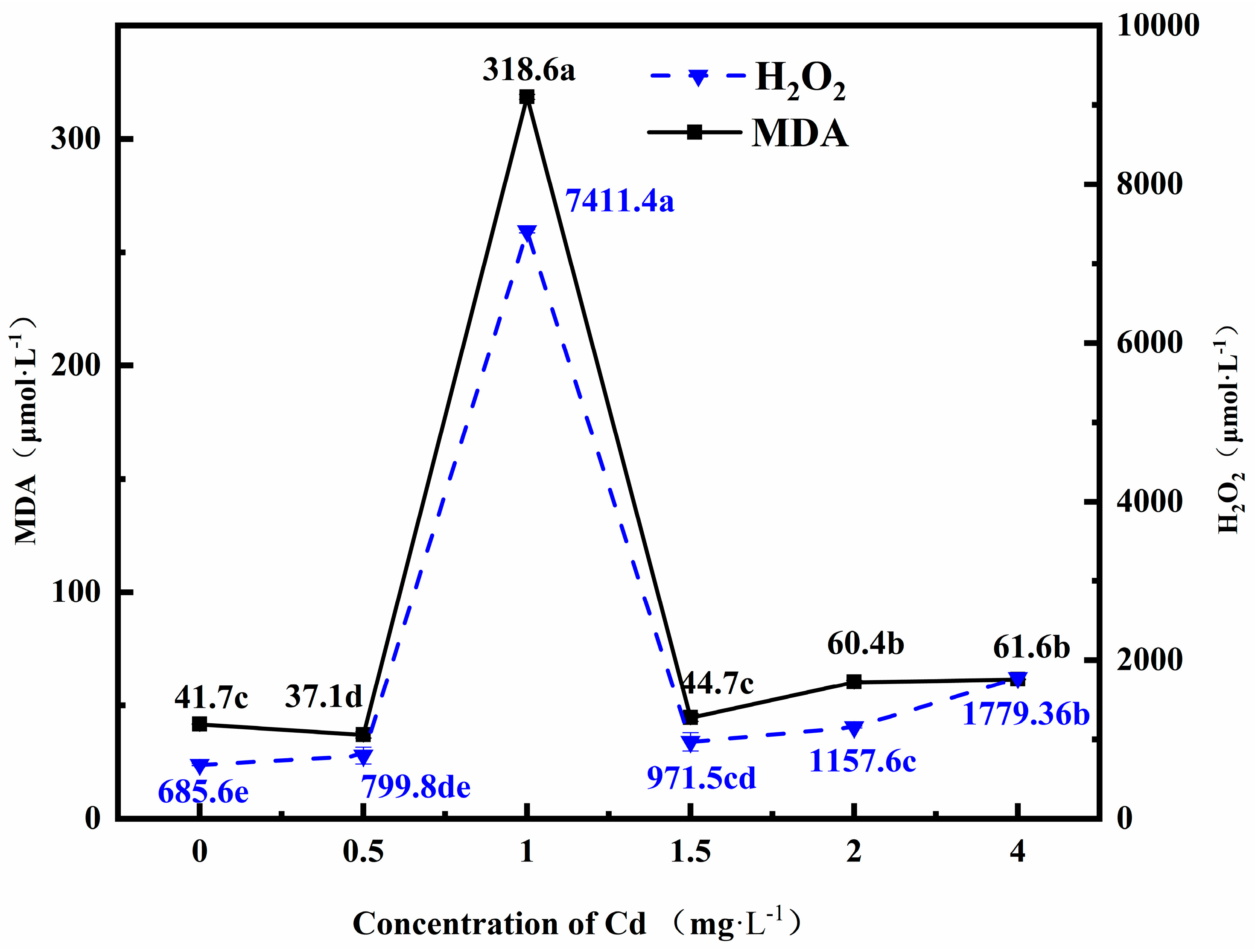
2.3. Determination of MDA and H2O2 Contents

The test fluid was obtained by 0.5 mL fermentation mixed with 0.1% (w/v) trichloroacetic acid (TCA), which was homogenized in an ice bath. After centrifugation at 13,780× g for 10 min, the supernatant was removed, and 0.2 mL test fluid was mixed with 0.2 mL phosphate buffer (pH 7.0) and 0.4 mL Potassium Iodide (KI). The absorbance of the supernatant was read at 390 nm, and H2O2 content was obtained from a standard curve for H2O2. Lipid peroxidation level was determined in terms of 2-thiobarbituric acid (TBA) reactive metabolite, chiefly MDA. We added 0.5 mL of test fluid mixed with 0.5 mL TBA, which quickly cooled after heating for 15 min, the absorbance of the supernatant was measured at 600 nm, 532 nm, and 450 nm. Formula for MDA is C (nmol/mL) = 6.45 (OD532–OD600) − 0.56 × OD450.

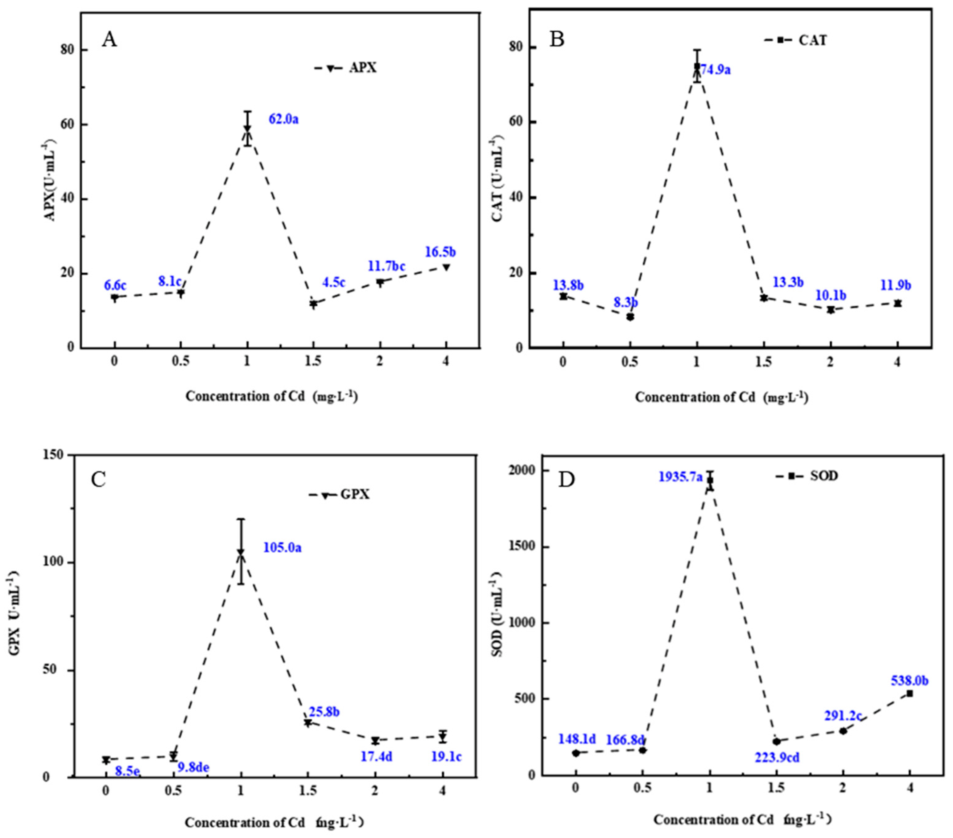
2.4. Determination of Antioxidant Enzyme Activity

Superoxide dismutase (SOD) activities were measured using the nitro-blue tetrazolium (NBT) reduction at 560 nm. Catalase (CAT) activity was determined by following the decrease in H2O2 at 240 m. Guaiacol peroxidase (GPX) activity was determined by the reduced glutathione depletion during the reaction. The reaction was initiated by the addition of H2O2 and was measured by the absorbance at 412 nm. APX activity was determined by measuring ascorbate consumption by absorbance at 290 nm. One unit of APX was defined as the amount of enzyme required to consume 1 µmol ascorbate min−1.

2.5. Calculation of the Amounts of MDA, H2O2, and Enzymes Produced in Mycelia

The amounts of MDA, H2O2, and enzymes produced were calculated from the concentration of MDA, H2O2, and enzymes contained in the fermentation broth and determined by previous experiments, multiplied by the total quantity of the fermentation broth, and then divided by the weight of mycelia.

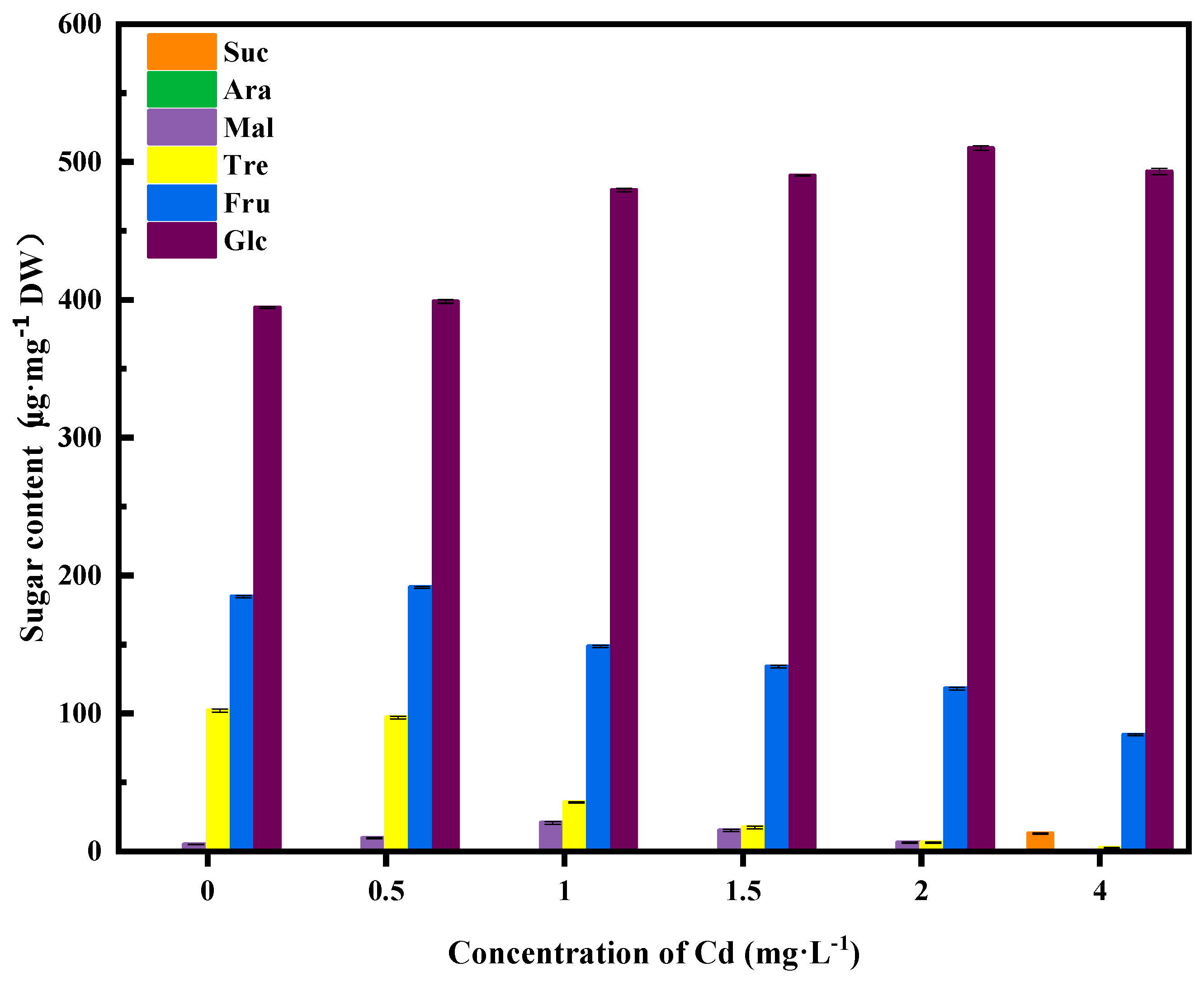
2.6. Measurement of Free Sugar Concentrations

The concentrations of free trehalose, arabinose, glucose, sucrose, fructose, and maltose in the fermentation broth were measured using a Thermo ICS5000 ion chromatography system and a CarboPac™ PA20 (250 × 3.0 mm) liquid chromatography column. The liquid chromatography parameters were mobile phase, solvent A: H2O, solvent B: 200 mM NaOH; the detector was electrochemical, the volume of fermentation was 25 µL, the flow velocity was 0.5 mL·min−1, and the temperature of the column was 30 °C. All samples were diluted by a factor of 50 and were tested directly on the machine.

2.7. Standard Curve Plots

Nine hundred microliters of 50% methanol in water (HPLC grade) were placed into a 15 mL centrifuge tube. We then added 100 mg of the following sugars: trehalose, arabinose, glucose, sucrose, fructose, and maltose. We added 50% methanol solution to make a total of 10 mL standard mixture. The mixture was shaken on a vortex mixer, and the final standard solution was obtained after diluting it with methanol by a factor of 100. The concentration of each sugar in the standard solution was 100 μg·mL−1. The solution was further diluted to the following concentrations: 1, 5, 10, 15, 20, 25, 30, and 40 g·mL−1 and placed in a 1.5 mL microcentrifuge tube for future use. The sugar concentrations were measured in the same way as Section 2.6, and all the readings were plotted to generate a standard curve.

2.8. Detection of the Expression of Antioxidant Enzymes and Sugar Metabolism Enzymes by Fluorescence Quantitative Reverse Transcription Polymerase Chain Reaction (qRT-PCR)

2.8.1. RNA Extraction and cDNA Preparation

Fermentation samples were placed in 1 mL Trizol and incubated at 25 °C for 5 min to fully decompose the samples. After adding liquid nitrogen, samples were ground, and 1 mL of Trizol was added for promoting cell disruption. The solution was centrifuged at 13,780× g at 4 °C for 5 min, and the supernatant was transferred to a new 1.5 mL centrifuge tube. We added 0.4 mL chloroform to each tube, mixed them by inversion for 30 s, and then placed them at room temperature for 5 min. Vigorous shaking was avoided to prevent genomic DNA rupture. The solution was centrifuged at 13,780 rpm at 4 °C for 15 min, and then the upper aqueous phase was transferred to a 1.5 mL microcentrifuge tube. One milliliter of isopropanol was added, the solution was mixed by inversion and then placed at room temperature for 10 min.
The mixture was centrifuged at 13,780× g at 4 °C for 10 min. We washed the RNA pellet twice with 2 mL 75% ethanol. The pellet was allowed to dry at room temperature. An appropriate amount of RNA samples was used to dissolve in diethyl pyrocarbonate (DEPC), and the concentration of 1 L of the extracted RNA was determined using a spectrophotometer. Five hundred nanograms of RNA were electrophoresed in 1% agarose to visualize high-quality RNA, which consisted of three bands (18S, 16S, and 5S). The RNA obtained was reverse transcribed into cDNA, using the reaction system shown in Table 2.

2.8.2. Primer Design and Fluorescence Quantitative Reverse Transcription Polymerase Chain Reaction (qRT-PCR)

We chose five genes related to oxidation resistance and six genes related to sugar metabolism, respectively [10,11]. These genes were obtained from the NCBI database. Primers used to amplify the following genes were designed based on gene sequences from oyster mushroom, white ferula mushroom, and Pleurotus: trehalase2 (MH645361.1), trehalose-6-phosphate synthase1 (MF674013.1), fructose-diphosphate aldolase (XM_036775205.1), chitin synthase (XM_036774923.1), UTP-1-glucoglucuryltransferase phosphate (XM_036776944.1), glyceraldehyde-3-phosphate dehydrogenase (AB690875.1), catalase1 (MF491446.1), ascorbic peroxidase1 (MH645355.1), glutathione peroxidase1 (MH645356.1), NADPH oxidase isoform 1 (XM_007298845.1), and SOD1 (KF768153.1) [12,13]. Oyster mushroom (AY772706.1)18S RNA was used as a reference. The fluorescence quantitative PCR system is shown in Table 3, and fluorescence quantitative PCR primers are shown in Table 4. The cycle threshold Ct was calculated from the detected Ct values with the internal reference, which consisted of the ACTIN1-1 gene. Then, 2△△CT analysis was performed on the samples without Cd stress as the control samples. Expression levels of the target genes are expressed relative to the value of the control.

3. Results

3.1. Changes in the Cell Membrane Damage Index in Fungi Grown under Cadmium (Cd) Stress

Cd stress enhances lipid peroxidation in the cell membrane of mycelia; it also enhances MDA production. As the final product of membrane lipid peroxidation, the MDA content reflects the degree of cell injury [14]. Figure 1 shows that at a Cd concentration of 1 mg·L−1, MDA production by mycelia reached a maximum of 318.6 mol·L−1, whereas the H2O2 level reached 7411.4 μmol·L−1, a value significantly higher than those in other treatments. These results can be explained by Cd stress that tends to increase the content of reactive oxygen species (ROS), such as H2O2 in plants, which in turn increases the degree of lipid peroxidation of cell membranes. Nevertheless, gradually increasing Cd concentrations in the fermentation broth resulted in decreased levels of MDA and H2O2 in the mycelia. These decreases are consistent with the decreased mycelial growth rate and biomass accumulation as Cd concentrations increased. High Cd concentrations cause the mycelia of P. eryngii to produce and accumulate high levels of ROS, which are cytotoxic, resulting in an imbalance in plant cell metabolism. Therefore, the mycelia of P. eryngii grown under Cd stress are in an abnormal physiological state that results in slow or stagnant growth.

3.2. Changes in Antioxidant Enzyme Activities in the Fermentation Mycelia under Cd Stress

In plants, SOD, GPX, CAT, and APX comprise the antioxidant system that plays an important role in protecting cells from oxidative damage [15]. Figure 2 shows that changes in the SOD, GPX, CAT, and APX concentrations followed the same trends; that is, as the Cd concentration increased, the levels of all enzymes tended to increase at Cd concentrations of less than 1 mg·L−1. At 1 mg·L−1 Cd, enzyme concentrations peaked. As Cd concentrations increased further, the levels of APX and SOD decreased and then increased, whereas those of CAT and GPX decreased continuously. In general, plants initially adapt to heavy-metal stress by increasing their antioxidant enzyme activity. This result is similar to that of a study conducted by Liang et al. [16], who investigated the effects of salt stress on wheat. They reported that increasing the duration of stress tends to decrease the activity of enzymes, indicating that the cumulative effect of long-term stress results in damage to cells that exceeds the scavenging capacity of antioxidant enzymes for ROS.

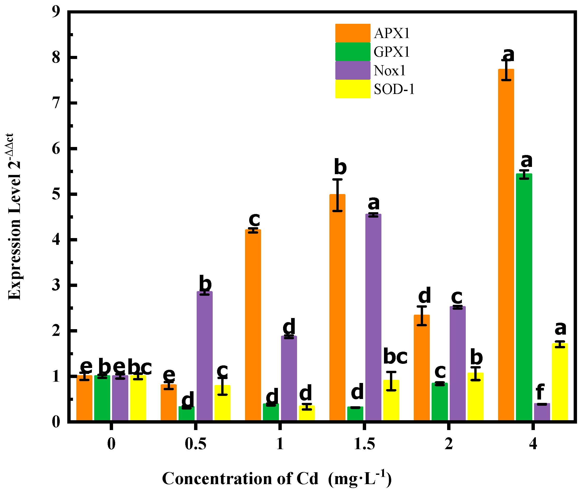
3.3. Changes in Antioxidant Enzyme Gene Expression during Cadmium (Cd) Stress

In this study, we tested the gene expression levels of CAT1, APX1, GPX1, Nox1, and SOD1 in response to Cd stress. Figure 3 shows that the expression levels of APX1, GPX1, and SOD1 peaked significantly at a Cd concentration of 4 mg·L−1, whereas they were the lowest at a Cd concentration of 0.5 mg·L−1. As Cd concentration increased, the expression levels of APX1 tended to first increase, then decrease, and finally increase again; GPX1 expression rose continuously, and SOD1 expression first decreased and then increased. The expression level of Nox1 peaked at 1.5 mg·L−1 Cd, and unlike the other genes, Nox1 expression first increased and then decreased. The changes in the expression levels of enzyme-related genes differed from those of the enzyme levels. Enzyme levels peaked at a Cd concentration of 1 mg·L−1, whereas gene expression levels at this Cd concentration were low. At a Cd concentration of 4 mg·L−1, enzyme levels were decreased significantly, whereas their gene expression levels were high.

3.4. Variation in Levels of Soluble Sugars in the Fermentation Broth during Cd Stress

The levels of trehalose, arabinose, glucose, sucrose, fructose, and maltose in the fermentation broth under different Cd concentrations show that the main sugars produced by P. eryngii during fermentation were trehalose, glucose, and fructose. As the Cd concentration increased, the levels of these sugars in the fermentation broth decreased. This was caused either by the decreasing production or increasing consumption of these sugars by mycelia under Cd stress. In the case of glucose, higher Cd concentrations in the fermentation broth resulted in higher glucose concentrations. Figure 4 shows that glucose levels in the fermentation broth peaked at 510.1 μg·mg−1, which was observed at a Cd concentration of 2 mg·L−1. The two possible sources of glucose in the fermentation broth were first, the medium itself, which contained glucose as an original component, and second, the fungal mycelia, which can produce glucose in response to Cd stress. Meanwhile, maltose levels reached 20.4 μg·mg1 in the presence of 1 mg·L−1 Cd in the fermentation broth. We could not detect sucrose in the fermentation broth, except at 4 mg·L−1 Cd, at which point we detected 12.9 μg·mg−1 sucrose. It is likely that this level of Cd stress caused the mycelia of P. eryngii to produce sucrose, which can act as a protective agent or serve as an energy source for mycelia under Cd stress.

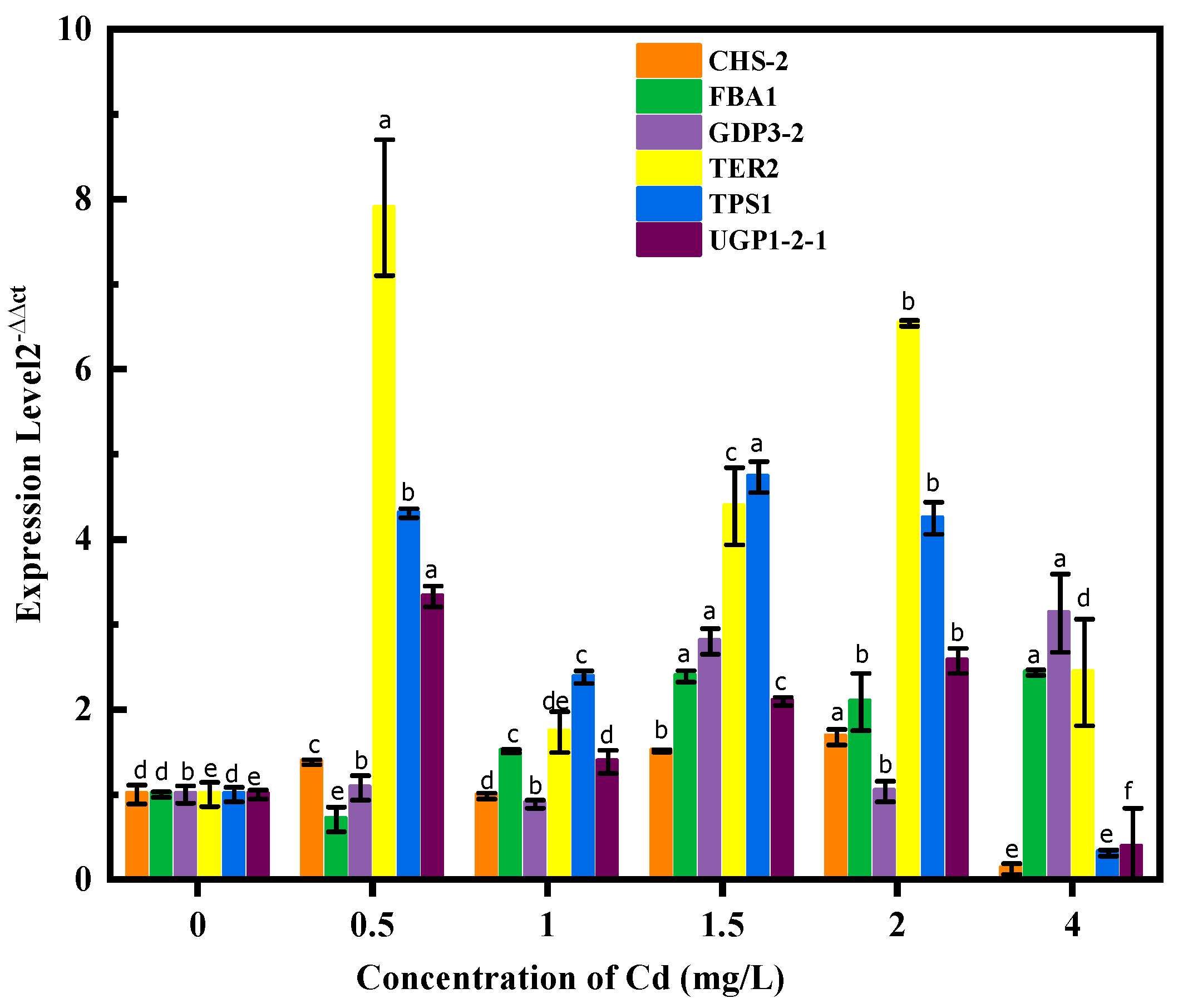
3.5. The Gene Expression of Soluble Sugar Metabolism–Related Enzymes during Cd Stress

The gene expression level of chitin synthase (CHS-2) peaked significantly at 2 mg·L−1 of Cd; that is, it was 1.67 and 13.9 times that of the control group (without Cd) and at 2 mg·L−1 of Cd, respectively. Chitin is a component of the fungal cell wall, and it plays an important role in maintaining cell integrity, hyphal growth, host infection, and asexual and sexual reproduction. CHS-2 is a key enzyme gene in chitin synthesis [17]. Compared with the control group, only the expression of CHS-2 at 2 mg·L−1 of Cd decreased significantly in response to Cd stress, whereas the expression levels of other genes were similar to or significantly higher than those of the control group. We conclude that the cell wall of P. eryngii mycelia is damaged by Cd stress, initiating chitin synthesis. Excessively high concentrations of heavy metals can cause severe damage to mycelia, which inhibits gene expression.
At Cd concentrations of 1.5 and 4 mg·L−1, the expression levels of fructose-diphosphate aldolase (FBA1-1) were significantly higher than those of other Cd concentrations; that is, they were 2.4 times that of the control group and 3.8 times that at a Cd concentration of 0.5 mg·L−1, respectively. FBA is a newly described bifunctional enzyme that catalyzes two different gluconeogenesis reactions to produce fructose-1, 6-bisphosphate (FBP), and fructose-6-phosphate [18]. At a Cd concentration of 0.5 mg·L−1, the levels of fructose and FBA1-1 expression were the highest and lowest, respectively. Increasing Cd concentrations resulted in decreasing levels of fructose in the fermentation broth, whereas the expression levels of FBA1-1 increased. Fructose is a soluble sugar that tends to accumulate in large amounts as P. eryngii grows. Cd stress may cause the quantity of fructose to decline while boosting the expression of FBA1-1.
At Cd concentrations of 1.5 and 4 mg·L−1, the expression levels of GDP3-2 were 2.8 and 3.1 times that of the control group, respectively. The other Cd concentrations did not significantly affect the expression levels of GDP3-2. GDP is one of the key enzymes involved in sugar metabolism, and it is an essential enzyme to maintain the energy levels needed to support life processes [19]. Thus, its sequence is highly conservative in nature. Moreover, GDP is a multifunctional enzyme that plays roles in glycolysis, the tricarboxylic acid cycle, DNA damage repair, plant antioxidant stress, and in responding to redox signal transduction [20]. Our results show that GDP3-2 expression increased at a Cd concentration of 1.5 mg·L−1 and then decreased to the control level at 2 mg·L−1 Cd. Its expression level increased again at 4 mg·L−1 Cd. These results reflect the tolerance of GDP3-2 to Cd stress, as well as the relative stability of GDP3-2 expression.
At a Cd concentration of 0.5 mg·L−1, UGP1-2-1 expression levels were 3.3 and 8.8 times that of the control group and at a Cd concentration of 2 mg·L−1, respectively. UGP1-2-1 is an enzyme involved in carbohydrate metabolism, and it is a key participant in glycogen and cell wall synthesis [21]. Our results show UGP1-2-1 expression peaks significantly at the initial stage of Cd stress. At a Cd concentration of 4 mg·L−1, UGP1-2-1 expression dropped to its minimum. The changes in UGP1-2-1 expression reflect those of CHS-2, which is also involved in cell wall synthesis. Maintaining normal metabolic activities, osmotic pressure inside and outside the cell, and cell wall integrity are the most basic requirements to resist stress.
As shown in Figure 5, when the concentration of Cd was 0.5 mg·L−1 and 1 mg·L−1, TRE2 expression levels were 7.9 times and 4.5 times higher than that of the control group, respectively. The TRE2 expression level reached its maximum when the concentration of Cd was 0.5 mg·L−1. When the concentration of Cd is between 1–2 mg·L−1, the TRE2 expression level increases along with the Cd concentration; the TRE2 expression level decreases again when the Cd concentration is higher than 2 mg·L−1. TRE2 expression levels were consistently higher than those of the control group at all Cd concentrations. At a Cd concentration of 1.5 mg·L−1 and 2 mg·L−1, the expression levels of TPS1 were 4.7 and 15.2 times that of the control group, respectively. When the concentration of Cd is between 0.5–2 mg·L−1, the expression levels of TPS1 were higher than that of the control group. When the concentration of Cd is 4 mg·L−1, the TPS1 expression level steeply decreases as it exceeds the Cd tolerance of mycelia. In general, macro fungi accumulate large amounts of small-molecular-weight carbohydrates during growth. In P. eryngii, trehalose is considered the main storage source of carbon [22], and it plays a role in fungal resistance to external stresses, such as dehydration, high temperature, salinity, nutrient deficiency, and oxidative free radical damage [23]. This sugar also acts as an effective cell protectant against radiation. Results of our experiments show that both TRE2 and TPS1 are highly expressed under the full range of Cd concentrations that the mycelia can tolerate. Therefore, trehalose, as a kind of soluble sugar, performs an important function to resist external stress in fungal mycelia.

4. Discussion

Here, we studied the expression of antioxidant enzymes, sugar metabolism, and the expression of sugar-metabolism-related enzymes in abalone mycelia under different Cd concentrations. As the Cd concentration increased, the levels of MDA and H2O2 in mycelia first increased and then decreased; the maximum levels reached were 318.61 and 7411.4 mol·L−1, respectively, at a Cd concentration of 1 mg·L−1. The increasing Cd concentration resulted in decreasing levels of MDA and H2O2. According to the research of Faroza Nazir, H2O2 can significantly reduce the level of Cd absorbed by plants [24], and in the early stage of Cd stress, H2O2 levels increase to resist stress. The increasing Cd concentration also tends to raise the MDA content slowly (at low concentrations) and significantly more at high Cd concentrations [25]. In Catharanthus roseus, Cd stress increases the levels of MDA and H2O2 [26]. In Arabidopsis thaliana, Cd stress also increases the H2O2 content of roots and leaves, with levels tending to be higher in the former [27]. These previous studies show that plants and fungi have the capacity to uptake heavy metals and resist stress by increasing the levels of H2O2 and MDA when the heavy-metal concentration is too high. At the same time, the antioxidant enzyme system is stimulated to resist stress. The MDA and H2O2 levels are an important indicator of the oxidative damage of cell membranes, and the increasing Cd concentration promotes the accumulation of MDA and H2O2 in plants and fungi [28]. Increasing Cd concentrations resulted in similar responses in SOD, GPX, CAT, and APX of the antioxidant enzyme system. At Cd concentrations less than 1 mg·L−1, the levels of these enzymes increased. At 1 mg·L−1 Cd, enzyme activity levels were far higher than those at other Cd concentrations. As the Cd concentration increased, APX and SOD levels tended to initially decrease and then increase, whereas those of CAT and GPX decreased continuously. These results are similar to those of Chen et al. [26], who examined the activities of POD, SOD, and CAT in response to 0–10 and 10–30 mg·L−1 of Cd stress. The responses of POD and CAT were similar; that is, their levels tended to increase first and then decrease, whereas SOD enzyme activity tended to increase more at 0–10 mg·L−1 of Cd stress than at 10–30 mg·L−1of Cd stress. In Potamogeton crispus, SOD and CAT enzyme activities tended to decrease and then increase in response to Pb concentrations ranging from 5 to 50 mL·L−1 [28]. Previous research studies on SOD tend to agree with our experimental results. However, the highest level of antioxidant enzyme gene expression did not occur at the same Cd concentration producing the highest level of the antioxidant enzyme. As the Cd concentration increased, expression levels of the antioxidant enzyme gene also either increased continuously, or increased, then decreased, and finally stopped. Alaraidh et al. [29] studied the expression levels of antioxidant enzyme genes in fenugreek (Trigonella foenum-graecum L.) in response to stress imposed by heavy metals (Cd, Cr, and Pb). The mRNA expression levels of CAT, POD, and APX increased significantly in response to heavy-metal stress.
The function of Cd stress is considered a sustaining process. Our results show that different Cd concentrations cause various types of damage to the mycelia. Higher Cd concentrations tend to damage the cell walls of the mycelia as well as the antioxidant enzyme system, proteins, and carbohydrates. In our experiments, the mycelia did not die in response to Cd stress; thus, the mycelia actively expressed its antioxidant enzyme system to resist the continuous stress. In mushrooms, melatonin enhances Cd tolerance by activating related metabolites and enzymes [30].
Analyses of trehalose, arabinose, glucose, sucrose, fructose, and maltose levels in the fermentation broth under different Cd concentrations showed that the main sugars produced by P. eryngii were trehalose, glucose, and fructose, followed by maltose and fructose. Sucrose was detected only at 4 mg·L−1 Cd. As the Cd concentration increases, changes in the levels of free sugars reflect those of antioxidant enzymes; that is, the levels of free sugars either decrease or first increase and then decrease. Analyses of the expression levels of trehalase 2, trehalose-6-phosphate synthase1, fructose-diphosphate aldolase, chitin synthase, UTP-1-phosphate glucosuryltransferase, and glyceraldehyde-3-phosphate dehydrogenase showed that under stress conditions, the enzymes activated at the initial stage were mainly trehalase 2, trehalose-6-phosphate synthase1, and UTP-1-phosphate glucuradyltransferase. The products of these three enzymes, trehalose and UDP glucose, are also the main components of the mycelia that resist stress. As the Cd concentration increased, the expression levels of these three enzymes also increased.
Increasing levels of Cd stress that were imposed continuously resulted in the continuous expression of genes related to antioxidant enzymes in the mycelia. This phenomenon is consistent with changes in the mycelial antioxidant enzyme system. Analyses of sugar metabolism changes in soybean seedlings in response to drought stress showed increased levels of glucose in the soybean roots and leaves [31]. Analyses of the expression levels of sucrose phosphate synthase (SPS) and sucrose synthase (SuSy) showed that increased duration of stress time resulted in increased expression levels of SPS and SuSy. At 4 mg·L−1 Cd, the following enzymes were not expressed: chitin synthase and UTP-1-phosphate glucosuryltransferase (cell wall synthesis–related enzymes), and trehalose-6-phosphate synthase1 (trehalose synthesis–related enzyme). Both Pleurotus and pulmonarius accumulated trehalose in the face of heat stress and proved that the addition of exogenous trehalose could reduce the degree of lipid peroxidation. Liu et al. [32] compared and studied the changes of the trehalose content and related gene and enzyme facing heat stress in the two strains.
TPS and TP catalyze reactions for trehalose synthesis. The levels of TPS and TP tended to first increase and then decrease through heat stress. TP levels decreased and then increased within 6 h. This phenomenon was due to the intensity of stress, as the mycelial cell wall was damaged severely, and the expression of related enzymes was limited. However, the mycelia continued to highly express TER2, FBA1-1, glyceraldehyde-3-phosphate dehydrogenase, and other enzymes related to energy metabolism, indicating that the mycelia were still maintaining normal physiological activities and resisting stress.
There are few studies in China on the mechanism of heavy-metal stress on the mycelia of edible fungi undergoing fermentation. Mycelia were selected as the subject of research because they can uptake heavy metals, and the mycelial fruiting body has a low capacity for enrichment [7]. Fungal mycelia fermentation is a relatively fast process, with the heavy metal easily recycled, and the process is scalable to industrial levels. Therefore, mycelia are an ideal material for environmental heavy-metal pollution control.
In conclusion, the cell membrane of P. eryngii mycelia is clearly damaged by Cd stress. Correspondingly, the levels of the enzymes related to antioxidant activity in the mycelia (SOD, CAT, GPX, and APX) first increased and then decreased. APX1, GPX1, and SOD1 (genes encoding antioxidant enzymes) reached their highest expression levels at a Cd concentration of 4 mg·L−1. The levels of trehalose, arabinose, and fructose in the fermentation broth decreased continuously. Moreover, the levels of glucose increased continuously, whereas those of maltose first increased and then decreased. Sucrose was detected at a Cd concentration of 4 mg·L−1. Measurements of the levels of enzymes related to soluble sugar metabolism show that an increasing Cd concentration is related to increased levels of enzymes involved in cell wall synthesis (CHS-2, UGP1-2-1) and resistance to stress (TPS1). The expression levels of these enzymes tend to increase until their expression is restricted. However, enzymes involved in energy metabolism (TRE2, FBA1-1, and GDP3-2) are expressed continuously at high levels because they are required to maintain normal physiological activities and resist stress in mycelia.

Author Contributions

Conceptualization, D.X.; Formal analysis, N.X.; Funding acquisition, D.X.; Investigation, L.-G.F.; Methodology, D.-N.L. and X.G.; Project administration, D.X.; Resources, F.W.; Writing—original draft, X.-H.H. All authors have read and agreed to the published version of the manuscript.

Funding

We would like to thank the finacial support from Hunan Provincial Natural Science Foundation of China (2019JJ40167), National Agricultural Products Quality and Safety Risk Assessment Project (Grant Nos. GJFP202002), the National Natural Science Foundation of China (21505162), Hunan Provincial Key Laboratory of Food Safety Monitoring and Early Warning (No.2020KFJJ02), Training Program for Excellent Young Innovators of Changsha (kq1905061).

Institutional Review Board Statement

Not applicable.

Informed Consent Statement

Not applicable.

Data Availability Statement

Not applicable.

Conflicts of Interest

The authors declare that they have no conflict of interest.

References

  1. Liu, X.Y.; Wu, C.Y.; Zhang, G.C. Study on heavy metal pollution and control of vegetables. J. Anhui Agric. Sci. 2019, 47, 10–12. (In Chinese) [Google Scholar]
  2. Wang, M.; Zou, J.; Duan, X.; Jiang, W.; Liu, D. Cadmium accumulation and its effects on metal uptake in maize. Bioresour. Technol. 2007, 98, 82–88. (In Chinese) [Google Scholar] [CrossRef] [PubMed]
  3. Zhu, G.W.; Zhang, J.; Du, J. Research progress of treatment of cadmium and chromium wastewater by microbiological technology. Biot. Resour. 2020, 42, 313–321. (In Chinese) [Google Scholar]
  4. Chatterjee, S.; Chatterjee, N.C.; Dutta, S.J. Bioreduction of chromium (Ⅵ) to chromium (Ⅲ) by a novel yeast strain Rhodotorula mucilaginosa (MTCC 9315). Afr. J. Biotechnol. 2012, 11, 14920–14929. [Google Scholar]
  5. Bahafid, W.; Joutey, N.T.; Sayel, H.; Mohamed, I.; Naïma, E. Chromium adsorption by three yeast strains isolated from sediments in Morocco. Geomicrobiol. J. 2013, 30, 422–429. [Google Scholar] [CrossRef]
  6. Fernández, P.M.; Cabral, M.E.; Delgado, O.D. Textile-dye polluted waters as a source for selecting chromate-reducing yeasts through Cr(Ⅵ)- enriched mi-crocosms. Int. Biodeterior. Biodegrad. 2013, 79, 28–35. [Google Scholar] [CrossRef]
  7. Huang, X.H.; Wang, C.H.; Xu, N. Tolerance and accumulation of four heavy metals in mycelium of Flammulina velutipes and Pleurotus eryngii. North. Hortic. 2019, 3, 143–150. (In Chinese) [Google Scholar]
  8. Zhang, Y.R.; Zhao, Y.; Song, P.P. Studies on growth and antioxidant system of Stropharia rugosoannulata mycelia by cadmium stress. Mol. Plant Breed. 2020, 3, 61–62. (In Chinese) [Google Scholar]
  9. Yu, H.; Zhang, J.Y.; Hu, Y.Q. Study on physiological response of Flammulina velutipes under stress of Mn2+ and Pb2+. North. Hortic. 2020, 18, 132–136. (In Chinese) [Google Scholar]
  10. Zhao, X.; Song, X.X.; Li, Y.P.; Li, Y.; Yu, C.; Zhao, Y.; Gong, M.; Shen, X.; Chen, M. Gene expression related to trehalose metabolism and its effect on Volvariella volvacea under low temperature stress. Sci. Rep. 2018, 8, 11011. [Google Scholar] [CrossRef]
  11. Zhou, S.; Zhang, X.Y.; Tang, C.H. Change of polyol and trehalose content and expression of their related metabolism enzymes in Ganoderma lingzhi SH during fruiting body growth. Mycosystema 2018, 37, 1090–1099. (In Chinese) [Google Scholar]
  12. Bai, Y.; Wang, J.; Han, J.; Han, J.; Xie, X.L.; Ji, C.G.; Lei, J.Y.; Yin, J.; Chen, L.; Wang, C.K.; et al. BCL2L10 inhibits growth and metastasis of hepatocellular carcinoma both in vitro and in vivo. Mol. Carcinogen. 2016, 56, 1137–1149. [Google Scholar] [CrossRef]
  13. Ding, A.Q.; Zheng, P.; Zhang, M.; Zhang, Q.Q. Impacts of electron donor and acceptor on the performance of electrotrophic denitrification. Environ. Sci. Pollut. Res. 2017, 24, 19693–19702. [Google Scholar] [CrossRef]
  14. Nie, J. Study on the Remediation Potential and Tolerance Mechanism of the Cadmium Contaminated Soil Treated by BH; Hunan University: Changsha, China, 2016. (In Chinese) [Google Scholar]
  15. Roxas, V.P.; Lodhi, S.A.; Garrett, D.K.; Allen, R.D. Stress tolerance in transgenic tobacco seedlings that overexpress glutathione S-transferase/glutathione peroxidase. Plant Cell Physiol. 2000, 41, 1229–1234. [Google Scholar] [CrossRef]
  16. Liang, C.; Wang, C.; Yang, X.F. Salt-tolerant Physiological Characters of Wheat Variety Dekang 961. Acta Bot. Boreali-Occident. Sin. 2019, 26, 2075–2082. (In Chinese) [Google Scholar]
  17. Li, M.; Zhao, Z.T.; Xu, J.R. Evolutionary genomic analysis of the chitin synthase gene family in fungi. In Proceedings of the 2014 Annual Conference of the Chinese Plant Pathology Society, Shenyang, China, 30 July 2014; p. 198. (In Chinese). [Google Scholar]
  18. Hou, Q.Q.; Liu, S.J.; Liu, C.B. Study on the catalytic mechanism of a novel bifunctional enzyme, fructose acetalase/phosphatase. In Proceedings of the 13th National Chemical Kinetics Conference of the Chinese Chemical Society, Wuhu, China, 23–28 August 2013; pp. 139–140. (In Chinese). [Google Scholar]
  19. Wu, Y.; Wu, M.; He, G.; Zhang, X.; Li, W.; Gao, Y.; Li, Z.; Wang, Z.; Zhang, C. Glyceraldehyde-3-phosphate dehydrogen-ase:a universal internal control for western blots in prokaryotic andeukaryotic cells. Anal. Biochem. 2012, 423, 15–22. [Google Scholar] [CrossRef]
  20. Lu, Q.; Mi, X.J.; Cui, J. Research Advances on the Mechanism of Glyceraldehydes-3-phosphate Dehydrogenase in Plant. Biotechnol. Bull. 2013, 8, 28. (In Chinese) [Google Scholar]
  21. Sandhoff, K.; Echten, G.V.; Schröder, M.; Schnabel, D.; Suzuki, K. Metabolism of glycolipids: The role of glycolipid-binding proteins in the function and pathobiochemistry of lysosomes. Biochem. Soc. Trans. 1992, 20, 695–699. [Google Scholar] [CrossRef]
  22. Vinocur, B.; Altman, A. Recent advances in engineering plant tolerance to abiotic stress: Achievements and limitations. Curr. Opin. Biotechnol. 2015, 16, 123–132. [Google Scholar] [CrossRef]
  23. Al-Bader, N.; Vanier, G.; Liu, H.; Gravelat, F.N.; Urb, M.; Hoareau, C. Role of trehalose biosynthesis in Aspergillus fumigatus development, stress response, and virulence. Infect. Immun. 2010, 78, 3007–3018. [Google Scholar] [CrossRef] [Green Version]
  24. Nazir, F.; Fariduddin, Q.Z.; Khan, T.A. Hydrogen peroxide as a signalling molecule in plants and its crosstalk with other plant growth regulators under heavy metal stress. Chemosphere 2020, 252, 126486–126505. [Google Scholar] [CrossRef] [PubMed]
  25. Chen, P.; Chen, T.; Li, Z.Q.; Jia, R.X.; Luo, D.J.; Tang, M.Q.; Huang, Z. Transcriptome analysis revealed key genes and pathways related to cadmium-stress tolerance in Kenaf (Hibiscus cannabinus L.). Ind. Crop. Prod. 2020, 158, 112970–112982. [Google Scholar] [CrossRef]
  26. Chen, Q.; Lu, X.; Guo, X.; Pan, Y.; Yu, B.; Tang, Z.; Guo, Q. Differential responses to Cd stress induced by exogenous application of Cu, Zn or Ca in the medicinal plant Catharanthus roseus. Ecotoxicol. Environ. Saf. 2018, 157, 266–275. [Google Scholar] [CrossRef] [PubMed]
  27. Mahmood, M.; Mansour, G.; Khalil, K. Physiological and antioxidative responses of medicinal plants exposed to heavy metals stress. Plant Gene 2017, 11, 247–254. [Google Scholar]
  28. Wang, M.Y.; Fu, Y.Q.; Yin, T. Study on growth and metabolites accumulation of Ganoderma lucidum mycelium by cadmium stress. Chin. Tradit. Herb. Drugs 2019, 50, 3444–3452. (In Chinese) [Google Scholar]
  29. Alaraidh, I.A.; Alsahli, A.A.; Abdel Razik, E.S. Alteration of antioxidant gene expression in response to heavy metal stress in Trigonella foenum-graecum L. S. Afr. J. Bot. 2018, 115, 90–93. [Google Scholar] [CrossRef]
  30. Gao, Y.Y.; Wang, Y.; Qian, J.; Si, W.S.; Tan, Q.; Xu, J.Y.; Zhao, Y.C. Melatonin enhances the cadmium tolerance of mushrooms through antioxidant-related metabolites and enzymes. Food Chem. 2020, 330, 127263–127293. [Google Scholar] [CrossRef]
  31. Du, Y.L.; Zhao, Q.; Chen, L.R.; Yao, X.D.; Zhang, W.; Zhang, B.; Xie, F.T. Effect of drought stress on sugar metabolism in leaves and roots of soybean seedlings. Plant Physiol. Biochem. 2020, 146, 1–12. [Google Scholar] [CrossRef]
  32. Liu, X.M.; Wu, X.L.; Gao, W.; Qu, J.B.; Chen, Q.; Huang, C.Y.; Zhang, J.X. Protective roles of trehalose in Pleurotus pulmonarius during heat stress response. J. Integr. Agric. 2019, 18, 428–437. [Google Scholar] [CrossRef] [Green Version]
Figure 1. Changes in levels of MDA and H2O2 under different Cd concentrations. (different lowercase letters indicate significant differences at the level of p < 0.05).
Sustainability 14 04125 g001
Figure 2. Changes in levels of APX (A), CAT (B), GPX (C), and SOD (D) under different Cd concentrations. (different lowercase letters indicate significant differences at the level of p < 0.05).
Sustainability 14 04125 g002
Figure 3. Relative expression of oxidase enzymes gene at different cadmium (Cd) concentrations. (different lowercase letters indicate significant differences at the level of p < 0.05).
Sustainability 14 04125 g003
Figure 4. Soluble carbohydrate levels at different cadmium (Cd) concentrations.
Sustainability 14 04125 g004
Figure 5. Relative gene expression levels of soluble sugar enzymes at different cadmium (Cd) concentrations. (different lowercase letters indicate significant differences at the level of p < 0.05).
Sustainability 14 04125 g005
Table 1. The concentration of Cd metals in d liquid fermentation (mg·L−1).
Treatment
ElementCK12345
Cd00.511.524
Table 2. RT-PCR reaction system.
ReagentVolume (μL)Final Concentration
5 × PrimeScript RT Master Mix (Perfect Real Time)2 μL
Total RNA-500 ng
RNase Free dH2Oup to 10 μL
Reaction liquid is prepared on ice; reverse transcription: 37 °C, 15 min; the deactivation reaction: 85 °C, 5 s.
Table 3. Quantitative PCR reaction system.
ReagentVolume (μL)
TB Green Premix Ex Taq II (Tli RNaseH Plus) (2×)5
Primer F0.4
Primer R0.4
ROX Reference Dye (50×)0.2
DNA Sample1
ddH2O3
Total Volume10
95 °C predenaturation 30 s, 95 °C denaturation 5 s, 60 °C annealing and elongating 30 s, 40 cycles.
Table 4. Primers for quantitative reverse transcription polymerase chain reaction (qRT-PCR).
NameForward Primer (5′–3′)Reverse Primer (5′–3′)
Trehalase 2 (TRE2)CTACGTACTACAGCGGCGAGCCTTGGACGAAGATGCCAGT
Trehalose-6-phosphate synthase1 (TPS1)TGATGACGTAGCAGATCGCCAAGTTCATCTCGCCTGGGTG
Fructose-diphosphate aldolase (FBA1-1)CACTCAGCCGGAGGACATTTTTTGCTTGCTCGTGAACTGC
Chitin synthase (CHS-2)GCAGCAATGAGAGCTCCAACGCCGTGTATCTCCTCGTCTG
UTP—glucose-1-phosphate uridyl transferase (UGP1-2-1)TCAGTCGTCTTGTCAGCACCCCAAGTAGCGGGTGAAGAGG
Glyceraldehyde-3-phosphate dehydrogenase (GDP3-2)TCGCCGTTATCAAGGAAGCCAAGTCGGTGGAGACTACGGA
Catalase1 (CAT1)TTAATCACCATCCGCCAGCACCAACTCCTCGGGCCATATC
Ascorbate peroxidase1 (Apx1)TGGTGTTTGCGCAATTCAGGGTCAGCACCTTGGGTAGCAT
Glutathione peroxidase1 (GPX1)CGAGAAGGGCCTCGAGATTCGGCCATGAGTGGGAAAGTCA
NADPH oxidase isoform 1 (Nox1)AGTCGCTGTCCTCATTGGTGCTCCTGCAGCAGACTTTGGA
Superoxide dismutase (SOD-1)ACGAATTCAGACTCATTCCCGTAATATCGAGCAGTTGCGGGT
ACTIN1-1CCCCTGAGCGAAAGTACTCCGCCGGACTCGTCGTATTCTT
Publisher’s Note: MDPI stays neutral with regard to jurisdictional claims in published maps and institutional affiliations.

