Article

The Effects of Varying Combinations of Dietary Selenium, Vitamin E, and Zinc Supplements on Antioxidant Enzyme Activity, and Developmental and Histological Traits in Testicular Tissues of 1-Year-Old Native Turkish Ganders

1
Department of Biology, Faculty of Arts and Science, Yozgat Bozok University, Yozgat 66900, Turkey
2
Department of Animal Science, Faculty of Agriculture, Yozgat Bozok University, Yozgat 66900, Turkey
3
Department of Animal Science, Faculty of Agriculture, Ondokuz Mayıs University, Samsun 55139, Turkey
4
Department of Animal Production, Faculty of Agriculture, Aristotle University of Thessaloniki, 54124 Thessaloniki, Greece
5
Research Institute of Animal Science, Hellenic Agricultural Organization Demeter, 58100 Giannitsa, Greece
*
Author to whom correspondence should be addressed.
Sustainability 2023, 15(16), 12245; https://0-doi-org.brum.beds.ac.uk/10.3390/su151612245
Submission received: 11 July 2023 / Revised: 1 August 2023 / Accepted: 8 August 2023 / Published: 10 August 2023
(This article belongs to the Special Issue Recent Advances in Poultry Management)

Abstract

:
The aim of this study was to determine the effects of varying combinations of dietary selenium (Se), vitamin E (Vit E), and zinc (Zn) supplements on antioxidant enzyme activity, and developmental and histological traits in testicular tissues of 1-year-old native Turkish ganders. A total of 48 animals were used and randomly assigned to 8 treatment groups (control, Se, Vit E, Zn, Se + Vit E, Se + Zn, Vit E + Zn, and Se + Vit E + Zn), with 6 birds in each group. In addition to the control (basic) diet, specific levels of supplements (0.3 mg/kg Se, 100 mg/kg Vit E, and 100 mg/kg Zn) were added to the diet of each treatment group. Antioxidative enzymes (superoxide dismutase, catalase, glutathione peroxidase, glutathione-S-transferase activities, and malondialdehyde level) were more advantageous in the testicular tissue of ganders fed with Se + Vit E + Zn. Malondialdehyde (MDA), which is an important indicator of lipid peroxidation, was not significantly affected by the dietary treatments. However, it was negatively correlated with the seminiferous tubule area (−0.34) and diameter (−0.35). Compared to the control, the highest seminiferous tubule area and germinative epithelial thickness were determined as being fed with Se + Vit E + Zn. The lowest seminiferous tubule diameter was determined in the control and Zn groups, while the highest was in the group fed with Se + Vit E + Zn and Se + Vit E. This study showed that the simultaneous supplementation of Se + Vit and E + Zn into the diet of native Turkish ganders had positive effects on the testicular tissue, by reducing oxidative damage and improving histological parameters without affecting their physiological status.

1. Introduction

In Türkiye, goose breeding is generally carried out in rural areas, and in addition to the traditional production structure of small-scale, recently, open grazing family farms and commercial intensive production have started [1,2,3,4]. However, low fertility and hatchability are major problems, especially when associated with low egg production in native Turkish geese. Goose owners often use 1-year-old ganders for breeding purposes in Türkiye. However, Boz et al. [5] reported that 1-year-old Turkish ganders had significantly lower fertility than 2-year-olds, alongside a lower semen quality.
Antioxidants are substances that prevent or delay oxidative stress, and this phenomenon is called antioxidant defense [6,7]. Cells have developed various defense mechanisms against oxidative stress, which are divided into enzymatic and non-enzymatic [8]. It is known that these defense mechanisms can be strengthened by the intake of non-enzymatic antioxidant substances, such as vitamins, Zn, and Se [9,10].
Free radicals are chemicals with an unpaired electron in their final orbit and are highly reactive. Although small amounts of free radicals are necessary during cell defense mechanisms, high levels can damage tissues and cause cell death [11]. Since free radicals easily exchange electrons with other molecules, such as lipids, proteins, and DNA, they cause changes in their structures [12], which disrupts the functioning of many organs [13].
Cells living in aerobic conditions are exposed to excessive amounts of oxidants. However, under normal conditions, there is a balance in living organisms between free radicals and antioxidant defense systems [14]. Oxidative stress is the disruption of the balance between the number of oxidants and antioxidant defense, in favor of the oxidants [15]. Moreover, owing to the tasks undertaken by antioxidant substances, cells are protected against damage by free radicals. When the number of free radicals exceeds the defense system, oxidative stress, and subsequent cell damage occur, which limits the functioning of organs [16].
Lipid peroxidation (LPO) is the major event that plays an important role in free radical toxicity. Free radicals attack the polyunsaturated fatty acids in the phospholipid layers in membranes to produce malondialdehyde (MDA), the end product of LPO [13]. Therefore, increased MDA is an important indicator of LPO [8]. Superoxide dismutase (SOD), catalase (CAT), glutathione-S-transferase (GST), and glutathione peroxidase (GPx) are enzymatic antioxidants that scavenge free radicals [10]. These enzymatic antioxidants in tissues neutralize the oxidative stress that occurs due to the formation of free radicals [17]. Therefore, if antioxidant enzyme activity in the cell is insufficient, an increase in the level of free radicals occurs, which promotes oxidative stress in cells [8]. SOD reduces the superoxide radical to hydrogen peroxide (H2O2) by donating an electron [12]. Then, the CAT enzyme converts H2O2 to water and molecular oxygen [15]. The main task of GPx, which uses GSH as a substrate, is to reduce H2O2 and alkyl peroxides [18]. Since GPx is responsible for the conversion of H2O2 to water, it reduces H2O2 levels [19]. GST is the enzyme responsible for the conjugation of various electrophilic substances to the GSH thiol group, meaning fewer toxic forms are created [12].
In male poultry, both testes are functional and located symmetrically on either side of the cervical roof midline in the abdominal cavity [20], which is the region of the body where they develop and perform their functions since the body temperature is 41–42 °C [21]. As in mammals, the testes in poultry have both endocrine and exocrine functions. While sperm production in the seminiferous tubules is an exocrine function, testosterone production by the Leydig cells in the interstitial tissue is an endocrine function [22].
Compared to other poultry species, overall reproductive performance is relatively low in native Turkish geese. The most important reason for this is low egg production on the female side and low sperm quality on the male side [5,23]. For this reason, by revealing the histological mechanisms in the testes where sperm production takes place, various strategies can be developed to improve sperm quality, especially in 1-year-old Turkish ganders. The aim of this study was to determine the effects of various combinations of dietary Se, Vit E, and Zn supplements on the antioxidant activity, and developmental and histological traits in testicular tissues of 1-year-old native Turkish ganders.

2. Materials and Methods

2.1. Animals and Experimental Design

This study was carried out at Yozgat Bozok University Research and Application Center, Yerköy Goose Production Farm. The animal material was from 48 1-year-old native Turkish ganders, with an average body weight of 3976.5 g. In this study, 8 treatment groups (control; Se; Vit E; Zn; Se + Vit E; Se + Zn; Zn + Vit E; Se + Vit E + Zn) were formed with 6 ganders in each.

2.2. Rearing and Feeding

The research was carried out in a house with 48 individual wire mesh cages (each 100 × 100 × 100 cm in size). The bottom of the cages was covered with plastic litter material, which does not harm the ganders. The house was naturally ventilated, yet fans were used when needed, and no additional heating was used. The temperature was kept between 18–24 °C during the study. Natural lighting was also provided through windows and no additional lighting was provided. An increase in day length was noted during the study, with daytime being approximately 11 h in March 2022 and 15 h in June 2022.
Since most of the goose production in Turkey is carried out under natural lighting conditions, the aim of this study was to simulate this situation. One feeder and one drinker were provided for each individual cage and the accumulated litter was cleaned every day. The ganders were fed the diets specified in the control and treatment groups for a total of 90 days (end of March to early June), in accordance with the reproduction periods of female geese in Turkey. At the end of this period, the ganders were sacrificed.
The ganders in the control group were fed the basic diet presented in Table 1, which was obtained from a private company. As shown in Table 2, the treatment groups were formed by adding specific levels of Se, Zn, and Vit E to the basic diet, with reference to Amem and Al-Daraji [24], Al-Daraji [25], and Jerysz and Lukaszewicz [26]. Individual ganders in each group were fed with 200 g/day feed, while water was provided ad libitum.

2.3. Determination of Antioxidant Enzyme Activity

At the end of the study period, the ganders were dissected to investigate their malondialdehyde (MDA) levels and antioxidant enzymes (superoxide dismutase (SOD), catalase (CAT), glutathione peroxidase (GPx), and glutathione-S-transferase (GST)) activities. Testicular tissues were separated to determine the enzyme activities and MDA levels and were stored at −80 °C until they were studied. These testicular tissue samples stored at the time of the study were homogenized in homogenization buffer (pH 7.4) for 3 min and the concentration and activity were determined by measuring the absorbance of the samples using “Biotech Engineering/Spectroscan 60 DV” brand spectrophotometer [27]. For SOD, CAT, GPx, GST, and MDA values, absorbance values were measured by the spectrophotometer at 440 nm [28], 240 nm [29], 340 nm [30], 340 nm [31], and 532 nm [32], respectively.

2.4. Measurement of Body and Testicular Weights

Body weights of the ganders were measured on the experimental day of sacrifice with a 0.01 precision balance. Testes were quickly removed from the dissected ganders and weighed, after cleaning from neighboring tissues. Testicular weight was obtained by accumulating the weights of both testes (right and left testes). Relative testicular weights were calculated using the following formula:
relative testicular weight = (testicular weight (g)/body weight (g)) × 100

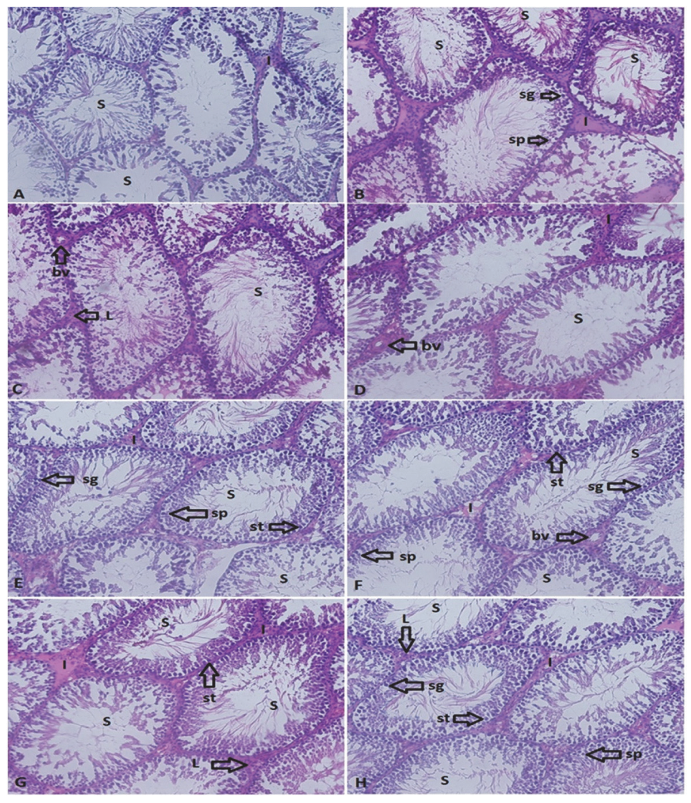
2.5. Light Microscope Investigations

Testicular tissues removed from the dissected ganders were washed in buffer and placed in formaldehyde fixative for light microscopy examination. After the fixation stage, washing and dehydration procedures were performed. Then, the tissues were paraffin blocked. Sections of 6–7 µ in thickness were removed from the prepared blocks. The sections were stained with hematoxylin–eosin, examined, and photographed using a microscope with a camera attachment (Olympus BX 51 light microscope; Olympus Corp., Tokyo, Japan).
The archived images were analyzed morphometrically using the cell D Soft Imaging System (Olympus Corp., Tokyo, Japan). The internal diameter of the seminiferous tubules, the thickness of the germinative epithelium (without tunica propria), and the area of the seminiferous tubule and interstitial tissue were measured in every image. The relative area (%) of the interstitial tissue, defined as the ratio of the area occupied by the interstitium to the total area (interstitial and tubular area) within the archived view field under 200 magnification, was calculated for each individual [33].

2.6. Statistical Analysis
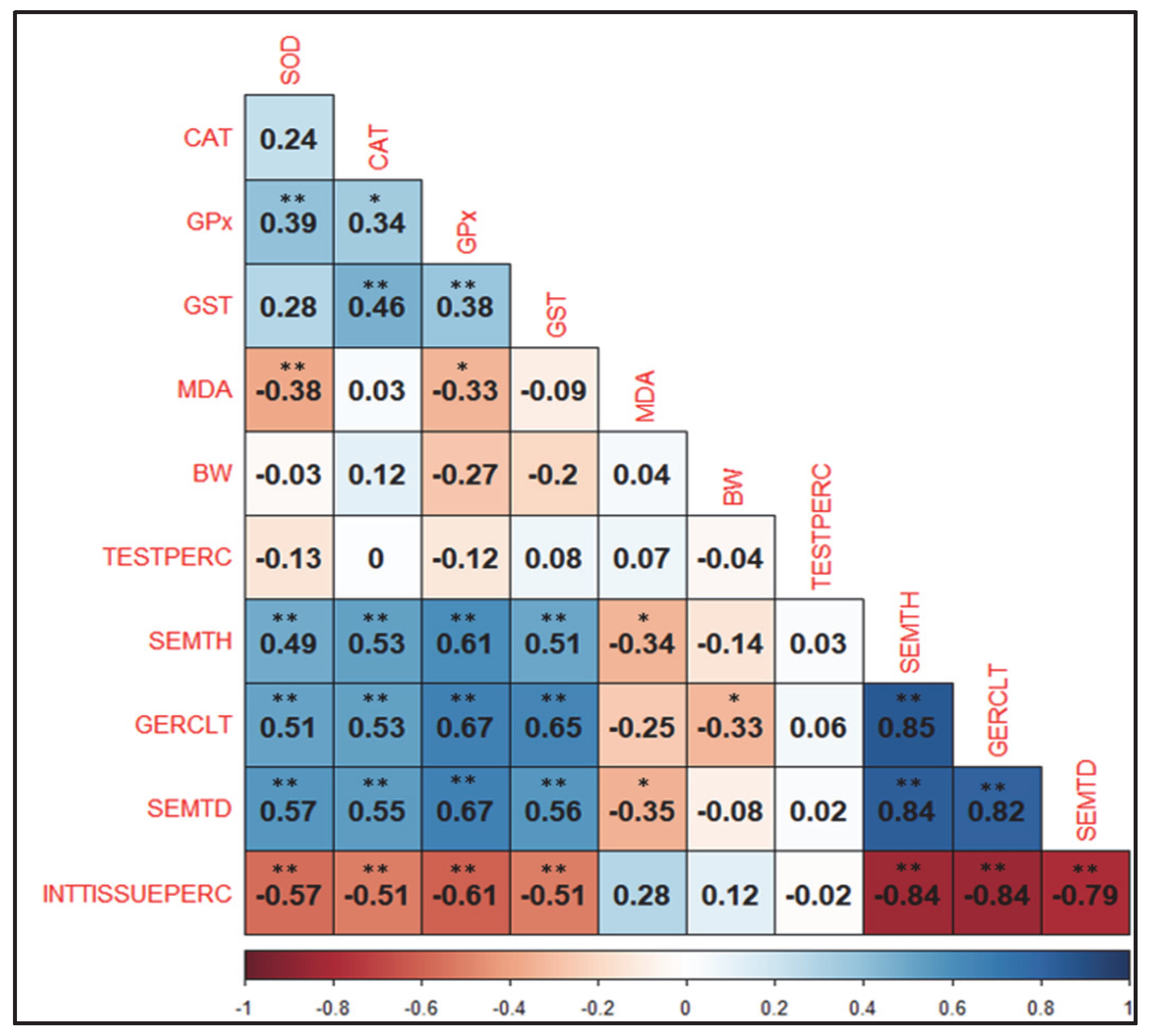
Statistical analysis was performed using SPSS 25.0 statistical software (SPSS, Inc., Chicago, IL, USA). Differences between varying combinations of the dietary Se, Zn, and Vit E supplements were analyzed by one-way ANOVA and Tukey’s test. Significance was set at p < 0.05. Relationships between the various oxidative enzyme activity, and developmental and histology parameters were analyzed by calculating Pearson correlation coefficients (r). For the purposes of discussion, the following descriptors were used to describe the relative strength of the correlations: very weak (r < 0.20), weak (r = 0.20–0.39), moderate (r = 0.40–0.59), strong (r = 0.60–0.79), and very strong (r = 0.80–0.99) [34].

3. Results

The effects of varying combinations of dietary Se, Zn, and Vit E supplements on antioxidant enzyme activity in the testicular tissues of 1-year-old native Turkish ganders are presented in Table 3. Superoxide dismutase enzyme (SOD) activity, catalase enzyme (CAT) activity, glutathione peroxidase enzyme (GPx) activity, and glutathione-S-transferase enzyme (GST) activity were highest in the Se + Vit E + Zn combination and lowest in the control group (Table 3, p < 0.05). Malondialdehyde (MDA), which is an important indicator of lipid peroxidation, was not significantly affected by the dietary treatments.
The effects of varying combinations of dietary Se, Zn, and Vit E supplements on the body weight at sacrifice and testicular weights of 1-year-old native Turkish ganders are shown in Table 4. There was no significant effect by the varying combinations of Se, Zn, and Vit E supplements on either the right, left, or overall total testicular weights, and relative testis weight. The weight of the ganders at sacrifice in the experimental groups was also found to be similar (Table 4).
Pearson correlation coefficients between oxidative enzyme activity, and developmental and histological traits in the testes of 1-year-old native Turkish ganders are shown in Figure 1. The SOD, CAT, GPx, and GST enzyme activities were moderately positively correlated with SEMTH, GERCLT, and SEMTD, while negatively correlated with INTTISSUEPERC. The MDA activity was negatively correlated with SEMTH (−0.34) and SEMTD (−0.35). While no correlation was found between the BW and TESTPERC traits and antioxidant enzyme activities, yet there was a weak correlation (−0.33) between BW and GERCLT. Strong correlations were revealed between testicular histological characteristics. Moreover, INTTISSUEPERC and SEMTH, GERCLT, and SEMTD traits were strongly negatively correlated at −0.84, −0.84, and −0.79, respectively. A very strong positive correlation was observed between SEMTD, SEMTH, and GERCLT, ranging from 0.82 to 0.85.
Histological images of testicular tissues in the treatment groups are shown in Figure 2 and morphometric measurements of some areas in these images are shown in Table 5. The effects of varying combinations of dietary Se, Zn, and Vit E supplements on seminiferous tubule area (mm2), germinative epithelial thickness (mm), seminiferous tubule diameter (mm), and relative interstitial tissue area (%) were found to be significant (Table 5, p < 0.05). The highest seminiferous tubule area and germinative epithelial thickness were determined in ganders fed with a combination of Se + Vit E + Zn compared to the control group (p < 0.05). The lowest seminiferous tubule diameter was determined in ganders fed a control plus Zn diet, with the highest in the Se + Vit E + Zn and Se + Vit E combination (p < 0.05). The relative interstitial tissue area ratio was the highest in the control and Zn group ganders and the lowest was in ganders fed a Se + Vit E + Zn and Se + Vit E combination (p < 0.05).

4. Discussion

Reactive oxygen substances (ROS) accumulate in the testes with advancing age and cause continuous oxidative stress in the cells. As a result, reproductive performance, which is of vital importance in males, decreases. Even if the testicular antioxidant capacity is low, antioxidant compounds in the testicular tissue can protect sperm against ROS [35]. Therefore, many studies have been conducted to increase and improve the antioxidant capacity in the testes [36,37,38]. Feeding programs provided to animals can affect reproductive activities and alter sexual behavior, morphology, and function of reproductive organs [39]. Se, Vit E, and Zn affect many biochemical and physiological systems in animal organisms, including reproduction [40,41,42,43]. Antioxidant enzymes limit the harmful effects of oxidant molecules in tissues and provide a defense against oxidative stress by scavenging free radicals [44]. Antioxidant enzymes work together to perform this task, and even small deviations in their activity can cause undesirable effects on cellular structures [45]. Therefore, it is important to determine the oxidative status in testes histology by determining the activity of SOD, CAT, GST, and GPx enzymes and measuring the MDA level.
Although MDA levels tended to be low in the control and Zn groups, SOD, CAT, GST, and GPx levels were the highest in the testes of ganders fed with a Se + Vit E + Zn supplementary diet compared to the control group. This triple combination has come to the fore as the dietary treatment that combats oxidative stress in testicular tissues of 1-year-old native Turkish ganders at the highest level. At the same time, the partial decrease in MDA values supports this situation, whereby the MDA level was already weak but significantly negatively correlated with SOD (−0.38) and GPx (−0.33) enzymes. Especially in ganders fed with the Se + Vit E combination, the MDA level tended to be slightly lower. Wan et al. [46] found lower levels of MDA in the blood serum of geese fed diets supplemented with Se. It is reported that the supplementation of Se and Vit E to the diet of roosters had a significant stimulatory effect on GPx activity in the testes [47]. Even if testicular antioxidant capacity is low, antioxidants in testicular tissue and seminal plasma can protect sperm against ROS. Improving testicular antioxidant capacity can be achieved with additional supplements to the diet, especially at older ages [7,36,37,38]. Since the data obtained in our study coincides with the end of the reproductive period, our findings support this situation.
In ganders, the testes are shaped like beans and located in the abdominal cavity. Their main function is to produce spermatozoa and secrete testosterone. The vasculature, size, and position of the testes vary according to whether the gander is sexually active or not [48]. Testes can reach an average weight of 1.67 g in the non-reproductive period and 12.3 g during the reproduction season. Akhtar et al. [49] determined testicular weights between 5.2 and 9.4 g and relative testicular weights between 0.40 and 0.23% in Yangzhou ganders between 181 and 227 days of age. Leska et al. [33] determined the testicular weights of ganders during the reproduction period, non-breeding period, and at the beginning of the breeding period as 12.3, 0.48, and 1.67 g, respectively. Opalka et al. [39] determined testicular weights as 5.5, 3.6, and 0.78 g in March, May, and July, respectively. In our study, total testicular weights varied between 0.8 and 2.5 g, the relative testis weight varied between 0.024 and 0.064 (%), and neither body weights nor testicular traits were affected by the dietary treatments. These low weight values are due to the fact that the ganders were at the end of the reproductive season, in line with Opalka et al. [39]. Moreover, especially in the groups where vitamins and minerals were added, they were higher than the values during the same period of other studies [33]. The differences revealed by other studies may have been influenced by many other environmental conditions, especially different dietary treatments and genotypes. Neither body weight nor testicular percentage was significantly associated with any antioxidant enzyme or histological parameter. This also indicates that various combinations of dietary Se, Vit E, and Zn supplements improved the sperm production function in ganders through antioxidant enzyme mechanisms at the cellular level, without affecting their general physiological status.
The seminiferous tubule area, germinative epithelial thickness, and seminiferous tubule diameter of the ganders fed a Se + Vit E + Zn supplementary diet seem to be advantageous in terms of continuity in the semen production compared to the control group. The strong negative correlation between these histological parameters and the relative interstitial tissue area also supports this. During the non-reproductive season, the seminiferous tubules and germinal epithelium are in a rudimentary state and develop as the reproduction season progresses. While the percentage of testicular interstitial tissue is high in the non-reproductive season, the rate of tubular tissue increases during the season [33,50]. In our study, the highest seminiferous tubule area was 0.027 mm2, the highest germinative epithelial thickness was 0.076 mm, the highest seminiferous tubule diameter was 0.144 mm, and the lowest relative interstitial tissue area was 1.60%. Leska et al. [33] determined the seminiferous tubule area, germinative epithelial thickness, seminiferous tubule diameter, and relative interstitial tissue area characteristics during the reproductive period, non-reproductive period, and at the beginning of the reproductive period as 0.109, 0.102, 0.366, 0.17 mm2, 0.014, 0.04, 0.127, 1.80 mm, and 0.023, 0.05, 0.148, 1.68%, respectively. Although our study was conducted at the end of the reproductive period, it found similar values to the beginning of the reproductive period in the study by Leska et al. [33], and higher values than the non-reproductive period, especially in the groups supplemented with vitamins and minerals. This highlights that the varying combinations of dietary Se, Vit E, and Zn supplements had positive results in terms of the ability to continually produce semen in native Turkish ganders, despite advancements in age.
Reproduction in geese is seasonal and cyclical, and reproductive functions are also known to decline towards the end of the laying period. This may be a result of the weakening of the physiological functions in the testicular tissue [29]. The results of our study also support this situation. In addition, Sabzian-Melei et al. [51], in their study on broiler breeder males, found higher seminiferous tubule area and seminiferous tubule diameter in birds fed with 30 mg/kg and 45 mg/kg Se added to the diet, similar to our study. The increase in Sertoli and Leydig cells probably results in higher ejaculate volume and is associated with higher spermatogenesis. This also contributes to the increase in seminiferous tubule diameter and epithelium thickness [51,52]. In our study, moderate to strong phenotypic correlations (0.49 to 0.67) observed between antioxidative enzyme activity and histological parameters revealed the stimulating effect of dietary treatments, such as Se, Vit E, and Zn, to increase and maintain an ability to produce sperm in testicular cells, despite advancements in age.

5. Conclusions

This study showed that the simultaneous supplementation of dietary Se + Vit E + Zn in native Turkish ganders had positive effects on testicular tissues, reducing oxidative damage and improving histological parameters. The fact that the seminiferous tubule area, germinative epithelial thickness, and seminiferous tubule diameter are higher than in the control group is an important finding for the continuity of sperm-producing ability. It has been shown that antioxidant vitamins and minerals (Se, Vit E, and Zn) increase sperm production ability in the testes without impairing the physiological traits of ganders. However, further studies are needed to reveal the fertilization ability and quality of this sperm produced for total reproductive efficiency in 1-year-old native Turkish ganders.

Author Contributions

Conceptualization, H.B., M.A.B. and K.E.; methodology, H.B., M.A.B. and K.E.; software, K.E.; validation, H.B., H.O.T., M.A.B., M.S. and K.E.; formal analysis, K.E. and M.A.B.; investigation, H.B., M.A.B. and H.O.T.; resources, H.B., M.A.B.; data curation, H.B., M.A.B. and K.E.; writing—original draft preparation, H.B., H.O.T., M.A.B., M.S. and K.E.; writing—review and editing, H.B., H.O.T., M.A.B., M.S., K.E., V.D. and G.S.; visualization, M.A.B. and K.E.; supervision, M.A.B. and M.S.; project administration, H.B. and M.A.B.; funding acquisition, H.B., H.O.T. and M.A.B. All authors have read and agreed to the published version of the manuscript.

Funding

This study was supported by Yozgat Bozok University (Project no: 6602b-FEN/20-412).

Institutional Review Board Statement

All procedures performed in the experiment were approved by the Erciyes University Experimental Animals Ethics Committee (Date: 1 July 2020, No: 07; Decision No: 20/095).

Informed Consent Statement

Not applicable.

Data Availability Statement

All the relevant data are available in the paper.

Acknowledgments

The authors are thankful to the Application and Research Center of Science and Technology of Yozgat Bozok University.

Conflicts of Interest

The authors declare no conflict of interest.

References

  1. Boz, M.A.; Sarıca, M.; Yamak, U.S. Goose production in province Yozgat. J. Poult. Res. 2014, 11, 16–20. [Google Scholar]
  2. Boz, M.A.; Sarıca, M.; Yamak, U.S. Economic evaluation of natural and artificial incubated geese in intensive and free-range production systems. Turk. J. Agric. Food Sci. Technol. 2016, 4, 981–986. [Google Scholar]
  3. Önder, H.; Boz, M.A.; Sarıca, M.; Abacı, S.H.; Yamak, U.S. Comparison of growth curve models in Turkish native geese. Eur. Poult. Sci. 2017, 81, 193. [Google Scholar] [CrossRef]
  4. Boz, M.A. Effect of classified rearing according to live weight on growth, carcass and some meat quality characteristics in geese. Turk. J. Agric. Food Sci. Technol. 2019, 7, 1429–1434. [Google Scholar]
  5. Boz, M.A.; Baş, H.; Sarıca, M.; Erensoy, K. The effects of natural mating and artificial insemination on reproductive traits of 1-and 2-year-old native Turkish geese. Vet. Res. Commun. 2021, 45, 211–221. [Google Scholar] [CrossRef] [PubMed]
  6. Çavdar, C.; Sifil, A.; Çamsarı, T. Reactive oxygen particles and antioxidant defence. Off. J. Turk. Nephrol. 1997, 3, 92–95. (In Turkish) [Google Scholar]
  7. Partyka, A.; Nizanski, W. Supplementation of Avian Semen Extenders with Antioxidants to Improve Semen Quality—Is It an Effective Strategy? Antioxidants 2021, 10, 1927. [Google Scholar] [CrossRef]
  8. Baş, H.; Kara, Ö.; Kara, M.; Pandır, D. Protective effect of vardenafil on ischemia-reperfusion injury in rat ovary. Turk. J. Med. Sci. 2013, 43, 684–689. [Google Scholar] [CrossRef]
  9. Brenneisen, P.; Steinbrenner, H.; Sies, H. Selenium, oxidative stress, and health aspects. Mol. Asp. Med. 2005, 26, 256–267. [Google Scholar] [CrossRef]
  10. Xu, Z.J.; Liu, M.; Niu, Q.J.; Huang, Y.X.; Zhao, L.; Lei, X.G.; Sun, L.H. Both selenium deficiency and excess impair male reproductive system via inducing oxidative stress-activated PI3K/AKT-mediated apoptosis and cell proliferation signaling in testis of mice. Free Radic. Biol. Med. 2023, 197, 15–22. [Google Scholar] [CrossRef]
  11. Ozbek, E. Induction of oxidative stress in kidney. Int. J. Nephrol. 2012, 2012, 465897. [Google Scholar] [CrossRef] [PubMed] [Green Version]
  12. Çaylak, E. Oxidative stress and antioxidants in the animals and the plants. Tıp Araştırmaları Derg. 2011, 9, 73–83. (In Turkish) [Google Scholar]
  13. Mansour, S.A.; Mossa, A.H. Adverse effects of lactational exposure to chlorpyrifos in suckling rats. Hum. Exp. Toxicol. 2010, 29, 77–92. [Google Scholar] [CrossRef] [PubMed]
  14. Codandabany, U. Erythrocyte lipid peroxidation and antioxidants in cigarette smokers. Cell Biochem. Funct. 2000, 18, 99–102. [Google Scholar] [CrossRef]
  15. Derviş, E. Oral antioksidanlar. Dermatoz 2011, 2, 263–267. (In Turkish) [Google Scholar]
  16. Lakshmi, B.V.S.; Sudhakar, M.; Aparna, M. Protective potential of black grapes against lead induced oxidative stress in rats. Environ. Toxicol. Pharmacol. 2013, 35, 361–368. [Google Scholar] [CrossRef] [PubMed]
  17. Mates, J.M. Effects of antioxidant enzymes in the molecular control of reactive oxygen species toxicology. Toxicology 2000, 153, 83–104. [Google Scholar] [CrossRef] [PubMed]
  18. Ajiboye, T.O. Redox status of the liver and kidney of 2,2-dichlorovinyl dimethyl phosphate (DDVP) treated rats. Chem.-Biol. Interact. 2010, 185, 202–207. [Google Scholar] [CrossRef] [PubMed]
  19. Aly, H.A.A.; Domènech, Ò.; Banjar, Z.M. Effect of nonylphenol on male reproduction: Analysis of rat epididymal biochemical markers and antioxidant defense enzymes. Toxicol. Appl. Pharmacol. 2012, 261, 134–141. [Google Scholar] [CrossRef] [PubMed]
  20. Mattsson, A.; Mura, E.; Brunström, B.; Panzica, G.; Halldin, K. Selective activation of estrogen receptor alpha in Japanese quail embryos affects reproductive organ differentiation but not the male sexual behavior or the parvocellular vasotocin system. Gen. Comp. Endocrinol. 2008, 159, 150–157. [Google Scholar] [CrossRef]
  21. Kahvecioğlu, O.; Çalışlar, T. Ürogenital ve Endokrin Sistemi. Evcil Kuşların Anatomisi; Dursun, N., Ed.; Medisan Publication: Ankara, Türkiye, 2002; pp. 103–128. (In Turkish) [Google Scholar]
  22. Deviche, P.; Hurley, L.L.; Fokidis, H.B. Avian testicular structure, function, and regulation. Horm. Reprod. Vertebr. 2011, 4, 27–69. [Google Scholar]
  23. Lukaszewicz, E.; Kruszynski, W. Evaluation of fresh and frozen-thawed semen of individual ganders by assessment of spermatozoa motility and morphology. Theriogenology 2003, 59, 1627–1640. [Google Scholar]
  24. Amem, M.H.; Al-Daraji, H.J. Zinc improves egg quality in Cobb500 broiler breeder females. Int. J. Poult. Sci. 2011, 10, 471–476. [Google Scholar] [CrossRef] [Green Version]
  25. Amem, M.H.; Al-Daraji, H.J. Effect of dietary zinc on semen quality of Cobb 500 broiler breeder males. Int. J. Poult. Sci. 2011, 10, 477–482. [Google Scholar] [CrossRef] [Green Version]
  26. Jerysz, A.; Lukaszewicz, E. Effect of Dietary Selenium and Vitamin E on Ganders’ Response to Semen Collection and Ejaculate Characteristics. Biol. Trace Elem. Res. 2013, 153, 196–204. [Google Scholar] [CrossRef] [PubMed] [Green Version]
  27. Bas, H.; Eroglu, H.E.; Dogan, H.; Uskutoglu, T.; Cosge Senkal, B.; Cesur, C. Evaluation of the chemical composition, genotoxic and cytotoxic effects of cocklebur (Xanthium strumarium L.) seed oil on human blood cells. Int. J. Agric. Life Sci. 2022, 6, 1–7. [Google Scholar]
  28. Marklund, S.; Marklund, G. Involvement of the superoxide anion radical in the autoxidation of pyrogallol and a convenient assay for superoxide dismutase. Eur. J. Biochem. 1974, 47, 469–474. [Google Scholar] [CrossRef] [PubMed]
  29. Aebi, H. Catalase in vitro. Methods Enzym. 1984, 105, 121–126. [Google Scholar]
  30. Paglia, D.E.; Valentine, W.N. Studies on the quantative and qualitative characterization of glutathione peroxidase. J. Lab. Med. 1987, 70, 158–165. [Google Scholar]
  31. Habig, W.H.; Pabst, M.J.; Jakoby, W.B. Glutathione-S-transferases: The first enzymatic step in mercapturic acid formation. J. Biol. Chem. 1974, 249, 7130–7139. [Google Scholar] [CrossRef] [PubMed]
  32. Ohkawa, H.; Ohishi, N.; Yagi, K. Assay for lipid peroxides in animal tissues by thiobarbituric acid reaction. Anal. Biochem. 1979, 95, 351–358. [Google Scholar] [CrossRef]
  33. Leska, A.; Kiezun, J.; Kaminska, B.; Dusza, L. Seasonal changes in the expression of the androgen receptor in the testes of the domestic goose (Ancer ancer f. domestica). Gen. Comp. Endocrinol. 2012, 179, 63–70. [Google Scholar] [CrossRef]
  34. Bowker, B.; Zhuang, H. Detection of razor shear force differences in broiler breast meat due to the woody breast condition depends on measurement technique and meat state. Poult. Sci. 2019, 98, 6170–6176. [Google Scholar] [CrossRef]
  35. Long, C.; Wang, Z.; Guo, Y.; Sheng, X.; Xing, K.; Ni, H.; Wang, X.; Xiao, L.; Qi, X. Research Note: Dietary supplementation with pyrroloquinoline quinone disodium (PQQ.Na2) improves oxidative status and semen quality in aging layer breeder roosters. Poult. Sci. 2022, 101, 101812. [Google Scholar] [CrossRef] [PubMed]
  36. Akhlaghi, A.; Ahangari, Y.J.; Navidshad, B.; Pirsaraei, Z.A.; Zhandi, M.; Deldar, H.; Rezvani, M.R.; Dadpasand, M.; Hashemi, S.R.; Poureslami, R.; et al. Improvements in semen quality, sperm fatty acids, and reproductive performance in aged Cobb 500 breeder roosters fed diets containing dried ginger rhizomes (Zingiber officinale). Poult. Sci. 2014, 93, 1236–1244. [Google Scholar] [CrossRef]
  37. Qi, X.; Shang, M.; Chen, C.; Chen, Y.; Hua, J.; Sheng, X.; Wang, X.; Xing, K.; Ni, H.; Guo, Y. Dietary supplementation with linseed oil improves semen quality, reproductive hormone, gene and protein expression related to testosterone synthesis in aging layer breeder roosters. Theriogenology 2019, 131, 9–15. [Google Scholar] [CrossRef] [PubMed]
  38. Yan, W.; Kanno, C.; Oshima, E.; Kuzuma, Y.; Kim, S.W.; Bai, H.; Takahashi, M.; Yanagawa, Y.; Nagano, M.; Wakamatsu, J.-I.; et al. Enhancement of sperm motility and viability by turmeric by-product dietary supplementation in roosters. Anim. Reprod. Sci. 2017, 185, 195–204. [Google Scholar] [CrossRef]
  39. Opalka, M.; Kaminska, B.; Piskula, M.K.; Puchajda-Skowronska, H.; Dusza, L. Effects of phytoestrogens on testosterone secretion by Leydig cells from Biłgoraj ganders (Anser anser). Br. Poult. Sci. 2006, 47, 237–245. [Google Scholar] [CrossRef]
  40. Surai, P.F.; Blesbois, E.; Grasseau, I.; Chalah, T.; Brillard, J.P.; Wishart, G.; Cerolini, S.; Sparks, N.H.C. Fatty acid composition, glutathione peroxidase and superoxide dismutase activity and total antioxidant activity of avian semen. Comp. Biochem. Physiol. 1998, 120, 527–533. [Google Scholar] [CrossRef] [PubMed]
  41. Zubair, M. Effects of dietary vitamin E on male reproductive system. Asian Pac. J. Reprod. 2017, 6, 145–150. [Google Scholar]
  42. Surai, P.F. Selenium in Poultry Nutrition and Health; Wageningen Academic Publishers: Wageningen, The Netherlands, 2018; ISBN 978-90-8686-317-4. [Google Scholar]
  43. Huang, L.; Li, X.; Wang, W.; Yang, L.; Zhu, Y. The Role of Zinc in Poultry Breeder and Hen Nutrition: An Update. Biol. Trace Elem. Res. 2019, 192, 308–318. [Google Scholar] [CrossRef] [PubMed]
  44. Fouad, A.M.; Kasem El-Senousey, H.A.; Ruan, D.; Xia, W.; Chen, W.; Wang, S.; Zheng, C. Nutritional modulation of fertility in male poultry. Poult. Sci. 2020, 99, 5637–5646. [Google Scholar] [CrossRef] [PubMed]
  45. Goel, A.; Dani, V.; Dhawan, D.K. Protective effects of zinc on lipid peroxidation, antioxidant enzymes and hepatic histoarchitecture in chlorpyrifos-induced toxicity. Chem.-Biol. Interact. 2005, 156, 131–140. [Google Scholar] [CrossRef]
  46. Wan, X.L.; Ju, G.Y.; Xu, L.; Yang, H.M.; Wang, Z.Y. Dietary selenomethionine increases antioxidant capacity of geese by improving glutathione and thioredoxin systems. Poult. Sci. 2019, 98, 3763–3769. [Google Scholar] [CrossRef] [PubMed]
  47. Surai, P.F.; Fujihara, N.; Speake, B.K.; Brillard, J.P.; Wishart, G.J.; Sparks, N.H.C. Polyunsaturated fatty acids, lipid peroxidation and antioxidant protection in avian semen. Asian Australas. J. Anim. Sci. 2001, 14, 1024–1050. [Google Scholar] [CrossRef]
  48. Buckland, R.; Guy, G. Goose Production Systems; Chapter 5: Male and Female Reproduction Systems. In Goose Production; Buckland, R., Guy, G., Eds.; FAO: Rome, Italy, 2002; p. 17. [Google Scholar]
  49. Akhtar, M.F.; Wei, Q.; Zhu, H.; Chen, Z.; Ahmad, E.; Zhendan, S.; Shi, F. The role of active immunization against inhibin a-subunit on testicular development, testosterone concentration and relevant genes expressions in testis, hypothalamus and pituitary glands in Yangzhou goose ganders. Theriogenology 2019, 128, 122–132. [Google Scholar] [CrossRef]
  50. Akhtar, M.F.; Ahmad, E.; Ali, I.; Shafiq, M.; Chen, Z. The Effect of Inhibin Immunization in seminiferous epithelium of Yangzhou goose ganders: A Histological Study. Animals 2021, 11, 2801. [Google Scholar] [CrossRef]
  51. Sabzian-Melei, R.; Zare-Shahneh, A.; Zhandi, M.; Yousefi, A.R.; Rafieian-Naeini, H.R. Effects of dietary supplementation of different sources and levels of selenium on the semen quality and reproductive performance in aged broiler breeder roosters. Poult. Sci. 2022, 101, 101908. [Google Scholar] [CrossRef] [PubMed]
  52. Leal, M.; Becker-Silva, S.; Chiarini-Garcia, H.; Franca, L. Sertoli cell efficiency and daily sperm production in goats (Capra hircus). Anim. Reprod. Sci. 2018, 1, 122–128. [Google Scholar]
Figure 1. Pearson correlation coefficients between oxidative enzyme activity (SOD: superoxide dismutase; CAT: catalase; GPx: glutathione peroxidase; GST: glutathione-S-transferase; MDA: malondialdehyde), and histological characteristics in testicular tissue (SEMTH: seminiferous tubule area (mm2); GERCLT: germinal cell layer thickness (mm); SEMTD: seminiferous tubule diameter (mm); INTTISSUEPERC: relative interstitial tissue (%)) and developmental traits (BW: body weight; TESTPERC: relative total testicular percentage (%)) in 1-year-old native Turkish ganders. **: p < 0.01; *: p < 0.05.
Sustainability 15 12245 g001
Figure 2. The histological structure of testicular tissues in 1-year-old native Turkish ganders (n = 48). (A) Control, (B) Se, (C) Vit E, (D) Zn, (E) Se + Vit E, (F) selenyum + Zn, (G) Vit E + Zn, and (H) Se + Vit E + Zn. S: seminiferous tubule; I: interstitial space; sg: spermatagonium; sp: spermatocyte; L: Leydig cell; st: Sertoli cell; bv: blood vessel. Magnification: 200×.
Sustainability 15 12245 g002
Table 1. Basic diet components and calculated contents *.
IngredientUnitAmount
Corn%57.5
Sunflower seed meal%18.5
Soybean meal (CP 46%)%10.0
Limestone%8.0
Cotton seed meal (CP 26%)%5.0
Salt%0.75
Vit premix%0.25
Analyzed nutrient content *
Dry matter%88.76
Crude protein (CP)%15.50
MEMJ/kg10.29
Crude oil%3.30
Crude fiber%7.14
Crude ash%11.68
Semg/kg0.15
Znmg/kg60
Vit Emg/kg30
* Feed analyses were carried out at Yozgat Bozok University Science and Technology Application and Research Centre laboratory, Yozgat, Turkey.
Table 2. Se, Zn, and Vit E contents in the experimental groups.
GroupsSe (mg/kg)Vit E (mg/kg)Zn (mg/kg)
Control0.153060
Se0.453060
Vit E0.1513060
Zn0.1530160
Se + Vit E0.4513060
Se + Zn0.4530160
Zn + Vit E0.15130160
Se + Vit E + Zn0.45130160
Se: 67 mg/kg Se premix, 4.5% Sodium Selenite Na2SeO3; Vit E: 200 mg/kg E−50 Adsorbate Rovimix®, 50% Vit E; Zn: 131 mg/kg 76.4% Zn oxide.
Table 3. Effects of varying combinations of dietary Se, Vit E, and Zn supplements on antioxidant enzyme activity in testicular tissues of 1-year-old native Turkish ganders (n = 48).
Dietary TreatmentsSOD
(U/mg Protein)
CAT
(mmol/mg Protein)
GPx
(mmol/mg Protein)
GST
(mmol/mg Protein)
MDA
(mmol/mg Protein)
Control4.84 c1.21 b5.10 c0.83 c0.47
Se6.56 abc1.25 ab7.66 abc1.05 abc0.44
Vit E6.54 abc1.27 ab7.83 abc1.01 abc0.42
Zn5.30 bc1.20 b6.40 bc0.89 bc0.45
Se + Vit E6.98 ab1.29 ab9.50 a1.14 ab0.41
Se + Zn6.69 abc1.25 ab8.48 ab1.00 abc0.42
Vit E + Zn6.62 abc1.26 ab8.55 ab0.99 abc0.37
Se + Vit E + Zn7.71 a1.36 a10.27 a1.20 a0.39
SEM0.4640.0270.6410.0650.032
df7,407,407,407,407,40
F values3.913.016.633.381.01
p values0.0020.0120.0000.0060.438
Each treatment (n = 6 birds/treatment) is expressed as mean ± standard error of the mean (SEM) and the statistical analysis was conducted using a one-way ANOVA with Tukey’s post hoc test. a–c Different letters in the same column are significantly different by Tukey’s multiple comparison tests (p < 0.05).
Table 4. Effects of varying combinations of dietary Se, Vit E, and Zn supplements on sacrificial and testes weights (in g) and percentages (%) in 1-year-old native Turkish ganders (n = 48).
Dietary TreatmentsSlaughter Weight (g)Right Testicular Weight (g)Left Testicular Weight (g)Total Testicular Weight (g)Relative Total
Testicular
Percentage (%)
Control4298.50.370.651.020.02
Se4253.80.460.831.300.03
Vit E4254.50.581.161.740.04
Zn4255.60.821.682.500.06
Se + Vit E4224.00.781.952.730.06
Se + Zn3930.90.320.540.850.02
Vit E + Zn4108.10.400.711.110.03
Se + Vit E + Zn3910.70.470.801.270.03
SEM63.7680.1790.5260.6990.02
df7,407,407,407,407,40
F values0.701.080.961.000.94
p values0.6700.3910.4750.4480.485
Each treatment (n = 6 birds/group) is expressed as mean ± standard error of the mean (SEM) and the statistical analysis was performed by a one-way ANOVA.
Table 5. Effects of varying combinations of dietary Se, Vit E, and Zn supplements on histological parameters in testicular tissues of 1-year-old native Turkish ganders (n = 48).
Dietary TreatmentsSeminiferous Tubule Area
(mm2)
Germinal Cell Layer Thickness
(mm)
Seminiferous Tubule Diameter
(mm)
Relative Area of
Interstitial Tissue (%)
Control0.016 d0.041 d0.122 c1.845 a
Se0.019 c0.053 c0.131 b1.754 b
Vit E0.020 c0.055 c0.134 b1.748 b
Zn0.016 d0.042 d0.124 c1.843 a
Se + Vit E0.024 b0.066 b0.141 a1.651 c
Se + Zn0.019 c0.055 c0.131 b1.752 b
Vit E + Zn0.021 c0.056 c0.135 b1.746 b
Se + Vit E + Zn0.027 a0.076 a0.144 a1.600 c
SEM0.0010.0020.0010.015
df7,407,407,407,40
F values36.2640.4128.5930.95
p values0.0000.0000.0000.000
Each treatment (n = 6 birds/group) is expressed as mean ± standard error of the mean (SEM) and the statistical analysis was conducted by a one-way ANOVA with Tukey’s post hoc test. a–d Different letters in the same column are significantly different by Tukey’s multiple comparison tests (p < 0.05).
Disclaimer/Publisher’s Note: The statements, opinions and data contained in all publications are solely those of the individual author(s) and contributor(s) and not of MDPI and/or the editor(s). MDPI and/or the editor(s) disclaim responsibility for any injury to people or property resulting from any ideas, methods, instructions or products referred to in the content.

