
Biogenic Synthesis of Silver Nanoparticles, Characterization and Their Applications—A Review


1
Laboratory of Plant Pathology, Department of Botany, Mohanlal Sukhadia University Udaipur, Udaipur 313001, Rajasthan, India
2
Plant Bioenergetics and Biotechnology Laboratory, Department of Botany, Mohanlal Sukhadia University Udaipur, Udaipur 313001, Rajasthan, India
*
Author to whom correspondence should be addressed.
Surfaces 2022, 5(1), 67-90; https://0-doi-org.brum.beds.ac.uk/10.3390/surfaces5010003
Submission received: 12 February 2021
/
Revised: 25 December 2021
/
Accepted: 28 December 2021
/
Published: 31 December 2021
(This article belongs to the Special Issue Surface Modification of Nanoparticles for Biomedical Applications)
Abstract
:With the growing awareness for the need of sustainable environment, the importance of synthesizing and the application of green nanoparticles has gained special focus. Among various metal nanoparticles, silver nanoparticles (AgNPs) have gain significant attention. AgNPs are synthesized conventionally by physical and chemical methods using chemicals such as reducing agents, which are hazardous to environment due to their toxic properties, provoking a serious concern to create and develop environment friendly methods. Thus, biological alternatives are emerging to fill gaps, such as green syntheses that use biological molecules taken from plant sources in the form of extracts, which have shown to be superior to chemical and physical approaches. These biological molecules derived from plants are assembled in a highly regulated manner to make them suitable for metal nanoparticle synthesis. The current review outlines the wide plant diversity that may be used to prepare a rapid and single-step procedure with a green path over the traditional ones, as well as their antifungal activity.
1. Introduction
“Nanotechnology” is the newest and one of the most promising and active areas of modern research. The technology deals with the design, synthesis, and manipulation of particles size ranging from 1–100 nm [1]. Within this size range, the chemical, physical, and biological properties change in the fundamental way of both individual atoms and their corresponding bulk material [2]. This very small size increases the surface area-to-volume ratios of particles. The nanoparticles synthesized using plant extract have gained huge consideration in recent years due to their remarkable properties and wide range of applications in catalysis [3], plasmonic [4], optoelectronics [5], biological sensor [6], water treatment, pharmaceutical applications [7], and agriculture and crop protection [8]. Stunning growth in this emerging technology has opened novel fundamental and applied frontiers, including the synthesis of nanomaterial and utilization of their physicochemical and optoelectronic properties [9]. The application of nanotechnology has increased in large number of areas such as optics, mechanics, chemical industries, space industries, electronics, energy science, single electron transistors, light emitters, nonlinear optical devices, photo-electrochemical, catalysis, biomedical, cosmetics, drug/gene delivery, and food and feed [10,11,12,13,14]. Among various nanoparticles used for all the above-mentioned purposes, the metallic AgNPs are contemplated as the most promising, as they possess remarkable antimicrobial properties due to their greater surface area to volume ratio, which is of curiosity for researchers due to the growing microbial resistance against metal ions and antibiotics and the development of resistant strains [15]. Nanoparticles are seen as a solution to many technologies and environmental challenges [10,16,17,18]. The biological synthesis methods of NPs reduce hazards to the global efforts. The development and implementation of these sustainable processes should adopt the fundamental principles of green chemistry. These principles draw attention on maximizing the efficiency of chemical processes without compromising the safety concern of the products.
2. Silver Nanoparticles
Among the various metallic nanoparticles, AgNPs are one of the most promising products in the nanotechnology industry. The development of consistent processes for the synthesis of AgNPs is an important field of current nanotechnology research. AgNPs have a wide range of use because of their unique characteristics such as optical, electrical and magnetic properties, which can be incorporated into antibacterial, antiviral, and antifungal applications, composite fibers, biosensor materials, cosmetic products, food industry uses, and electronic components [19,20]. The AgNPs are also reported as medical and pharmaceutical agents that have directly encountered by a human system in such products as shampoos, detergents, soaps, toothpaste, and cosmetics [21]. The biomedical use of AgNPs includes their application as antibacterial [22], antifungal [23], anti-inflammatory [24,25], antiviral [26], and anti-diabetic agents [27]. In recent studies, AgNPs were also reported in the diagnosis and treatment of cancer and as drug carriers by either active or passive mechanisms [28]. The antibacterial effects of silver have been noticed since antiquity and, in a variety of applications, silver is currently used to control bacterial growth including in dental work, catheters, and burning wounds. It is widely known that Ag ions and Ag-based compounds are highly toxic to microorganisms, showing strong biocidal effects [29]. Due to the optical properties of AgNPs, these are mainly used in photonic devices and in molecular diagnostics. An increasing application is the use of AgNPs for antimicrobial coatings. Many textiles and biomedical devices contain AgNPs that continuously release a low level of silver ions to provide protection against bacteria [30]. AgNPs were used as catalysts for the reduction of many aqueous aromatic nitro compounds in which NaBH4 alone could not reduce aromatic nitro compounds. NaBH4 was reported to enhance the catalytic activity of AgNPs by reducing the oxide layer to regenerate the fresh surface of the silver particles [2].
3. History and Concept of Nanotechnology
The history of nanoparticles is quite long and, during the last two decades, major developments within nanoscience have taken place. In 1970, Norio Taniguchi coined the term “nanotechnology.” But the concept of nanotechnology was initially defined by Nobel Prize winner Richard Feynman in his renowned address at the California Institute of Technology on 29 December 1959. The topic of nanoparticles was mentioned in one of his articles published in 1960 titled “There is Plenty of Room at the Bottom.” He pointed out that if a single bit of information required only 100 atoms, then all the books ever written could be kept in a cube with sides measuring 0.02-inch in length [31].
The nanoparticles are being used in many sectors including in the biomedical field, water treatment, the electrical industry, biological textiles, chemistry, and human health. Depending upon shape and size, colloidal metal particles play a critical role in different applications, including in the preparation of magnetic, electronic devices, wound healing, antimicrobial gene expression, and the preparation of biocomposites, the noble metal colloids having the specific optical, catalytical, and electromagnetic properties [32]. Nanotechnology is a recent invention in scientific research, but its basic concepts have been developed over a long period of time [33], since people have been coating glass windows with tiny coloured metal particles for a long time, particularly silver, to create a glassy yellow colour.
4. Classification of Nanoparticles
Nanoparticles are classified mainly into two categories: inorganic and organic nanoparticles. Carbon nanoparticles (fullerenes) are one of the organic nanoparticles. On the other hand, inorganic nanoparticles contain gold and magnetic nanoparticles and silver and semiconductor nanoparticles such as titanium dioxide and zinc oxide. There is an increasing interest in inorganic nanoparticles, as they offer superior material properties with functional usefulness. Due to their small structure and benefits over chemical imaging agents and medications, they have been investigated as a possible instrument for medical applications as well as for treating diseases. Gold nanoparticles have been widely employed in imaging, medication delivery, and biological target thermotherapy [34]. Inorganic nanoparticles such as metallic and semiconductor nanoparticles show core optical properties which may augment the transparency of polymer-particle composites. For those reasons, inorganic nanoparticles have gained much interest in studies devoted to optical properties in composites [35].
5. Methods of AgNP Synthesis
In AgNPs synthesizing and stabilization, several physical and chemical methods have been used. In recent years, Nanoparticle synthesis is one of the most fascinating areas of scientific research, and there is an increasing focus on manufacturing nanoparticles in environmentally acceptable ways (green chemistry). Mixed-valence polyoxometalates, polysaccharides, Tollens, irradiation, and biological processes are examples of green synthesis approaches that have advantages over traditional methods which use chemical agents that are detrimental to the environment. This chapter addresses the synthesis of AgNPs using physical, chemical, and green methods. The various types of methods used for the synthesis of nanoparticles are represented in Figure 1.
5.1. Physical Methods
In physical processes, there are various methods including plasma arcing [36], ball milling [37], thermal evaporation [38], spray pyrolysis [39], ultra-thin films [40], electron irradiation [41], pulsed laser desorption [42], lithographic techniques [43], sputter deposition [44], layer by layer growth [45], and the diffusion flame [46] synthesis of nanoparticles. In these methods, the nanoparticles synthesized by evaporation condensation, which could be conducted in a tube furnace at atmospheric pressure and is used to make metal nanoparticles.
The starting material within a boat centered at the furnace is vaporized into a carrier gas. The evaporation/condensation technique has previously been used to synthesize various nanoparticles, including Au, PbS, Ag, and fullerene. However, utilizing a tube furnace to make AgNPs has a number of disadvantages, including the fact that a tube furnace occupies a large space and need a high energy input while increasing the environmental temperature around the source material, and it takes a lot of time to gain thermal stability. In addition, a typical tube furnace requires power using up to more than several kilowatts and a pre-heating time of several tens of minutes to attain a stable operating temperature. Furthermore, AgNPs have been synthesized with the laser ablation of metallic bulk materials in solution [47,48,49,50,51]. One of the biggest advantages of laser ablation compared to another physical methods for synthesizing metal nanoparticles is the absence of chemical reagents in solutions. Therefore, pure colloids can be produced by this method which will be useful for more applications [52].
5.2. Chemical Methods
In chemical processes, nanoparticles are synthesized using several methods such as electro-deposition, sol-gel process [53] chemical solution deposition [53], chemical vapor deposition [54,55], the Langmuir Blodgett method, the soft chemical method, catalytic route, hydrolysis [56], co-precipitation, and the wet chemical method. Chemical reduction is the most common method for synthesis of AgNPs. Various organic and inorganic chemicals have been used as reducing agents; sodium borohydride (NaBH4), elemental hydrogen, sodium citrate, ascorbate, Tollens reagent, N,N-dimethylformamide (DMF), polyol process, and poly (ethylene glycol)–block copolymers are used for the reduction of silver ions (Ag+) in non-aqueous or aqueous solutions. The above-mentioned chemicals reduce silver nitrate into AgNPs. Silver ions (Ag+) are reduced into metallic silver (Ag0) by reducing agents, which is followed by agglomeration into oligomeric clusters. Capping agents are also used for size stabilization of the nanoparticles. These clusters eventually lead to the formation of metallic colloidal silver particles [57,58]. A lot of nanoparticles can be synthesized in a short span of time, which is one of the biggest advantages of this method. It is needed to use protective reducing agents to stabilize nanoparticles to avoid their agglomeration during the course of AgNPs preparation and protect the nanoparticles that can be absorbed on or bind onto nanoparticle surfaces [54]. Surfactants having functionalities for interactions with particle surfaces (e.g., thiols, amines, acids, and alcohols) can stabilize particle development and protect particles from precipitation, agglomeration, or losing their surface features. Polymeric substances such as poly (vinyl alcohol), poly (vinyl pyrrolidone), poly (methacrylic acid), poly (ethylene glycol), and polymethyl methacrylate have been shown to be efficient nanoparticle stabilizers [59].
These conventional methods use high radiation and concentrated reducing and stabilizing agents that are hazardous to human health and non-ecofriendly by-products. As a result, biological nanoparticle synthesis is a one-step bio-reduction process that consumes less energy to synthesize eco-friendly NPs [60]. Biological approaches rely on environmentally favorable resources such as plant extracts, bacteria, fungus, and microalgae such as cyanobacteria, diatoms, and seaweed [61,62].
5.3. Biological Methods
Nanoparticle synthesis by biological methods is a growing area of nanotechnology [63]. Plant extracts [64], bacteria [65], and fungus [66] are some eco-friendly resources for the biological synthesis of nanoparticles. As this synthesis of AgNPs does not employ the use of hazardous chemicals, it provides a variety of advantages in terms of environmental benefits and compatibility for pharmaceutical and other biological applications. Green synthesis has been proven to be better to conventional approaches in term of being cost-effective, nontoxic to the environment, and easily scaled up for large production [67].
5.3.1. Synthesis of AgNPs Using Bacteria
In 1999, Klaus and colleagues reported the first evidence of bacterial-mediated AgNP production and detected AgNP aggregation inside the cells of Pseudomonas stutzeri AG259, a bacterium isolated from a silver mine [68].
Rapid synthesis of AgNPs using keratinase was obtained from a novel keratin-degrading bacterial strain, Bacillus safensis LAU 13. UV spectrophotometry showed the maximum absorbance at 409 nm. The FTIR results indicated that proteins were the capping and stabilization molecules in the synthesis of AgNPs. The XRD data showed that the particles are crystalline in nature with an average size of ~8.3 nm and have a face-centred cubic phase. These particles were used against clinical isolates of E. coli and showed effective antibacterial activity [69]. AgNPs of spherical shape were synthesized using Bacillus methylotrophic DC3, isolated from the soil of Korean ginseng, a traditionally known oriental medicinal plant in Korea. The synthesized AgNPs were characterized using FE-TEM and the particles showed sizes in the range of 10–30 nm. The UV-vis absorption spectrum showed the maximum absorbance peak at 416 nm. In recent contributions, the synthesis of nanoparticles using bacteria including Pseudomonas deceptionensis, Weissella oryzae [70], Bacillus amyloliquefaciens, Bacillus licheniformis, and Rhodobacter sphaeroides [71,72,73] was reported. The extracellular synthesis of AgNPs was reported with the use of bacteria such as Acinetobacter calcoaceticus [74], Gluconobacter roseus [75], Klebsiella pneumonia [76], Salmonella typhimurium [77], Pseudomonas aeruginosa [78], Xanthomonas oryzae [79], and Rhodococcus sp. [80].
5.3.2. Synthesis of AgNPs Using Fungi
Extracellular synthesis of AgNPs using Fusarium oxysporum can be carried out in several kinds of materials, such as cloths [81]. The filamentous fungus Aspergillus fumigates is known for the rapid synthesis of AgNPs ranging from 5 to 25 nm [82]. The fungus, Aspergillus flavus, accumulated AgNPs on the surface of its cell wall in 72 h when challenged with a silver nitrate solution. After being dispersed by ultrasonication, these nanoparticles had an absorption peak in the UV-visible spectrum at 420 nm, which corresponded to the plasmon resonance of AgNPs. According to transmission electron micrographs of nanoparticles in aqueous solution, the fungus produced relatively monodisperse AgNPs with an average particle size of 8.92 ± 1.61 nm.
The formation of metallic silver has been confirmed by X-ray diffraction analysis of the nanoparticles. The Fourier transform infrared spectroscopy confirmed the presence of protein as the stabilizing agent surrounding the AgNPs [83]. The intracellular synthesis of AgNPs using fungus Fusarium oxysporum has been investigated. It was observed that, when exposed to the fungus, the aqueous silver ions are reduced in the solution, thereby leading to the formation of an extremely stable silver hydrosol. The size of AgNPs is in the range of 5–15 nm, and the proteins secreted by the fungus stabilize them in solution [84]. Mukherjee describes a unique biological approach for the manufacture of AgNPs using the fungus Verticillium. When the fungal biomass was exposed to aqueous Ag+ ions, the metal ions were reduced intracellularly, resulting in the production of AgNPs with diameters of 25 ± 12 nm. According to electron microscope investigation of thin sections of fungal cells, the silver particles were generated below the cell wall surface, perhaps due to metal ion reduction by enzymes present in the cell wall membrane [85].
It is also proclaimed by the extracellular biosynthesis of AgNPs using a common fungus, Alternaria alternata. These nanoparticles were assessed for their part in increasing the antifungal activity of fluconazole against Candida albicans, Phoma glomerata, Trichoderma sp., Phoma herbarum, and Fusarium semitectum. In the study, it was concluded that the antifungal activity of fluconazole was increased against the test fungi in the presence of AgNPs [86].
5.3.3. Synthesis of AgNPs Using Algae
The aqueous extract of Pithophora oedogonia was used for the synthesis of AgNPs. The synthesis process was considerably more rapid, and the generation of silver was reached for a few minutes. In the UV-vis spectrum, the maximum absorbance peak was observed at 445 nm. The SEM and DLS analysis of colloidal AgNPs revealed a size of 34.03 nm. The EDX spectroscopy revealed strong signals in the silver region and confirmed the presence of Ag. The FTIR analysis of the nanoparticles indicated the presence of protein, which was regarded as a capping agent surrounding the AgNPs [64]. Fresh Caulerpa racemosa, marine algae, was used in the synthesis of AgNPs. The reduction of silver nitrate was conducted at room temperature by the extract. The surface plasmon absorbance peak observed at 413 nm was revealed by UV-visible absorption spectroscopy. The FT-IR analysis revealed the possible functional groups responsible for the reduction and stabilization of the nanoparticles. The particles are crystalline in nature and 5–25 nm in size, as found by XRD and TEM analyses [87]. Spirogyra was also used for the synthesis of AgNPs. In UV-vis absorption spectrum the peak was observed at 430 nm. The synthesized AgNPs were uniform and 17.6 nm in size, as revealed by SEM. AgNPs can act as a powerful antibacterial agent against various pathogenic bacteria, which was confirmed by MIC and MBC results [88].
5.3.4. Synthesis of AgNPs Using Plant Entities
The plant entities use as in production assembly of AgNPs has gained attention because of its rapid, non-pathogenic, eco-friendly, and economical protocol, and it provides a single step technique for the biosynthesis of nanoparticles. The reduction and stabilization of silver ions by combination of biomolecules which are already recognized in the plant extracts such as proteins, amino acids, enzymes, polysaccharides, tannins, phenolics, terpenoids, alkaloids, saponins, and vitamins are medicinally important and environmentally benign, yet chemically complex structures [89,90]. A large number of plants reported to the synthesis of AgNPs are mentioned (Table 1) and discussed concisely in the presented review (Figure 2). The following steps are involved in the production of nanoparticles: the plant of interest is collected from the accessible area, rinsed thoroughly two to three times with tap water to remove both epiphytes and necrotic plants, and then cleaned again with deionized water to remove related debris. The plant components are cleaned and dried in the shade for 10–15 days before being pulverized. Around 10 g of dry powder is soaked in 100 mL deionized distilled water and boiled using the hot percolation process to produce the plant extract. After that, the infusion is filtered until no insoluble material is left in the extract.
The addition of a few milliliters of plant extract to 10−3 M AgNO3 solution induces the reduction of pure Ag+ ions to Ag0, which can be surveilled by measuring the UV-visible spectra of the solution at regular intervals [146]. A huge range of plants and their respective plant parts had been utilized for the synthesis of AgNPs. The green synthesized spherical shaped AgNPs with a size of 31.83 nm was observed using the aqueous rhizome extract of Acorus calamus. The FTIR analysis of these particles revealed that the reduction and capping of the AgNPs are due to the presence of aromatic amine, phenol/alcohol, and carbonyl groups in Acorus calamus extract. These nanoparticles exhibit antibacterial activity against Bacillus cereus, Staphylococcus aureus, and Bacillus subtilis and also evaluate its antioxidant and anticancer activity [147].
Calliandra haematocephala leaf extract was used as reducing agent for green synthesis of AgNPs. The extracellular synthesis of AgNPs is rapid, simple, eco-friendly, and cheaper in cost. The reduction of particles was primarily characterized by UV-visible spectrophotometry, and the SPR peak was identified to be at 414 nm. The SEM and XRD analyses revealed an average particle size of 70 nm with a face-centred cubic structure. The presence of element silver was determined by energy-dispersive spectroscopy (EDS). The stability of AgNPs was characterized by zeta potential, which is an equally negative charge (−17.2 mV) on particle surfaces. The antibacterial activity of these nanoparticles was evaluated against E. coli pathogenic bacteria [148]. Lantana camara was also used for the synthesis of AgNPs. Colorless to a brown–yellow colour of the solution are the visual indications of the nanoparticle formation. The crystalline nature of the particles was confirmed by X-ray diffraction (XRD) and selected area electron diffraction (SAED) patterns. The TEM analysis revealed that the average particles size was 14 nm. The FTIR results showed the involvement of the functional groups of Lantana camara leaf extract in the reduction and capping of AgNPs. The formation of AgNPs was also confirmed by X-ray photoelectron spectroscopy (XPS). These nanoparticles also exhibit an effective antibacterial activity against Bacillus and Pseudomonas [149].
Dried roasted seed extract of the plant Coffea arabica was mixed with silver nitrate solution in order to obtain AgNPs. The reduction of the silver was complete when the colour of the solution (AgNO3 + extract) changed from light to dark brown. The spherical- and ellipsoidal- shaped particles ranging from 20–30 nm were exhibited in TEM observation. The primary characterization of these particles by UV-vis absorption spectroscopy showed the maximum absorption at 459 nm. AgNPs are highly crystalline in nature and a cubic face-centred lattice was revealed by the XRD analysis. The EDX spectrum confirmed the presence of elemental silver. The test for the antibacterial activity of AgNPs on E. coli and S. aureus indicated a reduced bacterial growth with the development of well-defined inhibition zones [150]. In the study of Ahmed et al., AgNPs were synthesized at room temperature using an aqueous extract of Skimmia laureola leaves. The colour of the mixture was changed from pale yellow to dark brown during the synthesis process. In UV-vis absorption spectroscopy evaluation, the synthesized AgNPs showed SPR at around 460 nm. The spherical and hexagonal shapes and crystalline nature of AgNPs with size 25 nm were confirmed by SEM and XRD analysis. Reduction of this particle was mainly due to bioactive compounds of leaf extract of S. laureola such as triterpenoids, skimmidiol, and coumarins. Moreover, AgNPs showed remarkable potential against various Gram-positive and Gram-negative human pathogenic bacteria [151].
The stable and spherical AgNPs were synthesized using Rosmarinus officinalis Linn. leaf extract. Leaf extracts of R. officinalis have dual properties of stability and emulsifying agents. The structure and morphology of the biologically synthesized AgNPs analyzed by UV-visible, FT-IR, SEM, TEM, and XRD. Maximum absorbance at 450 nm confirmed the formation of AgNPs. AgNPs were spherical and crystalline in nature, with sizes ranging within 10–33 nm, examined by SEM, TEM, and XRD analysis, respectively. These particles had highly antibacterial and antifungal activities [134]. AgNPs were also synthesized using plant leaf extracts of Ocimum tenuiflorum, Solanum tricobatum, Syzygium cumini, Centella asiatica, and Citrus sinensis. The average particles size of 28 nm, 26.5 nm, 65 nm, 22.3 nm, and 28.4 nm were measured by AFM analysis corresponding to Osmium tenuiflorum, Solanum tricobatum, Syzygium cumini, Centella asiatica, and Citrus sinensis, respectively. These quasi-spherical-shaped AgNPs showed the highest antimicrobial activities against Staphylococcus aureus, Pseudomonas aeruginosa, Escherichia coli, and Klebsiella pneumonia pathogenic bacteria [152]. Zizphora tenuior extract has been successfully used to made AgNPs, which are spherical shaped and 8–40 nm in size. Synthesized AgNPs were characterized by various techniques such as UV-visible spectroscopy, FTIR, XRD, SEM, and TEM. This biogenic reduction of silver ions is environmentally benign. FTIR spectroscopy revealed that the reduction of nanoparticles was due to the involvement of alkaloids, phenolic compounds, terpenoids, and other stabilizing functional groups [153]. AgNPs were synthesized by Dipankar et al. using Iresine herbstii leaf extract, and then the formation of the nanoparticles was observed when the color of the reaction mixture turned brownish grey after 7 days of incubation. UV-vis absorption spectroscopy showed maximum absorbance at 460 nm, indicating the formation of Ag nanoparticles. SEM and EDX images showed pure and polydispersed spherical particles 44–64 nm in size and face-centred cubic in shape. FTIR analysis revealed the presence of plant biomolecule that might be responsible for the reduction of the into the nanoparticles. The synthesized AgNPs via green route exhibited strong antioxidant activity as well as cytotoxicity against HeLa cervical cell lines [154]. The root extract of Panax ginseng was used to synthesize silver and gold nanoparticles. Reduction of silver nitrate was conducted within 2 h of reaction time at 80 °C. AgNPs were synthesized successfully within 5 min of reaction time by reduction of auric acid at 80 °C. Synthesized gold and AgNPs was spherical shaped and 10–30 nm in size, as evident by characterization using various techniques such UV-Vis absorption spectroscopy, FE-TEM, EDX, and XRD. In addition, AgNPs showed a potent antibacterial activity against Bacillus anthracis, Vibrio parahaemolyticus, Staphylococcus aureus, E. coli, and Bacillus cereus [155]. The leaf extract of Elephantopus scaber L. was used to synthesize AgNPs. By the TEM analysis, it was found that synthesized nanoparticles were in spherical shapes with average particles sized 78 nm. It was observed in the DPPH assay that the AgNPs possessed antioxidant activity. Thus, it can be used as a potential free radical scavenger [156]. AgNPs were rapidly synthesized by Krishnaraj et al. using the leaf extract of Acalypha indica, and the formation of nanoparticles was noticed within 30 min [157]. The formation of stable AgNPs at various concentrations of AgNO3 gives mostly spherical particles with a diameter ranging from 15 to 50 nm. In the pursuit of making the nanoscale-research greener, the orange peel (Citrus sinensis), a common by-product of the food processing industry, has been utilized; it has reductive potency i.e., has been reported to prepare biopolymer template ‘‘green’’ AgNPs. TEM imaging revealed well-dispersed spherical particles of 3–12 nm size. It was also interesting to note that the highest fraction of particles had a diameter of 6 nm [158]. The aqueous extract of Abutilon indicum leaves caused a rapid reduction of AgNO3 into AgNPs. Formation, stability, size, and shape of the AgNPs were monitored by UV-vis spectrophotometer, TEM, SEM, and XRD [159]. In recent reports, these nanoparticles have been synthesized using the leaf extract of Melia dubia and were tested on the human breast cancer (KB) cell line. AgNPs showed remarkable cytotoxicity against the KB cell line [120].
Stachys lavandulifolia and Lathyrus sp. was also used to synthesize AgNPs with an average size of 7 and 11 nm, respectively. Reduction of the silver nitrate solution was indicated by colour change into dark brown and auburn after treating with extract. UV-vis absorption spectroscopy showed a strong SPR peak at 440 and 420 nm for S. lavandulifolia and Lathyrus sp., respectively. For the synthesis of AgNPs proteins, polyhydroxy functional groups were involved, as highlighted by FTIR report. These nanoparticles were used as an antifungal agent against Dothiorella sarmentorum, and the mycelium growth was inhibited as increasing the density of AgNPs [118]. Dwivedi et al. reported a facile and rapid biosynthesis of AgNPs from an obnoxious weed Chenopodium album. The leaf extract was prepared and successfully used for the synthesis of AuNPs and AgNPs having the size in the range of 10–30 nm. The spherical nanoparticles were observed at a higher leaf extract concentration, as inferred from the TEM imaging [160]. The AgNPs were formed after three hours of incubation at 37 °C using an aqueous solution of 1 mM silver nitrate.
Cymbopogon citratus (commonly known as lemon grass), a native aromatic herb from India and also cultivated in other tropical and subtropical countries, showed a strong antibacterial effect against E. coli, P. aeruginosa, P. mirabilis, S. Somenei, Shigella flexaneri, and Klebsiella pneumonia [161]. AgNPs were synthesized through the reduction of silver nitrate solution by aqueous extract of Azadirachta indica leaves by Prathna et al., and the growth kinetics of AgNPs was scrutinized having a size of 10–35 nm. By an easy green method using the thermal treatment of aqueous solutions of silver nitrate and natural rubber latex extracted from Hevea brasiliensis, colloidal AgNPs were synthesized. The AgNPs presented diameters ranging from 2 nm to 10 nm and had a spherical shape with an FCC crystalline structure [162].
6. Techniques Used for Characterization of Nanoparticles
Nanoparticles are characterized according on their size, shape, surface area, and disparity [107]. In many applications, the uniformity of these parameters is essential. The following are the most prevalent techniques for identifying nanoparticles: dynamic light scattering, scanning electron microscopy, transmission electron microscopy, UV-visible spectrophotometry, atomic force microscopy, Fourier transform infrared spectroscopy, powder X-ray diffraction, and energy-dispersive spectroscopy [163].
For the characterization of silver and gold nanoparticles, wavelength ranges of 400–450 nm [164] and 500–550 nm [105] are used, respectively. The characterization of the surface charge and the size distribution of the particles suspended in a liquid solution were demonstrated by DLS [107]. Electron microscopy such as SEM and TEM are commonly used for morphological characterization at the nanometer-to-micrometer scale [165]. TEM has a 1000-time higher resolution compared with the SEM. FTIR spectroscopy is a useful technique for characterizing the surface chemistry [166]. Table 2 shows various nanoparticle characterization techniques. FTIR is used to identify organic functional groups of plants biomolecules (e.g., hydroxyls, carbonyls) adhering to the surface of nanoparticles, as well as other surface chemical residues. The XRD technique is used to determine the crystal structure, characterization, and phase identification of nanoparticles [167]. In this technique, X-rays penetrate the nanomaterial, and the generated diffraction pattern is compared to standards to obtain structural information. Using energy-dispersive spectroscopy, the elemental composition of metal nanoparticles is established [168].
7. Plant Biomolecules Responsible for Bio-Reduction of Silver Ions
Several secondary metabolites and enzymes have relatively advanced the formation of metallic nanoparticles from the corresponding ionic compounds. The reduction reaction mainly involved plant biomolecules (secondary metabolites) such as sugars (polysaccharides), proteins, pigments, organic compounds, and plant resins. Plants’ natural products are involved in the reduction reaction to synthesize green nanoparticles. These secondary metabolites are known as key sources for controlling various acute diseases [169]. The proposed reduction reaction validated that the secondary metabolites are the main factors for the biosynthesis of metallic nanoparticles. The plant extracts contain numerous functional groups such as C-C (Alkenyl), C-N (amide), OH (phenolic and alcohol), N-H (amine), C-H, and COOH (carboxylic group). It is mainly symbolized as plant-secondary metabolites and might be micro- and macro biomolecules [170]. These chemical compounds are extensively involved in the synthesis of nanoparticles. R. hymenosepalus leaf extract enhances nanoparticle synthesis at room temperature with rapid reaction kinetics. As a result, the solvent extract of R. hymenosepalus is high in polyphenols such catechins and stilbenes molecules, which function as reducing and stabilising agents in the synthesis of AgNPs [171]. The ecofriendly technique was used to synthesis nanoparticles from secondary metabolites such as phenolics [172], proteins [29], polysaccharides [164,173], flavonoids, and tannins [174].
8. Factors Influencing the Synthesis of Nanoparticles
The size and shapes of nanoparticles depend on the hydrogen ion concentration. It was reported that Aloe vera extract produced Au-Ag core nanoparticles in various sizes and shapes by fluctuating the pH of the solvent medium [175]. Highly stable AgNPs synthesized using plant extracts have been reported in some studies. Surprisingly, the synthesized nanoparticles were found to be stable under a wide pH range (2–10) due to their high zeta potential [160,176]. On the other hand, the temperature is also one of the most stimulating factors for the nanoparticle’s biosynthesis with different size and shapes. However, in the case of AgNPs, very few studies have reported that temperature controls the shape of the synthesized nanoparticles [177]. Generally, AgNPs were successfully synthesized only at room temperature [40,178]. The study of Raju et al. regarding the synthesis of AgNPs using Cymbopogon flexuosus extract concluded that at the high reaction temperature, particles will lead to higher spherical shaped, whereas at low reaction temperatures, they mostly increase the nano triangle formation [179]. Generally, the difference in morphology is due to the percentage/amount of the salt solution in the reaction mixture. The salt concentration could be changed with the reduction ability and sizes. Some environmental factors, such as chemical and physical parameters, controlled metallic crystal structure using the plant biomass as substrates [180]. In addition, to plant extract and substrate concentration, the reduction reaction time (minutes-hours) is one of the factors affecting the shape, size, and stability of AgNPs to some extent. For a better synthesis of AgNPs, reaction parameters such as plant extract concentration, metal ion solution concentration, plant extract to metal ion solution concentration ratio, exposure period, pH, and temperature were adjusted [127,181]. One can make uniform sized AgNPs with better particle stability by adjusting one or more parameters [182]. These findings clearly show that plant extracts generated AgNPs at a significantly quicker pace than other methods. The production methodology followed green chemistry methods, and the generated nanoparticles were shown to be more stable, biocompatible, and useful. In light of these advantages, it is generally agreed that plant extracts are superior candidates for the biogenic production of nanoparticles, particularly AgNPs [62,183]. Figure 3 shows different types of factors affecting the morphology of nanoparticles.
9. Application of AgNPs
9.1. Antifungal Activity
Antifungal activity of the AgNPs against fungal pathogens is mostly indefinite. Nevertheless, it was reported that these nanoparticles had significant antifungal activities against T. mentagrophytes and Candida species (such as C. albicans, C. tropical, C. glabrata, C. parapsilosis, and C. krusei). AgNPs disrupt fungal cell wall structure and lead to significant damage to fungal cells [37,184]. Aspergillus oryzae (plant disease caused) and Candida albicans were used for determining anti-fungal activities of AgNPs by the disk diffusion method on the surface of Sabouraud Dextrose Agar inoculated with 1.0 × 10⁵ (CFU/mL) of a spore suspension of fungi. The Petri plates cultured with A. oryzae were incubated at 30 °C for 24–48 h, while the plates cultured with C. albicans were incubated at 32 °C for 24–48 h. The disks impregnated with AgNPs suspension (solubilized in 5% DMSO) of AgNPs doses (20, 40 and 80 mg/disk) were dispensed at different positions on the agar plate [185]. At the end of the incubation period, antifungal activities of AgNPs were determined as the diameter of the zone of inhibition. AgNPs had potent antifungal agents determined using in vitro Petri dish assays of Bipolaris sorokiniana and Magnaporthe grisea. The assay revealed that AgNPs had a significant effect on these two fungal pathogens. Effective concentrations of the silver compounds inhibiting colony formation by 50% were higher for B. sorokiniana than for M. grisea. These two fungi causing diseases on perennial ryegrass (Lolium perenne) were significantly reduced by AgNPs, further confirmed by colony formation chamber inoculation assays. For the most part, silver ions and nanoparticles effectively reduced disease severity with an application at 3 h before spore inoculation, but their efficacy significantly weakened when applied at 24 h after inoculation. The in vitro evaluations of AgNPs influence the colony formation of spores and disease progress of plant-pathogenic fungi. The plant efficacy of silver ions and nanoparticles is much greater with the preventative application, which may promote the direct contact of silver with spores and germ tubes and inhibit their viability [184].
Narayanan and Park (2014) studied the antifungal activity of AgNPs synthesized using turnip leaf (Brassica rapa L.) extract. Agar disc diffusion method was used to demonstrate antifungal activity against Gloeophyllum abietinum, Chaetomium globosum, G. trabeum, and Phanerochaete sordida. In this method, the sterile paper disc loaded with 2 and 4 mg/10 mL of synthesized AgNPs showed slight to moderate inhibition of wood-degrading fungal growth by forming a crescent-shaped inhibition zone around discs combined with nanoparticles. However, the disc loaded with 4 mg of chemically-synthesized AgNPs showed significant inhibition of fungi [25].
Gracilaria corticata mediated synthesized AgNPs used to evaluate antifungal activity against candida species. An amount of 30 µL of the nanoparticle solution was well sufficient to inhibit Candida albicans and C. glabrata. AgNPs showed good antifungal activity as compared with standard reference antifungal drug Nystatin [109]. It is a well-known fact that antifungal activity of AgNPs is likely to be well associated with its high surface-to-volume ratio with enhanced antimicrobial effect [185,186]. Plant-based AgNPs are found to be involved in membrane destruction, damage to fungal intercellular components, and cell activity in Candida sp. [187]. Commercially available antifungal medications have limited therapeutic applicability, as well as higher side effects and a slower recovery from microbial illness. As a result, adverse effects such as liver damage, renal failure, nausea, elevated body temperature, and diarrhoea might occur after taking commercial medications. Nanoparticles were used to create a new and effective antimicrobial medication. The multifunctional AgNPs exhibit potential antifungal action and successfully destroy spore-producing fungi.
By treating with metallic nanoparticles, significant changes were observed in the fungal cell membrane structure [188]. Using a combination of AgNPs and fluconazole, Phoma glomerata and Trichoderma sp. were destroyed. Furthermore, the biosynthesized AgNPs have potent antifungal action against the various phytopathogenic fungi, such as Fusarium oxysporum [6], Alternaria alternata, Sclerotinia sclerotiorum, Macrophomina phaseolina, Rhizoctonia solani, Botrytis cinerea, Curvularia lunata [181], A. flavus, and A. fumigatus [189]. Placing fungal mycelia from the zone of inhibition on PDA plates was used to test the vitality of the fungus. Normal fungal mycelia untreated with AgNPs grew quickly after 48 h, but fungal mycelia from the zone of inhibition of newly produced AgNPs expanded poorly at both doses. Antifungal activity of AgNPs against some of the fungi are mentioned in the Table 3.
9.2. Application of Nanoparticles in Agriculture
Attempts to apply nanotechnology in agriculture have begun as a result of the rising knowledge that traditional agricultural approaches will not increase a farm’s production. Despite being the backbone of third-world countries, agriculture is regrettably experiencing a number of global difficulties, including climate change, population growth, urbanization, resource sustainability, and environmental concerns. Traditional agricultural methods erode soil quality, contribute to eutrophication of aquatic ecosystems, and necessitate additional fertilizer usage, irrigation, and higher energy inputs in order to sustain productivity on degraded soil. As a result, there is a lot of interest in employing nanotechnology in agriculture these days, since it has a lot of potential for revolutionizing traditional agricultural techniques and food production with revolutionary technologies. Nanobiosensors and other smart delivery systems will also aid the agricultural industry in the combat against various crop infections and provide a cost-effective way to release pesticides and fertilizers in a regulated and site-specific manner [203]. However, there is still a lack of knowledge on nanomaterial fate and impacts in agricultural systems. The introduction of nanosensors can aid in calculating the quantity of agricultural inputs required by signaling the nutrient or water status of crop plants, allowing farmers to apply nutrients, water, or crop protection (insecticide, fungicide, or herbicide) only where necessary [204]. Moreover, nano-pesticides, nano fungicides, and nano-herbicides are being used in agriculture [205]. In affluent countries, nanolabeled water filters have been employed to clean up waste areas [206]. According to Karn et al. (2009), the usage of nano-encapsulated fertilizers may regulate fertiliser consumption based on crop requirements and reduce pollution [207]. Bhattacharyya et al. (2011) explored nanotechnology’s uses in a variety of disciplines, including nano-food, nano-food packaging, and nano-farming, as well as its consequences on the environment. In general, scientific applications of nanotechnology have the potential to significantly alter the agricultural system through enhanced production [35]. This can only be accomplished by allowing for greater plant input management and conservation. The greater public knowledge of the benefits and problems of rising nanotechnology and its products, the more likely the technology will be accepted. As a result, nanotechnology may become a necessary and crucial component of future agriculture and food production.
Applicability and phytotoxicity of silver nanomaterials in agriculture are well debated by the EPA [6,208]. As reported by Bergeson (2010a), there are more than 100 pesticides that contain AgNPs as antimicrobial agents [208]. However, the toxicity of nanosilver to humans and ecosystem is a major concern. Lu et al. (2010) have reported that the citrate-coated colloidal AgNPs were not cytotoxic, genotoxic, or phototoxic to humans; however, citrate-coated AgNPs in powder form were toxic [209]. The authors argued that this could be because of the “chemical change of spherical AgNPs in the powder to form silver oxides or ions”. Interestingly, by coating powdered AgNPs with biocompatible polyvinyl pyrrole, the phototoxicity was repressed [207]. To reverse the toxicity of nanomaterials, exploring such biocompatible coatings would increase the chances of applying nanomaterials in plant germination and growth. Research is also needed to investigate the adverse effect of such coatings on the desired seed/plant properties and the effectiveness of nanomaterials. Figure 4 shows many other applications of biologically synthesized nanoparticles in different areas.
10. Conclusion and Future Perspective
Due to the diverse properties, functionalities and wide applications of silver nitrate nanoparticles, biogenic AgNPs stand out as one of the most versatile materials. The AgNPs synthesized from metallic silver by using various methods are generally used in food, consumer products, and medical products because of their antibacterial, antifungal, and antioxidant activity. Silver has been most considerably studied and used since ancient times to prevent spoilage and fight infections. Biogenic synthesis of AgNPs offers several advantages over chemical and physical methods, such as single pot, cost effectiveness, ecofriendliness, easily scaling up for large scale production, and no need to use high pressure, energy, temperature, and hazardous chemicals. The delivery of nanoparticles based on nanotechnology has given propitious results for enhanced plant growth, nutrition, and plant disease resistance, and this has been accomplished through the site-specific delivery of essential nutrients and fertilizers with the help of controlled-release formulations of nanoparticles. Nanoencapsulation allows for the slow and sustained release of the active substances and provides better penetration, thus improving the herbicide application. The yield of staple food crops is much lower while the food demand is increasing day by day. For sustainable agriculture, the commercialized nanoparticles need to be applied to augment the growth and yield of crops.
Author Contributions
S.R. conceived the idea of the manuscript, provided the general concept and inputs for each specific section, and drafted the manuscript. S.R., R.T. and V.S. edited, compiled, and finalized the final draft. All authors have read and agreed to the published version of the manuscript.
Funding
This research received no external funding.
Institutional Review Board Statement
Not applicable.
Informed Consent Statement
Not applicable.
Data Availability Statement
Data is available on request from the corresponding author.
Acknowledgments
The author gratefully acknowledges the UGC-BSR meritorious fellowship F. No. 25-1/2014-15 (BSR)/7-125/2007/(BSR), University Grants Commission, New Delhi, India for financial assistance. S.R. also thank to Tripti Gour, Hanwant Singh and Deepak Kumar for their assistance in revising the manuscript.
Conflicts of Interest
The authors declare that the work was conducted in the absence of any commercial or financial relationships that could be construed as a potential conflict of interest.
Abbreviations
AFM: Atomic Force Microscopy; Ag, Silver; AgNO3, Silver Nitrate; AgNPs, Silver nanoparticles; Au, Gold; CFU, Colony-Forming Unit; CR, Congo Red; DLS, Dynamic Light Scattering; DMF, N,N-dimethylformamide; DMSO, Dimethyl Sulfoxide; DPPH, 2,2-Diphenyl-1-Picrylhydrazyl; EDS, Energy-Dispersive Spectroscopy; EPA, Environment Protection Agency; FCC, Face-Centered Cubic; FE-SEM, Field Emission Scanning Electron Microscopy; FE-TEM, Field Emission Transmission Electron Microscopy; FTIR, Fourier Transformed Infrared Spectroscopy; HRTEM, High-Resolution Transmission Electron Microscopy; MB, Methylene Blue; MO, Methyl Orange; mV, Mili Volt; NaBH4, Sodium Borohydride; PbS, Lead Sulphate; SAED, Selected Area Electron Diffraction; SEM, Scanning Electron Microscopy; SPR, Surface Plasmon Resonance; STM, Scanning Tunneling Microscope; TEM, Transmission Electron Microscopy; UV-vis, Ultra Violet Visible Spectroscopy; XAS, X-ray Absorption Spectroscopy; XPS, X-ray Photoelectron Spectroscopy; XRF, X-Ray Fluorescence; 4NP, 4-Nitrophenol.
References
- Mahasneh, A.M. Bionanotechnology: The Novel Nanoparticles Based Approach for Disease Therapy. Jordan J. Biol. Sci. 2013, 6, 246–251. [Google Scholar] [CrossRef] [Green Version]
- Al-Thabaiti, S.A.; Aazam, E.S.; Khan, Z.; Bashir, O. Aggregation of Congo red with surfactants and Ag-nanoparticles in an aqueous solution. Spectrochim. Acta Part A Mol. Biomol. Spectrosc. 2016, 156, 28–35. [Google Scholar] [CrossRef] [PubMed]
- Paul, K.; Bag, B.G.; Samanta, K. Green coconut (Cocos nucifera Linn) shell extract mediated size controlled green synthesis of polyshaped gold nanoparticles and its application in catalysis. Appl. Nanosci. 2014, 4, 769–775. [Google Scholar] [CrossRef] [Green Version]
- Khlebtsov, N.G.; Dykman, L.A. Plasmonic Nanoparticles: Fabrication, Optical Properties, and Biomedical Applications. In Handbook of Photonics for Biomedical Science; CRC Press: Boca Raton, FL, USA, 2010; pp. 73–122. [Google Scholar]
- Muruganandam, S.; Anbalagan, G.; Murugadoss, G. Optical, electrochemical and thermal properties of Co 2+-doped CdS nanoparticles using polyvinylpyrrolidone. Appl. Nanosci. 2015, 5, 245–253. [Google Scholar] [CrossRef] [Green Version]
- Liu, X.-M.; Feng, Z.-B.; Zhang, F.-D.; Zhang, S.-Q.; He, X.-S. Preparation and testing of cementing and coating nano-subnanocomposites of slow/controlled-release fertilizer. Agric. Sci. China 2006, 5, 700–706. [Google Scholar] [CrossRef]
- Prathna, T.C.; Sharma, S.K.; Kennedy, M. Nanoparticles in household level water treatment: An overview. Sep. Purif. Technol. 2018, 199, 260–270. [Google Scholar]
- Pugazhendhi, A.; Prabakar, D.; Jacob, J.M.; Karuppusamy, I.; Saratale, R.G. Synthesis and characterization of silver nanoparticles using Gelidium amansii and its antimicrobial property against various pathogenic bacteria. Microb. Pathog. 2018, 114, 41–45. [Google Scholar] [CrossRef] [PubMed]
- Chatzigoulas, A.; Karathanou, K.; Dellis, D.; Cournia, Z. NanoCrystal: A web-based crystallographic tool for the construction of nanoparticles based on their crystal habit. J. Chem. Inf. Model. 2018, 58, 2380–2386. [Google Scholar] [CrossRef]
- Atuchin, V.V.; Galashov, E.N.; Khyzhun, O.Y.; Kozhukhov, A.S.; Pokrovsky, L.D.; Shlegel, V.N. Structural and electronic properties of ZnWO4 (010) cleaved surface. Cryst. Growth Des. 2011, 11, 2479–2484. [Google Scholar] [CrossRef]
- Korbekandi, H.; Iravani, S. Silver Nanoparticles; IntechOpen: London, UK, 2012. [Google Scholar]
- Kokh, K.A.; Atuchin, V.V.; Gavrilova, T.A.; Kuratieva, N.V.; Pervukhina, N.V.; Surovtsev, N. V Microstructural and vibrational properties of PVT grown Sb2Te3 crystals. Solid State Commun. 2014, 177, 16–19. [Google Scholar] [CrossRef]
- Mudavakkat, V.H.; Atuchin, V.V.; Kruchinin, V.N.; Kayani, A.; Ramana, C. V Structure, morphology and optical properties of nanocrystalline yttrium oxide (Y2O3) thin films. Opt. Mater. 2012, 34, 893–900. [Google Scholar] [CrossRef]
- Dorozhkin, K.V.; Dunaevsky, G.E.; Sarkisov, S.Y.; Suslyaev, V.I.; Tolbanov, O.P.; Zhuravlev, V.A.; Sarkisov, Y.S.; Kuznetsov, V.L.; Moseenkov, S.I.; Semikolenova, N.V.; et al. Terahertz dielectric properties of multiwalled carbon nanotube/polyethylene composites. Mater. Res. Express 2017, 4, 106201. [Google Scholar] [CrossRef] [Green Version]
- Khalil, K.A.; Fouad, H.; Elsarnagawy, T.; Almajhdi, F.N. Preparation and characterization of electrospun PLGA/silver composite nanofibers for biomedical applications. Int J. Electrochem. Sci. 2013, 8, 3483–3493. [Google Scholar]
- Garg, V.; Chawla, K.; Pawar, S.K. Nanotechnology controlled local drug delivery system for the treatment of periodontitisc. J. Adv. Med. Med. Res. 2018, 1–17. [Google Scholar] [CrossRef]
- Dyshlyuk, L.; Babich, O.; Ivanova, S.; Vasilchenco, N.; Atuchin, V.; Korolkov, I.; Russakov, D.; Prosekov, A. Antimicrobial potential of ZnO, TiO2 and SiO2 nanoparticles in protecting building materials from biodegradation. Int. Biodeterior. Biodegrad. 2020, 146, 104821. [Google Scholar] [CrossRef]
- Ramana, L.N.; Sethuraman, S.; Ranga, U.; Krishnan, U.M. Development of a liposomal nanodelivery system for nevirapine. J. Biomed. Sci. 2010, 17, 57. [Google Scholar] [CrossRef] [PubMed] [Green Version]
- Senapati, S.; Ahmad, A.; Khan, M.I.; Sastry, M.; Kumar, R. Extracellular biosynthesis of bimetallic Au–Ag alloy nanoparticles. Small 2005, 1, 517–520. [Google Scholar] [CrossRef] [PubMed]
- Klaus-Joerger, T.; Joerger, R.; Olsson, E.; Granqvist, C.-G. Bacteria as workers in the living factory: Metal-accumulating bacteria and their potential for materials science. Trends Biotechnol. 2001, 19, 15–20. [Google Scholar] [CrossRef]
- Banerjee, P.; Satapathy, M.; Mukhopahayay, A.; Das, P. Leaf extract mediated green synthesis of silver nanoparticles from widely available Indian plants: Synthesis, characterization, antimicrobial property and toxicity analysis. Bioresour. Bioprocess. 2014, 1, 3. [Google Scholar] [CrossRef] [Green Version]
- Sondi, I.; Salopek-Sondi, B. Silver nanoparticles as antimicrobial agent: A case study on E. coli as a model for Gram-negative bacteria. J. Colloid Interface Sci. 2004, 275, 177–182. [Google Scholar] [CrossRef]
- Kuppusamy, P.; Yusoff, M.M.; Maniam, G.P.; Govindan, N. Biosynthesis of metallic nanoparticles using plant derivatives and their new avenues in pharmacological applications--An updated report. Saudi Pharm. J. 2016, 24, 473–484. [Google Scholar] [CrossRef] [PubMed]
- Gurunathan, S.; Kalishwaralal, K.; Vaidyanathan, R.; Venkataraman, D.; Pandian, S.R.K.; Muniyandi, J.; Hariharan, N.; Eom, S.H. Biosynthesis, purification and characterization of silver nanoparticles using Escherichia coli. Colloids Surf. B Biointerfaces 2009, 74, 328–335. [Google Scholar] [CrossRef] [PubMed]
- Narayanan, K.B.; Park, H.H. Antifungal activity of silver nanoparticles synthesized using turnip leaf extract (Brassica rapa L.) against wood rotting pathogens. Eur. J. Plant Pathol. 2014, 140, 185–192. [Google Scholar] [CrossRef]
- Suriyakalaa, U.; Antony, J.J.; Suganya, S.; Siva, D.; Sukirtha, R.; Kamalakkannan, S.; Pichiah, P.B.T.; Achiraman, S. Hepatocurative activity of biosynthesized silver nanoparticles fabricated using Andrographis paniculata. Colloids Surf. B Biointerfaces 2013, 102, 189–194. [Google Scholar] [CrossRef]
- Olaleye, M.T.; Akinmoladun, A.C.; Ogunboye, A.A.; Akindahunsi, A.A. Antioxidant activity and hepatoprotective property of leaf extracts of Boerhaavia diffusa Linn against acetaminophen-induced liver damage in rats. Food Chem. Toxicol. 2010, 48, 2200–2205. [Google Scholar] [CrossRef]
- Akhtar, M.S.; Panwar, J.; Yun, Y.-S. Biogenic synthesis of metallic nanoparticles by plant extracts. ACS Sustain. Chem. Eng. 2013, 1, 591–602. [Google Scholar] [CrossRef]
- Sanghi, R.; Verma, P. Biomimetic synthesis and characterisation of protein capped silver nanoparticles. Bioresour. Technol. 2009, 100, 501–504. [Google Scholar] [CrossRef]
- Naidu Krishna, S.; Govender, P.; Adam, J.K. Nano silver particles in biomedical and clinical applications. J. Pure Appl. Microbiol. 2015. [Google Scholar]
- Taniguchi, N. On the basic concept of nanotechnology. In Proceedings of the International Conference on Production Engineering, Tokyo, Japan, 26–29 August 1974. [Google Scholar]
- Tolochko, N.K. History of nanotechnology. In Encyclopedia of Life Support Systems; UNESCO: Paris, France, 2009. [Google Scholar]
- Melnik, A.V.; Shagalina, O.V. History of Nanotechnology. 2011. Available online: http://elib.sfu-kras.ru/bitstream/handle/2311/5037/s22_024.pdf?sequence=1 (accessed on 20 October 2021).
- Cheon, J.; Horace, G. Inorganic nanoparticles for biological sensing, imaging and therapeutics. J. Mater. Chem. 2009, 19, 6249–6250. [Google Scholar]
- Bhattacharyya, A.; Datta, P.S.; Chaudhuri, P.; Barik, B.R. Nanotechnology-A new frontier for food security in socio economic development. In Proceedings of the Disaster risk Vulnerablity Conference, Kottayam, India, 16–18 February 2011; 2011; pp. 116–120. [Google Scholar]
- Ashkarran, A.A.; Ahadian, M.M.; Ardakani, S.A.M. Synthesis and photocatalytic activity of WO3 nanoparticles prepared by the arc discharge method in deionized water. Nanotechnology 2008, 19, 195709. [Google Scholar] [CrossRef]
- Ma, P.C.; Tang, B.Z.; Kim, J.-K. Effect of CNT decoration with silver nanoparticles on electrical conductivity of CNT-polymer composites. Carbon N. Y. 2008, 46, 1497–1505. [Google Scholar] [CrossRef]
- Métraux, G.S.; Mirkin, C.A. Rapid thermal synthesis of silver nanoprisms with chemically tailorable thickness. Adv. Mater. 2005, 17, 412–415. [Google Scholar] [CrossRef]
- Mueller, R.; Mädler, L.; Pratsinis, S.E. Nanoparticle synthesis at high production rates by flame spray pyrolysis. Chem. Eng. Sci. 2003, 58, 1969–1976. [Google Scholar] [CrossRef]
- Khan, M.A.M.; Kumar, S.; Ahamed, M.; Alrokayan, S.A.; AlSalhi, M.S. Structural and thermal studies of silver nanoparticles and electrical transport study of their thin films. Nanoscale Res. Lett. 2011, 6, 434. [Google Scholar]
- Bogle, K.A.; Dhole, S.D.; Bhoraskar, V.N. Silver nanoparticles: Synthesis and size control by electron irradiation. Nanotechnology 2006, 17, 3204. [Google Scholar] [CrossRef]
- Alonso, J.C.; Diamant, R.; Castillo, P.; Acosta–Garcia, M.C.; Batina, N.; Haro-Poniatowski, E. Thin films of silver nanoparticles deposited in vacuum by pulsed laser ablation using a YAG: Nd laser. Appl. Surf. Sci. 2009, 255, 4933–4937. [Google Scholar] [CrossRef]
- Jensen, T.R.; Malinsky, M.D.; Haynes, C.L.; Van Duyne, R.P. Nanosphere lithography: Tunable localized surface plasmon resonance spectra of silver nanoparticles. J. Phys. Chem. B 2000, 104, 10549–10556. [Google Scholar]
- Okazaki, K.; Kiyama, T.; Hirahara, K.; Tanaka, N.; Kuwabata, S.; Torimoto, T. Single-step synthesis of gold--silver alloy nanoparticles in ionic liquids by a sputter deposition technique. Chem. Commun. 2008, 691–693. [Google Scholar] [CrossRef] [PubMed]
- Dubas, S.T.; Kumlangdudsana, P.; Potiyaraj, P. Layer-by-layer deposition of antimicrobial silver nanoparticles on textile fibers. Colloids Surf. A Physicochem. Eng. Asp. 2006, 289, 105–109. [Google Scholar] [CrossRef]
- Sahm, T.; Mädler, L.; Gurlo, A.; Barsan, N.; Pratsinis, S.E.; Weimar, U. Flame spray synthesis of tin dioxide nanoparticles for gas sensing. Sensors actuators B Chem. 2004, 98, 148–153. [Google Scholar] [CrossRef]
- Mafuné, F.; Kohno, J.; Takeda, Y.; Kondow, T.; Sawabe, H. Formation of gold nanoparticles by laser ablation in aqueous solution of surfactant. J. Phys. Chem. B 2001, 105, 5114–5120. [Google Scholar] [CrossRef]
- Tsuji, T.; Iryo, K.; Nishimura, Y.; Tsuji, M. Preparation of metal colloids by a laser ablation technique in solution: Influence of laser wavelength on the ablation efficiency (II). J. Photochem. Photobiol. A Chem. 2001, 145, 201–207. [Google Scholar] [CrossRef]
- Chen, Y.-H.; Yeh, C.-S. Laser ablation method: Use of surfactants to form the dispersed Ag nanoparticles. Colloids Surf. A Physicochem. Eng. Asp. 2002, 197, 133–139. [Google Scholar] [CrossRef]
- Sylvestre, J.-P.; Kabashin, A.V.; Sacher, E.; Meunier, M.; Luong, J.H.T. Stabilization and size control of gold nanoparticles during laser ablation in aqueous cyclodextrins. J. Am. Chem. Soc. 2004, 126, 7176–7177. [Google Scholar] [CrossRef]
- Kabashin, A.V.; Meunier, M. Synthesis of colloidal nanoparticles during femtosecond laser ablation of gold in water. J. Appl. Phys. 2003, 94, 7941–7943. [Google Scholar] [CrossRef] [Green Version]
- Tsuji, T.; Iryo, K.; Ohta, H.; Nishimura, Y. Preparation of metal colloids by a laser ablation technique in solution: Influence of laser wavelength on the efficiencies of colloid formation. Jpn. J. Appl. Phys. 2000, 39, L981. [Google Scholar] [CrossRef]
- Tarimala, S.; Kothari, N.; Abidi, N.; Hequet, E.; Fralick, J.; Dai, L.L. New approach to antibacterial treatment of cotton fabric with silver nanoparticle--doped silica using sol-gel process. J. Appl. Polym. Sci. 2006, 101, 2938–2943. [Google Scholar] [CrossRef]
- Oliveira, M.M.; Ugarte, D.; Zanchet, D.; Zarbin, A.J.G. Influence of synthetic parameters on the size, structure, and stability of dodecanethiol-stabilized silver nanoparticles. J. Colloid Interface Sci. 2005, 292, 429–435. [Google Scholar] [CrossRef] [PubMed]
- Panigrahi, S.; Kundu, S.; Ghosh, S.; Nath, S.; Pal, T. General method of synthesis for metal nanoparticles. J. Nanoparticle Res. 2004, 6, 411–414. [Google Scholar]
- Pileni, M.P. Nanosized particles made in colloidal assemblies. Langmuir 1997, 13, 3266–3276. [Google Scholar] [CrossRef]
- Wiley, B.; Sun, Y.; Mayers, B.; Xia, Y. Shape-controlled synthesis of metal nanostructures: The case of silver. Chem. Eur. J. 2005, 11, 454–463. [Google Scholar] [CrossRef] [PubMed]
- Evanoff, D.D.; Chumanov, G. Size-controlled synthesis of nanoparticles. 2. Measurement of extinction, scattering, and absorption cross sections. J. Phys. Chem. B 2004, 108, 13957–13962. [Google Scholar] [CrossRef]
- Valizadeh, H.; Mohammadi, G.; Ehyaei, R.; Milani, M.; Azhdarzadeh, M.; Zakeri-Milani, P.; Lotfipour, F. Antibacterial activity of clarithromycin loaded PLGA nanoparticles. Pharm. Int. J. Pharm. Sci. 2012, 67, 63–68. [Google Scholar]
- Sathishkumar, M.; Sneha, K.; Won, S.W.; Cho, C.-W.; Kim, S.; Yun, Y.-S. Cinnamon zeylanicum bark extract and powder mediated green synthesis of nano-crystalline silver particles and its bactericidal activity. Colloids Surf. B Biointerfaces 2009, 73, 332–338. [Google Scholar] [CrossRef] [PubMed]
- Patel, V.; Berthold, D.; Puranik, P.; Gantar, M. Screening of cyanobacteria and microalgae for their ability to synthesize silver nanoparticles with antibacterial activity. Biotechnol. Reports 2015, 5, 112–119. [Google Scholar] [CrossRef] [PubMed] [Green Version]
- Iravani, S. Green synthesis of metal nanoparticles using plants. Green Chem. 2011, 13, 2638–2650. [Google Scholar] [CrossRef]
- Roy, N.; Barik, A. Green synthesis of silver nanoparticles from the unexploited weed resources. Int. J. Nanotechnol. 2010, 4, 95. [Google Scholar]
- Sinha, S.N.; Paul, D.; Halder, N.; Sengupta, D.; Patra, S.K. Green synthesis of silver nanoparticles using fresh water green alga Pithophora oedogonia (Mont.) Wittrock and evaluation of their antibacterial activity. Appl. Nanosci. 2015, 5, 703–709. [Google Scholar] [CrossRef] [Green Version]
- Seshadri, S.; Prakash, A.; Kowshik, M. Biosynthesis of silver nanoparticles by marine bacterium, Idiomarina sp. PR58-8. Bull. Mater. Sci. 2012, 35, 1201–1205. [Google Scholar] [CrossRef] [Green Version]
- Muhsin, T.M.; Hachim, A.K. Mycosynthesis and characterization of silver nanoparticles and their activity against some human pathogenic bacteria. World J. Microbiol. Biotechnol. 2014, 30, 2081–2090. [Google Scholar] [CrossRef]
- Goodsell, D.S. Bionanotechnology: Lessons from Nature; John Wiley & Sons: Hoboken, NJ, USA, 2004. [Google Scholar]
- Klaus, T.; Joerger, R.; Olsson, E.; Granqvist, C.-G. Silver-based crystalline nanoparticles, microbially fabricated. Proc. Natl. Acad. Sci. USA 1999, 96, 13611–13614. [Google Scholar] [CrossRef] [Green Version]
- Lateef, A.; Adelere, I.A.; Gueguim-Kana, E.B.; Asafa, T.B.; Beukes, L.S. Green synthesis of silver nanoparticles using keratinase obtained from a strain of Bacillus safensis LAU 13. Int. Nano Lett. 2015, 5, 29–35. [Google Scholar] [CrossRef] [Green Version]
- Srivastava, A.; Bhatt, N.M.; Patel, T.P.; Dadheech, N.; Singh, A.; Gupta, S. Anti-apoptotic and cytoprotective effect of Enicostemma littorale against oxidative stress in Islets of Langerhans. Pharm. Biol. 2016, 54, 2061–2072. [Google Scholar] [CrossRef] [Green Version]
- Singh, R.; Nalwa, H.S. Medical applications of nanoparticles in biological imaging, cell labeling, antimicrobial agents, and anticancer nanodrugs. J. Biomed. Nanotechnol. 2011, 7, 489–503. [Google Scholar] [CrossRef]
- Bai, H.; Zhang, Z.; Guo, Y.; Jia, W. Biological synthesis of size-controlled cadmium sulfide nanoparticles using immobilized Rhodobacter sphaeroides. Nanoscale Res. Lett. 2009, 4, 717–723. [Google Scholar] [CrossRef] [PubMed] [Green Version]
- Elbeshehy, E.K.F.; Elazzazy, A.M.; Aggelis, G. Silver nanoparticles synthesis mediated by new isolates of Bacillus spp., nanoparticle characterization and their activity against Bean Yellow Mosaic Virus and human pathogens. Front. Microbiol. 2015, 6, 453. [Google Scholar] [CrossRef] [Green Version]
- Singh, R.; Wagh, P.; Wadhwani, S.; Gaidhani, S.; Kumbhar, A.; Bellare, J.; Chopade, B.A. Synthesis, optimization, and characterization of silver nanoparticles from Acinetobacter calcoaceticus and their enhanced antibacterial activity when combined with antibiotics. Int. J. Nanomed. 2013, 8, 4277. [Google Scholar]
- Krishnaraj, R.N.; Berchmans, S. In vitro antiplatelet activity of silver nanoparticles synthesized using the microorganism Gluconobacter roseus: An AFM-based study. RSC Adv. 2013, 3, 8953–8959. [Google Scholar] [CrossRef]
- Kalpana, D.; Lee, Y.S. Synthesis and characterization of bactericidal silver nanoparticles using cultural filtrate of simulated microgravity grown Klebsiella pneumoniae. Enzyme Microb. Technol. 2013, 52, 151–156. [Google Scholar] [CrossRef]
- Ghorbani, H.R. Biosynthesis of silver nanoparticles using Salmonella typhirium. J. Nanostructure Chem. 2013, 3, 358–362. [Google Scholar] [CrossRef] [Green Version]
- Srivastava, S.K.; Constanti, M. Room temperature biogenic synthesis of multiple nanoparticles (Ag, Pd, Fe, Rh, Ni, Ru, Pt, Co, and Li) by Pseudomonas aeruginosa SM1. J. Nanoparticle Res. 2012, 14, 831. [Google Scholar] [CrossRef]
- Narayanan, K.B.; Sakthivel, N. Biosynthesis of silver nanoparticles by phytopathogen Xanthomonas oryzae pv. oryzae strain BXO8. J. Microbiol. Biotechnol. 2013, 23, 1287–1292. [Google Scholar] [CrossRef]
- Otari, S.V.; Patil, R.M.; Nadaf, N.H.; Ghosh, S.J.; Pawar, S.H. Green synthesis of silver nanoparticles by microorganism using organic pollutant: Its antimicrobial and catalytic application. Environ. Sci. Pollut. Res. 2014, 21, 1503–1513. [Google Scholar] [CrossRef] [PubMed]
- Durán, N.; Marcato, P.D.; De Souza, G.I.H.; Alves, O.L.; Esposito, E. Antibacterial effect of silver nanoparticles produced by fungal process on textile fabrics and their effluent treatment. J. Biomed. Nanotechnol. 2007, 3, 203–208. [Google Scholar] [CrossRef] [Green Version]
- Bhainsa, K.C.; D’souza, S.F. Extracellular biosynthesis of silver nanoparticles using the fungus Aspergillus fumigatus. Colloids Surf. B Biointerfaces 2006, 47, 160–164. [Google Scholar] [CrossRef] [PubMed]
- Vigneshwaran, N.; Ashtaputre, N.M.; Varadarajan, P.V.; Nachane, R.P.; Paralikar, K.M.; Balasubramanya, R.H. Biological synthesis of silver nanoparticles using the fungus Aspergillus flavus. Mater. Lett. 2007, 61, 1413–1418. [Google Scholar] [CrossRef]
- Ahmad, A.; Mukherjee, P.; Senapati, S.; Mandal, D.; Khan, M.I.; Kumar, R.; Sastry, M. Extracellular biosynthesis of silver nanoparticles using the fungus Fusarium oxysporum. Colloids Surf. B Biointerfaces 2003, 28, 313–318. [Google Scholar] [CrossRef]
- Prathna, T.C.; Chandrasekaran, N.; Raichur, A.M.; Mukherjee, A. Biomimetic synthesis of silver nanoparticles by Citrus limon (lemon) aqueous extract and theoretical prediction of particle size. Colloids Surf. B Biointerfaces 2011, 82, 152–159. [Google Scholar] [CrossRef] [PubMed]
- Gajbhiye, M.; Kesharwani, J.; Ingle, A.; Gade, A.; Rai, M. Fungus-mediated synthesis of silver nanoparticles and their activity against pathogenic fungi in combination with fluconazole. Nanomed. Nanotechnol. Biol. Med. 2009, 5, 382–386. [Google Scholar] [CrossRef] [PubMed]
- Kathiraven, T.; Sundaramanickam, A.; Shanmugam, N.; Balasubramanian, T. Green synthesis of silver nanoparticles using marine algae Caulerpa racemosa and their antibacterial activity against some human pathogens. Appl. Nanosci. 2015, 5, 499–504. [Google Scholar] [CrossRef] [Green Version]
- Salari, Z.; Danafar, F.; Dabaghi, S.; Ataei, S.A. Sustainable synthesis of silver nanoparticles using macroalgae Spirogyra varians and analysis of their antibacterial activity. J. Saudi Chem. Soc. 2016, 20, 459–464. [Google Scholar] [CrossRef] [Green Version]
- Kulkarni, N.; Muddapur, U. Biosynthesis of metal nanoparticles: A review. J. Nanotechnol. 2014, 2014. [Google Scholar] [CrossRef] [Green Version]
- Raj, S.; Singh, H.; Trivedi, R.; Soni, V. Biogenic synthesis of AgNPs employing Terminalia arjuna leaf extract and its efficacy towards catalytic degradation of organic dyes. Sci. Rep. 2020, 10, 47–77. [Google Scholar] [CrossRef] [PubMed]
- Krishnaraj, C.; Muthukumaran, P.; Ramachandran, R.; Balakumaran, M.D.; Kalaichelvan, P.T. Acalypha indica Linn: Biogenic synthesis of silver and gold nanoparticles and their cytotoxic effects against MDA-MB-231, human breast cancer cells. Biotechnol. Reports 2014, 4, 42–49. [Google Scholar] [CrossRef] [PubMed] [Green Version]
- López, J.L.; Baltazar, C.; Torres, M.; Ruiz, A.; Esparza, R.; Rosas, G. Biosynthesis of Silver Nanoparticles Using Extracts of Mexican Medicinal Plants. In Characterization of Metals and Alloys; Springer: Cham, Switzerland, 2017; pp. 157–166. [Google Scholar]
- Medda, S.; Hajra, A.; Dey, U.; Bose, P.; Mondal, N.K. Biosynthesis of silver nanoparticles from Aloe vera leaf extract and antifungal activity against Rhizopus sp. and Aspergillus sp. Appl. Nanosci. 2015, 5, 875–880. [Google Scholar] [CrossRef] [Green Version]
- Kumar, D.A.; Palanichamy, V.; Roopan, S.M. Green synthesis of silver nanoparticles using Alternanthera dentata leaf extract at room temperature and their antimicrobial activity. Spectrochim. Acta Part A Mol. Biomol. Spectrosc. 2014, 127, 168–171. [Google Scholar] [CrossRef] [PubMed]
- Abdullah, N.I.S.B.; Ahmad, M.B.; Shameli, K. Biosynthesis of silver nanoparticles using Artocarpus elasticus stem bark extract. Chem. Cent. J. 2015, 9, 61. [Google Scholar] [CrossRef] [Green Version]
- Balakrishnan, S.; Srinivasan, M.; Mohanraj, J. Biosynthesis of silver nanoparticles from mangrove plant (Avicennia marina) extract and their potential mosquito larvicidal property. J. Parasit. Dis. 2016, 40, 991–996. [Google Scholar] [CrossRef] [Green Version]
- Govindarajan, M.; Benelli, G. Facile biosynthesis of silver nanoparticles using Barleria cristata: Mosquitocidal potential and biotoxicity on three non-target aquatic organisms. Parasitol. Res. 2016, 115, 925–935. [Google Scholar] [CrossRef] [PubMed]
- Kumar, P.P.N.V.; Pammi, S.V.N.; Kollu, P.; Satyanarayana, K.V.V.; Shameem, U. Green synthesis and characterization of silver nanoparticles using Boerhaavia diffusa plant extract and their anti bacterial activity. Ind. Crops Prod. 2014, 52, 562–566. [Google Scholar] [CrossRef]
- Kora, A.J.; Sashidhar, R.B.; Arunachalam, J. Aqueous extract of gum olibanum (Boswellia serrata): A reductant and stabilizer for the biosynthesis of antibacterial silver nanoparticles. Process Biochem. 2012, 47, 1516–1520. [Google Scholar] [CrossRef]
- Bankalgi, S.C.; Londonkar, R.L.; Madire, U.; Tukappa, N.K.A. Biosynthesis, characterization and antibacterial effect of phenolics-coated silver nanoparticles using Cassia javanica L. J. Clust. Sci. 2016, 27, 1485–1497. [Google Scholar] [CrossRef]
- Keshari, A.K.; Srivastava, A.; Chowdhury, S.; Srivastava, R. Green synthesis of silver nanoparticles using Catharanthus roseus: Its antioxidant and antibacterial properties. Nanomedicine Res. J. 2021, 6, 17–27. [Google Scholar]
- Küünal, S.; Visnapuu, M.; Volubujeva, O.; Rosario, M.S.; Rauwel, P.; Rauwel, E. Optimisation of plant mediated synthesis of silver nanoparticles by common weed Plantago major and their antimicrobial properties. In Proceedings of the IOP Conference Series: Materials Science and Engineering, Miskolc-Lillafüred, Hungary, 8–12 October 2018; Volume 613, p. 12003. [Google Scholar]
- Ramkumar, G.; Karthi, S.; Suganya, R.; Shivakumar, M.S. Evaluation of silver nanoparticle toxicity of Coleus aromaticus leaf extracts and its Larvicidal toxicity against dengue and Filariasis vectors. Bionanoscience 2016, 6, 308–315. [Google Scholar] [CrossRef]
- Nayak, D.; Pradhan, S.; Ashe, S.; Rauta, P.R.; Nayak, B. Biologically synthesised silver nanoparticles from three diverse family of plant extracts and their anticancer activity against epidermoid A431 carcinoma. J. Colloid Interface Sci. 2015, 457, 329–338. [Google Scholar] [CrossRef] [PubMed]
- Suresh, S. Studies on the dielectric properties of CdS nanoparticles. Appl. Nanosci. 2014, 4, 325–329. [Google Scholar] [CrossRef] [Green Version]
- Khatami, M.; Mehnipor, R.; Poor, M.H.S.; Jouzani, G.S. Facile biosynthesis of silver nanoparticles using Descurainia sophia and evaluation of their antibacterial and antifungal properties. J. Clust. Sci. 2016, 27, 1601–1612. [Google Scholar] [CrossRef]
- He, Y.; Du, Z.; Ma, S.; Cheng, S.; Jiang, S.; Liu, Y.; Li, D.; Huang, H.; Zhang, K.; Zheng, X. Biosynthesis, antibacterial activity and anticancer effects against prostate cancer (PC-3) cells of silver nanoparticles using Dimocarpus Longan Lour. peel extract. Nanoscale Res. Lett. 2016, 11, 300. [Google Scholar] [CrossRef] [Green Version]
- Banasiuk, R.; Krychowiak, M.; Swigon, D.; Tomaszewicz, W.; Michalak, A.; Chylewska, A.; Ziabka, M.; Lapinski, M.; Koscielska, B.; Narajczyk, M.; et al. Carnivorous plants used for green synthesis of silver nanoparticles with broad-spectrum antimicrobial activity. Arab. J. Chem. 2020, 13, 1415–1428. [Google Scholar] [CrossRef]
- Pethakamsetty, L.; Kothapenta, K.; Nammi, H.R.; Ruddaraju, L.K.; Kollu, P.; Yoon, S.G.; Pammi, S.V.N. Green synthesis, characterization and antimicrobial activity of silver nanoparticles using methanolic root extracts of Diospyros sylvatica. J. Environ. Sci. 2017, 55, 157–163. [Google Scholar] [CrossRef]
- Ali, S.; Perveen, S.; Ali, M.; Jiao, T.; Sharma, A.S.; Hassan, H.; Devaraj, S.; Li, H.; Chen, Q. Bioinspired morphology-controlled silver nanoparticles for antimicrobial application. Mater. Sci. Eng. C 2020, 108, 110421. [Google Scholar] [CrossRef] [PubMed]
- Raj, S.; Chand Mali, S.; Trivedi, R. Green synthesis and characterization of silver nanoparticles using Enicostemma axillare (Lam.) leaf extract. Biochem. Biophys. Res. Commun. 2018, 503, 2814–2819. [Google Scholar] [CrossRef] [PubMed]
- Ramkumar, V.S.; Pugazhendhi, A.; Gopalakrishnan, K.; Sivagurunathan, P.; Saratale, G.D.; Dung, T.N.B.; Kannapiran, E. Biofabrication and characterization of silver nanoparticles using aqueous extract of seaweed Enteromorpha compressa and its biomedical properties. Biotechnol. Rep. 2017, 14, 1–7. [Google Scholar] [CrossRef] [PubMed]
- Kumar, P.; Selvi, S.S.; Govindaraju, M. Seaweed-mediated biosynthesis of silver nanoparticles using Gracilaria corticata for its antifungal activity against Candida spp. Appl. Nanosci. 2013, 3, 495–500. [Google Scholar] [CrossRef] [Green Version]
- Rajkumar, P.V.; Prakasam, A.; Rajeshkumar, S.; Gomathi, M.; Anbarasan, P.M.; Chandrasekaran, R. Green synthesis of silver nanoparticles using Gymnema sylvestre leaf extract and evaluation of its antibacterial activity. South Afr. J. Chem. Eng. 2020, 32, 1–4. [Google Scholar]
- Suman, T.Y.; Rajasree, S.R.R.; Jayaseelan, C.; Mary, R.R.; Gayathri, S.; Aranganathan, L.; Remya, R.R. GC-MS analysis of bioactive components and biosynthesis of silver nanoparticles using Hybanthus enneaspermus at room temperature evaluation of their stability and its larvicidal activity. Environ. Sci. Pollut. Res. 2016, 23, 2705–2714. [Google Scholar] [CrossRef]
- Kumar, B.; Smita, K.; Cumbal, L. Biosynthesis of silver nanoparticles using Lantana camara flower extract and its application. J. Sol-Gel Sci. Technol. 2016, 78, 285–292. [Google Scholar] [CrossRef]
- Madivoli, E.S.; Kareru, P.G.; Gachanja, A.N.; Mugo, S.M.; Makhanu, D.S.; Wanakai, S.I.; Gavamukulya, Y. Facile synthesis of silver nanoparticles using Lantana trifolia aqueous extracts and their antibacterial activity. J. Inorg. Organomet. Polym. Mater. 2020, 30, 2842–2850. [Google Scholar] [CrossRef]
- Azizi, Z.; Pourseyedi, S.; Khatami, M.; Mohammadi, H. Stachys lavandulifolia and Lathyrus sp. mediated for green synthesis of silver nanoparticles and evaluation its antifungal activity against Dothiorella sarmentorum. J. Clust. Sci. 2016, 27, 1613–1628. [Google Scholar] [CrossRef]
- Kazlagić, A.; Abud, O.A.; Ćibo, M.; Hamidović, S.; Borovac, B.; Omanović-Mikličanin, E. Green synthesis of silver nanoparticles using apple extract and its antimicrobial properties. Health Technol. 2020, 10, 147–150. [Google Scholar] [CrossRef]
- Kathiravan, V.; Ravi, S.; Ashokkumar, S. Synthesis of silver nanoparticles from Melia dubia leaf extract and their in vitro anticancer activity. Spectrochim. Acta Part A Mol. Biomol. Spectrosc. 2014, 130, 116–121. [Google Scholar] [CrossRef] [PubMed]
- Ibrahim, H.M.M. Green synthesis and characterization of silver nanoparticles using banana peel extract and their antimicrobial activity against representative microorganisms. J. Radiat. Res. Appl. Sci. 2015, 8, 265–275. [Google Scholar] [CrossRef] [Green Version]
- Zuas, O.; Hamim, N.; Sampora, Y. Bio-synthesis of silver nanoparticles using water extract of Myrmecodia pendan (Sarang Semut plant). Mater. Lett. 2014, 123, 156–159. [Google Scholar] [CrossRef]
- Odeniyi, M.A.; Okumah, V.C.; Adebayo-Tayo, B.C.; Odeniyi, O.A. Green synthesis and cream formulations of silver nanoparticles of Nauclea latifolia (African peach) fruit extracts and evaluation of antimicrobial and antioxidant activities. Sustain. Chem. Pharm. 2020, 15, 100197. [Google Scholar] [CrossRef]
- Jain, S.; Mehata, M.S. Medicinal plant leaf extract and pure flavonoid mediated green synthesis of silver nanoparticles and their enhanced antibacterial property. Sci. Rep. 2017, 7, 15867. [Google Scholar] [CrossRef] [PubMed]
- Rashidipour, M.; Heydari, R. Biosynthesis of silver nanoparticles using extract of olive leaf: Synthesis and in vitro cytotoxic effect on MCF-7 cells. J. Nanostructure Chem. 2014, 4, 112. [Google Scholar] [CrossRef] [Green Version]
- Khalil, M.M.H.; Ismail, E.H.; El-Baghdady, K.Z.; Mohamed, D. Green synthesis of silver nanoparticles using olive leaf extract and its antibacterial activity. Arab. J. Chem. 2014, 7, 1131–1139. [Google Scholar] [CrossRef] [Green Version]
- Veerasamy, R.; Xin, T.Z.; Gunasagaran, S.; Xiang, T.F.W.; Yang, E.F.C.; Jeyakumar, N.; Dhanaraj, S.A. Biosynthesis of silver nanoparticles using mangosteen leaf extract and evaluation of their antimicrobial activities. J. Saudi Chem. Soc. 2011, 15, 113–120. [Google Scholar] [CrossRef] [Green Version]
- Balachandar, R.; Gurumoorthy, P.; Karmegam, N.; Barabadi, H.; Subbaiya, R.; Anand, K.; Boomi, P.; Saravanan, M. Plant-mediated synthesis, characterization and bactericidal potential of emerging silver nanoparticles using stem extract of Phyllanthus pinnatus: A recent advance in phytonanotechnology. J. Clust. Sci. 2019, 30, 1481–1488. [Google Scholar] [CrossRef]
- Sadeghi, B.; Rostami, A.; Momeni, S.S. Facile green synthesis of silver nanoparticles using seed aqueous extract of Pistacia atlantica and its antibacterial activity. Spectrochim. Acta Part A Mol. Biomol. Spectrosc. 2015, 134, 326–332. [Google Scholar] [CrossRef] [PubMed]
- Hamelian, M.; Zangeneh, M.M.; Shahmohammadi, A.; Varmira, K.; Veisi, H. Pistacia atlantica leaf extract mediated synthesis of silver nanoparticles and their antioxidant, cytotoxicity, and antibacterial effects under in vitro condition. Appl. Organomet. Chem. 2020, 34, e5278. [Google Scholar] [CrossRef]
- Panneerselvam, C.; Murugan, K.; Roni, M.; Aziz, A.T.; Suresh, U.; Rajaganesh, R.; Madhiyazhagan, P.; Subramaniam, J.; Dinesh, D.; Nicoletti, M.; et al. Fern-synthesized nanoparticles in the fight against malaria: LC/MS analysis of Pteridium aquilinum leaf extract and biosynthesis of silver nanoparticles with high mosquitocidal and antiplasmodial activity. Parasitol. Res. 2016, 115, 997–1013. [Google Scholar] [CrossRef] [PubMed]
- Aygün, A.; Özdemir, S.; Gülcan, M.; Cellat, K.; Sen, F. Synthesis and characterization of Reishi mushroom-mediated green synthesis of silver nanoparticles for the biochemical applications. J. Pharm. Biomed. Anal. 2020, 178, 112970. [Google Scholar] [CrossRef] [PubMed]
- Gulbagca, F.; Ozdemir, S.; Gulcan, M.; Sen, F. Synthesis and characterization of Rosa canina-mediated biogenic silver nanoparticles for anti-oxidant, antibacterial, antifungal, and DNA cleavage activities. Heliyon 2019, 5, e02980. [Google Scholar] [CrossRef] [PubMed] [Green Version]
- Ghaedi, M.; Yousefinejad, M.; Safarpoor, M.; Khafri, H.Z.; Purkait, M.K. Rosmarinus officinalis leaf extract mediated green synthesis of silver nanoparticles and investigation of its antimicrobial properties. J. Ind. Eng. Chem. 2015, 31, 167–172. [Google Scholar] [CrossRef]
- Khatami, M.; Pourseyedi, S.; Khatami, M.; Hamidi, H.; Zaeifi, M.; Soltani, L. Synthesis of silver nanoparticles using seed exudates of Sinapis arvensis as a novel bioresource, and evaluation of their antifungal activity. Bioresour. Bioprocess. 2015, 2, 47. [Google Scholar] [CrossRef] [Green Version]
- Pant, G.; Nayak, N.; Prasuna, R.G. Enhancement of antidandruff activity of shampoo by biosynthesized silver nanoparticles from Solanum trilobatum plant leaf. Appl. Nanosci. 2013, 3, 431–439. [Google Scholar] [CrossRef] [Green Version]
- Yugandhar, P.; Savithramma, N. Biosynthesis, characterization and antimicrobial studies of green synthesized silver nanoparticles from fruit extract of Syzygium alternifolium (Wt.) Walp. an endemic, endangered medicinal tree taxon. Appl. Nanosci. 2016, 6, 223–233. [Google Scholar] [CrossRef] [Green Version]
- Elemike, E.E.; Onwudiwe, D.C.; Fayemi, O.E.; Ekennia, A.C.; Ebenso, E.E.; Tiedt, L.R. Biosynthesis, electrochemical, antimicrobial and antioxidant studies of silver nanoparticles mediated by Talinum triangulare aqueous leaf extract. J. Clust. Sci. 2017, 28, 309–330. [Google Scholar] [CrossRef]
- Ajitha, B.; Reddy, Y.A.K.; Reddy, P.S. Biogenic nano-scale silver particles by Tephrosia purpurea leaf extract and their inborn antimicrobial activity. Spectrochim. Acta Part A Mol. Biomol. Spectrosc. 2014, 121, 164–172. [Google Scholar] [CrossRef]
- Edison, T.N.J.I.; Lee, Y.R.; Sethuraman, M.G. Green synthesis of silver nanoparticles using Terminalia cuneata and its catalytic action in reduction of direct yellow-12 dye. Spectrochim. Acta Part A Mol. Biomol. Spectrosc. 2016, 161, 122–129. [Google Scholar] [CrossRef] [PubMed]
- Oluwaniyi, O.O.; Adegoke, H.I.; Adesuji, E.T.; Alabi, A.B.; Bodede, S.O.; Labulo, A.H.; Oseghale, C.O. Biosynthesis of silver nanoparticles using aqueous leaf extract of Thevetia peruviana Juss and its antimicrobial activities. Appl. Nanosci. 2016, 6, 903–912. [Google Scholar] [CrossRef]
- Rupiasih, N.N.; Aher, A.; Gosavi, S.; Vidyasagar, P.B. Green synthesis of silver nanoparticles using latex extract of Thevetia peruviana: A novel approach towards poisonous plant utilization. In Recent Trends in Physics of Material Science and Technology; Springer: New York, NY, USA, 2015; pp. 1–10. [Google Scholar]
- Veisi, H.; Azizi, S.; Mohammadi, P. Green synthesis of the silver nanoparticles mediated by Thymbra spicata extract and its application as a heterogeneous and recyclable nanocatalyst for catalytic reduction of a variety of dyes in water. J. Clean. Prod. 2018, 170, 1536–1543. [Google Scholar] [CrossRef]
- Hasnain, M.S.; Javed, M.N.; Alam, M.S.; Rishishwar, P.; Rishishwar, S.; Ali, S.; Nayak, A.K.; Beg, S. Purple heart plant leaves extract-mediated silver nanoparticle synthesis: Optimization by Box-Behnken design. Mater. Sci. Eng. C 2019, 99, 1105–1114. [Google Scholar] [CrossRef]
- Shafaghat, A. Synthesis and characterization of silver nanoparticles by phytosynthesis method and their biological activity. Synth. React. Inorganic, Met. Nano-Metal Chem. 2015, 45, 381–387. [Google Scholar] [CrossRef]
- Sahayaraj, K.; Rajesh, S. Bionanoparticles: Synthesis and antimicrobial applications. Sci. against Microb. Pathog. Commun. Curr. Res. Technol. Adv. 2011, 23, 228–244. [Google Scholar]
- Nakkala, J.R.; Mata, R.; Gupta, A.K.; Sadras, S.R. Biological activities of green silver nanoparticles synthesized with Acorous calamus rhizome extract. Eur. J. Med. Chem. 2014, 85, 784–794. [Google Scholar] [CrossRef]
- Raja, S.; Ramesh, V.; Thivaharan, V. Green biosynthesis of silver nanoparticles using Calliandra haematocephala leaf extract, their antibacterial activity and hydrogen peroxide sensing capability. Arab. J. Chem. 2017, 10, 253–261. [Google Scholar] [CrossRef] [Green Version]
- Ajitha, B.; Reddy, Y.A.K.; Reddy, P.S. Green synthesis and characterization of silver nanoparticles using Lantana camara leaf extract. Mater. Sci. Eng. C 2015, 49, 373–381. [Google Scholar] [CrossRef] [PubMed]
- Dhand, V.; Soumya, L.; Bharadwaj, S.; Chakra, S.; Bhatt, D.; Sreedhar, B. Green synthesis of silver nanoparticles using Coffea arabica seed extract and its antibacterial activity. Mater. Sci. Eng. C 2016, 58, 36–43. [Google Scholar] [CrossRef]
- Ahmed, M.J.; Murtaza, G.; Mehmood, A.; Bhatti, T.M. Green synthesis of silver nanoparticles using leaves extract of Skimmia laureola: Characterization and antibacterial activity. Mater. Lett. 2015, 153, 10–13. [Google Scholar] [CrossRef]
- Logeswari, P.; Silambarasan, S.; Abraham, J. Synthesis of silver nanoparticles using plants extract and analysis of their antimicrobial property. J. Saudi Chem. Soc. 2015, 19, 311–317. [Google Scholar] [CrossRef] [Green Version]
- Sadeghi, B.; Gholamhoseinpoor, F. A study on the stability and green synthesis of silver nanoparticles using Ziziphora tenuior (Zt) extract at room temperature. Spectrochim. Acta Part A Mol. Biomol. Spectrosc. 2015, 134, 310–315. [Google Scholar] [CrossRef] [PubMed]
- Dipankar, C.; Murugan, S. The green synthesis, characterization and evaluation of the biological activities of silver nanoparticles synthesized from Iresine herbstii leaf aqueous extracts. Colloids Surf. B Biointerfaces 2012, 98, 112–119. [Google Scholar] [CrossRef]
- Singh, P.; Kim, Y.J.; Singh, H.; Wang, C.; Hwang, K.H.; Farh, M.E.-A.; Yang, D.C. Biosynthesis, characterization, and antimicrobial applications of silver nanoparticles. Int. J. Nanomed. 2015, 10, 2567. [Google Scholar]
- Kharat, S.N.; Mendhulkar, V.D. Synthesis, characterization and studies on antioxidant activity of silver nanoparticles using Elephantopus scaber leaf extract. Mater. Sci. Eng. C 2016, 62, 719–724. [Google Scholar] [CrossRef]
- Krishnaraj, C.; Jagan, E.G.; Rajasekar, S.; Selvakumar, P.; Kalaichelvan, P.T.; Mohan, N. Synthesis of silver nanoparticles using Acalypha indica leaf extracts and its antibacterial activity against water borne pathogens. Colloids Surf. B Biointerfaces 2010, 76, 50–56. [Google Scholar] [CrossRef]
- Veeraputhiran, V. Bio-catalytic synthesis of silver nanoparticles. Int. J. Chem. Tech. Res. 2013, 5, 255–2562. [Google Scholar]
- Ashokkumar, S.; Ravi, S.; Kathiravan, V.; Velmurugan, S. Synthesis of silver nanoparticles using A. indicum leaf extract and their antibacterial activity. Spectrochim. Acta Part A Bol. Biomol. Spectroscopy 2015, 134, 34–39. [Google Scholar] [CrossRef]
- Dwivedi, A.D.; Gopal, K. Biosynthesis of silver and gold nanoparticles using Chenopodium album leaf extract. Colloids Surf. A Physicochem. Eng. Asp. 2010, 369, 27–33. [Google Scholar] [CrossRef]
- Geetha, N.; Geetha, T.S.; Manonmani, P.; Thiyagarajan, M. Green synthesis of silver nanoparticles using Cymbopogan Citratus (Dc) Stapf. Extract and its antibacterial activity. Aus J Basic Appl Sci 2014, 8, 324–331. [Google Scholar]
- Ramya, M.; Subapriya, M.S. Green synthesis of silver nanoparticles. Int. J. Pharm. Med. Biol. Sci. 2012, 1, 54–61. [Google Scholar]
- Sepeur, S. Nanotechnology: Technical Basics and Applications; Vincentz Network GmbH & Co KG: Hanover, Germany, 2008. [Google Scholar]
- Huang, H.; Yang, X. Synthesis of polysaccharide-stabilized gold and silver nanoparticles: A green method. Carbohydr. Res. 2004, 339, 2627–2631. [Google Scholar] [CrossRef]
- Schaffer, B.; Hohenester, U.; Trügler, A.; Hofer, F. High-resolution surface plasmon imaging of gold nanoparticles by energy-filtered transmission electron microscopy. Phys. Rev. B 2009, 79, 41401. [Google Scholar] [CrossRef] [Green Version]
- Chithrani, B.D.; Ghazani, A.A.; Chan, W.C.W. Determining the size and shape dependence of gold nanoparticle uptake into mammalian cells. Nano Lett. 2006, 6, 662–668. [Google Scholar] [CrossRef]
- Sun, Y.; Xia, Y. Shape-controlled synthesis of gold and silver nanoparticles. Science 2002, 298, 2176–2179. [Google Scholar] [CrossRef] [PubMed] [Green Version]
- Strasser, P.; Koh, S.; Anniyev, T.; Greeley, J.; More, K.; Yu, C.; Liu, Z.; Kaya, S.; Nordlund, D.; Ogasawara, H.; et al. Lattice-strain control of the activity in dealloyed core--shell fuel cell catalysts. Nat. Chem. 2010, 2, 454–460. [Google Scholar] [CrossRef] [PubMed]
- Dubey, S.P.; Lahtinen, M.; Särkkä, H.; Sillanpää, M. Bioprospective of Sorbus aucuparia leaf extract in development of silver and gold nanocolloids. Colloids Surf. B Biointerfaces 2010, 80, 26–33. [Google Scholar] [CrossRef] [PubMed]
- Jha, A.K.; Prasad, K.; Kumar, V.; Prasad, K. Biosynthesis of silver nanoparticles using Eclipta leaf. Biotechnol. Prog. 2009, 25, 1476–1479. [Google Scholar] [CrossRef]
- Awwad, A.M.; Salem, N.M.; Abdeen, A.O. Green synthesis of silver nanoparticles using carob leaf extract and its antibacterial activity. Int. J. Ind. Chem. 2013, 4, 227–233. [Google Scholar] [CrossRef] [Green Version]
- Moulton, M.C.; Braydich-Stolle, L.K.; Nadagouda, M.N.; Kunzelman, S.; Hussain, S.M.; Varma, R.S. Synthesis, characterization and biocompatibility of “green” synthesized silver nanoparticles using tea polyphenols. Nanoscale 2010, 2, 763–770. [Google Scholar] [CrossRef]
- Wei, D.; Qian, W. Facile synthesis of Ag and Au nanoparticles utilizing chitosan as a mediator agent. Colloids Surf. B Biointerfaces 2008, 62, 136–142. [Google Scholar] [CrossRef] [PubMed]
- Nam, K.T.; Lee, Y.J.; Krauland, E.M.; Kottmann, S.T.; Belcher, A.M. Peptide-mediated reduction of silver ions on engineered biological scaffolds. ACS Nano 2008, 2, 1480–1486. [Google Scholar] [CrossRef]
- Shankar, S.S.; Ahmad, A.; Sastry, M. Geranium leaf assisted biosynthesis of silver nanoparticles. Biotechnol. Prog. 2003, 19, 1627–1631. [Google Scholar] [CrossRef]
- Dubey, S.P.; Lahtinen, M.; Sillanpää, M. Green synthesis and characterizations of silver and gold nanoparticles using leaf extract of Rosa rugosa. Colloids Surf. A Physicochem. Eng. Asp. 2010, 364, 34–41. [Google Scholar] [CrossRef]
- Lukman, A.I.; Gong, B.; Marjo, C.E.; Roessner, U.; Harris, A.T. Facile synthesis, stabilization, and anti-bacterial performance of discrete Ag nanoparticles using Medicago sativa seed exudates. J. Colloid Interface Sci. 2011, 353, 433–444. [Google Scholar] [CrossRef] [PubMed]
- Njagi, E.C.; Huang, H.; Stafford, L.; Genuino, H.; Galindo, H.M.; Collins, J.B.; Hoag, G.E.; Suib, S.L. Biosynthesis of iron and silver nanoparticles at room temperature using aqueous sorghum bran extracts. Langmuir 2011, 27, 264–271. [Google Scholar] [CrossRef] [PubMed]
- Raju, D.; Mehta, U.J.; Hazra, S. Synthesis of gold nanoparticles by various leaf fractions of Semecarpus anacardium L. tree. Trees 2011, 25, 145–151. [Google Scholar] [CrossRef]
- Marchiol, L. Synthesis of metal nanoparticles in living plants. Ital. J. Agron. 2012, 7, e37. [Google Scholar] [CrossRef]
- Krishnaraj, C.; Ramachandran, R.; Mohan, K.; Kalaichelvan, P.T. Optimization for rapid synthesis of silver nanoparticles and its effect on phytopathogenic fungi. Spectrochim. Acta Part A Mol. Biomol. Spectrosc. 2012, 93, 95–99. [Google Scholar] [CrossRef]
- Kannan, N.; Mukunthan, K.S.; Balaji, S. A comparative study of morphology, reactivity and stability of synthesized silver nanoparticles using Bacillus subtilis and Catharanthus roseus (L.) G. Don. Colloids Surf. B Biointerfaces 2011, 86, 378–383. [Google Scholar] [CrossRef] [PubMed]
- Mittal, A.K.; Chisti, Y.; Banerjee, U.C. Synthesis of metallic nanoparticles using plant extracts. Biotechnol. Adv. 2013, 31, 346–356. [Google Scholar] [CrossRef] [PubMed]
- Jo, Y.-K.; Kim, B.H.; Jung, G. Antifungal activity of silver ions and nanoparticles on phytopathogenic fungi. Plant Dis. 2009, 93, 1037–1043. [Google Scholar] [CrossRef] [Green Version]
- Baker, C.; Pradhan, A.; Pakstis, L.; Pochan, D.J.; Shah, S.I. Synthesis and antibacterial properties of silver nanoparticles. J. Nanosci. Nanotechnol. 2005, 5, 244–249. [Google Scholar] [CrossRef] [PubMed]
- Chohan, Z.H.; Sumrra, S.H.; Youssoufi, M.H.; Hadda, T.B. Metal based biologically active compounds: Design, synthesis, and antibacterial/antifungal/cytotoxic properties of triazole-derived Schiff bases and their oxovanadium (IV) complexes. Eur. J. Med. Chem. 2010, 45, 2739–2747. [Google Scholar] [CrossRef]
- Zhou, W.; Ma, Y.; Yang, H.; Ding, Y.; Luo, X. A label-free biosensor based on silver nanoparticles array for clinical detection of serum p53 in head and neck squamous cell carcinoma. Int. J. Nanomedicine 2011, 6, 381. [Google Scholar] [CrossRef] [PubMed] [Green Version]
- Phaechamud, T.; Mahadlek, J.; Aroonrerk, N.; Choopun, C.; Charoenteeraboon, J. Antimicrobial activity of ZnO-doxycycline hyclate thermosensitive gel. Sci. Asia 2012, 38, 64–74. [Google Scholar] [CrossRef]
- Pinto, R.J.B.; Almeida, A.; Fernandes, S.C.M.; Freire, C.S.R.; Silvestre, A.J.D.; Neto, C.P.; Trindade, T. Antifungal activity of transparent nanocomposite thin films of pullulan and silver against Aspergillus niger. Colloids Surf. B Biointerfaces 2013, 103, 143–148. [Google Scholar] [CrossRef]
- Al-Otibi, F.; Al-Ahaidib, R.A.; Alharbi, R.I.; Al-Otaibi, R.M.; Albasher, G. Antimicrobial Potential of Biosynthesized Silver Nanoparticles by Aaronsohnia factorovskyi Extract. Molecules 2021, 26, 130. [Google Scholar] [CrossRef]
- Bahrami-Teimoori, B.; Nikparast, Y.; Hojatianfar, M.; Akhlaghi, M.; Ghorbani, R.; Pourianfar, H.R. Characterisation and antifungal activity of silver nanoparticles biologically synthesised by Amaranthus retroflexus leaf extract. J. Exp. Nanosci. 2017, 12, 129–139. [Google Scholar] [CrossRef] [Green Version]
- Akpomie, K.G.; Ghosh, S.; Gryzenhout, M.; Conradie, J. Ananas comosus peel-mediated green synthesized magnetite nanoparticles and their antifungal activity against four filamentous fungal strains. Biomass Convers. Biorefinery 2021, 234821703. [Google Scholar] [CrossRef]
- Thenmozhi, M.; Kannabiran, K.; Kumar, R.; Khanna, V.G. Antifungal activity of Streptomyces sp. VITSTK7 and its synthesized Ag2O/Ag nanoparticles against medically important Aspergillus pathogens. J. Mycol. Med. 2013, 23, 97–103. [Google Scholar] [CrossRef]
- Asghar, M.A.; Zahir, E.; Shahid, S.M.; Khan, M.N.; Asghar, M.A.; Iqbal, J.; Walker, G. Iron, copper and silver nanoparticles: Green synthesis using green and black tea leaves extracts and evaluation of antibacterial, antifungal and aflatoxin B1 adsorption activity. Lwt 2018, 90, 98–107. [Google Scholar] [CrossRef] [Green Version]
- Bakharev, P.V.; Huang, M.; Saxena, M.; Lee, S.W.; Joo, S.H.; Park, S.O.; Dong, J.; Camacho-Mojica, D.C.; Jin, S.; Kwon, Y.; et al. Chemically induced transformation of chemical vapour deposition grown bilayer graphene into fluorinated single-layer diamond. Nat. Nanotechnol. 2020, 15, 59–66. [Google Scholar] [CrossRef] [Green Version]
- Jebril, S.; Jenana, R.K.B.; Dridi, C. Green synthesis of silver nanoparticles using Melia azedarach leaf extract and their antifungal activities: In vitro and in vivo. Mater. Chem. Phys. 2020, 248, 122898. [Google Scholar] [CrossRef]
- Santhoshkumar, J.; Sowmya, B.; Kumar, S.V.; Rajeshkumar, S. Toxicology evaluation and antidermatophytic activity of silver nanoparticles synthesized using leaf extract of Passiflora caerulea. South Afr. J. Chem. Eng. 2019, 29, 17–23. [Google Scholar] [CrossRef]
- Alomar, T.S.; AlMasoud, N.; Awad, M.A.; El-Tohamy, M.F.; Soliman, D.A. An eco-friendly plant-mediated synthesis of silver nanoparticles: Characterization, pharmaceutical and biomedical applications. Mater. Chem. Phys. 2020, 249, 123007. [Google Scholar] [CrossRef]
- Nguyen, D.H.; Lee, J.S.; Park, K.D.; Ching, Y.C.; Nguyen, X.T.; Phan, V.H.; Hoang Thi, T.T. Green silver nanoparticles formed by Phyllanthus urinaria, Pouzolzia zeylanica, and Scoparia dulcis leaf extracts and the antifungal activity. Nanomaterials 2020, 10, 542. [Google Scholar] [CrossRef] [PubMed] [Green Version]
- Jaffri, S.B.; Ahmad, K.S. Augmented photocatalytic, antibacterial and antifungal activity of prunosynthetic silver nanoparticles. Artif. cells Nanomed. Biotechnol. 2018, 46, 127–137. [Google Scholar] [CrossRef] [PubMed] [Green Version]
- Ghojavand, S.; Madani, M.; Karimi, J. Green synthesis, characterization and antifungal activity of silver nanoparticles using stems and flowers of felty germander. J. Inorg. Organomet. Polym. Mater. 2020, 30, 2987–2997. [Google Scholar] [CrossRef]
- Valsalam, S.; Agastian, P.; Arasu, M.V.; Al-Dhabi, N.A.; Ghilan, A.-K.M.; Kaviyarasu, K.; Ravindran, B.; Chang, S.W.; Arokiyaraj, S. Rapid biosynthesis and characterization of silver nanoparticles from the leaf extract of Tropaeolum majus L. and its enhanced in-vitro antibacterial, antifungal, antioxidant and anticancer properties. J. Photochem. Photobiol. B Biol. 2019, 191, 65–74. [Google Scholar] [CrossRef] [PubMed]
- Lateef, A.; Ojo, S.A.; Folarin, B.I.; Gueguim-Kana, E.B.; Beukes, L.S. Kolanut (Cola nitida) mediated synthesis of silver-gold alloy nanoparticles: Antifungal, catalytic, larvicidal and thrombolytic applications. J. Clust. Sci. 2016, 27, 1561–1577. [Google Scholar] [CrossRef]
- DeRosa, M.C.; Monreal, C.; Schnitzer, M.; Walsh, R.; Sultan, Y.L. Nanotechnology in fertilizers Nat. Nanotechnology 2010, 5, 91. [Google Scholar]
- Prasad, R.; Kumar, V.; Prasad, K.S. Nanotechnology in sustainable agriculture: Present concerns and future aspects. Afr. J. Biotechnol. 2014, 13, 705–713. [Google Scholar]
- Owolade, O.; Ogunleti, D. Effects of titanium dioxide on the diseases, development and yield of edible cowpea. J. Plant Prot. Res. 2008, 48, 329–335. [Google Scholar] [CrossRef]
- Karn, B.; Kuiken, T.; Otto, M. Nanotechnology and in situ remediation: A review of the benefits and potential risks. Ciência Saúde Coletiva 2011, 16, 165–178. [Google Scholar] [CrossRef] [Green Version]
- Bergeson, L.L. Nanosilver: US EPA’s pesticide office considers how best to proceed. Environ. Qual. Manag. 2010, 19, 79–85. [Google Scholar] [CrossRef]
- Gurav, A.S.; Kodas, T.T.; Wang, L.-M.; Kauppinen, E.I.; Joutsensaari, J. Generation of nanometer-size fullerene particles via vapor condensation. Chem. Phys. Lett. 1994, 218, 304–308. [Google Scholar] [CrossRef]
Figure 1.
Different types of method used for the synthesis of nanoparticles.

Figure 2.
Graphical representation of the mechanism of plant-mediated synthesis of silver nanoparticles and their application.
Figure 2.
Graphical representation of the mechanism of plant-mediated synthesis of silver nanoparticles and their application.

Figure 3.
Different types of factors affecting the morphology of nanoparticles.

Figure 4.
Application of biologically synthesized nanoparticles in various fields.


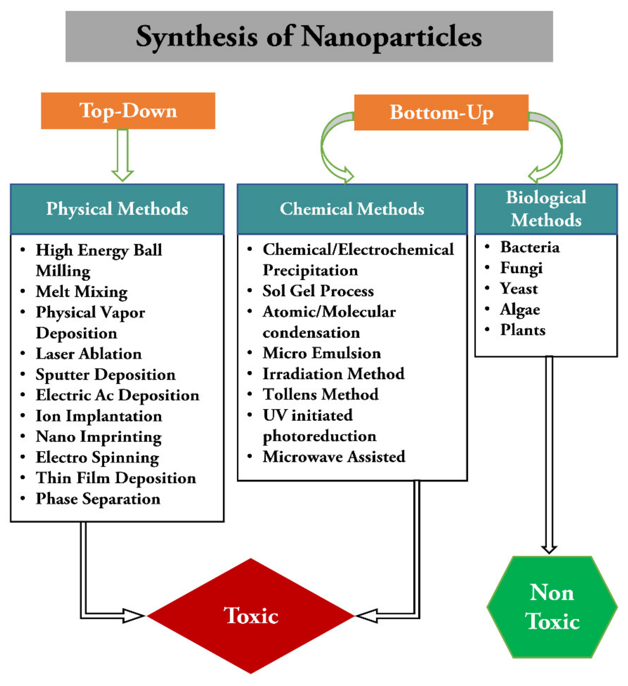
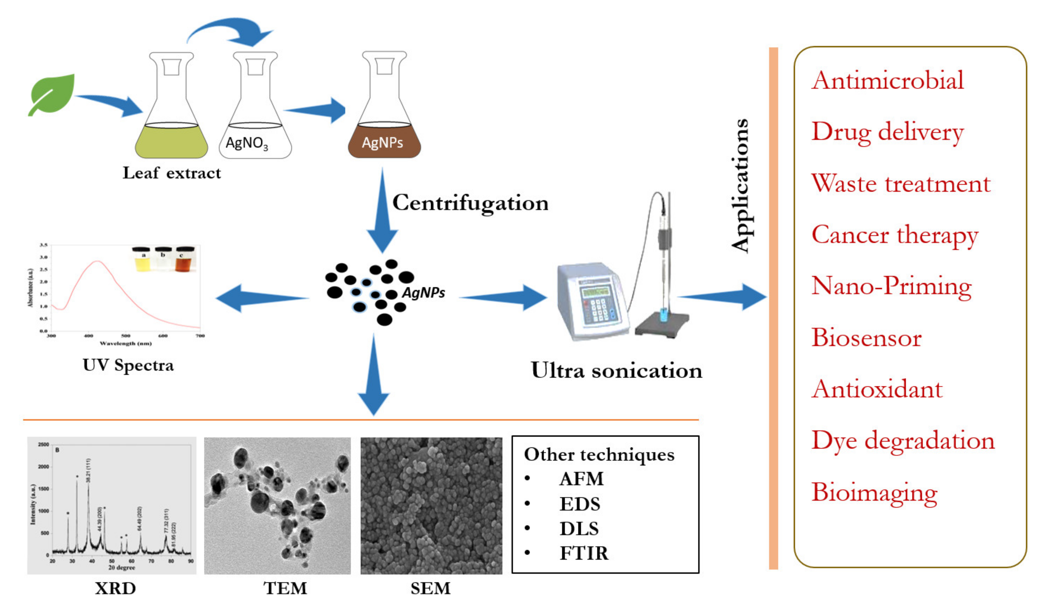
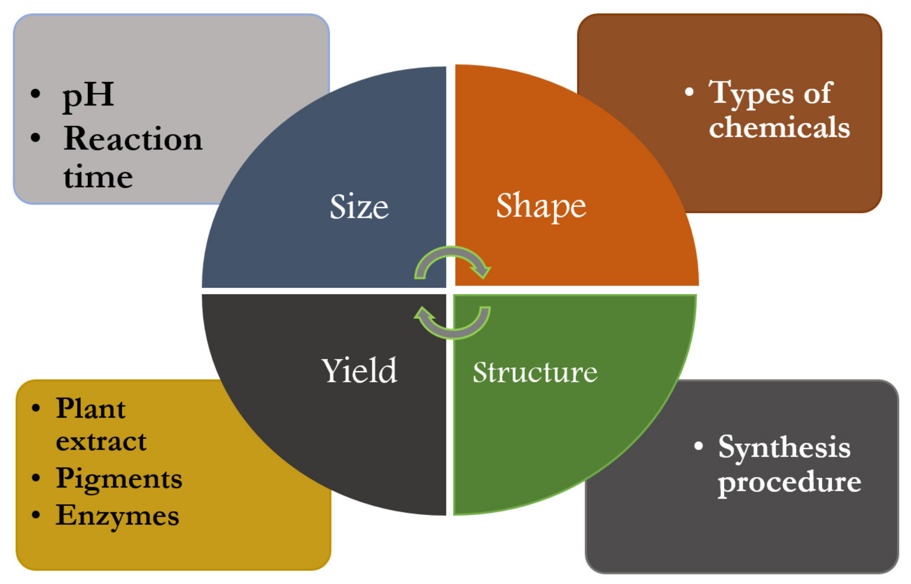
Table 1.
Silver nanoparticles synthesized using plants.
| Plants | Size (nm) | Plant’s Part | Shape | Applications | References |
|---|---|---|---|---|---|
| Acalypha indica | 10–50 | Leaves | - | Cytotoxic effects against MDA-MB-231, human breast cancer cells | [91] |
| Agastache mexicana and Tecoma stans | 16–20 | Leaves | Spherical | - | [92] |
| Aloe vera | 70 | Leaves | Spherical | Antifungal activity | [93] |
| Alternanthera dentate | 50–100 | Leaves | Spherical | Antimicrobial activity | [94] |
| Andrographis paniculata | 67–88 | Leaves | Spherical | Hepatocurative activity | [26] |
| Artocarpus elasticus | 5.81 | Stem bark | Spherical | - | [95] |
| Avicennia marina | 60–95 | Leaves | - | Mosquito larvicidal activity | [96] |
| Barleria cristata | 25–30 | Leaves | Spherical | Mosquitocidal and biotoxicity activity | [97] |
| Boerhaavia diffusa | 25 | Leaves | Spherical, FCC | Antibacterial activity | [98] |
| Boswellia serrate | 7–10 | Gum | Spherical | Antibacterial activity | [99] |
| Brassica rapa | 16.14 | Leaves | Anisotropic shaped | Antiangiogenic activity | [25] |
| Cassia javanica L. | 100 | Leaves | Spherical | Antibacterial activity | [100] |
| Cestrum nocturnum | 20 | Leaves | Spherical | Antioxidant and antibacterial | [101] |
| Cleome viscosa | 20–50 | Fruit | Spherical | Antimicrobial activity | [102] |
| Coleus aromaticus | 26–27 | Leaves | Spherical | Larvicidal toxicity against dengue and filariasis vectors | [103] |
| Cucurbita maxima, Moringa oleifera and Acorus calamus | 30–70 | Petals, leaves, rhizome | Roughly spherical | Anticancer activity against epidermoid A431 carcinoma | [104] |
| Delphinium denudatum | 85 | Roots | Spherical, FCC | Dielectric properties | [105] |
| Descurainia sophia | 30 | Seeds | Spherical | Antibacterial and antifungal activity | [106] |
| Dillenia indica | 11–24 | Fruit | Spherical | Antibacterial activity | [74] |
| Dimocarpus Longan Lour | 20–35 | Peel | - | Anticancer effects against prostate cancer (PC-3) cells | [107] |
| Dionaea muscipula | 5–10 | Whole plant | Quasi-spherical | Antioxidant | [108] |
| Diospyros sylvatica | 10 | Roots | Spherical | Antimicrobial activity | [109] |
| Elaeagnus umbellata | 40 | Whole plant | Spherical | Antimicrobial | [110] |
| Enicostemma axillare | 15–20 | Leaves | Spherical | - | [111] |
| Enteromorpha compressa | 4–24 | Seaweed | Spherical | Biomedical properties | [112] |
| Gracilaria corticata | 18–46 | Seaweeds | Spherical | Antifungal activity | [113] |
| Gymnema sylvestre | 20–30 | Leaves | Spherical | Antibacterial activity | [114] |
| Hybanthus enneaspermus | 25.2 | Whole plant | Spherical, hexagonal, triangular | Larvicidal activity | [115] |
| Lantana camara | 53.5 | Flower | - | - | [116] |
| Lantana trifolia | <50 | Leaves | Spherical | Antibacterial activity | [117] |
| Lathyrus sp. Stachys lavandulifolia | 117 | Seeds, Sepal | Spherical | Antifungal activity | [118] |
| Malus domestica | 16 | Fruit | - | Antimicrobial | [119] |
| Melia dubia | 7.3 | Leaves | Irregular | Anticancer activity | [120] |
| Musa paradisiaca | 23.7 | Banana peels | Spherical | Antimicrobial activity | [121] |
| Myrmecodia pendan | 10–20 | Leaves | Spherical, FCC | - | [122] |
| Nauclea latifolia | 12 | Fruit | Irregular | Antimicrobial and antioxidant | [123] |
| Ocimum Sanctum | 10–20 | Leaves | Spherical | Antibacterial activity | [124] |
| Olea europaea | 90 | Leaves | Spherical | Cytotoxic effect on MCF-7 cells | [125] |
| Olive leaf | 20–25 | Leaves | Spherical | Antibacterial activity | [126] |
| Parkia speciosa | 31 | Leaves | Spherical | Antibacterial, antioxidant and photocatalytic activity | [127] |
| Phyllanthus pinnatus | 61.45 | Stem | Spherical | Antibacterial activity | [128] |
| Pistacia atlantica | 10–50 | Seed | Spherical, FCC | Antibacterial activity | [129] |
| Pistacia atlantica | 40–60 | Leaves | Spherical | Antioxidant, cytotoxicity and antibacterial activity | [130] |
| Pteridium aquilinum | 30–40 | Leaves | Spherical | Mosquitocidal and antiplasmodial activity | [131] |
| Reishi Mushroom | 15–22 | Mushroom | Spherical | Antifungal activity | [132] |
| Rosa canina | 13–21 | Leaves | Spherical | Antioxidant, antibacterial, antifungal and DNA cleavage activity | [133] |
| Rosmarinus officinalis Linn. | 10–33 | Leaves | Spherical | Antimicrobial activity | [134] |
| Sinapis arvensis | 14 | Seeds | Spherical | Antifungal activity | [135] |
| Solanum trilobatum | 15–20 | Leaves | Cubic and hexagonal | Antidandruff activity | [136] |
| Syzygium alternifolium (Wt.) | 20 | Fruit | Spherical | Antimicrobial activity | [137] |
| Talinum triangulare | 13.86 | Leaves | Spherical | Electrochemical, antimicrobial and antioxidant activity | [138] |
| Tephrosia purpurea | 20 | Leaves | Spherical, FCC | Antibacterial activity | [139] |
| Terminalia cuneata | 25 | Barks | Distorted spherical | Catalytic action in reduction of direct yellow-12 dye | [140] |
| Terminalia arjuna | 10–50 | Leaves | Spherical | Catalytic action in reduction of MO, MB, 4NP and CR dyes | [90] |
| Thevetia peruviana | 18.1 | Leaves | Spherical | Antimicrobial activity | [141] |
| Thevetia Peruviana | 10–30 | Latex | Spherical, tubular, irregular | - | [142] |
| Thymbra spicata | 7 | Leaves | spherical | Catalytic reduction | [143] |
| Tradescantia pallida | 98 | Leaves | Spherical | Antibacterial activity | [144] |
| Viburnum lantana | 20–80 | Leaves | Spherical, FCC, uniform | Antibacterial Activity | [145] |
Table 2.
Different nanoparticles characterization techniques and techniques-related information.
| Techniques | Characterization Techniques | Information Provided |
|---|---|---|
| Microscopy | AFM | Size, morphology, surface texture, electrical, and mechanical properties |
| STM | Elemental and molecular composition | |
| SEM | Surface, size, shape morphology, and crystallographic composition | |
| TEM | Elemental composition and electrical conductivity | |
| X ray-based techniques | XAS, XRF, XPS, XRD | Surface, crystallographic, and elemental composition |
| Light Scattering Spectroscopic techniques | UV-visible DLS FTIR | Particle size, aggregation, structure, and surface chemistry |
Table 3.
Antifungal activity of biologically synthesized silver nanoparticles.
| Plant | Test Organism | Method | Reference |
|---|---|---|---|
| Aaronsohnia factorovskyi | Fusarium solani | Poisoned food technique | [190] |
| Aloe vera | Aspergillus sp. and Rhizopus sp. | Agar disc diffusion method | [93] |
| Amaranthus retroflexus | Macrophomina phaseolina, Alternaria alternata and Fusarium oxysporum | Poison food technique | [191] |
| Ananas comosus | Fusarium verticilliodes, Aspergillus flavus and Alternaria alternata | Well diffusion assay on malt extract agar | [192] |
| Argimone mexicana | Aspergillus flavus | Food poisoning | [193] |
| Brassica rapa L. | Gloeophyllum abietinum, G. trabeum, Chaetomium globosum, and Phanerochaete sordida | Agar disc diffusion assay | [79] |
| Camellia sinensis | Aspergillus flavus and A. parasiticu | Agar disc diffusion method | [194] |
| Gracilaria corticata (sea weeds) | Candida albicans and C. glabrata | Agar disc diffusion method | [109] |
| Ligustrum lucidum | Setosphaeria turcica | Paper Disk Diffusion Assay | [195] |
| Malva parviflora | Helminthosporium rostratum, Fusarium solani, Fusarium oxysporum, and Alternaria alternata | Poisoned food technique | [190] |
| Melia azedarach | Verticillium dahlia | Poisoned food technique | [196] |
| Ocimum sanctum | Candida albicans, Candida glabrata, and Candida tropicalis | Broth dilution method | [122] |
| Passiflora caerulea | Trichophyton mentagrophytes, T. rubrum, E. floccosum, M. audouinii, M. Canis | Agar disc diffusion method | [197] |
| Peganum harmala | Fusarium oxysporum, Alternaria alternata, and Tricoderma | Disc diffusion assay | [198] |
| Phyllanthus urinaria, Pouzolzia zeylanica, and Scoparia dulcis | Aspergillus niger, Aspergillus flavus, and Fusarium oxysporum | Poisoned food technique | [199] |
| Prunus cerasifer | Aspergillus niger, A. flavus, A. fumigatus, A. terreus, Penicillium chrysogenum, Fusarium solani and Lasiodiplodia theobromae | Kirby–Bauer disc diffusion assay | [200] |
| Sinapis arvensis | Neofusicoccum parvum | Mycelium growth inhibition test | [135] |
| Stachys lavandulifolia and Lathyrus sp. | Dothiorella sarmentorum | Agar dilution method | [118] |
| Teucrium polium L. | Fusarium oxysporum | Poisoned food technique | [201] |
| Tropaeolum majus L. | Trichoderma viridiae, Mucor spp., Aspergillus niger and Candida albicans | MIC | [202] |
Publisher’s Note: MDPI stays neutral with regard to jurisdictional claims in published maps and institutional affiliations. |
© 2021 by the authors. Licensee MDPI, Basel, Switzerland. This article is an open access article distributed under the terms and conditions of the Creative Commons Attribution (CC BY) license (https://creativecommons.org/licenses/by/4.0/).
Share and Cite
MDPI and ACS Style
Raj, S.; Trivedi, R.; Soni, V. Biogenic Synthesis of Silver Nanoparticles, Characterization and Their Applications—A Review. Surfaces 2022, 5, 67-90. https://0-doi-org.brum.beds.ac.uk/10.3390/surfaces5010003
AMA Style
Raj S, Trivedi R, Soni V. Biogenic Synthesis of Silver Nanoparticles, Characterization and Their Applications—A Review. Surfaces. 2022; 5(1):67-90. https://0-doi-org.brum.beds.ac.uk/10.3390/surfaces5010003
Chicago/Turabian StyleRaj, Shani, Rohini Trivedi, and Vineet Soni. 2022. "Biogenic Synthesis of Silver Nanoparticles, Characterization and Their Applications—A Review" Surfaces 5, no. 1: 67-90. https://0-doi-org.brum.beds.ac.uk/10.3390/surfaces5010003