Article

Viromics on Honey-Baited FTA Cards as a New Tool for the Detection of Circulating Viruses in Mosquitoes

1
Centre de Recerca en Sanitat Animal (CReSA), Institut de recerca en Tecnologies Agroalimentaries (IRTA), 08193 Barcelona, Spain
2
Institut Pasteur, Pathogen Discovery Laboratory, 75015 Paris, France
3
Servei de Control de Mosquits del Consell Comarcal del Baix Llobregat, 08820 Barcelona, Spain
4
Institut Pasteur – Bioinformatics and Biostatistics Hub—Computational Biology department, Institut Pasteur, USR 3756 CNRS—75015 Paris, France
5
National Veterinary School of Alfort, Paris-Est University, 94704 CEDEX, Maisons-Alfort, France
*
Author to whom correspondence should be addressed.
Submission received: 31 December 2019 / Revised: 25 February 2020 / Accepted: 27 February 2020 / Published: 29 February 2020
(This article belongs to the Special Issue Viromics: Approaches, Advances, and Applications)

Abstract

:
Worldwide, emerging and re-emerging infectious diseases (EIDs) are a major burden on public and animal health. Arthropod vectors, with mosquitoes being the main contributors of global disease, transmit more than 70% of the recognized EIDs. To assess new alternatives for arthropod-borne viral diseases surveillance, and for the detection of new viruses, honey-baited Flinders Technology Associates (FTA) cards were used as sugar bait in mosquito traps during entomological surveys at the Llobregat River Delta (Catalonia, Spain). Next generation sequencing (NGS) metagenomics analysis was applied on honey-baited FTA cards, which had been exposed to field-captured mosquitoes to characterize their associated virome. Arthropod- and plant-infecting viruses governed the virome profile on FTA cards. Twelve near-complete viral genomes were successfully obtained, suggesting good quality preservation of viral RNAs. Mosquito pools linked to the FTA cards were screened for the detection of mosquito-associated viruses by specific RT-PCRs to confirm the presence of these viruses. The circulation of viruses related to Alphamesonivirus, Quaranjavirus and unclassified Bunyavirales was detected in mosquitoes, and phylogenetic analyses revealed their similarities to viruses previously reported in other continents. To the best our knowledge, our findings constitute the first distribution record of these viruses in European mosquitoes and the first hint of insect-specific viruses in mosquitoes’ saliva in field conditions, demonstrating the feasibility of this approach to monitor the transmissible fraction of the mosquitoes’ virome. In conclusion, this pilot viromics study on honey-baited FTA cards was shown to be a valid approach for the detection of viruses circulating in mosquitoes, thereby setting up an alternative tool for arbovirus surveillance and control programs.

1. Introduction

Worldwide, two-thirds of all recognized emerging and re-emerging infectious diseases (EIDs) are of viral origin [1], with arthropod-borne viruses (arboviruses) being the causative agents of more than 30% of them [2]. Arboviruses circulate naturally between their vertebrate hosts and vectors. Nearly 135 arboviruses are known to infect humans, posing a significant threat to public health [3]. Globalization, together with anthropic activities and climate change, has facilitated the dispersal of pathogenic agents (arboviruses included), their hosts and vectors, extending the risk to more and newer areas [4,5]. Since the increased incidence of dengue [6], Zika [7,8], chikungunya [9,10] and West Nile viruses [11], there is a growing interest in understanding the viral diversity harbored by arthropod vectors, and a rising necessity to develop more effective surveillance and monitoring tools for circulating viruses.
Traditionally, for active surveillance and control purposes, samples from entomological surveys and/or from sentinel animals are subjected to laboratorial analyses to evidence arbovirus circulation. Despite these methodologies being considered the “gold standards”, many issues must be considered. For instance, in entomological surveys, specialized personnel are required to capture and classify specimens, and a cold chain must be maintained to prevent virus degradation until molecular processing [12,13]. Due to the low prevalence of infected individuals between inter-epidemic periods, large numbers of mosquitoes have to be analyzed to detect a virus [13]. When using sentinel animals, besides the necessary logistics, ethical considerations have to be taken into account, as the physical integrity of the animals, as well as that of the personnel, should be warranted [14]. Likewise, customary laboratorial techniques for virus detection present some limitations, for example in serological diagnosis, closely related viruses may produce cross-reactions [14], while PCR-based techniques target only those viral lineages that are already known, thereby underestimating the diversity of the sample while overlooking undescribed viruses that could potentially be pathogenic [15].
Since gold-standard strategies are time-consuming, logistically complex and potentially hazardous, honey-baited Flinders Technology Associates (FTA) cards have been used as an alternative tool for arbovirus surveillance as they inactivate pathogens and preserve nucleic acids on contact, thereby simplifying the labor [12,13,16]. In previous field trials, honey-soaked FTA cards have been used in combination with molecular techniques to detect several arboviruses, such as Ross River virus (RRV), Barmah Forest virus (BFV) [13,16,17,18] and West Nile virus strain Kunjin (WNVKUN) [13,17] in Australia, and Usutu virus (USUV) in Switzerland [19]. Moreover, while virological surveillance in mosquitoes is based mainly upon virus detection in entire mosquitoes, indicating that they might be infected, the detection of viruses expectorated within the saliva during sugar feeding and deposited directly on the FTA cards may identify infectious mosquitoes [18].
To overcome the detection bias of molecular-based techniques, deep sequencing technologies have been proven as a valid approach to detect, characterize and discover unknown or uncultured viruses within biological or environmental samples [20,21,22,23]. Recently, by high throughput sequencing, diverse and widely distributed novel non-taxonomic groups of RNA viruses that naturally infect insects have been discovered in mosquitoes. Between 2007 and 2017, 187 novel mosquito-associated viruses have been reported and classified within 25 families [24]; some of them commonly grouped with human/animal arboviral pathogens or plant viruses. The capacity to detect untargeted viruses enables metagenomics to act as a new and powerful approach to enhancing arbovirus surveillance programs [25].
To the best of our knowledge, for the first time, next generation sequencing (NGS) on honey-impregnated FTA cards used as sugar bait during entomological surveys has been tested as a new approach for the detection of viruses circulating in mosquitoes. Viromics results on FTA cards were confirmed by the detection of mosquito-associated viruses in field-captured mosquitoes. Additionally, near-complete viral genomes were obtained. Herein, we show that insect-specific viruses (ISVs) can be detected in saliva from field-captured mosquitoes and report some ISVs previously identified in other continents, as first-distribution records in European mosquitoes.

2. Materials and Methods

2.1. Study Area and Sampling Strategy

The present study was conducted at the Llobregat River Delta, North-Eastern Spain. In this Delta, densely populated areas coexist with natural habitats that serve as a strategic stopover on the route of migratory birds between Europe and Africa. For this reason, this area is considered to be of particular epidemiological interest and is targeted for arbovirus surveillance. In fact, sampling locations were chosen based on previous evidence of arbovirus circulation [26], and in places where the Servei de Control de Mosquits del Baix Llobregat performs regular mosquito monitoring and control activities. Peri-urban and rural biotopes within this area were sampled to provide variability and increase the probability of virus detection.
Every fortnight, from May to November 2015, host-seeking female mosquitoes were captured using CO2-baited EVS Mosquito Traps (Bioquip, Compton, CA, USA). Inside the collection bag of some traps, one honey-soaked Classic FTATM card (WhatmanTM, GE Healthcare UK limited, Buckinghamshire, UK) [15] was placed as a sugar-bait for the captured specimens. Only the honey-impregnated area of the card was left exposed to allow specimens feed on it while in the trap [27]. At each location, traps with and without honey-baited FTA cards were placed indiscriminately and kept operational from the early evening to the next morning (approximately 18 h). After sampling periods, FTA cards were removed, covered with Parafilm® (Bemis, Neenah, WI, USA) and coded according to location and sampling date. Only captured female mosquitoes were morphologically classified [28] and up to 30 individuals were pooled according to species, location and sampling date. A few non-culicid dipterans were also captured but not classified. The number of specimens with blue abdomens was recorded per species as evidence of feeding on the FTA cards. A cold chain was maintained through specimen transportation and handling to avoid RNA degradation [27]. FTA cards and specimens were preserved at −80 °C until molecular analysis.

2.2. RNA Extraction from FTA cards for NGS Analysis

Pre-extraction, frozen FTA cards were thawed at 4 °C, homogenized with 500 µL of cold sterile PBS by vortex and squeezed with a sterile pestle to extract its content. Total RNA was obtained from individual FTA cards (13 peri-urban and 23 rural) using the RNeasy Mini Kit (QIAGEN GmbH, Hilden, Germany) according to the manufacturer’s instructions. Extracted RNA was eluted in 50 µL of RNase-free water. A unique RNA sample per biotope was generated by pooling 15 µL of all the corresponding extracts of the given area.

2.3. Library Preparation, Sequencing and Bioinformatics Analysis

RNA samples were sequenced and analyzed as previously described with slight modifications [29]. Briefly, to obtain complementary DNA (cDNA), RNA samples were retro-transcribed using random hexamers and the SuperScript IV reverse transcriptase (Invitrogen, Vilnius, Lithuania). Random amplification of cDNAs was performed using the multiple displacement amplification (MDA) protocol with phi29 polymerase and random hexamers [30]. Libraries were sequenced at a depth of 60 to 80 million reads on an Illumina HiSeq2000 platform in a 150-base pairs (bp) single read format, outsourced to DNAvision Company (Charleroi, Belgium).
Raw reads were processed with an in-house bioinformatics pipeline as previously described [31]. Summarizing, it comprised quality check and trimming based on AlienTrimmer package [32] (Phred quality score cutoff = 80, min % of correctly called nt = 20) followed by read normalization using BBnorm program (https://jgi.doe.gov/data-and-tools/bbtools) (cut-off parameter of 100). De novo assemblies were performed using Megahit tool [33] (minimum contig length = 100 nt). For further ORF prediction ((https://figshare.com/articles/translateReads_py/7588592), minimum aa length = 15), a Diamond-based similarity search (v0.9.22.123) against the protein Reference Viral database (RVDB-prot 16.0 [34]) was conducted. Validation of viral taxonomic assignations was accomplished by a first Diamond-based search against the whole protein NCBI/nr database (1 November 2019 version) and a final search against the whole NCBI/nt nucleotide database (15 August 2019 version) to discard any putative non-viral intronic sequences that would, by chance, present a significant similarity with a viral protein. The pipeline used performs a protein blast for each viral contig and singleton, and then analyzes the taxonomic classification for all the co-best hits (meaning all the hits that have the same score). If all the hits were assigned to the same species, this species was reported as the closest hit. If the assembly had two or more different species or genera classifications, the last common ancestor was reported—genus or family, respectively. For low-level identities, taxonomic assignations were suggestive of putative new viral sequences. The quantification of abundance of each viral taxon was obtained by summing the length (in nucleotides) of all sequences being associated to this taxon, weighted by the k-mer coverage of each contig.

2.4. Primers Design and Virus Detection by Specific RT-PCRs

To confirm that viruses reported by metagenomics on FTA cards come solely from the captured specimens and not from the honey-bait, sequences assigned to mosquito-associated viruses were extracted. Among these, four viruses, with at least one assembly longer than 1000 nucleotides (nts) and with an identity higher than 90% were selected. Then, primers were designed from the extracted sequences of each chosen virus and conventional virus-specific reverse transcription polymerase chain reactions (RT-PCR) were set up. Viral RNA from mosquito pools and honey-baited FTA cards, which had not been exposed to mosquitoes, were then extracted using NucleoSpin® RNA Virus kit (Macherey–Nagel, Düren, Germany) following the manufacturer’s instructions. Using the OneStep RT-PCR kit (QIAGEN GmbH, Hilden, Germany), all the above-mentioned samples were screened for the detection of Alphamesonivirus 1, Bunyaviridae environmental sample, Dezidougou virus and Wuhan mosquito virus 7, adjusting the annealing temperatures to each set of primers (Table 1). As positive amplification controls, Dezidougou virus isolate and Alphamesonivirus cDNA were used (kindly provided, respectively, by Scott Weaver from the World Reference Centre for Emerging Viruses and Arboviruses at University of Texas Medical Branch (WRCEVA–UTMB), and Patricia Gil and Serafín Gutiérrez from Centre de Coopération Internationale en Recherche Agronomique pour le Développement (CIRAD) at Montpellier). Meanwhile, for other viruses, since viral isolates were not available, extracted RNA from the FTAs that had been subjected to metagenomics were used as positive amplification controls. Amplification products were visualized in 2% agarose gels with ethidium bromide (0.1 µg/mL) staining.

2.5. Sequencing and Phylogenetic Analyses

Virus-specific RT-PCR products were purified using the QIAquick® Gel Extraction Kit (QIAGEN GmbH, Hilden, Germany) and Sanger sequenced in both directions using the BigDye® Terminator v3.1 cycle Sequencing Kit (Life Technologies Corporation, Austin, TX, USA). At the Servei de Genomica i Bioinformatica at the Universitat Autonoma de Barcelona (SGB-UAB), amplicons were purified with the BigDye X Terminator kit (Applied Biosystems™, Waltham, MA, USA) and subjected to capillary electrophoresis in the Genetic Analyzer 3130xl (Applied Biosystems™, USA). Viral sequences were aligned using BioEdit Sequence Alignment Editor [35] and the identity of each virus was confirmed by comparing them to GenBank’s reference database using the nucleotide Basic Local Alignment Search Tool (BLASTn) algorithm. At least one viral sequence per geographic region and a year that exhibited high similarities in the BLAST analysis to our subject sequences was used to infer the phylogenetic relationship of each studied virus. Viral sequences were then pairwise aligned using ClustalW algorithm in the Molecular Evolutionary Genetics Analysis program version X (MEGAX) [36]. In the same program, phylogenetic trees were constructed using the Maximum Likelihood (ML) method. Based on the Bayesian information criterion (BIC) score [36,37] the best models were applied. Tamura-Nei (TN93+G) with gamma distributions showed to be the best fit for Alphamesonivirus/CAT and Wuhan mosquito/CAT viruses, and Hasegawa-Kishino-Yano (HKY+G) [38] with gamma distributions the best fit for Culex bunyavirus/CAT virus. In both cases, a 1000 replicate bootstrap was used.

2.6. Nucleotide Sequences Accession Numbers

The raw sequencing datasets for both batches of honey-baited FTA cards are available in the NCBI Sequence Read Archive (SRA) repository under the BioProject ID: PRJNA604676 (www.ncbi.nlm.nih.gov/biosample/13978317 and www.ncbi.nlm.nih.gov/biosample/13978318). All the viral genomes for which the complete CDS were obtained were deposited in the GenBank archive under the accession numbers: MT096515-MT096531. Sequences corresponding to the viruses detected in mosquito pools from the Llobregat River Delta are available under the accession numbers: MT063093-MT063099.

3. Results and Discussion

After sampling periods at the Llobregat River Delta, 1080 female mosquitoes were collected and classified into five species: Aedes albopictus (n = 20; 10 pools), Coquillettidia richiardii (n = 11; 5 pools), Culex pipiens (n = 755; 53 pools), Aedes caspius (n = 294; 24 pools) and Aedes detritus (n = 2; 1 pool) (Table S1). A total of 38 honey-baited FTA cards were recovered; 36 linked to mosquito captures and two from traps with no captures. Batches of 13 FTA cards from peri-urban and of 23 FTA cards from rural biotopes linked to mosquito captures constituted two independent samples for metagenomics analysis. Visual inspections depicted blue abdomens in 21% and 39% of the captured mosquitoes, respectively, for peri-urban and rural biotopes, confirming that they had fed on the FTA cards while in the trap. No evidence of blue dye was observed in Ae. detritus (Table S1).

3.1. Outputs on NGS on Honey-Baited FTA Cards

Next generation sequencing (NGS) on honey-baited FTA cards generated 61,362,209 and 80,631,320 of raw reads for rural and peri-urban biotopes, respectively. After filtering steps, 56,424,764 and 76,884,845 reads of 150 bases were assembled to produce 431,179 and 100,469 contigs respectively for rural and peri-urban datasets. Depurated reads also generated 3,128,224 and 846,017 singletons in each case.

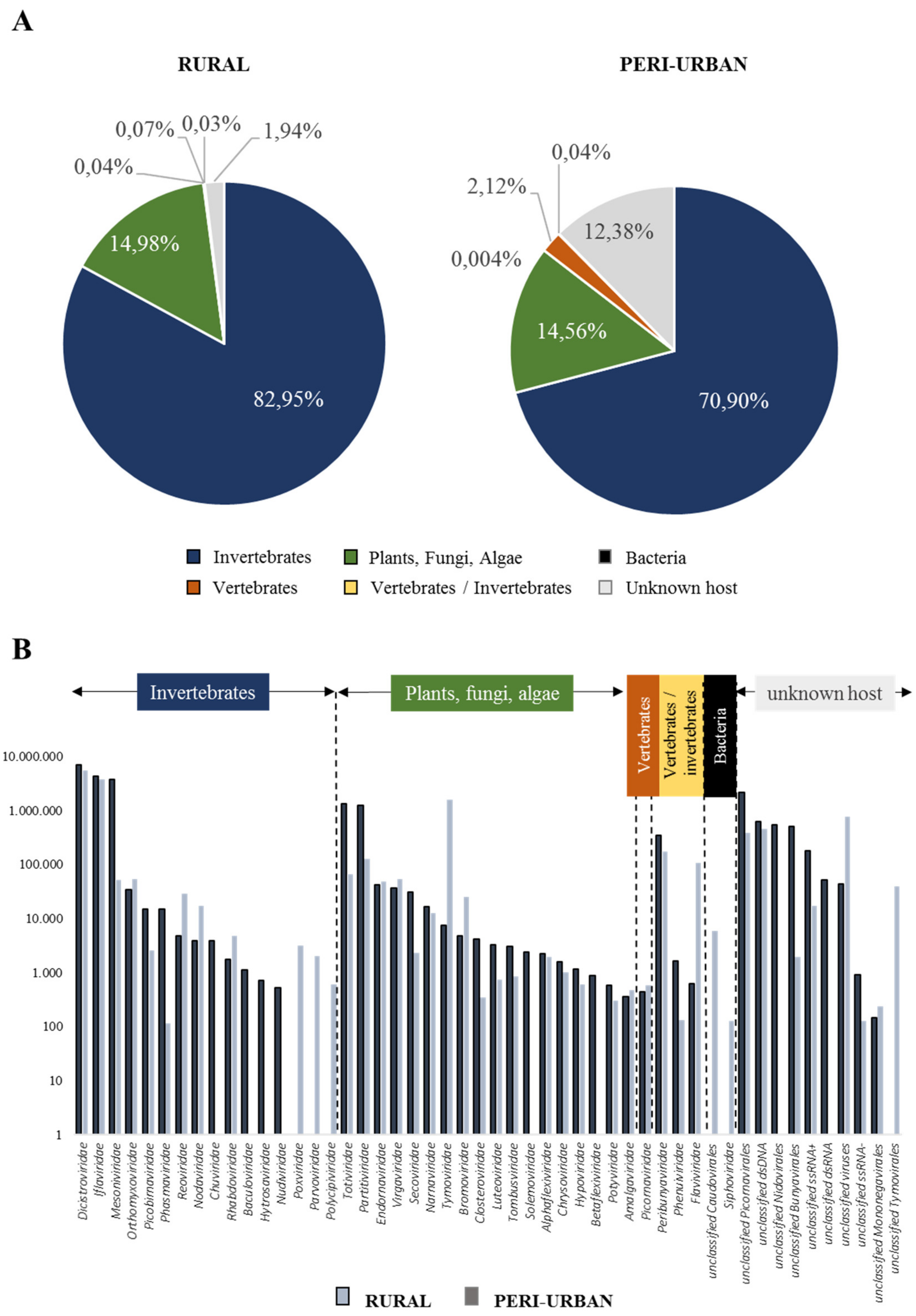
3.2. Virome Composition on Honey-Baited FTA Cards During Entomological Surveys

Taxonomic assignations of the viral sequences obtained by high throughput sequencing on honey-baited FTA cards revealed that more than 95% corresponded to RNA viruses. Picornavirales, Nidovirales and Tymovirales were the most represented single-stranded positive sense RNA (ssRNA+) viral orders; and Bunyavirales the most abundant single-stranded negative sense RNA (ssRNA-) order. Double-stranded RNA (dsRNA) viral families Partitiviridae and Totiviridae were also dominant. DNA and unclassified viruses comprised the remaining 5% of the viral diversity herein reported (Table S2). In agreement with previous virome studies, most of the taxa derived from honey-baited FTA cards have been identified in various invertebrates [39] and associated to mosquitoes [40]. Additionally, mosquito-specific viruses detected in FTA cards (Table 2) have been described as part of the viral communities harbored by several mosquito species in different geographic regions [41,42,43,44,45,46,47,48].
In the present study, taxonomic profiling revealed the prevalence of invertebrate-associated viruses (Figure 1A) with Dicistroviridae, Iflaviridae and Mesoniviridae being the most abundant families (Figure 1B). Sequences herein designated as Dicistroviridae and Iflaviridae (order Picornavirales) were mostly related to hymenopterans, in particular to the honeybee Apis mellifera. Since we could not sequence the honey used to impregnate the FTA cards as sugar bait, we cannot discard the possibility that these sequences might have come from it. However, recent virome studies have described these two families as the most abundant in culicid mosquitoes from the Yunnan province in China, and Zambezi province in Mozambique [49,50]. The additional description of honeybee-infecting virus Rhopalosiphum padi virus (Dicistroviridae, genus Cripavirus) in mosquito species from Hubei, China [51] and in Culex mosquitoes from California [41], together with the assembly of sequences linked to chronic bee paralysis virus (CBPV) (unclassified ssRNA+ virus) and Apis mellifera filamentous virus (dsDNA Hytrosaviridae family) from French Anopheles maculipennis [52] and from Culex mosquitoes from California [41] respectively, suggested that these viral families could be associated to mosquitoes as well. In addition, due to the low genetic identity of these viruses with their closest honeybee counterpart, the scarcity of mosquito-based sequences available in public databases, and the continuous discovery of new picorna-like viruses in insects [41,45,50,53,54], might suggest that we are dealing with novel mosquito picorna-like viruses. Based on the abovementioned findings, captured mosquitoes that fed on the FTA cards could have been the source of the identified viruses.
Besides invertebrate-related viruses, it was not surprising to find viral families usually detected in plants, fungi and algae (e.g., Tymoviridae, Totiviridae, Partitiviridae, Endornaviridae or Virgaviridae) as part of the viral diversity associated to honey-baited FTA cards (Figure 1B). Since, in Culex mosquitoes, sequences related to Totiviridae-like viruses have been found in Guadeloupe [46], Australia [25], China [49] and California [41]; Partitiviridae-like viruses have been detected in Sweden [45,48], Australia [25], Kenya [49] and California [41]; Endornaviridae-like viruses in Australia [25] and Tymoviridae-like viruses have been identified in Guadeloupe [46], Kenya [49], California [41], China [53], and Sweden [48]. Moreover, a Culex Tymoviridae-like virus (CuTLV) that was isolated from a Culex spp. pool from Xinjiang (China) was also shown to produce a cytopathic effect on Aedes albopictus C6/36 cell line [55], suggesting a potential plant/mosquito host-shift even when there is no record of mosquitoes as vectors of plant viruses [46]. Nonetheless, there is also the chance that: i) mosquitoes could have acquired these viruses while sap or nectar feeding prior to capture and deposited them on the FTA card along with saliva expectorations as mouthparts contaminants [49,56] while trapped; or ii) they could have been present in the honey used as bait.
To a lesser extent, the virome profile of FTA cards depicted sequences assigned to three dual-host (mosquito/vertebrate) virus families: Flaviviridae, Phenuiviridae and Peribunyaviridae. Flaviviridae-associated sequences were distantly related to two mosquito-specific viruses, Karumba virus (49% amino acid (aa) identity) and Calbertado virus (47–86% aa identities) (Table S2). Reads related to Phenuiviridae were assigned to a distant Phasi Charoen-like phasivirus with aa identities ranging from 58% to 77% (Table S2). Meanwhile, most of the Peribunyaviridae-associated sequences presented high homologies with Ganda bee virus (35–95% aa identity) (Table 2). Finally, no arboviruses were detected throughout the sampling period by NGS on honey-baited FTA cards. Despite six sequences matched with WNV (59–92% aa identity), these assignations were not taken into consideration due to the length (150 nt), nucleotide identity (<80%) and coverage (<80%) of the sequences.

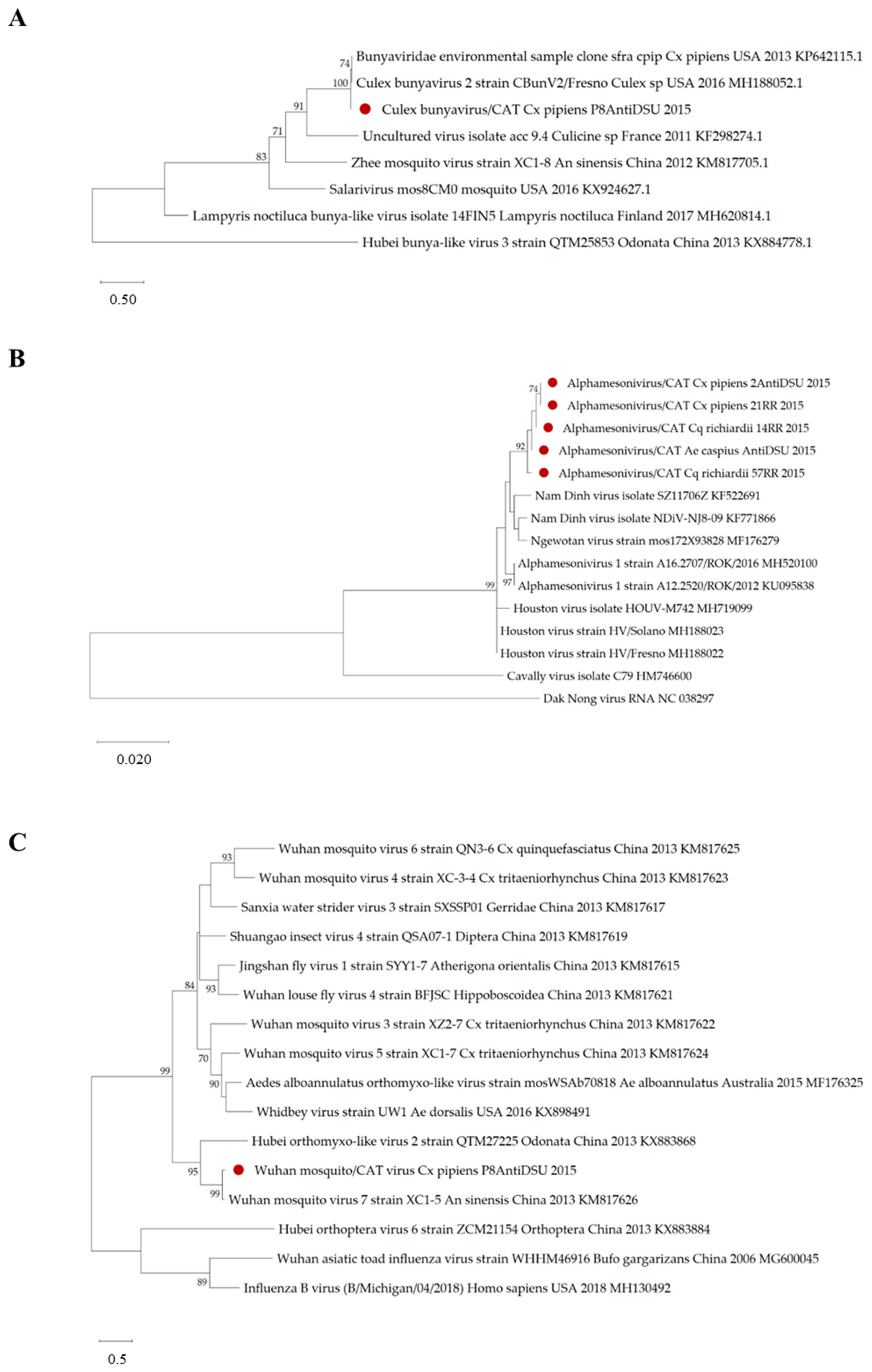
3.3. Viral Genomes Obtained from Honey-Baited FTA Cards

It is noteworthy that de novo assemblies of viral reads from both honey-baited FTA cards batches produced 12 near-complete viral genomes (>98% nucleotide coverage and >93% nucleotide identity) for which the 5′ and 3′ termini are incomplete since RACE-PCRs were not performed. Viral genomes within the orders Nidovirales (Alphamesonivirus 1: Ngewotan virus) and Picornavirales (e.g., Deformed wing virus and Culex Iflavi-like virus 4), and within unclassified RNA viruses (e.g., Hubei picorna-like virus 61 and Wenzhou soberno-like virus 4) were generated (Table 3). Obtaining near-complete genomes of viruses associated to mosquitoes, highlights the usefulness of FTA cards in preserving viral RNA. However, we cannot exclude that most of the honeybee-related virus genomes might come from the bait.

3.4. Virus Detection by Specific RT-PCRs on Honey-Baited FTA Cards Unexposed to Mosquitoes

To confirm virome results obtained through metagenomics analysis on honey-baited FTA cards, among all the mosquito-associated viruses (Table 2), Alphamesonivirus 1 (3.606.196 abundance in nucleotides), Dezidougou virus (1.424.472 abundance in nts), Bunyaviridae environmental sample (335.292 abundance in nts) and Wuhan mosquito virus 7 (43.351 abundance in nts) were selected to design specific primers and set up virus-specific RT-PCRs. All these selected viruses showed to have at least one contig with a matching sequence longer than 1000 nt and similarity above 90%. For identification matters, through the manuscript, these viruses would respectively be referred to as Alphamesonivirus/CAT virus, Culex bunyavirus/CAT virus, Dezidougou/CAT virus and Wuhan mosquito/CAT virus. Suffix “CAT” stands for the geographic region of detection, i.e., Catalonia.
Those honey-baited FTA cards, which were not exposed to mosquitoes recovered from entomological surveys, were then screened individually by virus-specific RT-PCRs to verify the source of the viruses detected by viromics. Screenings of both cards tested negative for Culex bunyavirus/CAT virus and Alphamesonivirus/CAT virus, and positive for Dezidougou/CAT virus and Wuhan mosquito/CAT virus. These detections could be explained by (i) the presence of non-culicid dipterans in the traps; they could have deposited these viruses while sugar feeding from the FTA cards, and/or (ii) the source of these viruses came from the honey impregnated on the cards.

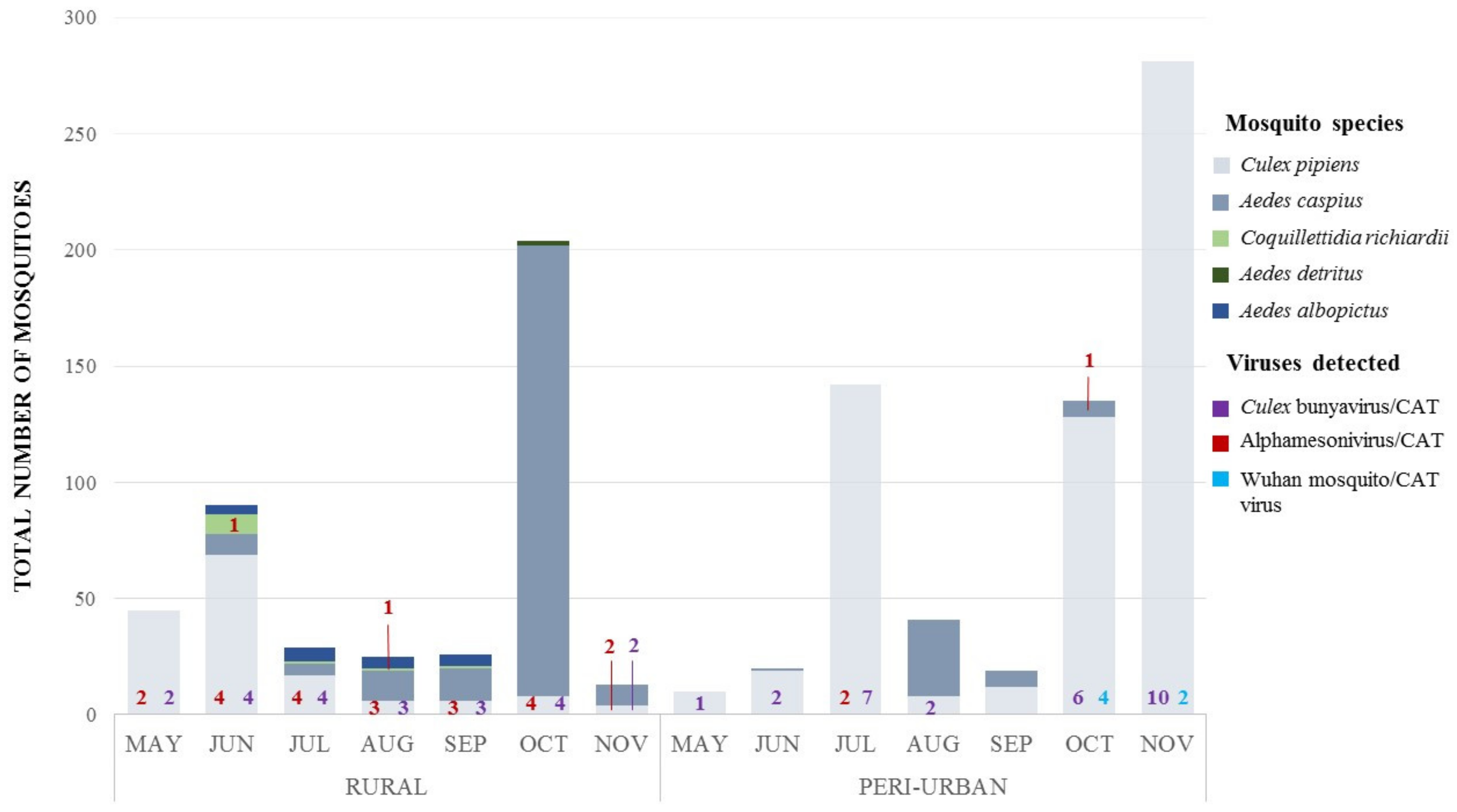
3.5. Virus Detection by Specific RT-PCRs on Field-Captured Mosquito Pools

Virus-specific screenings on mosquito pools confirmed virus circulation as depicted by NGS on FTA cards (Figure 2). Throughout sampling periods, Culex bunyavirus/CAT virus (unclassified Bunyavirales) was the most common and was recurrently detected in both biotopes (Figure 2). Out of 53 Cx. pipiens pools, 50 were found to be infected (including 14 pools unexposed to FTA cards), showing a high occurrence of this viral strain in Cx. pipiens mosquitoes from the Llobregat River Delta. BLASTn analysis of the amplified fragment of a RT-PCR positive pool showed a nucleotide similarity of 97.58% to Bunyaviridae environmental sample’s RNA-dependent RNA polymerase gene (RdRp). Phylogenetically, our strain clustered with Bunyaviridae environmental sample (2013) and Culex Bunyavirus 2 (2016), which have previously been detected in Culex spp. mosquitoes from the United States of America (USA) (Figure 3A). The discovery of Culex bunyavirus/CAT virus in Catalonian Cx. pipiens widens the range of known distribution for this mosquito-specific bunyaviruses from the USA in California [41,57] and Maryland [42], to Spain. Our findings might also suggest that these bunyaviruses could be genus-specific, as they have been detected only in Culex spp. mosquitoes.
Alphamesonivirus/CAT virus, the second most commonly detected virus (Figure 2), was identified in 24 Cx. pipiens pools, two Cq. richiardii and one Ae. caspius pools. Alphamesonivirus is the only recognized genus within the mosquito-restricted family Mesoniviridae (order Nidovirales) [58]. Strains herein reported, shared >98% nucleotide identity to Houston virus and Nam Dinh virus strains’ open reading frame 2 (ORF2) and were closely related to several alphamesonivirus strains that have been detected between 2008 and 2016 in Culex spp. mosquitoes. Houston virus (HOUV) and Nam Dinh virus (NDiV) in Culex quinquefasciatus from Mexico and China; Ngewotan virus in Culex australicus from Australia; NDiV, Alphamesonivirus-1 and HOUV in Culex spp. from China, South Korea, and the USA. It is worth mentioning that the viral strains detected in the present study demonstrated a closer relationship to each other than to the strains found in other geographic regions. Moreover, Alphamesonivirus/CAT strains found in Cx. pipiens, both rural and peri-urban biotopes, appeared to be more closely related to each other than to those found in other mosquito species from the same geographic area (Figure 3B), thereby suggesting co-evolution events within their host species. These findings, together with the detection of an alphamesonivirus in Cx. pipiens from Camargue, France [59], confirm the wide geographical distribution and host range described for the family Mesoniviridae [60]. Recently, viruses belonging to this family have been continually detected by virome metagenomics approaches in several mosquito species [41,47,49,53,61], therefore providing more support for this asseveration.
Finally, Wuhan mosquito/CAT virus was positively detected in six of 53 Cx. pipiens pools (Figure 2). Among these, five were captured in traps without honey-baited FTA cards and only one was exposed to a FTA card. Wuhan mosquito/CAT virus exhibited a high phylogenetic relationship (92.15% of nucleotide similarity) with Wuhan mosquito virus 7 strain’s PB1 gene detected in Anopheles sinensis from China in 2013 (Figure 3C). Wuhan mosquito virus 7 belongs to Quaranjavirus genus (family Orthomyxoviridae, order Articulavirales), which has been identified in a pool of Anopheles sinensis and Culex quinquefasciatus mosquitoes originating from Hubei, China [62]. Finally, throughout screenings, neither Ae. albopictus nor Ae. detritus were found to be infected by any of those viruses targeted. Detecting Culex bunyavirus/CAT and Wuhan mosquito/CAT viruses in Cx. pipiens pools, which were not exposed to honey-baited FTA cards, evidenced that these viruses were indeed infecting the mosquitoes and were not acquired while sugar feeding on the FTA cards.
The discovery of Culex bunyavirus/CAT, Alphamesonivirus/CAT and Wuhan mosquito/CAT viruses in culicid mosquitoes found in Catalonia, contributes to the knowledge of both the host range and their geographical distribution.

3.6. Overall Remarks of the Approach and Future Perspectives

The current study is a pioneer in applying viromics on honey-baited FTA cards during entomological surveys as a tool for the detection of circulating viruses in mosquitoes and the identification of virus in mosquitoes’ saliva. Through this approach, 19 ssRNA (+), six ssRNA (−), eight dsRNA, one ssDNA, five dsDNA viruses and several unclassified viruses were identified; and 12 near-complete viral genomes were obtained from FTA cards, among which seven were linked to mosquito species of sanitary relevance. Acquiring near-complete virus genomes is a clear advantage of metagenomics over classical surveillance based on PCR detection, since insights into the origin, evolution, and diversity of circulating viruses could be gained [25]. Further detection of Culex bunyavirus/CAT virus, Alphamesonivirus/CAT virus and Wuhan mosquito/CAT virus in mosquito pools confirmed the presence of these viruses in Europe, where previously their circulation had not been revealed. These findings highlight the value of honey-baited FTA cards combined with viromics in identifying a wide spectrum of viruses that may be associated to sylvan mosquitoes in susceptible areas for arbovirus transmission, without requiring previous knowledge of viral diversity. In future arbovirus surveillance, NGS on honey-baited FTA cards could be used as a guide for prevention and control strategies. In the case of arboviruses detection, entomological surveillance could be exhaustively carried out focusing on specimen classification and molecular analysis where the virus of interest has been previously detected in the FTA cards.
It is worth mentioning that, in spite of the advantages provided by NGS on honey-baited FTA cards, there are some drawbacks that need to be mentioned. Firstly, since FTA cards inactivate the viruses, and NGS provides only genetic information through this approach, no viable virus could be isolated for further characterization. Secondly, virus-bearing mosquito species could not be identified without complementary morphological and molecular analyses. Other possible constraints of this approach could be related to the feeding rate on FTA cards, the quantity of saliva expectorated by mosquitoes, and the number of viral copies liberated within the saliva while sugar feeding. The assumption of blue abdomens in mosquitoes, as the only proof of virus expectoration on FTA cards, might possibly overlook virus release while probing. This fact was evidenced with the detection of chikungunya virus (CHIKV) RNA in FTA cards exposed to experimentally infected Aedes aegypti despite the fact that there not been any record of blue dye in their abdomens [16]. Based on these findings, viruses identified by NGS in FTAs could also have been deposited by mosquitoes in which blue abdomen were not present. Furthermore, to improve the sensitivity and efficiency of our approach, honey-baited FTA cards could be placed inside Box gravid traps, as a recent study conducted in Switzerland demonstrated these to be the most effective traps for capturing females of different species when searching for an ovipositional site. In addition, these traps also exhibited the highest feeding success on honey-baited FTA cards [19].
The detection of ISVs through metagenomics on honey-baited FTA cards provides evidence that these viruses could be transmitted within mosquitoes’ expectorations, thereby contradicting previous beliefs that they could not be expelled with saliva [19]. Our findings are supported by the tissue tropism evidenced for Culex flavivirus (family Flaviviridae) and Phasi Charoen-like virus (PCLV) (genus Phasivirus, family Phenuiviridae), as they were also detected in salivary glands of Cx. pipiens from Iowa [63] and in Ae. aegypti from South China [64], respectively, and, most importantly, by the detection of Aedes flavivirus RNA in saliva from colonized Ae. albopictus [65]. Sequences distantly related to PCLV were also detected in our FTA cards. To date, ISVs transmission seemed to be primarily vertical from the adult female to its progeny and venereal from males to females [63,66]. However, horizontal transmission has been hypothesized on breeding sites by direct contact, through feeding in larvae and adults, and/or by copula [40]. Further studies are required to assess the transmission dynamics of the ISVs herein identified.
Furthermore, ISVs are a significant part of the mosquito’s virome. Due to their phylogenetic relationships, great abundance and high diversity, it is presumed that arboviruses might have been originated from arthropod-infecting viruses [67,68,69]. In addition, these viral symbionts are thought to alter the mosquito’s innate immune response, therefore modulating the vector competence for certain arboviruses, and so giving rise to new potential biotools for arbovirus control and prevention [69]. For instance, Culex flavivirus naturally infecting Cx. pipiens from Colorado possibly suppressed the early infection with West Nile virus (WNV) [70]. In Thailand, Zika virus (ZIKV) and dengue virus 1 (DENV-1) titers in head tissues of Aedes aegypti were reduced by intrathoracic inoculation of newly isolated cell fusing agent virus (CFAV) [71]. Likewise, a mosquito flavivirus of natural circulation in Aedes vexans form Catalonia seemed to decrease the susceptibility of infection to Rift Valley fever phlebovirus (RVFV) following experimental oral exposure [72].
As evidenced, and in spite of the continual discovery of novel mosquito-associated viruses, viral diversity harbored by vector species is still underestimated and little is known about their host range, distribution, ecology and evolution [67,73]. Further studies are required to isolate and fully characterize the genome of Alphamesonivirus/CAT, Culex bunyavirus/CAT and Wuhan mosquito/CAT viruses so as to assess their potential as vertebrate pathogens. Finding these ISVs in FTA cards, and therefore in mosquitoes saliva, rises concerns of the potential of these viruses to evolve from being insect-specific to dual-host viruses, acquiring the ability to infect vertebrate cells and become new emerging pathogens. Future surveillance strategies for emerging diseases could include NGS on honey-baited FTA cards to detect previously undiscovered and potentially transmissible viruses so as to prevent new arbovirus outbreaks.

4. Conclusions

The detection of viruses related to Alphamesonivirus, Quaranjavirus (Wuhan mosquito virus), and unclassified Bunyavirales in European field-captured mosquitoes using virus-specific primers derived from metagenomics results, demonstrated that viromics on honey-baited FTA cards is a valid approach for virological surveillance in mosquitoes. To the best of our knowledge, this is the first evidence of circulating ISVs in mosquitoes’ saliva under field conditions. Our study also constitutes the first distribution record of these viruses in the European continent, thereby demonstrating that they are widely distributed despite there being an information gap due to the majority of studies being focused primarily on arbovirus detection. Further studies are needed to better understand the evolutionary history of insect-specific viruses and their potential role in arbovirus transmission.

Supplementary Materials

The following are available online at https://0-www-mdpi-com.brum.beds.ac.uk/1999-4915/12/3/274/s1, Table S1: Species composition and feeding rates on honey-baited FTA cards in peri-urban and rural biotopes from the Llobregat River Delta; Table S2: Viral taxonomic assignations of assembled sequences associated to FTA cards linked to mosquito species during entomological surveys in Catalonia

Author Contributions

N.B. conceived the experimental design; L.B., C.A. and S.T. (Sandra Talavera) performed samplings and specimen morphological classification; L.B. analyzed the samples; S.T. (Sarah Temmam), T.B. and F.C.-F. performed the bioinformatics analysis; L.B. and N.B. prepared the original draft; S.T. (Sarah Temmam), T.B., F.C.-F., C.A., S.T. (Sandra Talavera) and M.E. review the manuscript. All authors have read and agreed to the published version of the manuscript.

Funding

This research was funded by the VMERGE Grant agreement ID: 613996, the European Commission, Horizon 2020 Infrastructures #731060 Infravec2, CERCA Programme, Generalitat de Catalunya and supported by Laboratoire d’Excellence “Integrative Biology of Emerging Infectious Diseases” (grant no. ANR-10-LABX-62-IBEID) and by the Direction Internationale de l’Institut Pasteur.

Acknowledgments

The authors would like to acknowledge the Servei de control de mosquits del Baix Llobregat for their professional and logistic collaboration during samplings. The World Reference Centre for Emerging Viruses and Arboviruses at University of Texas Medical Branch (WRCEVA – UTMB) especially Scott Weaver is acknowledged for providing Dezidougou virus isolate, and Patricia Gil and Serafín Gutiérrez from the Centre de Coopération Internationale en Recherche Agronomique pour le Développement (CIRAD) at Montpellier for providing cDNA of Alphamesonivirus used as positive amplification controls.

Conflicts of Interest

The authors declare no conflict of interest.

References

  1. Nii-Trebi, N.I. Emerging and neglected infectious diseases: Insights, advances and challenges. BioMed Res. Int. 2017, ID5245021. [Google Scholar] [CrossRef] [PubMed] [Green Version]
  2. Hollidge, B.S.; González-Scarano, F.; Soldan, S.S. Arboviral Encephalitides: Transmission, Emergence, and Pathogenesis. J. Neuroimmune Pharmacol. 2010, 5, 428–442. [Google Scholar] [CrossRef] [PubMed] [Green Version]
  3. Gubler, D.J. Human arbovirus infections worldwide. Ann. N. Y. Acad. Sci. 2001, 13–25. [Google Scholar] [CrossRef] [PubMed]
  4. Mayer, S.V. The emergence of arthropod-borne viral diseases: A global prospective, dengue, chikungunya and zika fevers. Acta Trop. 2017, 166, 155–163. [Google Scholar] [CrossRef]
  5. Franklinos, L.H.V.; Jones, K.E.; Redding, D.W.; Abubakar, I. The effect of global change in mosquito-borne diseases. Lancet Infect. Dis. 2019, 19, e302–e312. [Google Scholar] [CrossRef]
  6. Messina, J.P.; Brady, O.J.; Scott, T.W.; Zou, C.; Pigott, D.M.; Duda, K.A. Global spread of dengue virus types: Mapping the 70 year history. Trends Microbiol. 2014, 22, 138–146. [Google Scholar] [CrossRef] [Green Version]
  7. Vest, K.G. Zika virus: A basic overview of an emerging arboviral infection in the Western hemisphere. Disaster Med. Public. 2016, 10, 707–712. [Google Scholar] [CrossRef] [Green Version]
  8. Zinszer, K.; Morrison, K.; Brownstein, J.S.; Marinho, F.; Santos, A.F.; Nsoesie, E.O. Reconstruction of Zika virus in Brazil. Emerg. Infect. Dis. 2017, 23, 92–94. [Google Scholar] [CrossRef] [Green Version]
  9. Weaver, S.C.; Forrester, N.L. Chikungunya: Evolutionary history and recent epidemic spread. Antivir. Res. 2015, 120, 32–39. [Google Scholar] [CrossRef]
  10. Caglioti, C.; Lalle, E.; Castilletti, C.; Carletti, F.; Capobianchi, M.R.; Bordi, L. Chikungunya virus infection: An overview. New Microbiol. 2013, 36, 211–227. [Google Scholar]
  11. Gubler, D.J. The continuing spread of West Nile virus in the Western hemisphere. Clin. Infect. Dis. 2007, 45, 1039–1046. [Google Scholar] [CrossRef] [PubMed]
  12. Melanson, V.R.; Jochim, R.; Yarnell, M.; Bingham Ferlez, K.; Shashikumar, S.; Richardson, J.H. Improving Vector-Borne Pathogen Surveillance: A Laboratory-Based Study Exploring the Potential to Dengue Virus and Malaria Parasites in Mosquito Saliva. J. Vector Borne Dis. 2017, 301–310. [Google Scholar] [CrossRef] [PubMed]
  13. Ritchie, S.A.; Cortis, G.; Paton, C.; Townsend, M.; Shroyer, D.; Zborowski, P.; Hall-Mendelin, S.; van der Hurk, A.F. A Simple Non-Powered Pasive Trap for the Collection of Mosquitoes for Arbovirus Surveillance. J. Med. Entomol. 2013, 50, 185–194. [Google Scholar] [CrossRef] [PubMed]
  14. Johnson, B.J.; Kerlin, T.; Hall-Mendelin, S.; van der Hurk, A.F.; Cortis, G.; Doggett, S.L.; Toi, C.; Fall, K.; McMahon, J.L.; Townsend, M.; et al. Development and Field Evaluation of the Sentinel Mosquito Arbovirus Capture Kit (SMACK). Parasites Vectors 2015, 8, 509. [Google Scholar] [CrossRef] [PubMed] [Green Version]
  15. Zheng, Y.; Gao, S.; Padmanabhan, C.; Li, R.; Galvez, M.; Gutierrez, D. VirusDetect: An automated pipeline for efficient virus discovery using deep sequencing of small RNAs. Virology 2017, 500, 130–138. [Google Scholar] [CrossRef]
  16. Hall-Mendelin, S.; Ritchie, S.A.; Johansen, C.A.; Zborowski, P.; Cortis, G.; Dandridge, S.; Hall, R.A.; van der Hurk, A.F. Exploiting Mosquito Sugar Feeding to Detect Mosquito-Borne Pathogens. Proc. Natl. Acad. Sci. USA 2010, 107, 11255–11259. [Google Scholar] [CrossRef] [Green Version]
  17. Van der Hurk, A.F.; Hall-Mendelin, S.; Townsend, M.; Kurucz, N.; Edwards, J.; Ehlers, G.; Rodwell, C.; Moore, F.A.; McMahon, J.L.; Northill, J.A.; et al. Applications of a Sugar-Based Surveillance System to Track Arboviruses in Wild Mosquito Populations. Vector Borne Zoonotic Dis. 2014, 14, 66–73. [Google Scholar] [CrossRef]
  18. Flies, E.J.; Toi, C.; Weinstein, P.; Doggett, S.L.; Williams, C.R. Converting Mosquito Surveillance to Arbovirus Surveillance with Honey-Baited Nucleic Acid Preservation Cards. Vector Borne Zoonotic Dis. 2015, 15, 397–403. [Google Scholar] [CrossRef]
  19. Wipf, N.C.; Guidi, V.; Tonolla, M.; Ruinelli, M.; Müller, P.; Engler, O. Evaluation of honey-baited FTA cards in combination with different mosquito traps in an area of low arbovirus prevalence. Parasite Vectors. 2019, 12, 554. [Google Scholar] [CrossRef] [Green Version]
  20. Bibby, K. Metagenomic identification of viralpathogens. Trends Biotechnol. 2013, 31, 257–279. [Google Scholar] [CrossRef]
  21. Delwart, E.L. Viral metagenomics. Rev. Med. Virol. 2007, 17, 115–131. [Google Scholar] [CrossRef] [PubMed]
  22. Kristensen, D.M.; Mushegian, A.R.; Dolja, V.V.; Koonin, E.V. New dimensions of the virus world discovered by metagenomics. Trends Microbiol. 2010, 18, 11–19. [Google Scholar] [CrossRef] [PubMed] [Green Version]
  23. Greninger, A.L. A decade of RNA virus metagenomics is (not) enough. Virus Res. 2018, 244, 218–219. [Google Scholar] [CrossRef] [PubMed]
  24. Atoni, E.; Zhao, L.; Karungu, S.; Obanda, V.; Agwanda, B.; Xia, H.; Yuan, Z. The discovery and Global Distribution of Novel Mosquito-Associated Viruses in the Last Decade (2007–2017). Rev. Med. Virol 2019, 29, e2079. [Google Scholar] [CrossRef]
  25. Batovska, J.; Mee, P.T.; Lynch, S.E.; Sawbridge, T.I.; Rodoni, B.C. Sensitivity and Specificity of Metatranscriptomics as an Arbovirus Surveillance Tool. Sci. Rep. 2019, 9, 19398. [Google Scholar] [CrossRef] [Green Version]
  26. Busquets, N.; Alba, A.; Allepuz, A.; Aranda, C.; Nuñez, J.I. Usutu Virus Sequences in Culex pipiens (Diptera: Culicidae), Spain. Emerg Infect. Dis. 2008, 14, 861–863. [Google Scholar] [CrossRef]
  27. Van der Hurk, A.F.; Hall-Mendelin, S.; Johansen, C.A.; Warrilow, D.; Ritchie, S.A. Evolution of Mosquito-Based Arbovirus Surveillance Systems in Australia. J. Biomed. Biotechnol. 2012, 2012, 325659. [Google Scholar] [CrossRef] [Green Version]
  28. Schaffner, E.; Angel, G.; Geoffroy, B.; Hervey, J.P.; Rhaiem, A.; Brunhes, J. Les moustiques d’Europe. Logiciel d´identification et d´enseignement; IRD Editions: Paris, France, 2001. [Google Scholar]
  29. Moutailler, S.; Popovici, I.; Devillers, E.; Vayssier-Taussat, M.; Eloit, M. Diversity of Viruses in Ixodes ricinus, and characterization of a neurotropic strain of Eyach virus. New Microbes New Infect. 2016, 11, 71–81. [Google Scholar] [CrossRef] [Green Version]
  30. Vayssier-Taussat, M.; Moutailler, S.; Michelet, L.; Devillers, E.; Bonnet, S.; Cheval, J.; Hebert, C.; Eloit, M. Next Generation Sequencing Uncovers Unexpected Bacterial Pathogens in Ticks in Western Europe. PLoS ONE 2013, 8, e81439. [Google Scholar] [CrossRef]
  31. Temmam, S.; Chrétien, D.; Bigot, T.; Dufour, E.; Petres, S.; Desquesnes, M.; Devillers, E.; Dumarest, M.; Yousfi, L.; Moutailler, S.; et al. Monitoring silent spillovers before emergence: A pilot study at the tick/human interface in Thailand. Front. Microbiol. 2019. In press. [Google Scholar] [CrossRef]
  32. Criscuolo, A.; Brisse, S. AlienTrimmer removes adapter oligonucleotides with high sensitivity in short-insert paired-end reads. Commentary on Turner (2014) Assessment of insert sizes and adapter content in FASTQ data from NexteraXT libraries. Front. Genet. 2014, 5, 130. [Google Scholar] [CrossRef] [PubMed] [Green Version]
  33. Li, D.; Liu, C.M.; Luo, R.; Sadakane, K.; Lam, T.W. MEGAHIT: An ultra-fast single-node solution for large and complex metagenomics assembly via succinct de Bruijn graph. Bioinformatics 2015, 31, 1674–1676. [Google Scholar] [CrossRef] [PubMed] [Green Version]
  34. Bigot, T.; Temmam, S.; Pérot, P.; Eloit, M. RVDB-prot, a reference viral protein database and its HMM profiles [version 1; peer review: Awaiting peer review]. F1000 Res. 2019, 8, 530. [Google Scholar] [CrossRef] [Green Version]
  35. Hall, T.A. BioEdit: A user-friendly biological sequence alignment editor and analysis program for Windows 95/98/NT. Nucl. Acids. Symp. Ser. 1999, 41, 95–98. [Google Scholar]
  36. Kumar, S.; Stecher, G.; Li, M.; Knyaz, C.; Tamura, K. MEGA X: Molecular Evolutionary Genetics Analysis across computing platforms. Mol. Biol. Evol. 2018, 35, 1547–1549. [Google Scholar] [CrossRef] [PubMed]
  37. Nei, M.; Kumar, S. Molecular Evolution and Phylogenetics; Oxford University Press: New York, NY, USA, 2000. [Google Scholar]
  38. Hasegawa, M.; Kishino, H.; Yano, T. Dating the human-ape split by a molecular clock of mitochondrial DNA. J. Mol. Evol. 1985, 22, 160–174. [Google Scholar] [CrossRef]
  39. Shi, M.; Lin, X.-D.; Tian, J.-H.; Chen, L.-J.; Chen, X.; Li, C.-X.; Quin, X.-C.; Li, J.; Cao, J.-P.; Eden, J.-C.; et al. Redefining the Invertebrate RNA Virosphere. Nature 2016, 540, 539–543. [Google Scholar] [CrossRef]
  40. Agboli, E.; Leggewie, M.; Altinli, M.; Schnettler, E. Mosquito-Specific Viruses-Transmission and Interaction. Viruses 2019, 11, 873. [Google Scholar] [CrossRef] [Green Version]
  41. Sadeghi, M.; Altan, E.; Deng, X.; Barker, C.M.; Fang, Y.; Coffey, L.L. Virome of >12 thousand Culex mosquitoes from throughout California. Virology 2018, 74–88. [Google Scholar] [CrossRef]
  42. Frey, K.G.; Biser, T.; Hamilton, T.; Santos, C.J.; Pimentel, G.; Mokashi, V.P.; Bishop-Lilly, K.A. Bioinformatic characterization of Mosquito Viromes within the Eastern United States and Puerto Rico: Discovery of Novel Viruses. Evol. Bioinform. 2016, 12, 1–12. [Google Scholar] [CrossRef] [Green Version]
  43. Belda, E.; Nanfack-Minkeu, F.; Eiglmeier, K.; Carissimo, G.; Holm, I.; Diallo, M.; Diallo, D.; Vantaux, A.; Kim, S.; Sharakhov, I.V.; et al. De novo profiling of RNA viruses in Anopheles malaria vector mosquitoes from forest ecological zones in Senegal and Cambodia. Bmc Genom. 2019, 20, 664. [Google Scholar] [CrossRef] [Green Version]
  44. De Oliveira, G.; Costa, F.J.; da S Rego, M.O.; D’Athaide, E.S.; Funayama, D.; Montani, M.; Souza, R.; Vasconcelos, S.; Witkin, S.S.; Deng, X.; et al. Detection of RNA-Dependent RNA Polymerase of Hubei Reo-like Virus 7 by Next-Generation Sequencing in Aedes aegypti and in Culex quinquefasciatus Mosquitoes from Brazil. Viruses 2019, 11, 147. [Google Scholar] [CrossRef] [Green Version]
  45. Öhlund, P.; Hayer, J.; Lundén, H.; Hesson, J.C.; Blomström, A.L. Viromics Reveal a Number of Novel RNA Viruses in Swedish Mosquitoes. Viruses 2019, 11, 1027. [Google Scholar] [CrossRef] [PubMed] [Green Version]
  46. Shi, C.; Beller, L.; Deboutte, W.; Yinda, K.C.; Delang, L.; Vega-Rua, A. Failloux, A-B. ; Matthijnssens, J. Stable Distinct Core Eukaryotic Viromes in Different Mosquito Species from Guadeloupe, Using Single mosquito Viral Metagenomics. Microbiome 2019, 7, 121. [Google Scholar] [CrossRef] [PubMed]
  47. Sanborn, M.A.; Klein, T.A.; Kim, H.-C.; Fung, C.K.; Figueroa, K.L.; Yang, Y.; Asafo-adjei, E.A.; Jarman, R.G.; Hang, J. Metagenomics Analysis Reveals Three Novel and Prevalent Mosquito Viruses from a Single Pool of Aedes vexans niponni Collected in the Republic of Korea. Viruses 2019, 11, 222. [Google Scholar] [CrossRef] [Green Version]
  48. Pettersson, J.H.-O.; Shi, M.; Eden, J.-S.; Holmes, E.C.; Hesson, J.C. Meta-Transcriptomic Comparison of the RNA Viromes of Mosquito Vectors Culex pipiens and Culex torrentium in Northern Europe. Viruses 2019, 11, 1033. [Google Scholar] [CrossRef] [Green Version]
  49. Atoni, E.; Wang, Y.; Karungu, S.; Waruhiu, C.; Zohaib, A.; Obanda, V.; Agwanda, B.; Mutua, M.; Xia, H.; Yuan, Z. Metagenomic Virome Analysis of Culex Mosquitoes from Kenya and China. Viruses 2018, 10, 30. [Google Scholar] [CrossRef] [Green Version]
  50. Cholleti, H.; Hayer, j.; Fafetine, J.; Berg, M.; Blomström, A.-L. Genetic Characterization of a Novel Picrona-like Virus in Culex spp. Mosquitoes from Mozambique. Virol J. 2018, 15, 71. [Google Scholar] [CrossRef] [Green Version]
  51. Shi, C.; Liu, Y.; Hu, X.; Xiong, J.; Zhang, B.; Yuan, Z. A Metagenomic Survey of Viral Abundance and Diversity in Mosquitoes from Hubei Province. PLoS ONE 2015, 10, e0129845. [Google Scholar] [CrossRef]
  52. Cook, S.; Chung, B.Y.-W.; Bass, D.; Moureau, G.; Tang, S.; McAlister, E.; Culverwell, C.L.; Glücksman, E.; Wang, H.; Brown, T.D.K.; et al. Novel Viruses Discovery and Genome Reconstruction form Field RNA Samples Reveals Highly Divergent Viruses in Dipteran Hosts. PLoS ONE 2013, 8, e80720. [Google Scholar] [CrossRef] [Green Version]
  53. Xia, H.; Wang, Y.; Shi, C.; Atoni, E.; Zhao, L.; Yuan, Z. Comparative Metagenomic Profiling of Viromes Associated with Four Common Mosquito species in China. Virol Sin. 2018, 33, 59–66. [Google Scholar] [CrossRef] [PubMed]
  54. Habayeb, M.S.; Ekengren, S.K.; Hultmark, D. Nora Virus, a Persistent Virus in Drosophila, Defines a New Picorna-like Virus Family. J. Gen. Virol 2006, 87, 3045–3051. [Google Scholar] [CrossRef] [PubMed]
  55. Wang, L.; Lv, X.; Zhai, Y.; Fu, S.; Wang, D.; Rayner, S.; Tang, Q.; Liang, G. Genomic Characterization of a Novel Virus of the Family Tymoviridae Isolated from Mosquitoes. PLoS ONE 2012, 7, e39845. [Google Scholar] [CrossRef] [PubMed]
  56. Forrester, N.L.; Coffey, L.L.; Weaver, S.C. Arboviral Bottlenecks and Challenges to Maintaining Diversity and Fitness during Mosquito Transmission. Viruses 2014, 6, 3991–4004. [Google Scholar] [CrossRef] [PubMed] [Green Version]
  57. Chandler, J.A.; Liu, R.M.; Bennett, S.N. RNA Shutgun Metagenomic Sequencing of Northern California (USA) Mosquitoes Uncovers Viruses, Bacteria, and Fungi. Front. Microbiol. 2015, 6, 185. [Google Scholar] [CrossRef] [PubMed] [Green Version]
  58. Lauber, C.; Ziebuhr, J.; Junglen, S.; Drosten, C.; Zirkel, F.; Nga, P.T.; Morita, K.; Snijder, E.J.; Gorbalenya, A.E. Mesoniviridae: A Proposed New Family in the Order Nidovirales formed by a Single Species of Mosquito-Borne Viruses. Arch. Virol. 2012, 157, 1623–1628. [Google Scholar] [CrossRef] [Green Version]
  59. Gil, P.; Rakotoarivony, I.; Etienne, L.; Albane, M.; Benoit, F.; Grégory, L.; Busquets, N.; Birnberg, L.; Talavera, S.; Aranda, C.; et al. First Detection of a Mesonivirus in Culex pipiens in Five Countries Around the Mediterranean Basin. Abstract. In Proceedings of the EPIZONE—11th Annual Meeting ANSES, Paris, France, 19–21 September 2017. [Google Scholar]
  60. Vasilakis, N.; Guzman, H.; Firth, C.; Forrester, N.L.; Widen, S.G.; Wood, T.G.; Rossi, S.L.; Ghedin, E.; Popov, V.; Blasdell, K.R.; et al. Mesoniviruses are Mosquito-Specific Viruses with Extensive Geographic Distribution and Host Range. Virol. J. 2014, 11, 97. [Google Scholar] [CrossRef] [Green Version]
  61. Hang, J.; Klein, T.A.; Kim, H.-C.; Yang, Y.; Jima, D.D.; Richardson, J.H.; Jarman, R.G. Genome Sequences of Five Arboviruses in Field-Captured Mosquitoes in a Unique Rural Environment of South Korea. Genome Announc 2016, 4, e01644-15. [Google Scholar] [CrossRef] [Green Version]
  62. Li, C.X.; Shi, M.; Tian, J.-H.; Lin, X.-D.; Kang, Y.-J.; Chen, L.-J.; Qin, X.-C.; Xu, J.; Holmes, E.C.; Zhang, Y.-Z. Unprecedented Genomic Diversity of RNA Viruses in Arthropods Reveals the Ancestry of Negative-Sense RNA Viruses. eLife 2015, 4, e05378. [Google Scholar] [CrossRef]
  63. Saiyasombat, R.; Bolling, B.G.; Brault, A.C.; Bartholomay, L.C.; Blitvich, B.J. Evidence of Efficient Transovarial Transmission of Culex Flavivirus by Culex pipiens (Diptera: Culicidae). J. Med. Entomol 2011, 48, 1031–1038. [Google Scholar] [CrossRef]
  64. Zhang, X.; Huang, S.; Jin, T.; Lin, P.; Huang, Y.; Wu, C.; Peng, B.; Wei, L.; Chu, H.; Wang, M.; et al. Discovery and High Prevalence of Phasi Charoen-like Virus in Field-Captured Aedes aegypti in South China. Virology 2018, 523, 35–40. [Google Scholar] [CrossRef] [PubMed]
  65. Bolling, B.G.; Vasilakis, N.; Guzman, H.; Widen, S.G.; Wood, T.G.; Popov, V.L.; Thangamani, S.; Tesh, R.B. Insect-Specific Viruses Detected in Laboratory Mosquito Colonies and Their Potential Implications in Experiments Evaluating Arbovirus vector Competence. Am. J. Trop. Med. Hyg 2015, 92, 422–428. [Google Scholar] [CrossRef] [PubMed] [Green Version]
  66. Bolling, B.G.; Eisen, L.; Moore, C.G.; Blair, C.D. Insect-Specific Flaviviruses from Culex Mosquitoes in Colorado, with Evidence of Vertical Transmission. Am. J. Trop. Med. Hyg 2011, 85, 169–177. [Google Scholar] [CrossRef] [PubMed] [Green Version]
  67. Bolling, B.G.; Weaver, S.C.; Tesh, R.B.; Vasilakis, N. Insect-Specific Virus Discovery: Significance for the Arbovirus Community. Viruses 2015, 7, 4911–4928. [Google Scholar] [CrossRef] [PubMed] [Green Version]
  68. Dudas, G.; Obbard, D.J. Are Arthropods at the Heart of Virus Evolution? eLife 2015, 4, e05378. [Google Scholar] [CrossRef] [PubMed] [Green Version]
  69. Öhlund, P.; Lundén, H.; Blomström, A.-L. Insect-Specific Virus Evolution and Potential Effects in Vector Competence. Virus Genes 2019, 55, 127–137. [Google Scholar] [CrossRef] [Green Version]
  70. Bolling, B.G.; Olea-Popelka, F.J.; Eisen, L.; Moore, C.G.; Blair, C.D. Transmission Dynamics of an Insect-Specific Flavivirus in a Naturally Infected Culex pipiens Laboratory Colony and Effects of Co-Infection on Vector Competence for West Nile Virus. Virology 2012, 427, 90–97. [Google Scholar] [CrossRef] [Green Version]
  71. Baidaliuk, A.; Miot, E.F.; Lequime, S.; Moltini-Conclois, I.; Delaigue, F.; Dabo, S.; Dickson, L.B.; Aubry, F.; Merkling, S.H.; Cao-Lormeau, V.M.; et al. Cell-Fusing Agent Virus Reduces Arbovirus Dissemination in Aedes aegypti Mosquitoes In Vivo. J. Virol 2019, 93, e00705-19. [Google Scholar] [CrossRef] [Green Version]
  72. Birnberg, L.; Talavera, S.; Aranda, C.; Núñez, A.I.; Napp, S.; Busquets, N. Field-Captured Aedes vexans (Meigen, 1830) is a Competent Vector for Rift Valley Fever Phlebovirus in Europe. Parasit Vectors 2019, 12, 484. [Google Scholar] [CrossRef]
  73. Vasilakis, N.; Tesh, R.B. Insect-Specific Viruses and their Potential Impact on Arbovirus Transmission. Curr. Opin. Virol. 2015, 15, 69–74. [Google Scholar] [CrossRef] [Green Version]
Figure 1. Overview of viral composition of honey-baited FTA cards. (A) Shows the proportion of viral reads classified by host type. Proportions of bacteria and vertebrate/invertebrate are too small to be seen in the figure. (B) Abundance in nucleotides of each viral family estimated by summing sequence length in nucleotides weighted by the k-mer coverage of each contig.
Viruses 12 00274 g001
Figure 2. Mosquito species dynamics and virus occurrence in rural and peri-urban biotopes from the Llobregat River Delta. Cumulative bars represent the total number of female mosquitoes captured per month per sampling site. Numbers in color correspond to the total number of mosquito pools that tested positive for a given virus on a particular month and sampling site.
Viruses 12 00274 g002
Figure 3. Phylogenetic trees of viruses detected by virus-specific RT-PCR in Catalonian mosquitoes. Trees were drawn to scale, with branch lengths measured in the number of substitutions per site. The percentage of trees in which the associated taxa clustered together is shown next to the branches. Initial tree(s) for the heuristic search were obtained automatically by applying Neighbor-Join and BioNJ algorithms to a matrix of pairwise distances estimated using the Maximum Composite Likelihood (MCL) approach, and then selecting the topology with a superior log likelihood value. Codon positions included were 1st+2nd+3rd+Noncoding. A discrete Gamma distribution was used to model evolutionary rate differences among sites. (A) Culex bunyavirus/CAT virus, evolutionary history inferred by using the Maximum Likelihood (ML) method and Hasegawa-Kishino-Yano model (HKY+G). The tree with the highest log likelihood (−6117.36) is shown (five categories (+G, parameter = 1.2252)). There were 946 positions in the final dataset. (B) Alphamesonivirus/CAT evolutionary history inferred by using the ML method and Tamura-Nei (TN93+G) model. The tree with the highest log likelihood (−2006.57) is shown (five categories (+G, parameter = 0.3417)). There were a total of 839 positions in the final dataset. (C) Wuhan mosquito/CAT virus evolutionary history was inferred by using the ML method and Tamura-Nei model (TN93+G). The tree with the highest log likelihood (−8996.79) is shown (five categories (+G, parameter = 0.7704). There were a total of 729 positions in the final dataset.
Viruses 12 00274 g003
Table 1. Virus-Specific Primers for RT-PCRs.
Mosquito-Associated VirusesPrimer CodePrimer Nucleotide Sequence (5′→3′)Tm (°C)RT-PCR Fragment Size (bp)
Alphamesonivirus 1ALPMFGCGCCATTCTGCAGATCAAC581033
ALPMRGTGCCAATAAACGCGTGATG
Bunyaviridae environmental sampleBNYFGAGTCCTTGTCCATCCCYGC571059
BNYRGTGCAGGAAGAAGKAGCATGG
Dezidougou virusDZGFGTCCTGTTAAGCTGCAACCC56400
DZGRCGTAACAACGATAAGTGGCG
Wuhan mosquito virus 7 WHNFGCGGAGAGAGGYAAAATGGATC57572
WHNRCATTCCCATCAGGAACCCTG
Table 2. Mosquito-associated viruses identified in honey-baited Flinders’ Technology Associates (FTA) cards by next-generation sequencing (NGS) analysis. Taxonomic assignations with assembly lengths higher than 400 nt are shown. Abundance and contig length are expressed in nucleotides (nt). Viral identities are expressed in nucleotides and amino acids (aa).
Closest HitGene/ProductAbundanceaa Identity (%)Max. Contig Length% Coveragent Identity (%)Accession No.
RuralAlphamesonivirus 1Spike protein, hypothetical protein360619653–100132810099.18MF176279.1
Bunyaviridae environmental sampleRNA-dependent RNA polymerase33529249–9943549999.02KP642114.1
Culex bunya-like virusHypothetical protein28900747–1009209898.45MH188002.1
Culex iflavi-like virus 4Polyprotein100917571–10017069995.77NC_040574.1
Culex picorna-like virus 1Polyprotein80699864–100123810096.37MH703059.1
Culex-associated Luteo-like virusHypothetical protein, RNA-dependent RNA polymerase328567–1005669995.04MK440647.1
Dezidougou virusHypothetical protein 1936687–10063810094.34KY968698.1
Hubei picorna-like virus 61Hypothetical protein5357884–1009169995.63KX883915.1
Wenzhou soberno-like virus 4Hypothetical proteins 1 and 266885294–98228410096.67KX882831.1
Wuhan mosquito virus 5PB15460505801375.95KX898491.1
Peri-urbanAedes pseudoscutellaris reovirus VP1524469–1006679978.08DQ087276.1
Alphamesonivirus 1ORF1a, pp1a polyprotein2259060–10093210098.18MH520106.1
Culex Hubei-like virusHypothetical protein514285–1005109190.34MH188025.1
Culex iflavi-like virus 4Polyprotein16815497–100217010096.04NC_040574.1
Culex luteo-like virusRNA-dependent RNA polymerase 1668642–6712796567.49MF176386.1
Culex picorna-like virus 1Polyprotein10297977–100129010098.29MH703059.1
Culex pipiens associated Tunisia virusReplicase1131996–10014469889.11NC_040723.1
Culicine-associated Z virusVP1, RNA-dependent RNA polymerase1458477–977659683.33KF298283.1
Daeseongdong virus 1ORF1, putative RNA-dependent RNA polymerase61453775–9558319582.27KU095841.1
Dezidougou virusHypothetical protein 1142447285–100188210095.42KY968698.1
Karumba virusSimilar NS5 protein966874931602876.31JF707857.1
Hubei picorna-like virus 61Hypothetical protein581501870–100125210096.01KX883915.1
Negevirus nona 1 Hypothetical protein19083049–9527659987.11AB972669.1
Wuhan mosquito virus 6Nucleoprotein948072–10046810097.01MF176381.1
Wuhan mosquito virus 7PB14335153–100184610092.15KM817626.1
Bold type corresponds to the selected viruses for primers design.
Table 3. Near-complete viral genomes obtained by NGS on honey-baited FTA cards. Viral assignations with a genome coverage higher than 98% and identities higher than 95% are shown.
SampleOrderFamilyClosest virusNo ReadsMean coverage per ntCoverage (%)% Identity (nt)Accession No
RuralPicornaviralesDicistroviridaeKashmir bee virus28080401,29 X10096.74AY275710.1
Black queen cell virus isolate BQCV_MS311249,85 X10093.78MH267694.1
IflaviridaeDeformed wing virus isolate Hamilton392151,47 X10099.77MF623172.1
Culex iflavi-like 4 virus strain CIVL/Kern17787250,75 X10095.78NC_040574.1
NidoviralesMesoniviridaeNgewotan virus strain mos172×93828932663,03 X10098.88MF176279.1
Unclassified RNA virusesWenzhou soberno-like virus 4 strain mosZJ3539112059562,28 X9996.79KX882831.1
Peri-urbanPicornaviralesDicistroviridaeAphid lethal paralysis virus isolate ALPV-CE5728,42 X9994.75JX480861.1
IflaviridaeDeformed wing virus isolate Hamilton367047,57 X10099.75MF623172.1
Culex iflavi-like 4 virus strain CIVL/Kern143520,74 X10095.72NC_040574.1
Unclassified RNA virusesHubei picorna-like virus 61 strain mosHB2359031473772384,82 X10095.84KX883915.1
Hubei noda-like virus 11 strain arthropodmix224822102756 964,36 X10097.58KX883010.1
Dezidougou virus strain DEZI/Aedes africanus/SEN/DAK-AR-41524/1984493974,39 X9895.32KY968698.1

