Article

Bacteriophage Rescue Therapy of a Vancomycin-Resistant Enterococcus faecium Infection in a One-Year-Old Child following a Third Liver Transplantation

by
Kevin Paul
1,*,
Maya Merabishvili
2,
Ronen Hazan
3,*,
Martin Christner
4,
Uta Herden
5,
Daniel Gelman
3,
Leron Khalifa
3,
Ortal Yerushalmy
3,
Shunit Coppenhagen-Glazer
3,
Theresa Harbauer
1,
Sebastian Schulz-Jürgensen
1,
Holger Rohde
4,
Lutz Fischer
5,
Saima Aslam
6,
Christine Rohde
7,
Ran Nir-Paz
8,
Jean-Paul Pirnay
2,*,
Dominique Singer
1,† and
Ania Carolina Muntau
1,†
1
Department of Pediatrics, Kinder-UKE, University Medical Center Hamburg-Eppendorf, 20246 Hamburg, Germany
2
Burn Centre, Laboratory for Molecular and Cellular Technology (LabMCT), Queen Astrid Military Hospital, B-1120 Brussels, Belgium
3
Institute of Dental Sciences, School of Dentistry, Hebrew University of Jerusalem, Jerusalem 9112001, Israel
4
Institute of Medical Microbiology, Virology and Hygiene, University Medical Center Hamburg-Eppendorf, 20246 Hamburg, Germany
5
Department of Visceral Transplantation, University Medical Center Hamburg-Eppendorf, 20246 Hamburg, Germany
6
Center for Innovative Phage Applications and Therapeutics, Division of Infectious Diseases and Global Public Health, University of California, San Diego, CA 92093, USA
7
Leibniz Institute DSMZ—German Collection of Microorganisms and Cell Cultures GmbH, 38124 Braunschweig, Germany
8
Department of Clinical Microbiology and Infectious Disease, Hadassah University Medical Center, Faculty of Medicine, Hebrew University of Jerusalem, Jerusalem 9112001, Israel
*
Authors to whom correspondence should be addressed.
These authors contributed equally to this work.
Submission received: 30 July 2021 / Revised: 21 August 2021 / Accepted: 1 September 2021 / Published: 7 September 2021
(This article belongs to the Special Issue State-of-the-Art Phage Therapy Development in Europe)

Abstract

:
Phage therapy is an experimental therapeutic approach used to target multidrug-resistant bacterial infections. A lack of reliable data with regard to its efficacy and regulatory hurdles hinders a broad application. Here we report, for the first time, a case of vancomycin-resistant Enterococcus faecium abdominal infection in a one-year-old, critically ill, and three times liver transplanted girl, which was successfully treated with intravenous injections (twice per day for 20 days) of a magistral preparation containing two Enterococcus phages. This correlated with a reduction in baseline C-reactive protein (CRP), successful weaning from mechanical ventilation and without associated clinical adverse events. Prior to clinical use, phage genome was sequenced to confirm the absence of genetic determinants conferring lysogeny, virulence or antibiotic resistance, and thus their safety. Using a phage neutralization assay, no neutralizing anti-phage antibodies in the patient’s serum could be detected. Vancomycin-susceptible E. faecium isolates were identified in close relation to phage therapy and, by using whole-genome sequencing, it was demonstrated that vancomycin-susceptible E. faecium emerged from vancomycin-resistant progenitors. Covering a one year follow up, we provide further evidence for the feasibility of bacteriophage therapy that can serve as a basis for urgently needed controlled clinical trials.

1. Introduction

Biliary atresia is an obliterative cholangiopathy presenting in the neonatal period, and it is treated by Kasai portoenterostomy during the first months of life as first-line therapy [1]. However, progressive cholestasis and recurrent cholangitis, even after portoenterostomy, make biliary atresia the leading indication for liver transplantation in the pediatric cohort [2]. Abdominal site infections caused by enterococci belong to the most common complications after Kasai portoenterostomy [3] and liver transplantation [4,5]. The clinical management of infections with E. faecium is particularly challenging due to the high rate of multidrug resistance in this species [4,5,6]. In general, vancomycin-resistant enterococci are a major medical concern, with E. faecium being one of the ESKAPE pathogens classified as “high priority” in the WHO priority pathogens list [7].
Facing emerging rates of infections by multidrug-resistant (MDR) bacteria, phage therapy has gained worldwide attention to serve as a possible treatment option. The growing worldwide interest in phage therapy, which was predominantly used in Eastern Europe during the past century [8], is illustrated by recent reports of cases or case series of successful phage therapy in refractory infections, e.g., [9,10,11,12]. However, to our knowledge, to date no intravenous phage therapy of enterococcal infection has been reported. Although phage therapy is increasingly seen as a promising alternative or complement to antibiotic therapy, its efficacy has not been shown in controlled clinical trials. This results in regulatory hurdles, which contribute to limit a broader application of phage therapy [13,14]. Here we describe the successful phage therapy of a vancomycin-resistant E. faecium (VREfm) infection in a pediatric liver transplant recipient, underscoring the potential usefulness of phage therapy in the treatment of infections caused by VRE.

2. Materials and Methods

2.1. Phage Susceptibility Testing

Phage susceptibility testing was performed in triplicate using a modified double agar overlay method [15]. Briefly, 0.1 mL of an overnight culture of the target bacteria VREfm was added to 3.5 mL of agarose (0.5%) and plated on brain heart infusion (BHI, Difco, Detroit, MI) agar plates. Drops (0.05 mL) of 27 anti-enterococci phages from the Israeli Phage Bank (IBP) [16] in titers of ~108–109 plaque-forming units (PFU)/mL were spotted on the bacterial lawn. The plates were incubated for 18 h at 37 °C. Two phages, EFgrKN (GenBank Accession: MW004544) and EFgrNG (GenBank Accession: MW004545) [17], exerted clear plaques, and their efficacy was validated in liquid culture (Supplementary Figure S1, and also as schematical figure in [18]). Next, the efficacy of the phages was tested in triplicate in the presence of antibiotics to determine synergy or interference between them. To this end, untreated and treated bacteria were grown in a 96-well plate at 37 °C for 24 h. The 600 nm absorbance was recorded every 20 min after 5 s linear shaking. The bacteria were added in their logarithmic phase (1.5 × 10⁷ colony forming units (CFU)/mL). Phages and antibiotics were added at t = 0 as follows: EFGrNG: 7 × 10⁷ PFU/mL; EFGrKN: 2.4 × 10⁸ PFU/mL; ampicillin: 16 µg/mL (0.5 MIC); vancomycin: 16 µg/mL (0.5 MIC); chloramphenicol: 1 µg/mL (~0.5 MIC of linezolid); gentamicin: 12.5 µg/mL (~0.025 MIC because the bacteria were highly sensitive).

2.2. Phage DNA Analysis and Lysogeny

DNA of the phages was extracted, purified and sequenced as previously described [19]. Analysis of lysogeny, virulence and antibiotic resistance determinants was performed using Abricate (version 0.8.13, [Seemann T. Abricate Github https://github.com/tseemann/abricate (accessed on 30 July 2021)]), comparing to all of its databases.

2.3. Production of Phage Active Pharmaceutical Ingredients (APIs)

Phages EFgrKN and EFgrNG were propagated by the double agar overlay method [15] to a titer of 1010 PFU/mL using bacterial host strain VREfm isolated from the patient. The obtained lysates were centrifuged at 35,000× g for 1.5 h, and the phage pellet was resuspended in DPBS (Lonza, Verviers, Belgium) to obtain phage stocks with a titer of 1011 PFU/mL, which were further diluted to 109 PFU/mL and endotoxin purified by Endotrap HD (Lionex, Braunschweig, Germany) column mode affinity chromatography. Samples of the obtained phage Active Pharmaceutical Ingredients (APIs) were sent to Sciensano, the Belgian Scientific Institute of Public Health (Brussels, Belgium), for quality assessment and product certification [20]. Upon Sciensano approval, both phage APIs were mixed and diluted, in the form of a magistral preparation, to the titers of ~107 and ~108 PFU/mL in 0.9 % NaCl (Fresenius Kabi, Bad Homburg, Germany) and sent directly to the University Medical Center Hamburg—Eppendorf for application in the patient. As the two phages produce indistinguishable plaque morphologies, it was impossible to determine the titer of each phage separately when present in the mixture. The joint titers of the first and the second batches of magistral preparations were defined as 8.1 × 10⁷ and 5.2 × 10⁸ PFU/mL, respectively.

2.4. Stability of the Phages

The stability of the two batches of magistral preparations containing phages EFgrKN and EFgrNG was determined by the double agar overlay method [15]. The preparations were stored in 15 mL polypropylene tubes (Greiner Bio-One, Vilvoorde, Belgium) at 4 °C. As noted earlier, only joint titers were defined.

2.5. Phage Neutralization Assay

Phage neutralization by the patient’s serum was evaluated according to Adams 1959 [21] with some modifications. Blood samples were collected on 4, 28 and 49 days after initiation of phage therapy and were centrifuged, after clotting, at 2000× g for 10 min. The obtained serum samples (supernatant) were stored at −80 °C. For testing, 0.9 mL of the diluted (1:100) serum samples was mixed with 0.1 mL of phages EFgrKN or EFgrNG at the concentrations of 4.6 ± 2.4 × 10⁷ and 4.3 ± 2.7 × 10⁷ PFU/mL, respectively, and incubated at 37 °C for 30 min. After incubation, the phages were titered using the host strain VREfm, which was isolated from the patient, to determine the number of non-neutralized active phage particles. Each sample was tested in triplicate against each phage, and mean values and standard deviations were determined.
The rate of phage inactivation is calculated using the following equation: K = 2.3 D/t × log p0/p, in which D is the reciprocal of serum dilution, p0 the initial number of phages and p the final number of phages at time t min. The equation is only valid when the neutralization rate of the phage is within the range 90–99%.

2.6. Microbiological Methods and Whole Genome Sequencing of E. faecium Isolates

Culture, species identification and susceptibility testing were carried out as previously described [22]. For whole-genome analysis, Nextera XT libraries were sequenced on an Illumina NextSeq 500 platform to obtain paired 150 bp reads with at least 100-fold coverage. De novo assembly, annotation, MLST-typing, resistome analysis and core-genome snip distance calculation were performed with the nullarbor toolchain (version 2.0.20191013, [Seemann T, Goncalves da Silva A, Bulach DM, Schultz MB, Kwong JC, Howden BP. Nullarbor Github https://github.com/tseemann/nullarbor]) (accessed on 30 July 2021). Large-scale genome comparisons were performed with mauve (version 2.4.0) [23].

3. Results

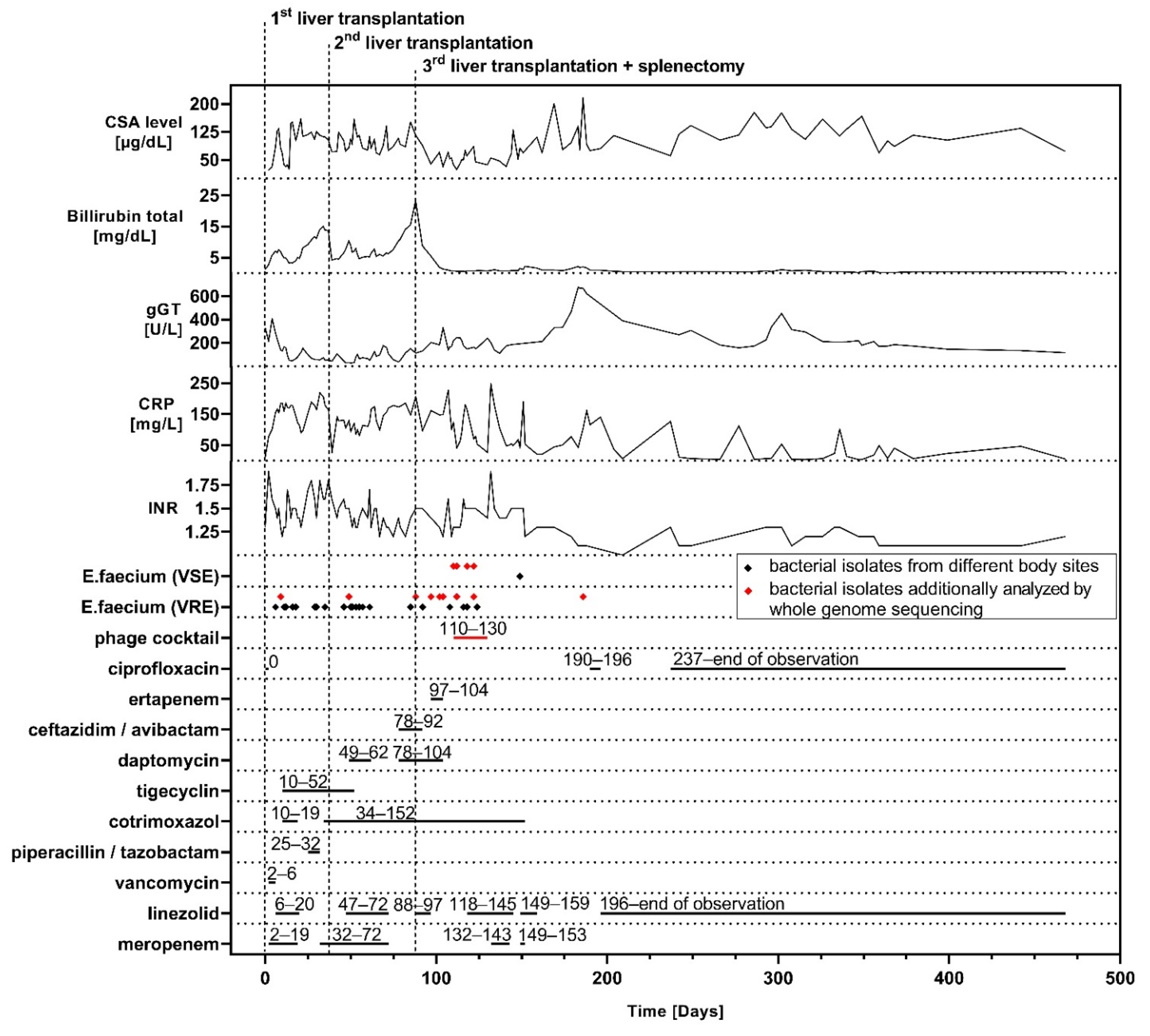
3.1. Case Presentation

The patient first presented to University Medical Center Hamburg—Eppendorf in 2019 at the age of 10 months and had undergone portoenterostomy due to biliary atresia at the age of 8 weeks in Iran.
At that time, her clinical picture was compatible with that of a failed Kasai, showing a severely cirrhotic liver and additional multiple liver abscesses after recurrent cholangitis. She was immediately evaluated for liver transplantation and, due to ongoing deterioration, subsequently received a left lateral liver split five weeks after admission as a high-urgency transplantation. For long-term immunosuppression, cyclosporin A (CSA) and prednisolone were used. Vast intrahepatic bacterial colonization led to a severe systemic infection post transplantation, which was, among other transiently detected bacteria, mainly caused by vancomycin-resistant E. faecium (VREfm), grown from various specimens (i.e., explanted organ, blood cultures, abdominal drainage, bile ducts of the transplanted organ). With extended life support and adaption of antibiotics (Figure 1), stabilization of her vital functions and resolution of the systemic infection was achieved.
However, even repetitive abdominal lavages and revision of the biliodigestive anastomosis did not resolve the VREfm-related abdominal site infection. Liver ischemia reperfusion injury with delayed graft function, together with a critical microvascular oxygen supply during the post-transplant period, resulted in superinfected necrotic areas.
Due to progressive organ damage, and to eradicate the reservoir of the infection, a re-transplantation was needed five weeks after the first transplantation, which was performed with a full organ. Unfortunately, after a temporary improvement in liver function and systemic inflammation, VREfm was repeatedly detected in abdominal swabs, and the patient’s medical condition deteriorated to a level similar to that before the re-transplantation, as demonstrated by blood CRP and bilirubin levels. Multiple antibiotic treatment schemes (Figure 1) and extensive surgical treatment were not sufficient to control the infection. Based on recent reports of successful treatments of refractory infections with bacteriophages in transplant patients [9,11] we explored this option for our patient. Meanwhile, because of terminal transplant organ damage in a mildly ventilated child, without need for vasopressors and a localized infection of the abdomen, listing of the patient for a third liver transplantation was decided after multiple interdisciplinary case conferences and thorough informed consent from the parents. The third transplantation with a split organ was performed one month after the second transplantation together with a splenectomy, since previous imaging studies were highly suggestive for multiple intra-splenic abscesses. Indeed, post-operative bacterial cultures confirmed a severe splenic colonization with VREfm.
The third transplantation plus splenectomy led to a persistent normalization of liver function and cholestasis, but VREfm was still detected in the abdomen, with ongoing elevated CRP levels.
Three weeks after the third liver transplantation (Figure 1 day 110), we applied an individualized two-phage cocktail prepared for use as salvage therapy. The indication of phage therapy was accompanied and approved by the Ethics Committee of the University Medical Center Hamburg–Eppendorf, and it was conducted under the umbrella of article § 37 (Unproven Interventions in Clinical Practice) of the Declaration of Helsinki after expert advice and informed consent of the family. We initiated a ten-day course of 2 mL/kg bodyweight (BW) of the magistral preparation (joint titer 8.1 × 10⁷ PFU/mL in NaCl 0.9%) administered intravenously over two hours, twice daily. To reduce the theoretical risk of an allergic reaction against the phage preparation, an H1-antagonist was administered before every phage application. In addition, the first phage dose was given over a duration of 4 h and after prior administration of methylprednisolone (2 mg/kg BW).
Initiation of phage therapy led to a rapid drop in CRP starting the day after the first dose. However, this was followed by an increase in inflammatory parameters together with an ongoing detection of VREfm in the abdomen and respiratory tract. Associated therewith, quality control of the magistral phage preparation showed a rapid drop of phage titer with one log after 9 days (also see Section 3.2), indicating that the applied phage titers might have been lower than expected and did not reach levels throughout the treatment that were reported as being effective [24]. It was decided to prolong the treatment for another 10 days with a newly produced batch of magistral phage preparation with an increased phage concentration (joint titer 5.2 × 10⁸ PFU/mL in NaCl 0.9%) and administered 2 mL/kg BW twice daily intravenously. Following this, a persistent reduction in baseline CRP together with constant improvement of the clinical status was achieved. During and after treatment, we did not observe any adverse events attributable to phage administration. With the limitation that the intra-abdominal compartment was not accessible after permanent closure of the abdominal wall and removal of all drainages, for the rest of the hospital course, VREfm was not detected in routine screening from rectal and tracheal (via tracheostoma) swabs.
After phage therapy, recovery of the patient was interrupted by a blood stream infection with Klebsiella pneumoniae (Figure 1, days 132–143) followed by a respiratory infection with Enterobacter cloacae (Figure 1, days 149–153). Both infections responded well to an escalation of the antibiotic treatment with meropenem. After five months of mechanical ventilation and unsuccessful attempts prior to phage therapy, the patient was successfully weaned from the respirator. Two weeks later, after a total of 167 days, she was discharged from the pediatric intensive care unit and was transferred to a rehabilitation center shortly after.
Long-term follow up was complicated by sustained intra-abdominal inflammation, which most likely was associated with ischemic-type biliary lesions after liver transplantation, which were present in sonography and represented by elevated gGT levels. This required a long-term oral therapy with empiric ciprofloxacin in combination with linezolid throughout the one year follow up. The latter antibiotic was added after routine screening had detected (one-time) the presence of VREfm in rectal and tracheal swabs. Afterwards, VREfm was not detected in any of the routine screenings, even after administration of linezolid was stopped, until submission of this work.

3.2. Phage Procurement and Quality Standards

Although phage therapy is increasingly identified as a promising tool for the treatment of MDR bacterial infections, the current lack of broadly available commercial (GMP-certified) phage preparations made us reach for a solution that consisted of an international academic collaboration, in order to obtain a personalized phage preparation with an acceptable quality.
An international call for suitable phages was coordinated by IPATH at UC San Diego, and bacterial isolates were sent to responding laboratories. Two lytic phages, EFgrKN (GenBank accession: MW004544.1) and EFgrNG (GenBank accession: MW004545.1) [17], were matched to the patient’s strain at the Israeli Phage Bank (IBP) at the Hebrew University of Jerusalem (Jerusalem, Israel).
Both phages delayed the growth of VREfm by more than 12 h (Supplementary Figure S1, and also as schematical figure in [18]). Additionally, sub-inhibitory concentration of the antibiotics gentamicin and chloramphenicol increased the inhibitory effect of both phages by 20 h and 4 h, respectively. The addition of sub-inhibitory concentration of vancomycin increased the inhibitory effect of phage EFGrKN, but not of EFGrNG, by an additional 10 h. Furthermore, in the case of EFGrKN with vancomycin and gentamycin, almost no regrowth was observed (Supplementary Figures S2 and S3 and also as schematical figure in [18]).
To minimize the risk of adding virulence factors in vivo, the absence of lysogeny was confirmed by BLAST analysis of the phage DNA sequence against known repressor or integrase sequences. In addition, the absence of sequences coding for known virulence factors, submitted to many databases, was tested using Abricate (version 0.8.13, Seemann T. Abricate Github https://github.com/tseemann/abricate (accessed on 30 July 2021)).
For the production of the phages as APIs, purified from the bacterial remnants (e.g., endotoxins) and fit for incorporation in magistral preparations for medical use (incl. intravenously) [20], the two phages were shipped to the Queen Astrid Military Hospital in Brussels (Brussels, Belgium). The produced phage APIs were diluted and aliquoted (magistral preparation) in vials with weight-adjusted dosages and were sent to the treating facility, the Department of Pediatrics at University Medical Center Hamburg Eppendorf (Hamburg, Germany), where they were stored at 4 °C until use.
The stability of the phage magistral preparations stored at 4 °C was evaluated over a 2-month period. The titer of the first batch (8.1 × 10⁷ PFU/mL) dropped with one log after 9 days and kept on decreasing, showing a more than 4 log reduction after 50 days. Thus, this preparation was probably not suitable for therapeutic application, as empirically an effective “therapeutic titer” for phage preparations is considered in the range of 106–108 PFU/mL [24]. The titer of the second batch (5.2 × 10⁸ PFU/mL) appeared to be more stable and dropped with one log PFU/mL after 63 days.

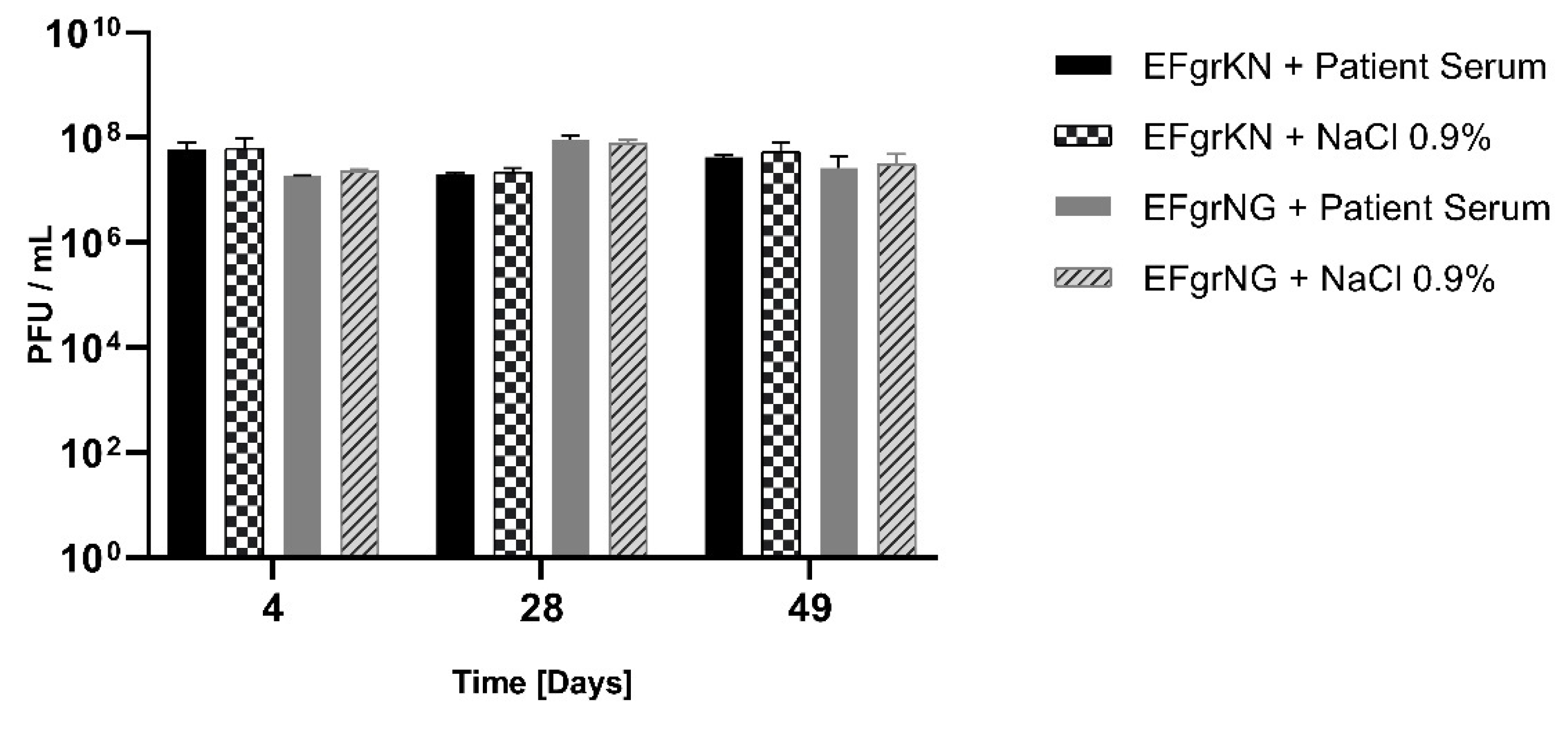
3.3. Humoral Immune Response

The response of the patient’s adaptive immune system towards the applied phages was tested using a classical phage neutralization assay [21] performed on serum samples collected 4, 28 and 49 days after initiation of phage therapy. Neutralizing phage antibodies were not detected (Figure 2). This implies that the phages EFgrKN and EFgrNG did not generate a strong immune response in the patient, particularly by stimulating synthesis of neutralizing antibodies. However, these results must be interpreted in the context of an overall immunosuppressed transplant patient with impaired capability of immunoglobulin synthesis.

3.4. Genetic Characterization of E. faecium Isolates

In vitro data of VREfm indicated a synergistic effect of phage EFGrKN in combination with vancomycin (Supplementary Figure S2). Importantly, over the course of the infection several additional VREfm were isolated from various body sites (Figure 1, Table 1). Shortly after phage application, E. faecium exhibiting a vancomycin-susceptible phenotype was identified (Figure 1, Table 1). Vancomycin-resistant and susceptible isolates all belonged to ST1299, and core genome analysis only found an SNP distance of 4–12, suggesting that all isolates emerged from the same vancomycin-resistant progenitor. In vancomycin-susceptible E. faecium_1–3 and E. faecium_4, a deletion of four contigs comprising 20.8 kb with >99.9% identity to van-cluster carrying E. faecium plasmid sequences (e.g., accession number KX810025.1) was identified, reasonably explaining the observed vancomycin-susceptible phenotype.
These results provide unambiguous genetic evidence for persistence of the same invasive E. faecium clone throughout the infectious course, which also experienced a loss of vancomycin resistance. However, the temporal relationship of phage therapy and van-loss has to be interpreted with caution and might just be coincidental, as van loss is frequently observed in VRE infections, e.g., [25], and the first vancomycin-susceptible isolate was retrieved already on the same day of first phage application.

4. Discussion

Abdominal site infections after portoenterostomy and orthotropic liver transplantation are a common complication [3,4,5], and, especially, post-transplant cholangitis reduces graft survival significantly [6]. Colonization and subsequent infections with VRE in liver transplant patients are overall associated with high mortality, and current antibiotic-based therapeutic strategies to achieve decolonization have low success rates [26]. In animal models, phages were shown to have a high therapeutic efficacy [27,28,29] and significant tissue penetration [30,31]. Taking into account the long history of clinical use in Eastern Europe and recent case reports, phage therapy qualifies as a promising tool in the treatment of MDR bacterial infections. Unfortunately, to date, no commercial phage therapy products have made it to the market. However, with this case we illustrated that the currently established international networks are capable to quickly respond in certain selected cases. In order to meet increasing demands, there is an urgent need for globally commercialized phage products, GMP-certified and tested in randomized controlled trials, but also for an approval of pragmatic manufacturing processes for the production of personalized phage products in combination within a broad collaboration network.

5. Conclusions

After an internationally coordinated effort, phage therapy was applied in a critically ill pediatric patient that had undergone three successive liver transplants and had a persistent VREfm infection. Over a one-year follow up, the treatment was not associated with any adverse events. Although the disease course was complex, clinical improvement was clearly linked to phage application. To our knowledge, we here present the first case of intravenous phage therapy against an infection with E. faecium. The clinical course and the data concerning immune response, phage stability and product safety provide evidence for the potential benefit arising from phage therapy. The recent interest in phage-therapy is driven by the growing number of refractory drug-resistant bacterial infections. However, there is a need for standardized data to analyze efficacy and safety of phage therapy as well as product quality. This would support decision making of health care providers and would help regulatory authorities to license phage products and adapt their regulatory framework to approve manufacturing processes of personalized (adapted) phage products, which would facilitate the use of phage therapy in a broader patient spectrum.

Supplementary Materials

The following are available online at https://0-www-mdpi-com.brum.beds.ac.uk/article/10.3390/v13091785/s1, Figure S1: The inhibition effects of phages and antibiotics on the target bacteria VREfm; Figure S2: Combined effect of phage EFGrKN and antibiotics on the target bacteria VREfm; Figure S3: Combined effect of phage EFGrNG and antibiotics on the target bacteria VREfm.

Author Contributions

Conceptualization, K.P., C.R., D.S. and A.C.M.; methodology, M.M., R.H. and R.N.-P.; validation, J.-P.P., R.H. and R.N.-P.; formal analysis, K.P., M.M., M.C., D.G., L.K., O.Y. and S.C.-G.; investigation, K.P., M.M., M.C., D.G., L.K., O.Y. and S.C.-G.; resources, K.P., R.H., S.A., R.N.-P., J.-P.P., H.R., T.H., S.S.-J., U.H., L.F., D.S. and A.C.M.; data curation, K.P., M.M., M.C., D.G., L.K., O.Y. and S.C.-G.; writing—original draft preparation, K.P., M.M., R.H. and M.C.; writing—review and editing, M.C., U.H., S.A., D.G., L.K., O.Y., S.C.-G., T.H., H.R., J.-P.P., S.S.-J., L.F., C.R., R.N.-P., D.S. and A.C.M.; visualization, K.P. and R.H.; supervision R.H., R.N.-P., J.-P.P., D.S. and A.C.M.; project administration, K.P.; funding acquisition, R.H., R.N.-P. and S.A. All authors have read and agreed to the published version of the manuscript.

Funding

Parts of this work were funded by the US–Israel Binational Science Foundation (grant #2017123), the Israel Science Foundation IPMP (grant #ISF_1349/20), the Rosetrees Trust (grant A2232) and the Milgrom Family Support Program to R.H. and R.N.-P.; S.A. received funding support from the UC San Diego Chancellor’s Innovation Fund.

Institutional Review Board Statement

The indication of phage therapy was accompanied and approved by the Ethics Committee of the University Medical Center Hamburg—Eppendorf.

Informed Consent Statement

Written informed consent has been obtained from the patient’s legal guardians to publish this paper.

Data Availability Statement

Phage Genome Sequences are accessible at GenBank; EFGrKN’s accession number is MW004544.1, and EFGrNG’s accession number is MW004545.1. Raw data are available over the NIH BioSample database project (BioProject number PRJNA706131)/Israeli Phage Bank (IPB), with accession numbers SAMN18137778 for EFGrKN and SAMN18191644 for EFGrNG. Further clinical data are available on request from the corresponding author. The data are not publicly available for protection of the patient’s identity.

Acknowledgments

The authors would like to thank the nurses and the whole medical team for their impressive, persevering professionality with which they provided security and emotional support even in critical situations. We also would like to thank the team from IPATH, which received funding from the Mallory Smith Legacy Fund, for coordinating this world-wide phage hunt. Without that effort, a quick response in such a limited time frame would not have been possible.

Conflicts of Interest

The authors declare no conflict of interest.

References

  1. Lakshminarayanan, B.; Davenport, M. Biliary atresia: A comprehensive review. J. Autoimmun. 2016, 73, 1–9. [Google Scholar] [CrossRef] [PubMed]
  2. Cuenca, A.G.; Kim, H.B.; Vakili, K. Pediatric liver transplantation. Semin. Pediatr. Surg. 2017, 26, 217–223. [Google Scholar] [CrossRef]
  3. Baek, S.H.; Kang, J.M.; Ihn, K.; Han, S.J.; Koh, H.; Ahn, J.G. The Epidemiology and Etiology of Cholangitis After Kasai Portoenterostomy in Patients With Biliary Atresia. J. Pediatr. Gastroenterol. Nutr. 2020, 70, 171–177. [Google Scholar] [CrossRef] [PubMed]
  4. Reid, G.E.; Grim, S.A.; Sankary, H.; Benedetti, E.; Oberholzer, J.; Clark, N.M. Early intra-abdominal infections associated with orthotopic liver transplantation. Transplantation 2009, 87, 1706–1711. [Google Scholar] [CrossRef]
  5. Viehman, J.A.; Clancy, C.J.; Clarke, L.; Shields, R.K.; Silveira, F.P.; Kwak, E.J.; Vergidis, P.; Hughes, C.; Humar, A.; Nguyen, M.H. Surgical Site Infections after Liver Transplantation: Emergence of Multidrug-Resistant Bacteria and Implications for Prophylaxis and Treatment Strategies. Transplantation 2016, 100, 2107–2114. [Google Scholar] [CrossRef] [PubMed]
  6. Yao, S.; Yagi, S.; Nagao, M.; Uozumi, R.; Iida, T.; Iwamura, S.; Miyachi, Y.; Shirai, H.; Kobayashi, A.; Okumura, S.; et al. Etiologies, risk factors, and outcomes of bacterial cholangitis after living donor liver transplantation. Eur. J. Clin. Microbiol. Infect. Dis. 2018, 37, 1973–1982. [Google Scholar] [CrossRef] [PubMed]
  7. De Oliveira, D.M.P.; Forde, B.M.; Kidd, T.J.; Harris, P.N.A.; Schembri, M.A.; Beatson, S.A.; Paterson, D.L.; Walker, M.J. Antimicrobial resistance in ESKAPE pathogens. Clin. Microbiol. Rev. 2020, 33, e00181-19. [Google Scholar] [CrossRef]
  8. Sulakvelidze, A.; Alavidze, Z.; Morris, G.J. Minireview Bacteriophage Therapy. Antimicrob. Agents Chemother. 2001, 45, 649–659. [Google Scholar] [CrossRef] [PubMed] [Green Version]
  9. Aslam, S.; Courtwright, A.M.; Koval, C.; Lehman, S.M.; Morales, S.; Furr, C.L.L.; Rosas, F.; Brownstein, M.J.; Fackler, J.R.; Sisson, B.M.; et al. Early clinical experience of bacteriophage therapy in 3 lung transplant recipients. Am. J. Transplant. 2019, 19, 2631–2639. [Google Scholar] [CrossRef]
  10. Aslam, S.; Lampley, E.; Wooten, D.; Karris, M.; Benson, C.; Strathdee, S.; Schooley, R.T. Lessons learned from the first 10 consecutive cases of intravenous bacteriophage therapy to treat multidrug-resistant bacterial infections at a single center in the United States. Open Forum Infect. Dis. 2020, 7, ofaa389. [Google Scholar] [CrossRef]
  11. Dedrick, R.M.; Guerrero-Bustamante, C.A.; Garlena, R.A.; Russell, D.A.; Ford, K.; Harris, K.; Gilmour, K.C.; Soothill, J.; Jacobs-Sera, D.; Schooley, R.T.; et al. Engineered bacteriophages for treatment of a patient with a disseminated drug-resistant Mycobacterium abscessus. Nat. Med. 2019, 25, 730–733. [Google Scholar] [CrossRef]
  12. Nir-Paz, R.; Gelman, D.; Khouri, A.; Sisson, B.M.; Fackler, J.; Alkalay-Oren, S.; Khalifa, L.; Rimon, A.; Yerushalmy, O.; Bader, R.; et al. Successful Treatment of Antibiotic-resistant, Poly-microbial Bone Infection with Bacteriophages and Antibiotics Combination. Clin. Infect. Dis. 2019, 69, 2015–2018. [Google Scholar] [CrossRef]
  13. Theuretzbacher, U.; Piddock, L.J.V. Non-traditional Antibacterial Therapeutic Options and Challenges. Cell Host Microbe 2019, 26, 61–72. [Google Scholar] [CrossRef] [PubMed]
  14. Pirnay, J.P.; De Vos, D.; Verbeken, G. Clinical application of bacteriophages in Europe. Microbiol. Aust. 2019, 40, 8–15. [Google Scholar] [CrossRef] [Green Version]
  15. Merabishvili, M.; Pirnay, J.P.; Verbeken, G.; Chanishvili, N.; Tediashvili, M.; Lashkhi, N.; Glonti, T.; Krylov, V.; Mast, J.; Van Parys, L.; et al. Quality-controlled small-scale production of a well-defined bacteriophage cocktail for use in human clinical trials. PLoS ONE 2009, 4, e4944. [Google Scholar] [CrossRef] [PubMed]
  16. Yerushalmy, O.; Khalifa, L.; Gold, N.; Rakov, C.; Alkalay-Oren, S.; Adler, K.; Ben-Porat, S.; Kraitman, R.; Gronovich, N.; Ginat, K.S.; et al. The israeli phage bank (IPB). Antibiotics 2020, 9, 269. [Google Scholar] [CrossRef] [PubMed]
  17. Alkalay-Oren, S.; Gold, N.; Khalifa, L.; Yerushalmy, O.; Coppenhagen-Glazer, S.; Nir-Paz, R.; Hazan, R. Complete Genome Sequences of Two Enterococcus faecalis Bacteriophages, EFGrKN and EFGrNG, Targeted to Phage Therapy. Microbiol. Resour. Announc. 2021, 10, 20–22. [Google Scholar] [CrossRef]
  18. Gelman, D.; Yerushalmy, O.; Ben-Porat, S.; Rakov, C.; Alkalay-Oren, S.; Adler, K.; Khalifa, L.; Abdalrhman, M.; Coppenhagen-Glazer, S.; Aslam, S. Clinical Phage Microbiology: A suggested framework and recommendations for the in-vitro matching steps of phage therapy. Lancet Microbe bioRxiv 2021. [Google Scholar] [CrossRef]
  19. Yerushalmy, O.; Coppenhagen-Glazer, S.; Nir-Paz, R.; Tuomala, H.; Skurnik, M.; Kiljunen, S.; Hazan, R. Complete Genome Sequences of Two Klebsiella pneumoniae Phages Isolated as Part of an International Effort. Microbiol. Resour. Announc. 2019, 8, 1–3. [Google Scholar] [CrossRef] [PubMed] [Green Version]
  20. Pirnay, J.P.; Verbeken, G.; Ceyssens, P.J.; Huys, I.; de Vos, D.; Ameloot, C.; Fauconnier, A. The magistral phage. Viruses 2018, 10, 64. [Google Scholar] [CrossRef] [Green Version]
  21. Adams, M.H. Methods of study of bacterial viruses. In Bacteriophages; Interscience Publishers, Inc.: New York, NY, USA, 1959; pp. 443–523. [Google Scholar]
  22. Olearo, F.; Both, A.; Belmar Campos, C.; Hilgarth, H.; Klupp, E.M.; Hansen, J.L.; Maurer, F.P.; Christner, M.; Aepfelbacher, M.; Rohde, H. Emergence of linezolid-resistance in vancomycin-resistant Enterococcus faecium ST117 associated with increased linezolid-consumption. Int. J. Med. Microbiol. 2021, 311, 151477. [Google Scholar] [CrossRef] [PubMed]
  23. Tritt, A.; Eisen, J.A.; Facciotti, M.T.; Darling, A.E. An Integrated Pipeline for de Novo Assembly of Microbial Genomes. PLoS ONE 2012, 7, e42304. [Google Scholar] [CrossRef] [PubMed] [Green Version]
  24. Merabishvili, M. Production of bacteriophages using bacterial suspension cultures for phage-therapy. In Industrial Scale Suspension Culture of Living Cells; Meyer, H.P., Schmidhalter, D.R., Eds.; Wiley-VCH Verlag: Weinheim, Germany, 2014; pp. 537–543. [Google Scholar]
  25. Chacko, K.I.; Sullivan, M.J.; Beckford, C.; Altman, D.R.; Ciferri, B.; Pak, T.R.; Sebra, R.; Kasarskis, A.; Hamula, C.L.; Van Bakel, H. Genetic basis of emerging vancomycin, linezolid, and daptomycin heteroresistance in a case of persistent Enterococcus faecium bacteremia. Antimicrob. Agents Chemother. 2018, 62, e02007-17. [Google Scholar] [CrossRef] [Green Version]
  26. Belga, S.; Chiang, D.; Kabbani, D.; Abraldes, J.G.; Cervera, C. The direct and indirect effects of vancomycin-resistant enterococci colonization in liver transplant candidates and recipients. Expert Rev. Anti. Infect. Ther. 2019, 17, 363–373. [Google Scholar] [CrossRef] [PubMed]
  27. Gelman, D.; Beyth, S.; Lerer, V.; Adler, K.; Poradosu-Cohen, R.; Coppenhagen-Glazer, S.; Hazan, R. Combined bacteriophages and antibiotics as an efficient therapy against VRE Enterococcus faecalis in a mouse model. Res. Microbiol. 2018, 169, 531–539. [Google Scholar] [CrossRef] [PubMed]
  28. Cheng, M.; Liang, J.; Zhang, Y.; Hu, L.; Gong, P.; Cai, R.; Zhang, L.; Zhang, H.; Ge, J.; Ji, Y.; et al. The bacteriophage EF-P29 efficiently protects against lethal vancomycin-resistant enterococcus faecalis and alleviates gut microbiota imbalance in a murine bacteremia model. Front. Microbiol. 2017, 8, 837. [Google Scholar] [CrossRef]
  29. Biswas, B.; Adhya, S.; Washart, P.; Paul, B.; Trostel, A.N.; Powell, B.; Carlton, R.; Merril, C.R. Erratum: Bacteriophage therapy rescues mice bacteremic from a clinical isolate of vancomycin-resistant Enterococcus faecium (Infection and Immunity (2002) 70:1 (204–210)). Infect. Immun. 2002, 70, 1664. [Google Scholar] [CrossRef] [Green Version]
  30. Rakhuba, D.V.; Kolomiets, E.I.; Szwajcer Dey, E.; Novik, G.I. Bacteriophage receptors, mechanisms of phage adsorption and penetration into host cell. Pol. J. Microbiol. 2010, 59, 145–155. [Google Scholar] [CrossRef]
  31. Huh, H.; Wong, S.; St. Jean, J.; Slavcev, R. Bacteriophage interactions with mammalian tissue: Therapeutic applications. Adv. Drug Deliv. Rev. 2019, 145, 4–17. [Google Scholar] [CrossRef]
Figure 1. Time course of major operations, representative lab values, detection of vancomycin susceptible (VSE) and resistant (VRE) E. faecium from different sites as well as antibacterial treatment starting from the day of the first liver transplantation. Phages were administered over ten days of 2 mL/kg BW as a magistral preparation (joint titer first batch 8.1 × 10⁷ PFU/mL in NaCl 0.9%) followed directly by another ten days of 2 mL/kg BW (joint titer second batch 5.2 × 10⁸ PFU/mL in NaCl 0.9%) twice daily. Exact days of treatments, where the date of the first liver transplantation is defined as day 0, are plotted above each timeline, and isolates that were further analyzed by whole-genome sequencing are highlighted.
Viruses 13 01785 g001
Figure 2. Phage neutralization assay was performed in triplicate on serum samples of the patient collected on days 4, 28 and 49 after initiation of phage therapy with the phages EFgrKN and EFgrNG. The resulting phage activity, after incubation with the serum samples, was expressed in PFU/mL and compared to a control consisting of phage particles in NaCl 0.9%, after incubation at 37 °C for 30 min. Results are plotted as mean ± SD. At no time point could relevant differences in phage titers, which would indicate neutralizing activity of patient serum, be observed.
Viruses 13 01785 g002
Table 1. VREfm and E. faecium isolates identified before and after phage therapy.
Isolate IDIsolation SiteIsolation Relative to Phage Application (Days)MIC 1MLST 2
Vancomycin (mg/L)Sequence Type
VREfm_1Blood culture−101>2561299
VREfm_2Central venous−61>2561299
catheter
VREfm_3Abdominal swab−21>2561299
VREfm_4Abdominal swab−12>2561299
VREfm_5Abdominal drainage−7>2561299
VREfm_6Upper respiratory tract swab−7>2561299
VREfm_7Intraoperative swab−5>2561299
E. faecium_1Abdominal drainage021299
E. faecium _2Abdominal drainage221299
E. faecium_3Abdominal drainage821299
E. faecium _4Abdominal drainage1021299
VREfm_8Abdominal drainage10>2561299
VREfm_9Inguinal swab76>2561299
1 MIC—minimum inhibitory concentration. 2 MLST—multi-locus sequence typing.
Publisher’s Note: MDPI stays neutral with regard to jurisdictional claims in published maps and institutional affiliations.

