Article

Pathophysiology of Influenza D Virus Infection in Specific-Pathogen-Free Lambs with or without Prior Mycoplasma ovipneumoniae Exposure

1
Department of Microbiology and Cell Biology, Montana State University, 2155 Analysis Drive, Bozeman, MT 59718, USA
2
Montana State Veterinary Diagnostic Laboratory, 1911 West Lincoln Street, Bozeman, MT 59718, USA
3
Division of Basic Biomedical Sciences, Sanford School of Medicine, University of South Dakota, Vermillion, SD 57069, USA
*
Author to whom correspondence should be addressed.
Submission received: 21 April 2022 / Revised: 23 June 2022 / Accepted: 27 June 2022 / Published: 28 June 2022
(This article belongs to the Special Issue Non-A Influenza 2.0)

Abstract

:
Polymicrobial pneumonias occur frequently in cattle, swine, and sheep, resulting in major economic losses. Individual pathogens comprising these complex infections may be mild on their own but can instead exhibit synergism or increase host susceptibility. Two examples of such pathogens, Mycoplasma ovipneumoniae (M. ovipneumoniae) and influenza D viruses (IDVs), naturally infect domestic sheep. In sheep, the role of M. ovipneumoniae in chronic nonprogressive pneumonia is well-established, but the pathogenesis of IDV infection has not previously been studied. We utilized a specific-pathogen-free sheep flock to study the clinical response to IDV infection in naïve vs. M. ovipneumoniae-exposed lambs. Lambs were inoculated intranasally with M. ovipneumoniae or mock infection, followed after four weeks by infection with IDV. Pathogen shedding was tracked, and immunological responses were evaluated by measuring acute phase response and IDV-neutralizing antibody titers. While lamb health statuses remained subclinical, M. ovipneumoniae-exposed lambs had significantly elevated body temperatures during IDV infection compared to M. ovipneumoniae-naïve, IDV-infected lambs. Moreover, we found a positive correlation between prior M. ovipneumoniae burden, early-infection IDV shedding, and IDV-neutralizing antibody response. Our findings suggest that IDV infection may not induce clinical symptoms in domestic sheep, but previous M. ovipneumoniae exposure may promote mild IDV-associated inflammation.

1. Introduction

Polymicrobial pneumonias in cattle, swine, and sheep result in major annual economic losses in the livestock industry. It is hypothesized that during these complex infections, the primary pathogen may drive disease severity by remodeling or suppressing the host immune response, thereby facilitating infection by additional pathogens and/or limiting the host’s ability to combat the new infections [1,2,3,4,5,6]. Understanding the immunological processes driving these events is critical for effective prevention and management. Our group has investigated influenza-associated polymicrobial infections and identified several processes by which influenza A virus infection modulates host susceptibility to subsequent bacterial secondary infection [7,8,9]. We subsequently extended these investigations to include influenza D virus (IDV)-Staphylococcus aureus sequential infection using a mouse model [10]. In the current study we utilized a recently developed specific-pathogen-free (SPF) sheep flock to investigate the impact of prior Mycoplasma ovipneumoniae (M. ovipneumoniae) infection on IDV pathogenesis in a natural host [11].
M. ovipneumoniae is a respiratory bacterium commonly detected in healthy and diseased lambs [12,13,14]. M. ovipneumoniae infection can induce chronic nonprogressive pneumonia, particularly in 2- to 12-month-old lambs [15,16,17,18]. While its prevalence in healthy domestic lambs demonstrates that M. ovipneumoniae infection can be asymptomatic, evidence suggests that asymptomatic carriage may reduce lamb growth rates [19,20,21,22]. Importantly, while domestic sheep tolerate M. ovipneumoniae infection, wild sheep species such as Rocky Mountain Bighorn sheep (Ovis canadensis) and argali (Ovis ammon) are highly susceptible to M. ovipneumoniae infection, which predisposes them to severe-to-lethal polymicrobial interstitial pneumonia [19,20,21,23,24,25]. The mechanisms through which M. ovipneumoniae promotes secondary infection are not fully understood. Ex vivo studies have demonstrated that M. ovipneumoniae damages tracheal epithelium through the generation of reactive oxygen species, which may increase susceptibility to a secondary pathogen [4,26]. M. ovipneumoniae also induces ciliostasis [27], promotes anti-ciliary autoantibody production [28], and may compromise the mucociliary escalator and enable the migration of inert upper respiratory tract resident microbes such as Mannheimia haemolytica into the lower respiratory tract, which can result in severe interstitial pneumonia [23]. In the lower respiratory tract, M. ovipneumoniae reduces the phagocytic capacity of alveolar macrophages and impairs pathogen clearance [5,29], while stimulating proinflammatory cytokine secretion, which damages host tissue [4,30]. M. ovipneumoniae may also impair the adaptive immune response as it bears a surface protein that directly inhibits mitogen-stimulated expansion of T-cells and B-cells [31,32]. Each of these mechanisms may confer an immunosuppressed state and contribute to the increased disease severity associated with M. ovipneumoniae colonization.
IDVs comprise a recently identified genus of Orthomyxoviridae about which little is known [33,34]. The natural host range is broad, including (in order of known seroprevalence) livestock like cattle (40–95%) [35,36,37,38], swine (6–12%) [33,39,40], horses (12%) [41], sheep (2–6%) [38,39,42,43,44], goats (1–4%) [38,42,44,45], dromedary camels (6–99%, possible cross-reactivity with influenza C virus) [44,46], as well as wild ungulates including feral swine [40,47] and water buffalo [45]. There is also serological evidence of human infection [33,48,49,50], and experimental infections have been carried out in model organisms of the human respiratory tract (mice [10,51], guinea pigs [52], and ferrets [33,53]). In most species, IDV primarily infects ciliated cells of the upper respiratory tract and soft palate [4,33,53,54]. Cellular tropism varies between species due in part to differences in respiratory system glycosylation patterns [54,55]. IDV infections depend on docking of the major surface glycoprotein (hemagglutinin-esterase fusion protein) to extracellular 9-O-acetylated sialic acid residues, and as such, cellular permissivity is dependent on the presence of these modified glycans [56,57]. IDV infections typically remain subclinical [35,53,58,59,60,61], but metagenomics studies suggest that IDVs may play an etiologic role in a bovine respiratory disease (BRD), a severe-to-lethal polymicrobial infection common in pre- and post-weaned calves [35,62,63]. In cattle, IDVs infect both the upper and lower respiratory tract and can cause mild-to-moderate respiratory symptoms [53,58,61,64]; however, respiratory tissue tropism has not been established for most hosts, including sheep. Until now, active IDV infection has never been documented in sheep, but serostudies in the United States [42], Ireland [43], France [38], and West Africa [39,44] report IDV-neutralizing antibodies in domestic sheep, suggesting that IDV infections in sheep occur commonly across multiple continents.
Despite evidence of circulation of both M. ovipneumoniae and IDV in sheep, no studies have characterized infections involving both pathogens. Our previous work and the work of others suggests that M. ovipneumoniae monoinfection does not consistently induce clinical illness in SPF lambs [11,19,65,66]. To date, experimental coinfection of an IDV with a bacterial pathogen has been limited to two studies in calves and one in mice [10,58,64]. In the current study, we infected SPF lambs with M. ovipneumoniae or mock treatment. After four weeks, we infected all animals with IDV to characterize IDV pathogenesis with and without a recent or ongoing M. ovipneumoniae infection. We found that on their own, both M. ovipneumoniae and IDV infections were asymptomatic. While we observed no significant difference in overt respiratory disease or shedding when M. ovipneumoniae-infected versus naïve lambs were inoculated with IDV, we found subclinical (<40 °C) elevation in rectal temperature, as well as elevated serum protein concentrations in lambs exposed to both pathogens. Our data also allowed for correlative analysis which suggests that the amount of recent M. ovipneumoniae burden may correspond to the degree of early-phase IDV replication and the endpoint IDV-neutralizing antibody titer. Because no causal relationship has been established, these data must be confirmed by additional experiments.

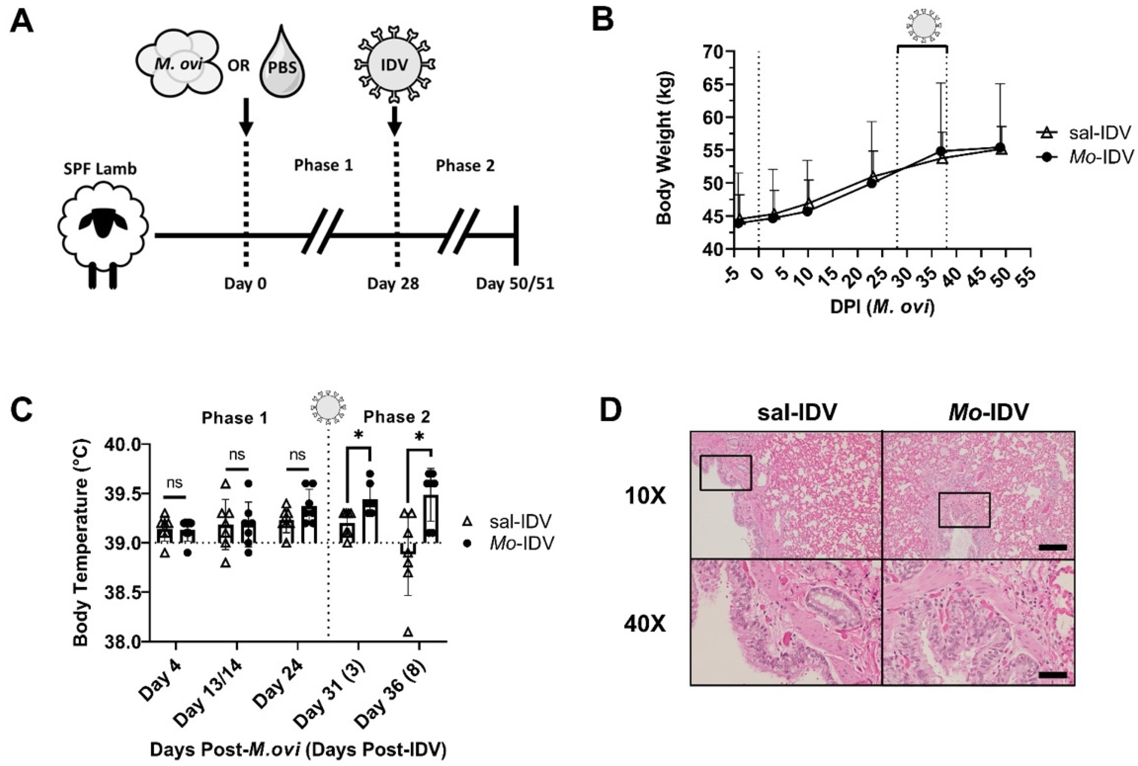
2. Materials and Methods

2.1. Animals and Husbandry

All procedures in this study were approved by the Institutional Animal Care and Use Committee (IACUC) of Montana State University, protocol# 2019-109. We previously reported the derivation of a SPF sheep flock through established motherless rearing methods using mixed-breed (Rambouillet/Targhee) founders (F0) [11]. The SPF flock was maintained under SPF conditions at the Johnson Family Livestock Facility (JFLF) in Bozeman, MT. Of the F1 SPF sheep, 2 rams and 11 ewes were bred, and of the resulting progeny (F2), 3 ewes and 4 wethers were selected for each experimental group in this study (n = 7 lambs/group).
Four days prior to the study start date, 12–15-week-old lambs were weaned and moved into a temperature-controlled room (15.5–16.8 °C) in the JFLF ABSL-2 facility for acclimation. Experimental groups were housed in different rooms, separated by a procedure area. To minimize the possibility of cross-contact or accidental exposures, all personnel showered and donned sterile personal protective equipment before and after entering either room. All protocols were performed on the M. ovipneumoniae-naïve group prior to the M. ovipneumoniae-inoculated group. Equipment was sterilized between experimental groups and, when possible, separate sets of procedural equipment were used for each group.

2.2. Health Monitoring

Lamb health status was monitored daily, and combined clinical scores were assigned daily using previously established metrics [11]. To summarize, lambs were scored in five categories: (1) behavior, (2) appetite, (3) respiratory symptoms, (4) clinical interventions, and (5) body temperature (measured rectally). Each category uses a scale from 0 to 5 with higher scores indicating a worsening state, giving a maximum possible combined clinical score of 25. Lambs were also weighed every 14 days. Trained animal husbandry staff examined lambs twice daily and monitored them by video feed throughout each day.

2.3. M. ovipneumoniae Preparation

M. ovipneumoniae inoculum was grown from a previously described nasal wash isolate (MSU NW-4) and expanded exactly as described in the original publication [11]. Briefly, inoculum was grown at 37 °C in Mycoplasma broth under microaerophilic conditions. On the day of inoculation, media was removed (10,000× g, 10 min, 4 °C) and culture was resuspended in sterile FACS buffer (2% fetal bovine serum and 0.1% sodium azide in DPBS) for counting by flow cytometry [67]. Cells were subsequently stained with SYBR Safe DNA Gel Stain (Invitrogen, Carlsbad, CA, USA) at 4 °C for 30 min and Absolute Counting Beads (Invitrogen, Carlsbad, CA, USA) were added to solution immediately prior to analysis using an Accuri C6 flow cytometer (BD Biosciences, Franklin Lakes, NJ, USA).

2.4. Virus Preparation

Influenza D virus (D/swine/Oklahoma/1334/2011) was propagated in embryonated chicken eggs, and inoculum titer was determined by plaque assay as previously described with modifications [68,69]. On the day of inoculation, an aliquot of inoculum was serially diluted in 1X inoculation media (1X MEM, 0.3% bovine serum albumin, 2 mM HEPES, with penicillin/streptomycin) containing 1 µg/mL TPCK-trypsin (Thermo Fisher Scientific, Waltham, MA, USA). Dilutions were plated in duplicate onto 90–100% confluent monolayers of Madin-Darby Canine Kidney cells (MDCK, ATCC# NBL-2), a kind gift from Dr. Benfeng Lei. After a 1 h infection period at 33 °C, cells were washed, and a semisolid overlay was added (1X inoculation media, 1 µg/mL TPCK-trypsin (Thermo Fisher Scientific, Waltham, MA, USA), 0.6% agarose, 100 µg/mL DEAE-dextran (Sigma Aldrich, St. Louis, MO, USA)). Plates incubated for 5 days at 33 °C. Plaques were visualized by tetrazolium dye vital staining by dispensing 0.5 mL dye solution (3 mg/mL 3-(4,5-Dimethylthiazol-2-yl)-2,5-Diphenyltetrazolium Bromide (MTT, Thermo Fisher Scientific, Waltham, MA, USA) in 150 mM NaCl) on top of each overlay as previously described [70]. Cells were incubated with the dye for 60 min at 37 °C, 5% CO2, or until blue puncta developed and plaques were enumerated immediately.
IDV for hemagglutinin inhibition assays was propagated in swine testicular cells (ST, ATCC# CRL-1746, a kind gift from Dr. Feng Li). Upon reaching 60% confluence, cells were inoculated (MOI = 0.1) in a minimal volume of inoculation media at 33 °C, 5% CO2 for 1 h. Cells were then cultured for 5 days at 37 °C, 5% CO2 in inoculation media with TPCK-trypsin reduced to 0.1 µg/mL. After outgrowth period, media was collected and virus was concentrated by ultracentrifugation over a 30% sucrose cushion at 100,000× g, 90 min, 4 °C. Pellets were resuspended in sterile DPBS and stored at −80 °C until use.

2.5. Intranasal Inoculations

Each experimental group consisted of 3 ewes and 4 wethers aged 12–15 weeks old. Among the entire cohort were 3 sets of twins, which were distributed evenly across groups. On the first day of the study, lambs were inoculated with either sterile DPBS (n = 7) or 3.5 × 10⁸ CFU M. ovipneumoniae (n = 7) by instilling 15 mL into each naris and 10 mL orally similar to what has previously been described [11,25,71,72,73]. On day 28 of the study, when in our previous study the peak M. ovipneumoniae infection occurred in SPF sheep [11], all lambs were inoculated intranasally with 1.86 × 10⁵ PFU egg grown IDV in DPBS as previously described, with modifications [33,74].

2.6. Sampling

2.6.1. Nasal and Rectal Swabs

At indicated timepoints, either both nares or rectum were swabbed, and swabs were transported in DPBS (2 mL). Swabs were vortexed briefly and transport media was aliquoted and stored at −20 °C until use.

2.6.2. Serum Collection

At the stated timepoints, whole blood was collected by jugular venipuncture using BD Vacutainer serum tubes (Becton, Dickson and Company, Franklin Lakes, NJ, USA) and serum was separated by centrifugation at 1000× g, 10 min, 4 °C. Aliquots were stored at −20 °C until use.

2.6.3. Fecal Samples

Fecal samples were obtained directly from the rectum and were transported on ice. Samples (80–120 mg) were subsequently homogenized in 500 µL TRIzolTM Reagent (Invitrogen, Carlsbad, CA, USA) using a bead beater, then stored at −80 °C until time of RNA extraction.

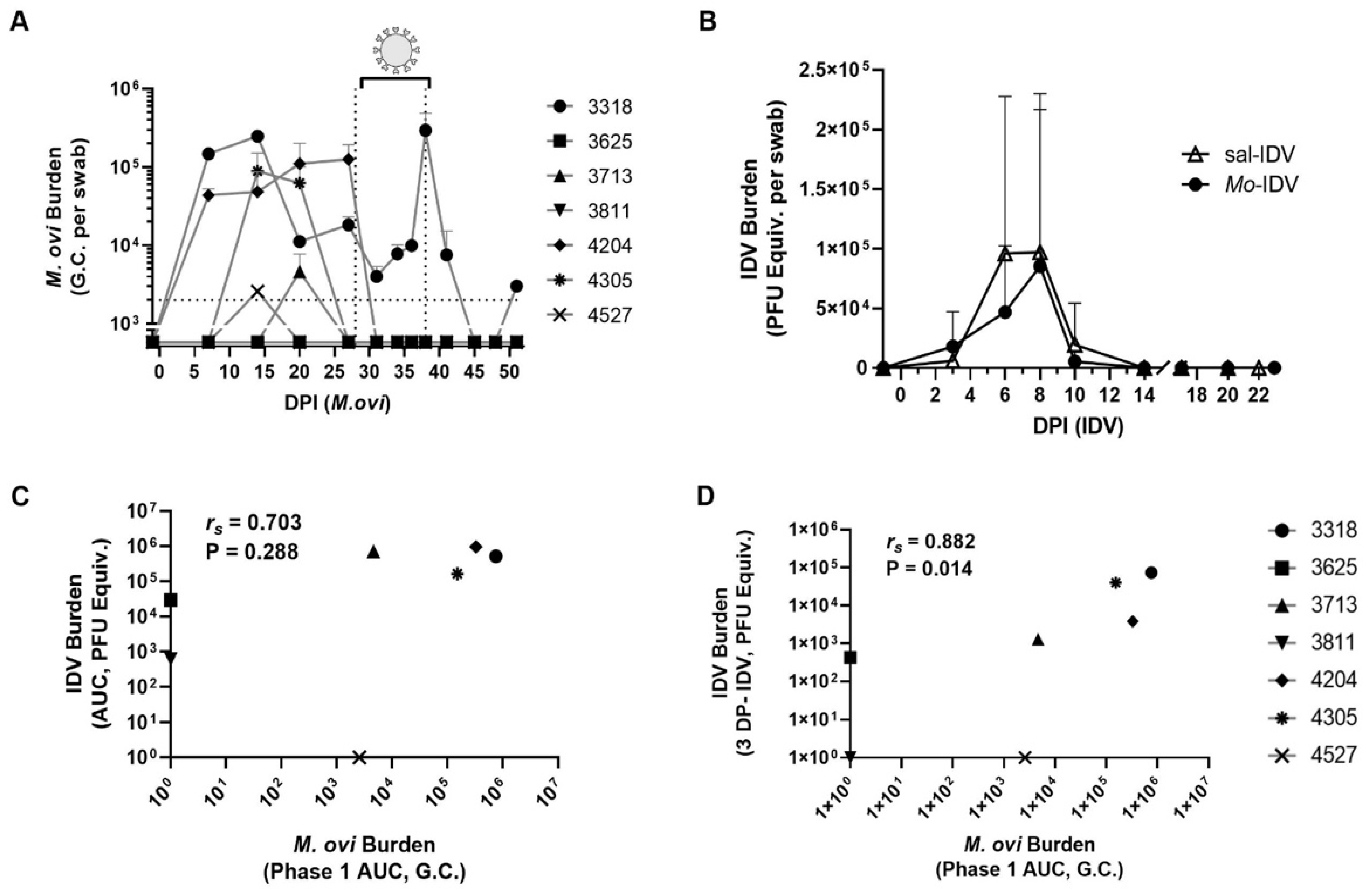
2.7. Determining Pathogen Burden

2.7.1. M. ovipneumoniae Quantification

DNA was extracted from nasal swab samples (500 µL) using the DNeasy Ultraclean Microbial Kit (Qiagen, Redwood City, CA, USA), with modifications. Briefly, cells were pelleted at 10,000× g for 10 min, resuspended in lysis buffer, and incubated at 95 °C for 10 min prior to proceeding with the remainder of the manufacturer’s protocol. Each sample was eluted in 50 µL of provided elution buffer. M. ovipneumoniae burden was determined by qPCR using SsoAdvanced Universal SYBR Green Supermix (Bio-Rad Laboratories, Hercules, CA, USA) and the following previously validated primers [75]: Fwd: 5′-TCTCCCAGATGATGCTAACC-3′; Rev: 5′-TGAAAATCAACTGGTCTAA-3′. Samples were volume-normalized using 0.5 µL DNA per reaction. Genome copy number was interpolated from a standard curve using a fragment of the p113 gene (Twist Bioscience, San Francisco, CA, USA; sequence listed below). A standard curve was analyzed in each sample plate. Thermal cycling reactions proceeded in 10 µL volumes using a CFX384 Touch Real-Time PCR Detection System (Bio-Rad Laboratories, Hercules, CA, USA) with the following cycling conditions: 3-min polymerase activation at 98 °C followed by 40 cycles of denaturation for 15 s at 98 °C and annealing/extension for 30 s at 60 °C.
p113 fragment sequence (5′->3′): AGCTACCAAGTCGACATTCTAGAAATTCTCCCAGATGATGCTAACCAAAATTTT AAAGTCAAATTTCAAGCTAGCCAAAAATTAGCAAACGGTGACATCGCCAAATCT GACATTTATGAACAAGTTGTTTCTTTTGTCAAAGAATCAACTATTTTAATTGCCGA ATTTAATTTTTCCTTACAAAAAATTACAAGCAGACTTAATCAACAAGTCCAAAAT TTAATTTCTGCTCGAACCGCCAATTTTGCTGATCAAAATTCAGCTACTTCAAATC CAACAGATCCTAGCACAATTAGACCAGTTGATTTTCAACATGACTTAAGAATTC ATAAAGCAAA

2.7.2. IDV Quantification

Viral RNA was extracted from nasal and rectal swabs (140 µL) with the QIAamp Viral RNA Mini Kit (Qiagen, Redwood City, CA, USA) as directed, using the provided carrier RNA, and eluted in 60 µL nuclease-free water. Fecal samples were processed exactly as previously described [76]. Blood samples (100 µL) were processed using the PureLink™ RNA Mini Kit (Invitrogen, Carlsbad, CA, USA) according to the manufacturer’s instructions. All RNA was reverse-transcribed using High-Capacity cDNA Reverse Transcriptase Kit (Applied BiosciencesTM, Cheshire, UK) using 10 µL RNA extract per reaction. Prior to IDV quantification, successful RNA extraction was confirmed using qRT-PCR to probe for pan-eukarya 18S rRNA (Hs99999901_s1; Applied Bioscience, Cheshire, UK). Extractions with Cq > 25 were excluded from analysis and extraction was repeated. IDV burden was determined by qRT-PCR using SsoAdvanced Universal Probes Supermix (Bio-Rad Laboratories, Hercules, CA, USA) and the following previously validated FAM-labeled primers targeting the segment encoding PB1 [76]: Fwd: 5′-CAGCTGCGATGTCTGTCATAAG-3′; Rev: 5′-ACAAATTCGCAGGGCCATTA-3′; Probe: 5′-FAM-AATGGACTTTCTCCTGGGACTGCT-TAMRA-3′. To generate a standard curve for interpolation of PFU equivalents (PFU Equiv.), IDV inoculum was serially diluted in sterile DPBS and RNA was extracted from each dilution as described for nasal and rectal swabs. A plaque assay was performed in parallel as described above to confirm the inoculum concentration. cDNA libraries were generated as described above and the resulting standard curve was analyzed in each sample plate. Thermal cycling reactions proceeded as for M. ovipneumoniae except with the following cycling conditions: 30 s polymerase activation at 95 °C followed by 40 cycles of denaturation for 15 s at 95 °C and annealing/extension for 30 s at 60 °C.

2.8. Hemagglutinin Inhibition (HI) Assay

IDV neutralizing antibody titers were determined by HI as previously described, with minor modifications [77]. Briefly, serum was treated with receptor-destroying enzyme II (Denka Seiken, Tokyo, Japan) at a 3:1 ratio for 18 h at 37 °C, followed by enzyme inactivation at 56 °C for 30 min. Sera were diluted to 0.1X in PBS and stored at −20 °C until use. HI was performed on sera diluted in series from 1:10 to 1:1280 using a 1% suspension of washed chicken red blood cells (Lampire Biological Laboratories, Pipersville, PA, USA) and in the presence of 4 HAUs of ST-grown IDV. The agglutination reaction proceeded for 45 min at room temperature in V-bottom plates and titers were read by tilting the plate 90° for 30 s. Viral HA activity was determined just prior to test and confirmed with an in-plate dilution series. Tests were performed on two independent days, twice each day. Partially agglutinated wells were considered agglutinated. Titers are reported as inverse dilution, and sera with titers < 10 were assigned a titer of 5 for statistical analysis and graphical representation.

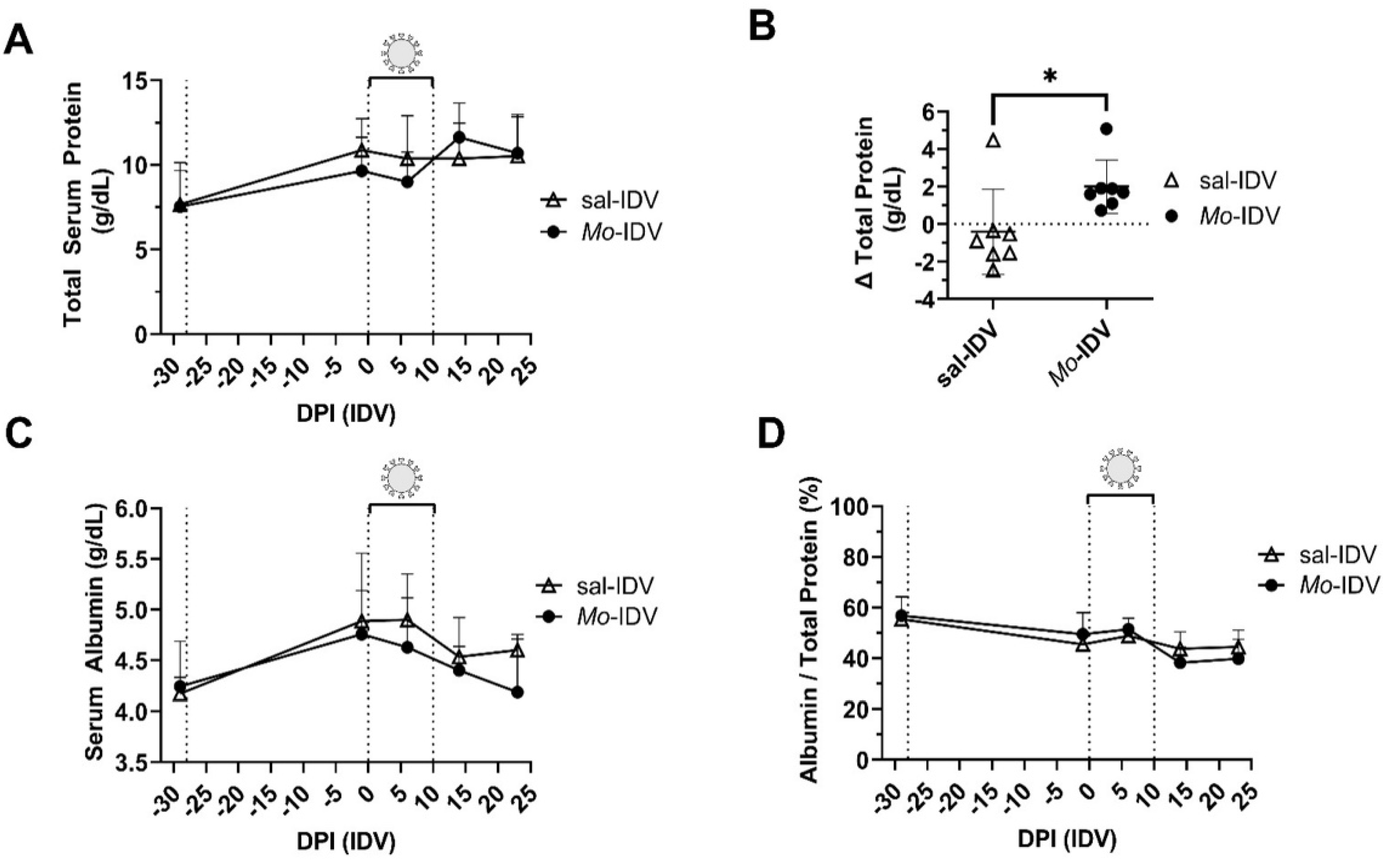
2.9. Acute Phase Protein and L-Lactate Quantification

Total protein was measured by BCA assay as directed (Thermo Fisher Scientific, Waltham, MA, USA). Albumin was determined using a modified bromocresol green (BCG) protocol. Briefly, 50 µL BCG reagent (Eagle Diagnostics, De Soto, TX, USA) was combined with 50 µL serum diluted appropriately in 0.9% saline. Reaction incubated 15 min at room temperature before measuring absorbance at 630 nm. Each test plate included a 125–1500 µg/mL standard curve using a commercially-available 2 mg/mL bovine serum albumin standard (Thermo Fisher Scientific, Waltham, MA, USA). Serum amyloid A was measured using a commercially available ELISA (PHASE™ RANGE Multispecies SAA ELISA kit, Tridelta Development, Ltd., Maynooth, Co., Kildare, Ireland) using the bovine standard. L-lactate was measured using the EnzyChrom™ L-lactate Assay Kit (BioAssay Systems, Hayward, CA, USA) as directed, scaled to a final volume of 100 µL/reaction. Sera exhibiting gross hemolysis (background-corrected absorbance at 520 nm > 0.5) were omitted from statistical analyses.

2.10. Statistical Analyses

Data are presented as means of at least two technical replicates per sample. Unless otherwise stated in figure legend, differences between treatment groups were analyzed using unpaired two-tailed Student’s t-test and all data are reported as means ± standard deviation (SD). When multiple comparisons were made, p-values were adjusted by the Bonferroni–Dunn method. Confidence in Spearman’s rank-order correlation analyses was expressed using a two-tailed p-value. All analyses were performed using GraphPad Prism 9 software.

3. Results

3.1. Prior M. ovipneumoniae Infection Elicits a Subclinical Response in IDV-Infected Lambs

To assess IDV pathogenesis with and without prior M. ovipneumoniae infection, fourteen SPF lambs were divided into two groups housed in separate rooms (n = 7 lambs/group). On day 0 (initiation of Phase 1), M. ovipneumoniae (3.5 × 10⁸ CFU) or phosphate-buffered saline sham were introduced intranasally. After 4 weeks, all lambs were intranasally inoculated with IDV (1.86 × 10⁵ PFU), resulting in a double-infected group (Mo-IDV) and an M. ovipneumoniae-naïve group infected only with IDV (sal-IDV) (Figure 1A). Lambs were monitored for an additional 22 (sal-IDV) or 23 (Mo-IDV) days before necropsy. For clarity, the first four weeks of the study during which time we evaluated the difference between M. ovipneumoniae-inoculated lambs and immunologically naïve saline controls is denoted as phase 1, and the time period after IDV infection, comparing IDV pathogenesis with versus without prior M. ovipneumoniae exposure, is denoted as phase 2 (Figure 1A).
Overall health, respiratory symptoms, demeanor, weight, and rectal temperature were documented throughout the study to identify signs of illness (Table 1 and Figure 1B,C). For reasons deemed unrelated, the experimental conditions, one sal-IDV wether (4104) was euthanized 13 days post-IDV after exhibiting signs of abdominal discomfort and anorexia starting 8 days post-IDV. Data collected from this lamb after 7 days post-IDV were omitted from analysis.
As expected, based on our previous work, during phase 1 of the study, M. ovipneumoniae-infected and sham-treated lambs remained asymptomatic, gained weight at the same rate, and showed no significant difference in rectal temperatures (Table 1 and Figure 1B,C) [11]. Following IDV infection (phase 2), we observed no clinical manifestations in any lamb, and no difference in growth between groups (Table 1 and Figure 1B). We did find that Mo-IDV lambs had significantly higher rectal temperatures versus sal-IDV lambs at both 3 and 8 days post-IDV (Table 1 and Figure 1C). While all temperatures remained in the normal range (<40 °C) [78], this finding could indicate that recent M. ovipneumoniae infection promotes a mild inflammatory response during subsequent IDV infection.
Necropsy was performed on 5–6 lambs per group at 22 (sal-IDV) or 23 (Mo-IDV) days post-IDV. All lambs in both groups were apparently healthy at necropsy, with no abnormalities or signs of pathogen-associated inflammation. Lung tissue featured mild-to-moderate congestion, petechiae, hemorrhage, edema, and/or atelectasis, all of which were deemed agonal by a board-certified veterinary pathologist. Accordingly, histological evaluation showed no evidence of microscopic lesions, and no evidence of inflammation-associated immune cell recruitment (Figure 1D). Similarly, concurrent evaluation of bronchoalveolar lavages collected at necropsy did not reveal any differences in either numbers or composition of cellular infiltrates between sal-IDV and Mo-IDV infected lambs (data not shown).

3.2. Recent M. ovipneumoniae Infection Has a Minimal Impact on IDV Shedding

Nasal swabs were collected throughout the study to determine bacterial and/or viral burden. Pathogen burden (M. ovipneumoniae: Genome Copies, G.C.; IDV: plaque-forming unit equivalents, PFU Equiv., see methods for details) was quantified by qPCR or RT-qPCR using the standard curve method (Figure 2A,B and Supplementary Figure S1A,B). We found that after intranasal inoculation, nasal M. ovipneumoniae shedding varied widely between lambs and over time (Figure 2A). Shedding was never detected for two wethers (3625 and 2811). One ewe and one wether (4527 and 3713) were M. ovipneumoniae-positive at only one timepoint (day 14 or 20, respectively), and one wether (4305) shed only at the day 14 and day 20 timepoints. Two ewes (3318 and 4205) shed M. ovipneumoniae throughout phase 1 (until 1 day prior to IDV inoculation) and were presumably shedding at the time of IDV infection. Of those two, M. ovipneumoniae titers dropped sharply at the next collection timepoint (3 days post-IDV) and remained undetectable for the duration of the study (4205) or remained relatively low (3318) until IDV shedding resolved (Figure 2B and Supplementary Figure S1A). Presence of IDV in blood (at day 3 post-IDV) and rectal swabs (at day 7 post-IDV) was determined based on the pattern of nasal IDV shedding in Figure 2B. At these selected time points, we found no signs of viremia or rectal shedding (data not shown).
All lambs were inoculated intranasally with IDV at the start of phase 2 (28 days post-M. ovipneumoniae). We found no significant difference in IDV shedding between groups at any discrete collection timepoint and no difference in total shedding as reflected by area under the curve (AUC) (Figure 2B and Supplementary Figure S1A,B). IDV remained undetectable for one ewe from each group (sal-IDV: 4016, Mo-IDV: 4527) (Supplementary Figure S1A,B). The remaining 6 sal-IDV lambs and 5 of the remaining 6 Mo-IDV lambs shed IDV at 3, 6, 8, and 10 days post-IDV. One Mo-IDV ewe (3811) shed only at 6, 8, and 10 days post-IDV. While not significant, Mo-IDV lambs showed a trend towards decreased IDV burden compared to sal-IDV animals at 6 days post-IDV (sal-IDV = 9.60 × 10⁴ ± 1.32 × 10⁵ PFU Equiv.; Mo-IDV = 4.71 × 10⁴ ± 5.55 × 10⁴ PFU Equiv.; ratio-paired t-test, adjusted p-value = 0.08, Figure 2B).
The wide distribution in degree and timing of shedding of both pathogens allowed us to assess whether the amount of M. ovipneumoniae shedding correlated to the amount of IDV shedding within the Mo-IDV lambs. To test this, we utilized a Spearman’s rank-order correlation analysis, which separately ranked lambs by order of their degree of IDV shedding and by order of their M. ovipneumoniae shedding, then determined how similar those rankings are between the two rank orders. This test describes both the strength and directionality of the correlation (Spearman’s coefficient, rs; positive correlation: 0 to 1, negative correlation: 0 to −1, with 1 or −1 being perfect correlations) as well as the certainty of that strength (a standard p-value, P). We found a strong positive correlation between total M. ovipneumoniae shedding during phase 1 of the study (days 0–28 post-M. ovipneumoniae) and total IDV shedding, but this correlation had weak certainty (rs = 0.703, p = 0.228) (Figure 2C). Interestingly, we found a very strong positive correlation with high certainty between phase 1 M. ovipneumoniae shedding and subsequent IDV shedding at 3 days post-IDV (rs = 0.882, p = 0.014) (Figure 2D). While causation cannot be determined, these data suggest that M. ovipneumoniae infection is associated with subsequent accelerated early-infection IDV replication.

3.3. Total Serum Protein and Acute Phase Response

Increased total protein in serum can occur during prolonged infection due to the generation of acute-phase proteins during an inflammatory response. We collected serum every 7 or 14 days throughout the study and found no difference in total serum protein levels between treatment groups at any serum collection timepoint (Table 2 and Figure 3A). Because the total serum protein response occurs over time and may not be reflected by single-timepoint or even sequential-timepoint comparisons, we also compared total serum protein concentrations from serum collected before versus after the IDV shedding period (1 day prior to IDV and 14 days post-IDV) (Figure 1B). Consistent with our earlier findings that recent M. ovipneumoniae infection may increase IDV-associated body temperature and early viral replication, we found that Mo-IDV lambs also showed an increase in total serum protein concentration after IDV infection, while sal-IDV lambs showed a small decrease in total serum protein during this time period (Mo-IDV: 2.00 ± 1.43 g/dL versus sal-IDV: −0.413 ± 2.27 g/dL; adjusted p-value = 0.035) (Figure 3B).
We also measured acute-phase response via serum albumin (a negative acute-phase protein), serum amyloid A (a positive acute-phase protein), and L-lactate (a metabolic marker of critical illness). As expected, each group displayed decreased serum albumin levels following IDV infection (14 days post-IDV), although this change was not statistically significant (Table 2 and Figure 3C). Albumin fraction (albumin/total protein) remained largely stable, and no significant differences were observed between groups at any timepoint, indicating overall relative health (Figure 3D). Serum amyloid A levels varied over time and between lambs but were not affected by treatment (Table 2).
While it has been used as a prognostic indicator in some species, the predictive power of L-lactate in sheep infection outcomes remains unexplored in a controlled infection setting. To investigate whether L-lactate was a predictor of IDV-associated pathology, we compared serum L-lactate at 1 day prior to and 6 days post-IDV and found no significant difference between experimental groups at either timepoint (Table 2). L-lactate also failed to predict viral load or reflect past (phase 1) or ongoing bacterial burden (correlative data not shown).

3.4. IDV Infection Elicits a Neutralizing Antibody Response in SPF Lambs

To date, studies have reported domestic sheep seroconversion against D/OK, D/660, and D/IM lineage IDVs [42]. To investigate the induction of a neutralizing antibody response in sal-IDV and Mo-IDV lambs, we evaluated titers by HI assay (Figure 4A). Lambs showed seropositivity beginning 14 days post-IDV, with all but 2 sal-IDV (3525 and 4016) and all but 2 Mo-IDV (3318 and 3811) lambs reaching the defined seropositivity threshold (geometric mean titer (GMT) ≥ 10; sal-IDV GMT = 21.5; Mo-IDV GMT = 17.2). Interestingly, overall titers were diminished at the experimental endpoint (22–23 days post-IDV; sal-IDV GMT = 14.1; Mo-IDV GMT = 11.2), when all but 2 sal-IDV (3525 and 3418) and all but 3 Mo-IDV (3713, 3811, and 4527) lambs remained above the seropositivity threshold. No IDV-neutralizing antibody response was detected in one IDV-positive wether from the Mo-IDV group (3811; HI ≤ 5). This lamb shed only low levels of IDV at 6, 8, and 10 DP-IDV (6 DP-IDV: 9.0 ± 17.0 PFU/mL; 8 DP-IDV: 99.0 ± 64.1 PFU/mL; 10 DP-IDV: 86.8 ± 65.2 PFU/mL) and never shed M. ovipneumoniae, despite having been inoculated. Conversely, both lambs with subdetectable IDV shedding (sal-IDV: 4016; Mo-IDV: 4527) showed a measurable neutralizing antibody response at either 14 days post-IDV (4527; GMT = 16.8) or 22 days post-IDV (4016; GMT = 7.1). Interestingly, Mo-IDV lamb 4527 exhibited the lowest shedding of any lamb with detectable M. ovipneumoniae and no detectable IDV, but still produced IDV-neutralizing antibodies. Notably, these data support serosurveillance data which suggests that in natural IDV infections, sheep mount a neutralizing antibody response, but that HI titers remain lower than those in some other species such as cattle and swine [33,34,42,47,79].
As with the viral and bacterial shedding data, the HI results were heterogeneous, providing an opportunity for correlative analysis. We found a moderate positive correlation between IDV shedding at 3 days post-IDV and neutralizing antibody response at the endpoint of the study (22 or 23 days post-IDV) (rs = 0.671, p = 0.012). Interestingly, this effect was strongly driven by the Mo-IDV lambs (rs = 0.954, p = 0.031) and was not observed within the sal-IDV group (rs = 0.031, p = 0.967) (Figure 4B). Moreover, we also found a strong positive correlation between phase 1 M. ovipneumoniae burden and endpoint IDV neutralizing antibody titers (rs = 0.817, p = 0.037) (Figure 4C). Taken together, these correlative data imply a connection between recent M. ovipneumoniae burden, early IDV infection, and neutralizing antibody response. Moreover, differential responses between Mo-IDV versus sal-IDV lambs suggest that while subtle, recent M. ovipneumoniae infection does in some way modify the immunological response to IDV in SPF lambs.

4. Discussion

IDV is a recently discovered pathogen, and so far, its pathogenesis has only been studied in a small subset of its natural hosts [33,47,53,58,64,74,80]. The primary goal of this study was to characterize IDV infection in immunologically naïve domestic lambs, which to our knowledge, has not been studied previously. Because lambs are commonly transiently colonized by M. ovipneumoniae early in life (2–12 months), we also sought to establish whether IDV progression is altered in the context of a recent or ongoing M. ovipneumoniae infection [81]. Our experimental design also allowed us to compare our current findings regarding M. ovipneumoniae infection to those we obtained with the previous year’s flock [11]. Finally, we found variable shedding, acute phase responses, and neutralizing antibody titers from lamb to lamb, which we leveraged to identify correlations between recent M. ovipneumoniae burden, early-infection IDV replication, and neutralizing antibody response (Figure 2C,D and Figure 4B,C).
In phase 1 of this study, lambs were infected intranasally with a cultured M. ovipneumoniae isolate (NW-4) or mock-infected with phosphate-buffered saline, whereas in our previous study, lambs were infected using fresh nasal washes from confirmed M. ovipneumoniae-positive sheep [11]. While all nasal wash-inoculated lambs in our previous study sustained a detectable M. ovipneumoniae infection for 12 weeks, only 5 of 7 isolate-inoculated lambs in the current study ever detectably shed M. ovipneumoniae. Moreover, 6 of 7 lambs in the current study apparently cleared the infection by week 5 (Figure 1A). Importantly, lambs in neither study showed overt clinical signs in response to M. ovipneumoniae infection [11]. While our inoculum was a higher dose than that which was previously used by others [25,65,71,72,82,83], it is well established that individual M. ovipneumoniae strains vary widely in virulence and that M. ovipneumoniae is readily culture-attenuated, both of which could have contributed to our comparatively early M. ovipneumoniae clearance and complete lack of respiratory symptoms [28,66,81,84].
In phase 2, all lambs were inoculated with IDV. Similar to M. ovipneumoniae, IDV shedding was variable, and 2 lambs failed to detectibly shed virus at any point. Our shedding data show that in sheep, IDV maintains a replication pattern similar to IDVs in other species, inducing an acute infection which persists for less than 2 weeks (Figure 2B and Supplemental Figure S1A,B) [33,47,51,52,53,61]. Moreover, this experiment demonstrated that IDV infection can remain subclinical in lambs, even after recent or ongoing M. ovipneumoniae infection, and that virus readily sheds during asymptomatic infection (Figure 2B and Supplemental Figure S1A,B). Although others have observed rectal shedding and viremia in cattle and swine, we found no evidence of either at the chosen timepoints (rectal sample: day 7 post-IDV; blood samples: day 3 post-IDV) [45,47,58]. To date, there are no reported IDV isolates from small ruminant respiratory samples, although there is one report of IDV recovered from a rectal swab from a goat that was critically ill with severe diarrhea [45]. This goat isolate belongs to the same phylogenetic lineage as the IDV used in this study (D/OK) and is closely related to both bovine and swine IDVs, suggesting that some IDVs may contribute to illness (albeit gastric) in sheep under certain circumstances [45]. A more thorough investigation of possible gastrointestinal transmission of IDVs should be pursued in the future to gain insight into this possible mechanism of transmission.
IDV is often asymptomatic in cattle but may predispose its hosts to secondary infections by facultative pathogens, including Mycoplasma spp. This effect was demonstrated in a study by Lion and colleagues which found that in immunologically naïve calves, simultaneous IDV-Mycoplasma bovis (M. bovis) infection increased early-infection M. bovis nasal shedding and promoted M. bovis colonization in the lower respiratory tract compared to calves that were not coinfected with IDV [64]. Moreover, Nissly and colleagues found that among IDV-positive cattle, those coinfected by additional respiratory pathogens had a higher IDV burden than those solely infected with IDV [63]. While etiology remains unclear, these data suggest that IDV may act synergistically with other respiratory pathogens. In this experiment, M. ovipneumoniae was introduced first and had cleared for most lambs by the time IDV was introduced, so for most lambs, any impact of M. ovipneumoniae would be due to recent rather than ongoing infection, and therefore, true synergism would not be possible.
Accordingly, in phase 2, we found no difference in clinical symptoms or overall viral shedding between groups. Despite this, we found several indicators that recent M. ovipneumoniae infection impacted IDV pathogenesis. For instance, during IDV infection Mo-IDV lambs had significantly elevated rectal temperatures compared to the sal-IDV (Figure 1C). While these temperatures always remained subclinical, elevated body temperature is a hallmark response to influenza infection in many species. Interestingly, in our study, Mo-IDV group showed a strong positive correlation between total phase 1 M. ovipneumoniae burden and early-infection IDV shedding (3 days post-IDV) (Figure 2D). While these data are correlative, one possible explanation is that recent M. ovipneumoniae infection expedites the establishment of a subsequent IDV infection. In an immunocompromised host, this might result in a more severe infection, but if the immune system is functioning optimally, as viral burden rises, there is a proinflammatory response which in turn contains the infection to result in asymptomatic carriage, as we observed in these lambs. During an inflammatory response, serum protein levels rise as acute phase proteins are produced. Consistent with our observation of elevated body temperature in Mo-IDV lambs during viral infection, we found that Mo-IDV total serum protein level was elevated at 14 days post-IDV as compared to 1 day before IDV (Figure 3B). Conversely, serum protein levels in sal-IDV lambs fell for all but one animal, suggesting that recent M. ovipneumoniae infection promoted inflammation during IDV infection, whereas IDV alone did not. Collectively, these data suggest the presence of a subclinical immune response to IDV which is enhanced by recent or ongoing M. ovipneumoniae infection.
While an innate immunity-driven inflammatory response may partially mitigate an infection, neutralizing antibodies are typically required for complete clearance of a pathogen. Interestingly, hemagglutinin inhibition assays on serum from naturally infected sheep tend to show lower neutralizing antibody titers than those from naturally infected cattle. In a U.S. retrospective study, Quast et al. reported that more than half of seropositive sheep (defined as HI ≥ 1:40) had titers of 1:40, with a maximum documented titer of 1:320 [42]. This is not specific to the United States; in France, no reported titers have exceeded 1:160, and in West Africa, the maximum reported titer to date is 1:80 [38,39,44]. By comparison, HI results in 2400 randomly sampled Irish cattle reported only 7% of seropositive cattle to have titers as low as 1:40, with over 15% of titers ≥ 1:640–1:10,240 [43]. A recent study established that a 1:10 HI cutoff had equivalent specificity and sensitivity to viral microneutralization-based IDV diagnostics, and it may therefore be appropriate to consider whether a lower seropositivity cutoff value should be employed for reporting purposes [85]. Herein we used a cutoff value of GMT ≥ 1:10, i.e., excluding samples for which any replicate is well agglutinated. Mirroring surveillance data, we observed a maximum GMT of 1:67, with a minority of responses exceeding the traditional influenza seropositivity threshold of ≥ 1:40 (sal-IDV = 3/7, Mo-IDV = 2/7). Lambs failed to maintain a GMT ≥ 1:40 to the endpoint of the study (22 or 23 days post-IDV), and only 3/6 sal-IDV and 4/7 Mo-IDV maintained GMT ≥ 1:10 through the endpoint. These findings are in contrast to three separate calf studies, in which animals sustained or increased their titers between the same timepoints [53,61,64]. Notably, no lambs in this study seroconverted at 6 days post-IDV, which diverges from the response reported in intranasally-inoculated feral swine and calves [47,53], but was consistent with colostrum-deprived calves as well as calves infected by contact transmission [61].
In the abovementioned recent study by Lion and colleagues, increased disease severity in calves coinfected with M. bovis and IDV was associated with overexpression of interferon-γ during coinfection as compared to single infections, which the authors attributed to differences in lymphocytic infiltrate between treatment groups [64]. In our study, neither the quality nor the quantity of cellular lung infiltrates at 23 days post-IDV were affected by prior M. ovipneumoniae infection. Interestingly, we also found that M. ovipneumoniae infection alone does not induce transcription of inflammatory cytokines in PBMCs (data not shown). Nonetheless, our observation of a positive correlation between M. ovipneumoniae burden, early-infection IDV shedding, and IDV-neutralizing antibody response suggest that prior M. ovipneumoniae infection could promote anti-IDV inflammation. Thus, future studies will examine the quality of anti-viral immune response to IDV infection in lambs and whether and how this response could be modified by persistence of M. ovipneumoniae.
Overall, we observed low and unstable neutralizing antibody response to IDV, which may not be uncommon to sheep. Despite this, we found a correlation between IDV shedding at 3 days post-IDV with endpoint HI titer (Figure 4B), suggesting that those lambs with higher early-infection viral loads may mount a more robust secondary immune response and the strength of this correlation was almost exclusively driven by the Mo-IDV group (rs = 0.954, p = 0.031). This finding is somewhat surprising because M. ovipneumoniae has been shown to directly suppress mitogenic expansion of T-cells and B-cells, which could plausibly result in a delayed or impaired antibody response [31,32]. Interestingly, the lamb with the highest overall M. ovipneumoniae burden also had the highest endpoint HI titer (lamb 3318; GMT = 33.6), and M. ovipneumoniae burden had a strong positive correlation with endpoint titer, suggesting that recent or ongoing M. ovipneumoniae infection may not suppress a heterologous antibody response in lambs (Figure 4C). Taken together, our serological data support prior evidence that sheep fail to either mount or maintain a robust neutralizing antibody response against IDV, and the data indicate that this response may also wane more rapidly than with other hosts, but that recent or ongoing M. ovipneumoniae infection does not inhibit antibody production or efficacy.
In conclusion, this study confirms that IDV replicates in sheep and elicits a neutralizing antibody response, but IDV does not confer pathology or strong acute-phase responses even in sheep infected with M. ovipneumoniae. Our findings corroborate serosurveillance data that indicate that IDV neutralizing antibody titers appear relatively weak in comparison to other species’ responses, suggesting that they were sufficient to clear the pathogen within two weeks of infection [38,39,42,43,44]. We also noted rapid waning of the antibody response, which could impact serosurveillance study outcomes. Although subclinical, we found multiple indicators that recent M. ovipneumoniae infection stimulated an immunological response, featuring elevated body temperature and an increase in total serum protein in response to IDV infection only in lambs previously infected with M. ovipneumoniae. Moreover, we found that at day 3, post-IDV viral titers and endpoint HI titers correlated to prior M. ovipneumoniae burden. We posit that on their own, both M. ovipneumoniae and IDV are minimally immunostimulatory in sheep, but that M. ovipneumoniae infection may prime the lamb immune system to respond more robustly to secondary infection by IDV. In the context of two subclinical infections, this may not lead to overt respiratory symptoms, but virulence varies widely between M. ovipneumoniae strains [66,81,86], and IDV is not genetically stable [74,87], it readily reassorts [88], and its tissue tropism and host range can vary by strain [74,89]. Changes in any of these parameters could dramatically alter outcomes to coinfection of both of these pathogens in sheep. Finally, recent work has demonstrated that interspecies transmission is possible and has suggested a model wherein cattle are the primary host of IDV, but periodic spillover events result in contained outbreaks in other species [74]. Still, it is unclear whether sheep are an intermediate host and whether sheep adapted IDVs can infect other species. Therefore, it is important not only that IDV surveillance continues, but also that a standardized HI titer cutoff is implemented, and a sheep IDV isolate(s) is obtained and sequenced for reference.

Supplementary Materials

The following supporting information can be downloaded at: https://0-www-mdpi-com.brum.beds.ac.uk/article/10.3390/v14071422/s1, Figure S1: IDV nasal shedding patterns in SPF lambs varies within both experimental groups.

Author Contributions

Conceptualization and methodology: all authors; validation, E.R. and C.S.; formal analysis, E.R. and C.S.; investigation, K.J., E.R., C.S., B.T.J. and J.S.; resources, A.R.-A., D.B. and K.J.; data curation, E.R.; writing—original draft preparation, E.R. and A.R.-A.; writing—review and editing, A.R.-A., D.B., M.J. and K.J.; visualization, E.R.; supervision, A.R.-A., D.B., M.J. and V.H.; funding acquisition, A.R.-A. All authors have read and agreed to the published version of the manuscript.

Funding

This research was funded by an Institutional Development Award (IDeA) from the National Institute of General Medical Sciences (NIGMS) of the NIH (P20GM103474) with additional support from the Montana Agricultural Experiment Station projects number 1019382 and 1026146 (A.R.-A., D.B., respectively), Department of Microbiology and Cell Biology at MSU (A.R.-A), Molecular Biosciences Program at MSU (E.R.), USDA-NIFA AFRI 2019-67016-29907 (D.B.). Any opinions, findings, and conclusions or recommendations expressed in this material are those of the author(s) and do not necessarily reflect the views of the National Institutes of Health.

Institutional Review Board Statement

The animal study protocol was approved by the Institutional Animal Care and Use Committee of the Montana State University (protocol code 164-109, approved 22 May 2019).

Informed Consent Statement

Not applicable.

Data Availability Statement

Not applicable.

Acknowledgments

We would like to thank Feng Li for his generous donation of ST cells and guidance on IDV outgrowth and purification, and Seth Walk for his generosity with space and equipment use. We would also like to thank Steve Jones and Abrielle Grandbois for their assistance in animal handling, the Johnson Family Livestock Facility staff for year-round care of the SPF flock, and Thea Johnson for her integral role in establishing the flock.

Conflicts of Interest

The authors declare no conflict of interest. The funders had no role in the design of the study; in the collection, analyses, or interpretation of data; in the writing of the manuscript, or in the decision to publish the results.

References

  1. Verma, A.K.; Bansal, S.; Bauer, C.; Muralidharan, A.; Sun, K. Influenza Infection Induces Alveolar Macrophage Dysfunction and Thereby Enables Noninvasive Streptococcus Pneumoniae to Cause Deadly Pneumonia. J. Immunol. 2020, 205, 1601–1607. [Google Scholar] [CrossRef] [PubMed]
  2. Sender, V.; Hentrich, K.; Henriques-Normark, B. Virus-Induced Changes of the Respiratory Tract Environment Promote Secondary Infections with Streptococcus Pneumoniae. Front. Cell. Infect. Microbiol. 2021, 11, 643326. [Google Scholar] [CrossRef] [PubMed]
  3. Chen, Z.; Zeng, Y.; Wei, Y.; Wang, Q.; Liu, M.; Zhang, B.; Liu, J.; Zhu, Q.; Xu, S. Influenza D Virus Matrix Protein 1 Restricts the Type I Interferon Response by Degrading TRAF6. Virology 2022, 568, 1–11. [Google Scholar] [CrossRef] [PubMed]
  4. Xue, D.; Li, Y.; Jiang, Z.; Deng, G.; Li, M.; Liu, X.; Wang, Y. A ROS-Dependent and Caspase-3-Mediated Apoptosis in Sheep Bronchial Epithelial Cells in Response to Mycoplasma Ovipneumoniae Infections. Vet. Immunol. Immunopathol. 2017, 187, 55–63. [Google Scholar] [CrossRef]
  5. Niang, M.; Rosenbusch, R.F.; Lopez-Virella, J.; Kaeberle, M.L. Expression of Functions by Normal Sheep Alveolar Macrophages and Their Alteration by Interaction with Mycoplasma Ovipneumoniae. Vet. Microbiol. 1997, 58, 31–43. [Google Scholar] [CrossRef]
  6. Khan, M.A.; Kaeberle, M.L. Mitogenic Activity of Mycoplasma Ovipneumoniae Antigens for Sensitized Ovine Lymphocytes. Pak. Vet. J. 1995, 15, 117–120. [Google Scholar]
  7. Shepardson, K.M.; Larson, K.; Johns, L.L.; Stanek, K.; Cho, H.; Wellham, J.; Henderson, H.; Rynda-Apple, A. IFNAR2 Is Required for Anti-Influenza Immunity and Alters Susceptibility to Post-Influenza Bacterial Superinfections. Front. Immunol. 2018, 9, 2589. [Google Scholar] [CrossRef]
  8. Shepardson, K.; Larson, K.; Cho, H.; Johns, L.L.; Malkoc, Z.; Stanek, K.; Wellhman, J.; Zaiser, S.; Daggs-Olson, J.; Moodie, T.; et al. A Novel Role for PDZ-Binding Motif of Influenza A Virus Nonstructural Protein 1 in Regulation of Host Susceptibility to Postinfluenza Bacterial Superinfections. Viral Immunol. 2019, 32, 131–143. [Google Scholar] [CrossRef]
  9. Shepardson, K.M.; Larson, K.; Morton, R.V.; Prigge, J.R.; Schmidt, E.E.; Huber, V.C.; Rynda-Apple, A. Differential Type I Interferon Signaling Is a Master Regulator of Susceptibility to Postinfluenza Bacterial Superinfection. mBio 2016, 7, e00506-16. [Google Scholar] [CrossRef] [Green Version]
  10. Skelton, R.M.; Shepardson, K.M.; Hatton, A.; Wilson, P.T.; Sreenivasan, C.; Yu, J.; Wang, D.; Huber, V.C.; Rynda-Apple, A. Contribution of Host Immune Responses Against Influenza D Virus Infection Toward Secondary Bacterial Infection in a Mouse Model. Viruses 2019, 11, 994. [Google Scholar] [CrossRef] [Green Version]
  11. Johnson, T.; Jones, K.; Jacobson, B.T.; Schearer, J.; Adams, N.; Thornton, I.; Mosdal, C.; Jones, S.; Jutila, M.; Rynda-Apple, A.; et al. Experimental Infection of Specific-Pathogen-Free Domestic Lambs with Mycoplasma Ovipneumoniae Causes Asymptomatic Colonization of the Upper Airways That Is Resistant to Antibiotic Treatment. Vet. Microbiol. 2022, 265, 109334. [Google Scholar] [CrossRef] [PubMed]
  12. USDA APHIS. Mycoplasma Ovipneumoniae. Available online: https://www.aphis.usda.gov/aphis/ourfocus/animalhealth/sa_animal_disease_information/sheep-goat/movi/mycoplasma-ovipneumoniae (accessed on 26 February 2022).
  13. Alley, M.R.; Quinlan, J.R.; Clarke, J.K. The Prevalence of Mycoplasma Ovipneumoniae and Mycoplasma Arginini in the Respiratory Tract of Sheep. N. Z. Vet. J. 1975, 23, 137–141. [Google Scholar] [CrossRef] [PubMed]
  14. Manlove, K.; Branan, M.; Baker, K.; Bradway, D.; Cassirer, E.F.; Marshall, K.L.; Miller, R.S.; Sweeney, S.; Cross, P.C.; Besser, T.E. Risk Factors and Productivity Losses Associated with Mycoplasma Ovipneumoniae Infection in United States Domestic Sheep Operations. Prev. Vet. Med. 2019, 168, 30–38. [Google Scholar] [CrossRef] [PubMed]
  15. Alley, M.R.; Clarke, J.K. The Experimental Transmission of Ovine Chronic Non-Progressive Pneumonia. N. Z. Vet. J. 1979, 27, 217–220. [Google Scholar] [CrossRef]
  16. St. George, T.D.; Carmichael, L.E. Isolation of Mycoplasma Ovipneumoniae from Sheep with Chronic Pneumonia. Vet. Rec. 1975, 97, 205–206. [Google Scholar] [CrossRef]
  17. Fernández, S.; Galapero, J.; Rey, J.; Pérez, C.J.; Ramos, A.; Rosales, R.; Ayling, R.; Alonso, J.M.; Gómez, L. Investigations into the Seasonal Presence of Mycoplasma Species in Fattening Lambs. Vet. J. 2016, 212, 80–82. [Google Scholar] [CrossRef]
  18. Alley, M. The Effect of Chronic Non-Progressive Pneumonia on Weight Gain of Pasture-Fed Lambs. N. Z. Vet. J. 1987, 35, 163–166. [Google Scholar] [CrossRef]
  19. Jones, G.E.; Gilmour, J.S.; Rae, A.G. The Effect of Mycoplasma Ovipneumoniae and Pasteurella Haemolytica on Specific Pathogen-Free Lambs. J. Comp. Pathol. 1982, 92, 261–266. [Google Scholar] [CrossRef]
  20. Brogden, K.; Lehmkuhl, H.; Cutlip, R. Pasteurella Haemolytica Complicated Respiratory Infections in Sheep and Goats. Vet. Res. 1998, 29, 233–254. [Google Scholar]
  21. DaMassa, A.J.; Wakenell, P.S.; Brooks, D.L. Mycoplasmas of Goats and Sheep. J. Vet. Diagn. Invest. 1992, 4, 101–113. [Google Scholar] [CrossRef] [Green Version]
  22. Besser, T.E.; Levy, J.; Ackerman, M.; Nelson, D.; Manlove, K.; Potter, K.A.; Busboom, J.; Benson, M. A Pilot Study of the Effects of Mycoplasma Ovipneumoniae Exposure on Domestic Lamb Growth and Performance. PLoS ONE 2019, 14, e0207420. [Google Scholar] [CrossRef] [Green Version]
  23. Dassanayake, R.P.; Shanthalingam, S.; Herndon, C.N.; Subramaniam, R.; Lawrence, P.K.; Bavananthasivam, J.; Cassirer, E.F.; Haldorson, G.J.; Foreyt, W.J.; Rurangirwa, F.R.; et al. Mycoplasma Ovipneumoniae Can Predispose Bighorn Sheep to Fatal Mannheimia Haemolytica Pneumonia. Vet. Microbiol. 2010, 145, 354–359. [Google Scholar] [CrossRef]
  24. Li, Z.; Du, Z.; Li, J.; Sun, Y. Comparative Analysis on Lung Transcriptome of Mycoplasma Ovipneumoniae (Mo)-Infected Bashbay Sheep and Argali Hybrid Sheep. BMC Vet. Res. 2021, 17, 327. [Google Scholar] [CrossRef]
  25. Besser, T.E.; Cassirer, E.F.; Potter, K.A.; Lahmers, K.; Oaks, J.L.; Shanthalingam, S.; Srikumaran, S.; Foreyt, W.J. Epizootic Pneumonia of Bighorn Sheep Following Experimental Exposure to Mycoplasma Ovipneumoniae. PLoS ONE 2014, 9, e110039. [Google Scholar] [CrossRef]
  26. Niang, M.; Rosenbusch, R.F.; DeBey, M.C.; Niyo, Y.; Andrews, J.J.; Kaeberle, M.L. Field Isolates of Mycoplasma Ovipneumoniae Exhibit Distinct Cytopathic Effects in Ovine Tracheal Organ Cultures. J. Vet. Med. Ser. A 1998, 45, 29–40. [Google Scholar] [CrossRef]
  27. Jones, G.E.; Keir, W.A.; Gilmour, J.S. The Pathogenicity of Mycoplasma Ovipneumoniae and Mycoplasma Arginini in Ovine and Caprine Tracheal Organ Cultures. J. Comp. Pathol. 1985, 95, 477–487. [Google Scholar] [CrossRef]
  28. Niang, M.; Rosenbusch, R.F.; Andrews, J.J.; Lopez-Virella, J.; Kaeberle, M.L. Occurrence of Autoantibodies to Cilia in Lambs with a “Coughing Syndrome”. Vet. Immunol. Immunopathol. 1998, 64, 191–205. [Google Scholar] [CrossRef]
  29. Al-Kaissi, A.; Alley, M.R. Electron Microscopic Studies of the Interaction between Ovine Alveolar Macrophages and Mycoplasma Ovipneumoniae In Vitro. Vet. Microbiol. 1983, 8, 571–584. [Google Scholar] [CrossRef]
  30. Chen, J.; Zhou, Y.; Zhu, E.; Yang, P.; Li, M.; Zhang, S.; Yue, J.; Wen, M.; Wang, K.; Cheng, Z. Mycoplasma Ovipneumoniae Induces Caspase-8-Dependent Extrinsic Apoptosis and P53- and ROS-Dependent Intrinsic Apoptosis in Murine Alveolar Macrophages. Virulence 2021, 12, 2703–2720. [Google Scholar] [CrossRef]
  31. Shahzad, W.; Ajuwape, A.T.P.; Rosenbusch, R.F. Global Suppression of Mitogen-Activated Ovine Peripheral Blood Mononuclear Cells by Surface Protein Activity from Mycoplasma Ovipneumoniae. Vet. Immunol. Immunopathol. 2010, 136, 116–121. [Google Scholar] [CrossRef]
  32. Einarsdottir, T.; Gunnarsson, E.; Hjartardottir, S. Icelandic Ovine Mycoplasma Ovipneumoniae Are Variable Bacteria That Induce Limited Immune Responses In Vitro and In Vivo. J. Med. Microbiol. 2018, 67, 1480–1490. [Google Scholar] [CrossRef]
  33. Hause, B.M.; Ducatez, M.; Collin, E.A.; Ran, Z.; Liu, R.; Sheng, Z.; Armien, A.; Kaplan, B.; Chakravarty, S.; Hoppe, A.D.; et al. Isolation of a Novel Swine Influenza Virus from Oklahoma in 2011 Which Is Distantly Related to Human Influenza C Viruses. PLoS Pathog. 2013, 9, e1003176. [Google Scholar] [CrossRef] [Green Version]
  34. Hause, B.M.; Collin, E.A.; Liu, R.; Huang, B.; Sheng, Z.; Lu, W.; Wang, D.; Nelson, E.A.; Li, F. Characterization of a Novel Influenza Virus in Cattle and Swine: Proposal for a New Genus in the Orthomyxoviridae Family. mBio 2014, 5. [Google Scholar] [CrossRef] [Green Version]
  35. Mitra, N.; Cernicchiaro, N.; Torres, S.; Li, F.; Hause, B.M. Metagenomic Characterization of the Virome Associated with Bovine Respiratory Disease in Feedlot Cattle Identified Novel Viruses and Suggests an Etiologic Role for Influenza D Virus. J. Gen. Virol. 2016, 97, 1771–1784. [Google Scholar] [CrossRef]
  36. Ahlgren, E. Retrospective Serological and Virological Survey of Influenza D Virus among Cattle in Sweden. 2019. Available online: http://www.diva-portal.org/smash/get/diva2:1337747/FULLTEXT01.pdf (accessed on 26 February 2022).
  37. Ducatez, M.F.; Pelletier, C.; Meyer, G. Influenza D Virus in Cattle, France, 2011–2014. Emerg. Infect. Dis. 2015, 21, 368–371. [Google Scholar] [CrossRef]
  38. Oliva, J.; Eichenbaum, A.; Belin, J.; Gaudino, M.; Guillotin, J.; Alzieu, J.-P.; Nicollet, P.; Brugidou, R.; Gueneau, E.; Michel, E.; et al. Serological Evidence of Influenza D Virus Circulation among Cattle and Small Ruminants in France. Viruses 2019, 11, 516. [Google Scholar] [CrossRef] [Green Version]
  39. Sanogo, I.N.; Kouakou, C.; Batawui, K.; Djegui, F.; Byarugaba, D.K.; Adjin, R.; Adjabli, K.; Wabwire-Mangen, F.; Erima, B.; Atim, G.; et al. Serological Surveillance of Influenza D Virus in Ruminants and Swine in West and East Africa, 2017–2020. Viruses 2021, 13, 1749. [Google Scholar] [CrossRef]
  40. Gorin, S.; Fablet, C.; Quéguiner, S.; Barbier, N.; Paboeuf, F.; Hervé, S.; Rose, N.; Simon, G. Assessment of Influenza D Virus in Domestic Pigs and Wild Boars in France: Apparent Limited Spread within Swine Populations Despite Serological Evidence of Breeding Sow Exposure. Viruses 2019, 12, 25. [Google Scholar] [CrossRef] [Green Version]
  41. Nedland, H.; Wollman, J.; Sreenivasan, C.; Quast, M.; Singrey, A.; Fawcett, L.; Christopher-Hennings, J.; Nelson, E.; Kaushik, R.S.; Wang, D.; et al. Serological Evidence for the Co-Circulation of Two Lineages of Influenza D Viruses in Equine Populations of the Midwest United States. Zoonoses Public Health 2018, 65, e148–e154. [Google Scholar] [CrossRef]
  42. Quast, M.; Sreenivasan, C.; Sexton, G.; Nedland, H.; Singrey, A.; Fawcett, L.; Miller, G.; Lauer, D.; Voss, S.; Pollock, S.; et al. Serological Evidence for the Presence of Influenza D Virus in Small Ruminants. Vet. Microbiol. 2015, 180, 281–285. [Google Scholar] [CrossRef] [Green Version]
  43. O’Donovan, T.; Donohoe, L.; Ducatez, M.F.; Meyer, G.; Ryan, E. Seroprevalence of Influenza D Virus in Selected Sample Groups of Irish Cattle, Sheep and Pigs. Ir. Vet. J. 2019, 72, 11. [Google Scholar] [CrossRef] [Green Version]
  44. Salem, E.; Cook, E.A.J.; Lbacha, H.A.; Oliva, J.; Awoume, F.; Aplogan, G.L.; Hymann, E.C.; Muloi, D.; Deem, S.L.; Alali, S.; et al. Serologic Evidence for Influenza C and D Virus among Ruminants and Camelids, Africa, 1991–2015. Emerg. Infect. Dis. 2017, 23, 1556–1559. [Google Scholar] [CrossRef] [Green Version]
  45. Zhai, S.-L.; Zhang, H.; Chen, S.-N.; Zhou, X.; Lin, T.; Liu, R.; Lv, D.-H.; Wen, X.-H.; Wei, W.-K.; Wang, D.; et al. Influenza D Virus in Animal Species in Guangdong Province, Southern China. Emerg. Infect. Dis. 2017, 23, 1392–1396. [Google Scholar] [CrossRef]
  46. Murakami, S.; Odagiri, T.; Melaku, S.K.; Bazartseren, B.; Ishida, H.; Takenaka-Uema, A.; Muraki, Y.; Sentsui, H.; Horimoto, T. Influenza D Virus Infection in Dromedary Camels, Ethiopia. Emerg. Infect. Dis. 2019, 25, 1224–1226. [Google Scholar] [CrossRef]
  47. Ferguson, L.; Luo, K.; Olivier, A.K.; Cunningham, F.L.; Blackmon, S.; Hanson-Dorr, K.; Sun, H.; Baroch, J.; Lutman, M.W.; Quade, B.; et al. Influenza D Virus Infection in Feral Swine Populations, United States. Emerg. Infect. Dis. 2018, 24, 1020–1028. [Google Scholar] [CrossRef] [Green Version]
  48. White, S.K.; Ma, W.; McDaniel, C.J.; Gray, G.C.; Lednicky, J.A. Serologic Evidence of Exposure to Influenza D Virus among Persons with Occupational Contact with Cattle. J. Clin. Virol. 2016, 81, 31–33. [Google Scholar] [CrossRef]
  49. Trombetta, C.M.; Marchi, S.; Manini, I.; Kistner, O.; Li, F.; Piu, P.; Manenti, A.; Biuso, F.; Sreenivasan, C.; Druce, J.; et al. Influenza D Virus: Serological Evidence in the Italian Population from 2005 to 2017. Viruses 2020, 12, 30. [Google Scholar] [CrossRef] [Green Version]
  50. Trombetta, C.M.; Montomoli, E.; Di Bartolo, I.; Ostanello, F.; Chiapponi, C.; Marchi, S. Detection of Antibodies against Influenza D Virus in Swine Veterinarians in Italy in 2004. J. Med. Virol. 2021, 94, 2855–2859. [Google Scholar] [CrossRef]
  51. Oliva, J.; Mettier, J.; Sedano, L.; Delverdier, M.; Bourgès-Abella, N.; Hause, B.; Loupias, J.; Pardo, I.; Bleuart, C.; Bordignon, P.J.; et al. Murine Model for the Study of Influenza D Virus. J. Virol. 2020, 94, e01662-19. [Google Scholar] [CrossRef] [Green Version]
  52. Sreenivasan, C.; Thomas, M.; Sheng, Z.; Hause, B.M.; Collin, E.A.; Knudsen, D.E.B.; Pillatzki, A.; Nelson, E.; Wang, D.; Kaushik, R.S.; et al. Replication and Transmission of the Novel Bovine Influenza D Virus in a Guinea Pig Model. J. Virol. 2015, 89, 11990–12001. [Google Scholar] [CrossRef] [Green Version]
  53. Ferguson, L.; Olivier, A.K.; Genova, S.; Epperson, W.B.; Smith, D.R.; Schneider, L.; Barton, K.; McCuan, K.; Webby, R.J.; Wan, X.-F. Pathogenesis of Influenza D Virus in Cattle. J. Virol. 2016, 90, 5636–5642. [Google Scholar] [CrossRef] [Green Version]
  54. Mazzetto, E.; Bortolami, A.; Fusaro, A.; Mazzacan, E.; Maniero, S.; Vascellari, M.; Beato, M.S.; Schiavon, E.; Chiapponi, C.; Terregino, C.; et al. Replication of Influenza D Viruses of Bovine and Swine Origin in Ovine Respiratory Explants and Their Attachment to the Respiratory Tract of Bovine, Sheep, Goat, Horse, and Swine. Front. Microbiol. 2020, 11, 1136. [Google Scholar] [CrossRef]
  55. Nemanichvili, N.; Berends, A.J.; Wubbolts, R.W.; Gröne, A.; Rijks, J.M.; de Vries, R.P.; Verheije, M.H. Tissue Microarrays to Visualize Influenza D Attachment to Host Receptors in the Respiratory Tract of Farm Animals. Viruses 2021, 13, 586. [Google Scholar] [CrossRef]
  56. Song, H.; Qi, J.; Khedri, Z.; Diaz, S.; Yu, H.; Chen, X.; Varki, A.; Shi, Y.; Gao, G.F. An Open Receptor-Binding Cavity of Hemagglutinin-Esterase-Fusion Glycoprotein from Newly-Identified Influenza D Virus: Basis for Its Broad Cell Tropism. PLoS Pathog. 2016, 12, e1005411. [Google Scholar] [CrossRef] [Green Version]
  57. Liu, R.; Sreenivasan, C.; Yu, H.; Sheng, Z.; Newkirk, S.J.; An, W.; Smith, D.F.; Chen, X.; Wang, D.; Li, F. Influenza D Virus Diverges from Its Related Influenza C Virus in the Recognition of 9-O-Acetylated N-Acetyl- or N-Glycolyl-Neuraminic Acid-Containing Glycan Receptors. Virology 2020, 545, 16–23. [Google Scholar] [CrossRef]
  58. Zhang, X.; Outlaw, C.; Olivier, A.K.; Woolums, A.; Epperson, W.; Wan, X.-F. Pathogenesis of Co-Infections of Influenza D Virus and Mannheimia Haemolytica in Cattle. Vet. Microbiol. 2019, 231, 246–253. [Google Scholar] [CrossRef]
  59. Zhang, M.; Hill, J.E.; Fernando, C.; Alexander, T.W.; Timsit, E.; van der Meer, F.; Huang, Y. Respiratory Viruses Identified in Western Canadian Beef Cattle by Metagenomic Sequencing and Their Association with Bovine Respiratory Disease. Transbound. Emerg. Dis. 2019, 66, 1379–1386. [Google Scholar] [CrossRef]
  60. Mekata, H.; Yamamoto, M.; Hamabe, S.; Tanaka, H.; Omatsu, T.; Mizutani, T.; Hause, B.M.; Okabayashi, T. Molecular Epidemiological Survey and Phylogenetic Analysis of Bovine Influenza D Virus in Japan. Transbound. Emerg. Dis. 2018, 65, e355–e360. [Google Scholar] [CrossRef]
  61. Salem, E.; Hägglund, S.; Cassard, H.; Corre, T.; Näslund, K.; Foret, C.; Gauthier, D.; Pinard, A.; Delverdier, M.; Zohari, S.; et al. Pathogenesis, Host Innate Immune Response, and Aerosol Transmission of Influenza D Virus in Cattle. J. Virol. 2019, 93, e01853-18. [Google Scholar] [CrossRef] [Green Version]
  62. Ng, T.F.F.; Kondov, N.O.; Deng, X.; Van Eenennaam, A.; Neibergs, H.L.; Delwart, E. A Metagenomics and Case-Control Study to Identify Viruses Associated with Bovine Respiratory Disease. J. Virol. 2015, 89, 5340–5349. [Google Scholar] [CrossRef] [Green Version]
  63. Nissly, R.H.; Zaman, N.; Ibrahim, P.A.S.; McDaniel, K.; Lim, L.; Kiser, J.N.; Bird, I.; Chothe, S.K.; Bhushan, G.L.; Vandegrift, K.; et al. Influenza C and D Viral Load in Cattle Correlates with Bovine Respiratory Disease (BRD): Emerging Role of Orthomyxoviruses in the Pathogenesis of BRD. Virology 2020, 551, 10–15. [Google Scholar] [CrossRef]
  64. Lion, A.; Secula, A.; Rançon, C.; Boulesteix, O.; Pinard, A.; Deslis, A.; Hägglund, S.; Salem, E.; Cassard, H.; Näslund, K.; et al. Enhanced Pathogenesis Caused by Influenza D Virus and Mycoplasma Bovis Coinfection in Calves: A Disease Severity Linked with Overexpression of IFN-γ as a Key Player of the Enhanced Innate Immune Response in Lungs. Microbiol. Spectr. 2021, 9, e01690-21. [Google Scholar] [CrossRef]
  65. Foggie, A.; Jones, G.E.; Buxton, D. The Experimental Infection of Specific Pathogen Free Lambs with Mycoplasma Ovipneumoniae. Res. Vet. Sci. 1976, 21, 28–35. [Google Scholar] [CrossRef]
  66. Jones, G.E.; Gilmour, J.S.; Rae, A.G. The Effects of Different Strains of Mycoplasma Ovipneumoniae on Specific Pathogen-Free and Conventionally-Reared Lambs. J. Comp. Pathol. 1982, 92, 267–272. [Google Scholar] [CrossRef]
  67. Brown, M.R.; Hands, C.L.; Coello-Garcia, T.; Sani, B.S.; Ott, A.I.G.; Smith, S.J.; Davenport, R.J. A Flow Cytometry Method for Bacterial Quantification and Biomass Estimates in Activated Sludge. J. Microbiol. Methods 2019, 160, 73–83. [Google Scholar] [CrossRef] [PubMed]
  68. Littlefield, K. Evaluating the Zoonotic Potential of Non-Human Influenza D Virus. Master’s Thesis, Johns Hopkins University, Baltimore, MD, USA, 2021. [Google Scholar]
  69. Ishida, H.; Murakami, S.; Kamiki, H.; Matsugo, H.; Takenaka-Uema, A.; Horimoto, T. Establishment of a Reverse Genetics System for Influenza D Virus. J. Virol. 2020, 94, e01767-19. [Google Scholar] [CrossRef]
  70. Klebe, R.J.; Harriss, J.V. A Technically Simple “Non-Lethal” Vital Staining Procedure for Viral Plaque and Cell Transformation Assays. Brief Report. Arch. Virol. 1984, 81, 359–362. [Google Scholar] [CrossRef] [PubMed]
  71. Li, J.; Liu, H.; Zhao, N.; Wang, J.; Yang, Y.; Sun, Y. Therapeutic Effects of Recombinant SPLUNC1 on Mycoplasma Ovipneumoniae-Infected Argali Hybrid Sheep. Res. Vet. Sci. 2020, 133, 174–179. [Google Scholar] [CrossRef]
  72. Du, Z.; Sun, Y.; Wang, J.; Liu, H.; Yang, Y.; Zhao, N. Comprehensive RNA-Seq Profiling of the Lung Transcriptome of Bashbay Sheep in Response to Experimental Mycoplasma Ovipneumoniae Infection. PLoS ONE 2020, 15, e0214497. [Google Scholar] [CrossRef]
  73. Khan, M.A. Respiratory Infection of Lambs with Mycoplasma Ovipneumoniae. Ph.D. Thesis, Iowa State University, Ames, IA, USA, 1993. [Google Scholar]
  74. Kaplan, B.S.; Falkenberg, S.; Dassanayake, R.; Neill, J.; Velayudhan, B.; Li, F.; Vincent, A.L. Virus Strain Influenced the Interspecies Transmission of Influenza D Virus between Calves and Pigs. Transbound. Emerg. Dis. 2021, 68, 3396–3404. [Google Scholar] [CrossRef]
  75. Yang, F.; Dao, X.; Rodriguez-Palacios, A.; Feng, X.; Tang, C.; Yang, X.; Yue, H. A Real-Time PCR for Detection and Quantification of Mycoplasma Ovipneumoniae. J. Vet. Med. Sci. 2014, 76, 1631–1634. [Google Scholar] [CrossRef] [PubMed] [Green Version]
  76. Britton, G.J.; Chen-Liaw, A.; Cossarini, F.; Livanos, A.E.; Spindler, M.P.; Plitt, T.; Eggers, J.; Mogno, I.; Gonzalez-Reiche, A.S.; Siu, S.; et al. Limited Intestinal Inflammation despite Diarrhea, Fecal Viral RNA and SARS-CoV-2-Specific IgA in Patients with Acute COVID-19. Sci. Rep. 2021, 11, 13308. [Google Scholar] [CrossRef] [PubMed]
  77. Kaufmann, L.; Syedbasha, M.; Vogt, D.; Hollenstein, Y.; Hartmann, J.; Linnik, J.E.; Egli, A. An Optimized Hemagglutination Inhibition (HI) Assay to Quantify Influenza-Specific Antibody Titers. J. Vis. Exp. 2017, 55833. [Google Scholar] [CrossRef] [PubMed]
  78. Table: Normal Rectal Temperature Ranges. Available online: https://www.merckvetmanual.com/multimedia/table/normal-rectal-temperature-ranges (accessed on 19 April 2022).
  79. Chiapponi, C.; Faccini, S.; De Mattia, A.; Baioni, L.; Barbieri, I.; Rosignoli, C.; Nigrelli, A.; Foni, E. Detection of Influenza D Virus among Swine and Cattle, Italy. Emerg. Infect. Dis. 2016, 22, 352–354. [Google Scholar] [CrossRef] [Green Version]
  80. Guan, M.; Blackmon, S.; Olivier, A.K.; Zhang, X.; Liu, L.; Woolums, A.; Crenshaw, M.A.; Liao, S.F.; Webby, R.; Epperson, W.; et al. Time-Dependent Proinflammatory Responses Shape Virus Interference during Coinfections of Influenza A Virus and Influenza D Virus. Viruses 2022, 14, 224. [Google Scholar] [CrossRef] [PubMed]
  81. Ionas, G.; Mew, A.J.; Alley, M.R.; Clarke, J.K.; Robinson, A.J.; Marshall, R.B. Colonisation of the Respiratory Tract of Lambs by Strains of Mycoplasma Ovipneumoniae. Vet. Microbiol. 1985, 10, 533–539. [Google Scholar] [CrossRef]
  82. Thirkell, D.; Spooner, R.K.; Jones, G.E.; Russell, W.C. The Humoral Immune Response of Lambs Experimentally Infected with Mycoplasma Ovipneumoniae. Vet. Microbiol. 1990, 24, 143–153. [Google Scholar] [CrossRef]
  83. Buddle, B.M.; Herceg, M.; Davies, D.H. Experimental Infection of Sheep with Mycoplasma Ovipneumoniae and Pasteurella Haemolytica. Vet. Microbiol. 1984, 9, 543–548. [Google Scholar] [CrossRef]
  84. Niang, M.; Rosenbusch, R.F.; Andrews, J.J.; Kaeberle, M.L. Demonstration of a Capsule on Mycoplasma Ovipneumoniae. Am. J. Vet. Res. 1998, 59, 557–562. [Google Scholar]
  85. Saegerman, C.; Salem, E.; Ait Lbacha, H.; Alali, S.; Zouagui, Z.; Meyer, G.; Ducatez, M.F. Formal Estimation of the Seropositivity Cut-off of the Hemagglutination Inhibition Assay in Field Diagnosis of Influenza D Virus in Cattle and Estimation of the Associated True Prevalence in Morocco. Transbound. Emerg. Dis. 2021, 68, 1392–1399. [Google Scholar] [CrossRef]
  86. Ionas, G.; Norman, N.G.; Clarke, J.K.; Marshall, R.B. A Study of the Heterogeneity of Isolates of Mycoplasma Ovipneumoniae from Sheep in New Zealand. Vet. Microbiol. 1991, 29, 339–347. [Google Scholar] [CrossRef]
  87. He, W.-T.; Lu, M.; Xing, G.; Shao, Y.; Zhang, M.; Yang, Y.; Li, X.; Zhang, L.; Li, G.; Cao, Z.; et al. Emergence and Adaptive Evolution of Influenza D Virus. Microb. Pathog. 2021, 160, 105193. [Google Scholar] [CrossRef] [PubMed]
  88. Saegerman, C.; Gaudino, M.; Savard, C.; Broes, A.; Ariel, O.; Meyer, G.; Ducatez, M.F. Influenza D Virus in Respiratory Disease in Canadian, Province of Québec, Cattle: Relative Importance and Evidence of New Reassortment between Different Clades. Transbound. Emerg. Dis. 2021, 69, 1227–1245. [Google Scholar] [CrossRef] [PubMed]
  89. Nemanichvili, N.; Berends, A.J.; Tomris, I.; Barnard, K.N.; Parrish, C.R.; Gröne, A.; Rijks, J.M.; Verheije, M.H.; de Vries, R.P. Influenza D Binding Properties Vary amongst the Two Major Virus Clades and Wildlife Species. Vet. Microbiol. 2022, 264, 109298. [Google Scholar] [CrossRef]
Figure 1. IDV with or without recent M. ovipneumoniae infection does not induce clinical symptoms in SPF lambs. (A) Schematic representation of experimental design. On day 0, seven lambs were administered M. ovipneumoniae (3.5 × 10⁸ CFU) or PBS control (n = 7 lambs/group); at day 28, all lambs were inoculated with IDV (D/Oklahoma/1337/2011; 1.86 × 10⁵ PFU). (B) Weights and (C) rectal temperatures were monitored throughout the study. (D) Representative histological analysis of distal lungs from each treatment group. Boxes on 10× images denote 40× field. 10× scale bar: 500 μm; 40× scale bar: 100 μm; M. ovi: M. ovipneumoniae; comparisons evaluated by Student’s t-test; * Bonferroni–Dunn-adjusted p < 0.05, ns: p > 0.05. Vertical dashed lines denote IDV shedding period (B) or inoculation (C), horizontal line indicates average clinically normal lamb rectal temperature (39 °C). Data are represented as mean ± standard deviation (SD).
Viruses 14 01422 g001
Figure 2. Recent M. ovipneumoniae infection correlates to early IDV shedding. Nasal shedding was monitored over time for (A) M. ovipneumoniae, individual lambs and (B) IDV, mean for each group. (C,D) Scatterplot showing correlation between total M. ovipneumoniae burden and (C) total IDV shedding, and (D) IDV shedding at 3 days post-IDV. Dotted horizontal line represents assay detection limit. Phase 1 AUC: area under the curve for phase 1 section of traces in (A); G.C.: enumeration of genome copies by qPCR; PFU Equiv.: enumeration of plaque-forming units by qRT-PCR; rs: Spearman correlation coefficient; P: p-value. Data presented as mean ± SD.
Viruses 14 01422 g002
Figure 3. Prior infection with M. ovipneumoniae has a minimal effect on IDV-associated acute-phase response. (A) Total serum protein was measured throughout duration of study. (B) Change in total serum protein between 1 day prior to IDV infection and 14 days post-infection. (C) Serum albumin concentration were measured throughout duration of study. (D) Ratio of serum albumin:total protein. Vertical dotted lines signify inoculation with M. ovipneumoniae or IDV, or the last observed timepoint of IDV shedding, respectively. DPI: days post-infection. Data are presented as mean ± SD. Comparisons were evaluated by Student’s t-test. * Bonferroni–Dunn adjusted p < 0.05.
Viruses 14 01422 g003
Figure 4. IDV antibody response correlates to prior M. ovipneumoniae burden and early-IDV-infection viral load. (A) Neutralizing antibody titer by HI assay. (B,C) Correlations between HI titer and (B) viral burden at 3 days post-IDV or (C) total M. ovipneumoniae burden during phase 1 (Phase 1 AUC). AUC: area under the curve; rs: Spearman correlation coefficient; P: p-value. Horizontal line denotes HI cutoff value (10).
Viruses 14 01422 g004
Table 1. Clinical parameters from each group at indicated timepoints.
DPI (M. ovi)DPI (IDV)sal-IDV (SD)Mo-IDV (SD)
Attitude a2-0 (0)0.14 (0.38)
3-0.14 (0.38)0 (0)
14-0 (0)0.17 (0.41)
21-0 (0)0.14 (0.38)
23-0 (0)0.14 (0.38)
42140 (0)0.14 (0.38)
Body Condition b2-3.29 (0.27)3.36 (0.24)
9-3.29 (0.39)3.29 (0.39)
16-3.21 (0.27)3.07 (0.35)
23-3.36 (0.38)3.57 (0.45)
3023.64 (0.38)3.5 (0.29)
3793.86 (0.24)3.79 (0.39)
44163.42 (0.2)3.57 (0.35)
Clinical Signs c2-0 (0)0.14 (0.38)
3-0.14 (0.38)0 (0)
23-0 (0)0.14 (0.38)
25-0.14 (0.38)0 (0)
27-0 (0)0.14 (0.38)
2800 (0)0.14 (0.38)
2800 (0)0.14 (0.38)
a Attitude. 0: bright, alert, responsive (BAR, ideal state); 1: less BAR; 2: beginning to appear visibly ill; 3: obviously ill but alert and responsive; 4: depressed, unresponsive; 5: appears critically ill. b Body condition. 1: very lean/bony; 2: lean; 3: ideal; 4: fat; 5: obese. c Clinical signs. 0: healthy; 1: mild discomfort and/or minimal nasal/ocular discharge, panting; 2: clear nasal discharge, attitude change, with or without coughing 1–5 times; 3: major attitude change, constant coughing with decreased appetite and/or moderate nasal or ocular discharge; 4: appears very ill, prominent nasal or ocular discharge, still eating; 5: anorexic, lethargic, unresponsive. DPI: days post-infection; ND: not done.
Table 2. Total serum protein and acute-phase response. Start denotes 1 day prior to M. ovipneumoniae infection. All other days relative to IDV infection. Endpoint denotes 22 days post-IDV (sal-IDV) or 23 days post-IDV (Mo-IDV). SAA: serum amyloid A; P: Bonferroni–Dunn adjusted p-value. Comparisons evaluated by Student’s t-test.
Timepointsal-IDVMo-IDV
MeanMedianRangeMeanMedianRangep
Total
Serum Protein (g/dL)
Start7.667.36.2–8.87.67.66.5–8.7>0.999
Day-110.8119.1–11.79.69.79.1–10.10.244
Day 610.49.97.8–13.199.56.8–100.236
Day 1410.410.38–13.511.611.410.5–14.20.581
Endpoint10.510.78.6–12.210.710.58.8–13>0.999
Albumin (g/dL)Start4.24.23.9–4.44.24.33.3–4.7>0.999
Day-14.94.94.6–5.54.84.83.3–5.9>0.999
Day 64.94.94.7–5.34.64.83.3–5.5>0.999
Day 144.44.43.9–4.64.44.43.6–5.2>0.999
Endpoint4.64.64.4–4.74.24.33.1–50.546
SAA (ng/mL)Day-1618.558.53.4–2902.2499.742.40–2936.8>0.999
Day 6158.430.80–838667.3231.236.2–2793.50.587
L-lactate (mM)Day-11.921.4–2.21.71.81.1–2.30.495
Day 61.81.90.3–31.91.80.8–2.80.838
Publisher’s Note: MDPI stays neutral with regard to jurisdictional claims in published maps and institutional affiliations.

