Article

Risk of Pulmonary Fibrosis and Persistent Symptoms Post-COVID-19 in a Cohort of Outpatient Health Workers

by
Rosario Fernández-Plata
1,†,
Anjarath-Lorena Higuera-Iglesias
1,†,
Luz María Torres-Espíndola
2,
Arnoldo Aquino-Gálvez
3,
Rafael Velázquez Cruz
4,
Ángel Camarena
5,
Jaime Chávez Alderete
6,
Javier Romo García
1,
Noé Alvarado-Vásquez
7,*,
David Martínez Briseño
1,*,
Manuel Castillejos-López
1,* and
Research Working Group
1
Department of Epidemiology and Statistics, National Institute of Respiratory Diseases “Ismael Cosío Villegas”, Tlalpan 4502, Mexico City 14080, Mexico
2
Laboratory of Pharmacology, National Institute of Pediatrics, Insurgentes Sur 3700, Mexico City 04530, México
3
Laboratory of Molecular Biology of Emerging Diseases and COPD, National Institute of Respiratory Diseases “Ismael Cosío Villegas”, Tlalpan 4502, Mexico City 14080, Mexico
4
Genomics of Bone Metabolism Laboratory, National Institute of Genomic Medicine (INMEGEN), Mexico City 14610, Mexico
5
Laboratory of HLA, National Institute of Respiratory Diseases “Ismael Cosío Villegas”, Tlalpan 4502, Mexico City 14080, Mexico
6
Department of Bronchial Hyperreactivity, National Institute of Respiratory Diseases “Ismael Cosío Villegas”, Tlalpan 4502, Mexico City 14080, Mexico
7
Department of Biochemistry, National Institute of Respiratory Diseases “Ismael Cosío Villegas”, Tlalpan 4502, Mexico City 14080, Mexico
*
Authors to whom correspondence should be addressed.
These authors contributed equally to this work.
Carlos Alberto Carrasco Rueda, Ilan Vinitzky Brener, Ivette Buendía Roldan, Ireri Thirión Romero, Yazmin Godínez Zavala, Citlaltepetl Salinas Lara, Israel Torres Ramírez de Arellano and Rogelio Pérez Padilla.
Submission received: 29 July 2022 / Revised: 19 August 2022 / Accepted: 20 August 2022 / Published: 23 August 2022
(This article belongs to the Special Issue Post-COVID Syndrome)

Abstract

:
Background: Infection by SARS-CoV-2 has been associated with multiple symptoms; however, still, little is known about persistent symptoms and their probable association with the risk of developing pulmonary fibrosis in patients post-COVID-19. Methods: A longitudinal prospective study on health workers infected by SARS-CoV-2 was conducted. In this work, signs and symptoms were recorded of 149 health workers with a positive PCR test for SARS-CoV-2 at the beginning of the diagnosis, during the active infection, and during post-COVID-19 follow-up. The McNemar chi-square test was used to compare the proportions and percentages of symptoms between the baseline and each follow-up period. Results: The signs and symptoms after follow-up were cardiorespiratory, neurological, and inflammatory. Gastrointestinal symptoms were unusual at the disease onset, but unexpectedly, their frequency was higher in the post-infection stage. The multivariate analysis showed that pneumonia (HR 2.4, IC95%: 1.5–3.8, p < 0.001) and positive PCR tests still after four weeks (HR 5.3, IC95%: 2.3-12.3, p < 0.001) were factors associated with the diagnosis of post-COVID-19 pulmonary fibrosis in this study group. Conclusions: Our results showed that pneumonia and virus infection persistence were risk factors for developing pulmonary fibrosis post-COVID-19, after months of initial infection.

1. Introduction

COVID-19 is an infectious disease caused by the SARS-CoV-2 coronavirus, which was responsible for the outbreak in Wuhan, China, in December 2019 [1]. The identified symptoms caused by SARS-CoV-2 include cough, fever, dyspnea, headache, myalgia, arthralgia, odynophagia, chills, chest pain, rhinorrhea, anosmia, dysgeusia, and conjunctivitis [2,3]. Moreover, recently, balance disorders were reported in patients post-COVID-19; while olfactory and gustatory systems are affected particularly, vestibular and auditory symptoms were not significant [3]. Although most of these symptoms are usually mild in 80% of outpatients [4], some studies reported that 70–80% of patients who recovered from infection by the virus present one or more persistent symptoms [5] and that 1 to 5% of them progress to severe forms of the disease, requiring ventilatory support. Infections developing into severe conditions occur more frequently in older people or people with pre-existing chronic diseases [4,6], and recently, some reports suggest the probability of developing pulmonary fibrosis [5].
Most studies have focused on treating hospitalized or post-discharge patients [7,8,9,10,11,12]. Nonetheless, few studies have followed outpatients with a diagnosis previous to COVID-19, a reason why there is still insufficient knowledge about their sequels in this type of patient. Some studies have reported the persistence of at least one symptom after 125 days and neurological indicators after 98 days from the onset of symptoms in non-hospitalized patients [13,14]. Current evidence reported an impaired olfactory and gustatory system stemmed in patients post-COVID-19 after follow-up for six months [3]. Another study showed that 69% of non-hospitalized adults had to attend more than one outpatient visit after diagnosis during a follow-up period of 28–180 days, suggesting that health care is needed for a long time in these patients [15].
For that reason, the main objective of our work was to describe the clinical characteristics of ambulatory health workers infected previously with SARS-CoV-2 and to evaluate the possible association between persistent symptoms and the risk of developing pulmonary fibrosis after six months of initial infection.

2. Materials and Methods

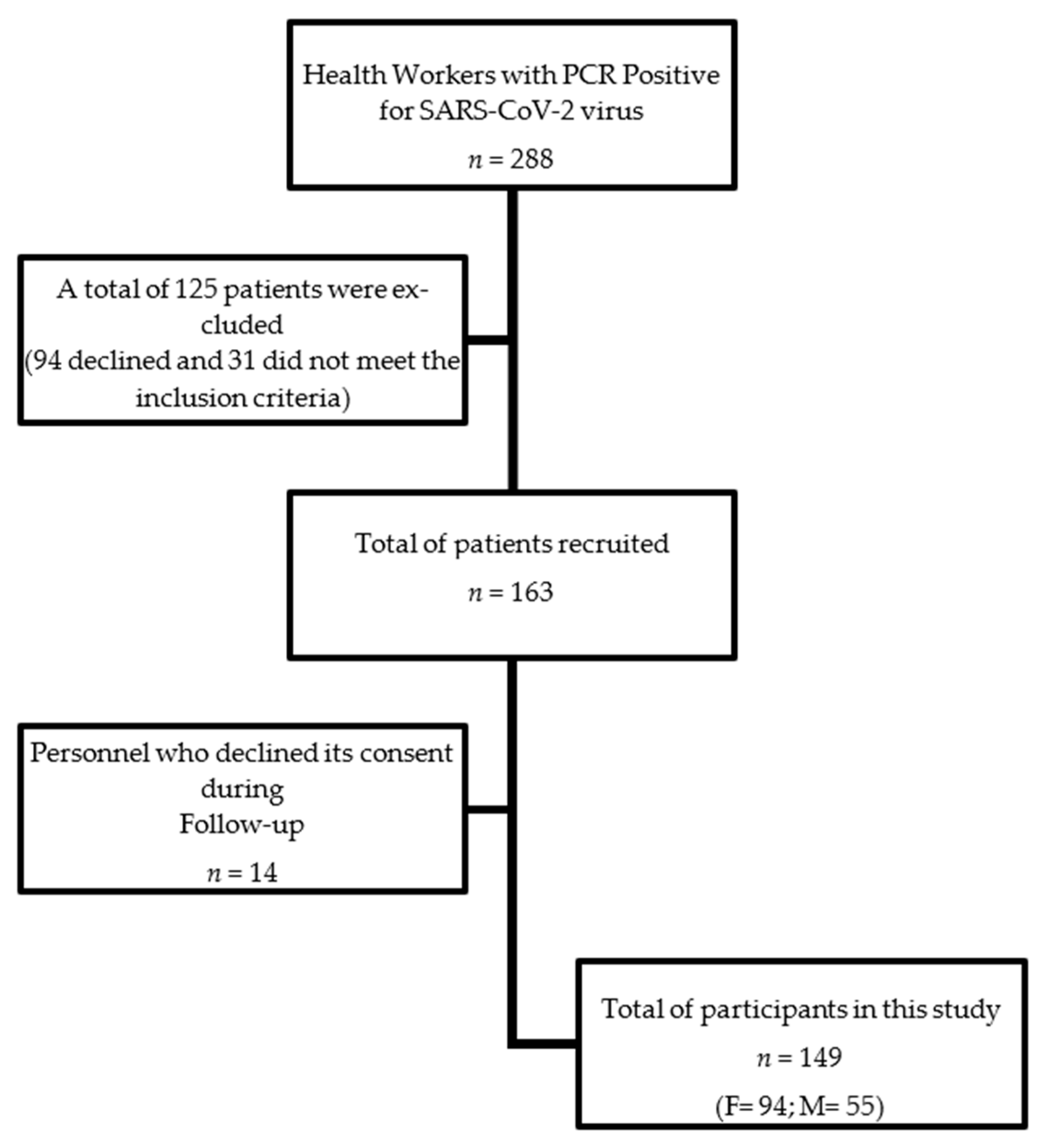
2.1. Design and Study Population

A longitudinal prospective study was conducted from April 6 to December 14, 2021, at the National Institute of Respiratory Diseases “Ismael Cosío Villegas” (INER). All workers who presented clinical symptoms or had contact with a family member or co-worker who tested positive for the SARS-CoV-2 virus were considered for enrollment in this study. However, only workers with a positive viral-RNA test by RT-PCR were invited to participate. Those who agreed signed informed consent. We evaluated the clinical symptoms at three moments:
  • At baseline, when workers attend to be tested for the first time.
  • Three or five days after a positive RT-PCR test (active infection).
  • Minimally, six months after a negative RT-PCR test (post-infection period).
The protocol and informed consent were approved by the INER Research, Bioethics, and Biosafety Committee. The approval number was E05–20.

2.2. Procedures

At baseline, all workers were tested and asked about their clinical symptoms. In
Mexico, all suspected cases of COVID-19 are registered in the Epidemiological Surveillance System for Respiratory Diseases (SISVER) of the Directorate General of Epidemiology (DGE). In the SISVER, risk factors, comorbidities, and initial symptoms detected as characteristic symptoms of the SARS-CoV-2 virus disease are recorded. A standardized questionnaire was designed to obtain the clinical symptoms from participants during all evaluation phases, but during the second and third measurements, the participants were reached by mobile phone. The questionnaire was applied through a Google drive document sent by an instant messaging application for smartphones. Patients were asked about the clinical information previously included in the Epidemiological Surveillance System for Respiratory Diseases (SISVER), and variables associated with new symptoms were added. Additionally, the symptoms were classified into four categories: neurological, gastric, inflammatory, and cardiorespiratory.

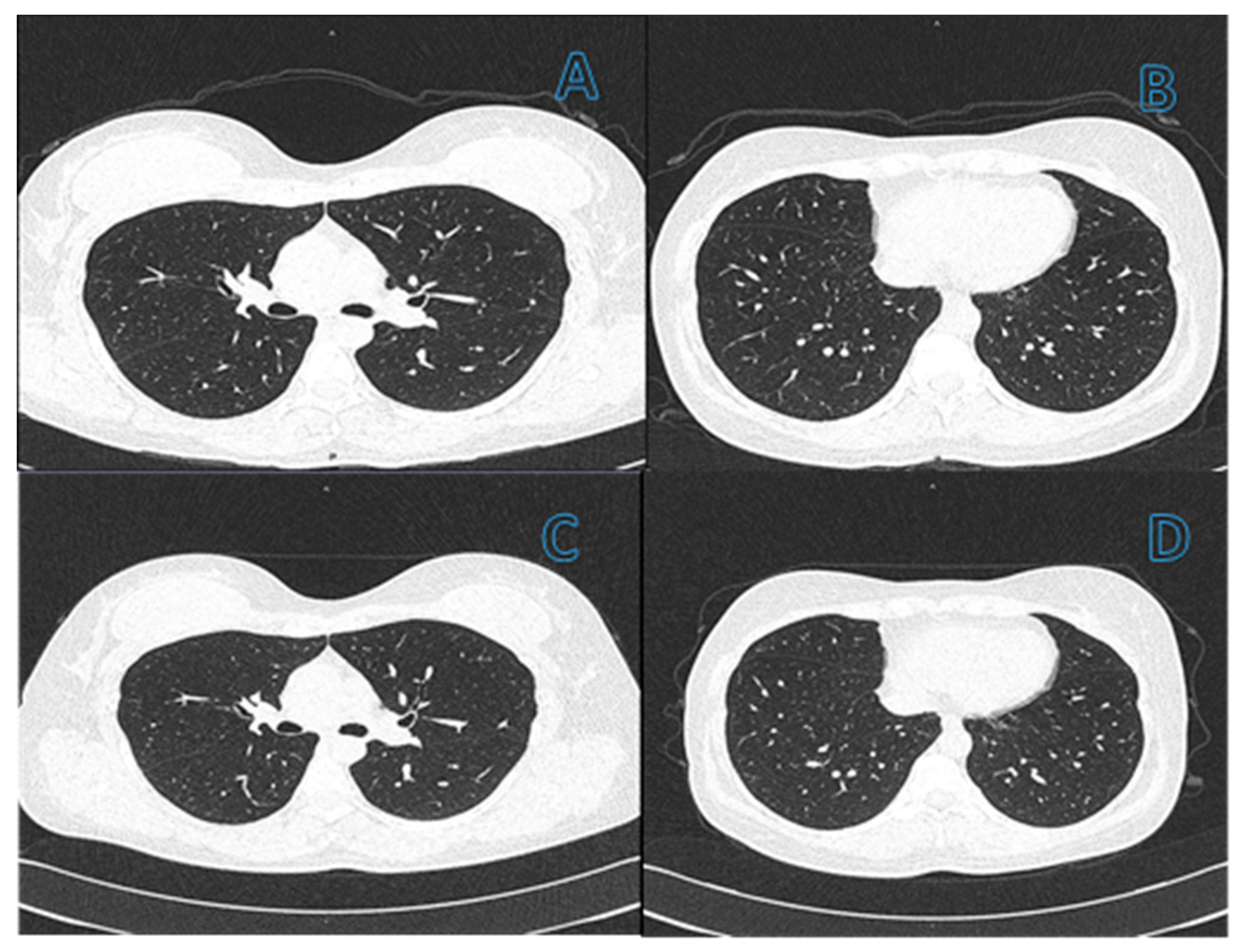
2.3. Post-COVID Assessment of Pulmonary Fibrosis

Although CT is not considered within the standard treatment, some evidence [5] suggested the probability of developing pulmonary fibrosis in these patients. This gives support to deciding to perform the CT three or five days after a positive RT-PCR test and six months after a negative RT-PCR test in these patients. According to 2015 consensus diagnostic guidelines for PF by the American Thoracic Society (ATS), pulmonary fibrosis was defined as a combination of tomographic findings, including parenchymal bands, irregular interfaces, a thick reticular pattern, and bronchiectasis, which was confirmed by the clinical assessment of a pulmonologist.

2.4. Statistical Analysis

Categorical variables were presented as frequency rates and percentages, and quantitative variables were described using the median and interquartile range. Characteristics of the study population were described for each measurement. We used McNemar’s chi-square to compare proportions among measurements. The univariate and multivariate Cox proportional hazards analyses were used to assess the independent prognosis factors for post-COVID-19 pulmonary fibrosis. A result was considered statistically significant if its CI95% did not include the null value. Statistical analysis was performed using STATA software (version 14; Stata Corp, College Station, TX, USA).

3. Results

3.1. Group of Study

According to international guidelines, 288 health workers were confirmed positive for SARS-CoV-2. One hundred twenty-five were excluded from participating in this study, leaving one hundred sixty-three eligible participants who gave their consent to enroll. Later, 14 more left the study after the first or second measurement, leaving a final study population of 149 patients (Figure 1). Of the 149 health workers who met the inclusion criteria, 63% were female. The study population’s age median (p25, p75) was 35 years (29, 45). Sixty-two percent of the participants were medical personnel: 14% administrative staff, and 24% came from different hospital areas. Forty-six (31%) of the patients reported three or more symptoms at the beginning of the study.

3.2. Clinical Characteristics

A total of 17% of the participants indicated at least one comorbidity, among which obesity was the most frequent with 5.4%, followed by hypertension with 4.7%, and diabetes with 3.4% (Table 1). In addition, 4.7% of patients were smokers, 95.3 % had the BCG vaccine, while 81.2% had received the influenza vaccine. A total of 58 patients (38.9%) presented pneumonia during the first 7–10 days of infection, of which 21 scored 2 points and the rest 1 point according to the CURB65 scale. Regarding the elapsed days from a positive to a negative PCR test, the median (p25, p75) was 17 (15, 30), while the median (p25, p75) from a positive test to the last questionnaire applied was 128 days (99, 153).

3.3. Signs and Symptoms Prevalence

Signs and symptoms reported by the patients showed high variability in the three times they were evaluated. The most frequent baseline symptoms were headache (43.6%), cough (38.9%), and rhinorrhea (26.9%), followed by arthralgia (22.2%) and fever (20.8%). While in active infection, fatigue or weakness were the most frequent symptoms (65.1%), followed by headache (59.7%), anosmia (55.0%), myalgia (53.7%), and dysgeusia/ageusia (51.0%), principally; however, a great number of other symptoms was described too (e.g., arthralgia, odynophagia, sleeping problems, etc.) (Table 2). During the post-infection period, fatigue (36.9%) and dyspnea (22.8%) were the most prevalent symptoms, followed by headache (21.5%) and hair loss (21.5%) (Table 2). Comparing baseline reports versus active infection, the highest increments over the prevalence of signs and symptoms were fatigue (0.7 vs. 65%, p-value < 0.0001), anosmia (4.7 vs. 55%, p-value < 0.0001), and dysgeusia/ageusia (5.4 vs. 51%, p-value < 0.0001).
Additionally, the unreported baseline symptoms that manifested during active infection and persisted during post-infection (Table 2) were:
(1)
Neurological: fatigue or weakness, difficulty concentrating, blurred vision, hair loss, cramps, ear disorders, sleeping problems, and anxiety;
(2)
Gastric: bite alteration and weight changes;
(3)
Inflammatory and cardiorespiratory: dermatitis, dyspnea, and tachycardia.
Statistical analysis showed significant differences in all cases (1 vs. 2 or 3; p < 0.0001) (Table 2).

3.4. Diagnosis of Pulmonary Fibrosis and Risk Factors for the Development of Post-COVID-19 Pulmonary Fibrosis

Pulmonary fibrosis was diagnosed in 31/149 (21%) patients based on a combination of tomographic findings, including parenchymal bands, irregular interfaces, a thick reticular pattern, and bronchiectasis confirmed by a pulmonologist. Figure 2 and Figure 3 shows computed tomography scans from patients without (Figure 2) or with (Figure 3) diagnosis of pulmonary fibrosis after a follow-up of six months from initial infection by SARS-CoV-2.
The univariate Cox regression analysis showed that patients with pneumonia had a significantly higher risk of developing post-COVID-19 pulmonary fibrosis than those without pneumonia (hazard ratio (HR) 2.2, IC95%: 1.4–3.5, p = 0.0007). Similarly, patients with positive PCR test > 4 weeks had a significantly higher risk than patients without pneumonia (HR 4.4, IC95%: 2.1–8.7, p < 0.001). In comparison, age (HR 1.01, IC95%: 0.97–1.05, p < 0.46) and sex (HR 1.2, IC95%: 0.59–1.99, p < 0.55) did not show statistically significant differences.
The univariate analysis included both comorbidities and vaccination status. However, we decided not to include them in the multivariate model to maintain parsimonious models that permit the prediction of fibrosis. The confounding factors such as sex and age were adjusted by Cox regression despite having p-values greater than 0.10 (Table 3).

4. Discussion

This study is the first prospective cohort with an extended follow-up period to assess symptom persistence and risk of developing post-COVID-19 pulmonary fibrosis in outpatient healthcare workers previously infected by the SARS-CoV-2. Our work contributes to the generation of knowledge since it shows the consequences of acute illness in ambulatory health personnel six or more months after the apparition of the first symptoms. Results showed that a significant proportion of patients presented persistent symptoms of the nature of cardiorespiratory, neurological, gastrointestinal, or inflammatory, which were reported since the early stages of the disease and maintained at the six-month follow-up. In addition, some signs and symptoms did not show statistically significant differences between the initial and final assessment, indicating that these symptoms persist after the initial infection.
Our data also show that respiratory symptoms prevailed in most patients. This finding is important because it is one of the few viral infections that cause a high percentage of long-term persistent respiratory symptoms in outpatients. Respiratory sequelae have been commonly observed in hospitalized patients with severe influenza but rarely in other infections [16]. Several studies have reported the persistence of neurological symptoms in patients with COVID-19 that can manifest as a neurological syndrome with diverse and complex characteristics [17]. Among these neurological manifestations, the most common are headaches, dizziness, loss of taste and smell, encephalitis, and cerebrovascular disease [2,3,18]. Central and peripheral nervous system disorders associated with acute COVID-19 are usually transient; however, neurological sequelae may persist for months [19]. In this sense, whether the chronic sequelae can become reversible remains without a definitive answer and is one question that can be answered with prolonged follow-up of these patients.
RNA from the SARS-CoV-2 virus has been identified in stool samples from infected patients [20], and the viral receptor is expressed on gastrointestinal epithelial cells, indicating that this virus can actively infect and replicate in the gastrointestinal tract [21], even without developing gastrointestinal symptoms [22]. Recent evidence shows that the incidence rate of vomiting and diarrhea found by us during infection is similar to that reported by other studies [23]. However, the ageusia/dysgeusia ratio was higher than that reported in a systematic review by Mehraeen [24]. Here, we show that gastrointestinal symptoms are rare at disease onset in outpatients with COVID-19, but surprisingly, their frequency was higher in the post-infection stage of the disease.
It is essential to note several persistent inflammatory symptoms in this study. As far as we know, the mechanisms that lead to the persistence of inflammatory symptoms post-COVID are unknown. However, like complications in other organs or systems, cell damage and a robust innate immune response associated with the production of inflammatory cytokines can contribute to the development of this persistence. The risk of persistent symptoms such as conjunctivitis, dermatitis, and hair loss in the post-acute phase of COVID-19 is likely related to the duration and severity of a hyper-inflammatory state, although how long it persists is unknown. More research is needed to provide insight into the immunological mechanisms of the disease.
In this context, it could be assumed that there is a very high probability that there will be a transition from acute cases of COVID-19 to cases of patients with post-COVID sequelae, which could associate with the development of pulmonary fibrosis. Therefore, it is appropriate to focus on the medium- and long-term consequences in recovered patients since it is becoming a public health problem, which will require current and future multidisciplinary efforts to treat these patients. Our study agrees with other works, where a proportion of critically ill patients who recovered from pneumonia associated with the COVID-19 infection were reported to have early lung damage, which derived from the chronic deterioration of lung function and its evolution to pulmonary fibrosis [25]. However, despite mentioning and suggesting the probable development of fibrosis in patients with COVID-19, the presence of pulmonary fibrosis in outpatients has still rarely been studied [5,25]. In our study, 21% of the cases had fibrosis as a severe sequela.
Although previous works had suggested the possibility of developing pulmonary fibrosis in post-COVID patients, our results were unexpected. The above underlines the importance of follow-up with these patients, whereby our next objective is to extend its monitoring period to at least 12 months. Pneumonia at the beginning of the acute condition and positivity in the PCR test for SARSCoV2 for four weeks or more were risk factors for the fibrosis-associated damage. Our findings are consistent with previous studies, which indicate that mild SARS-CoV-2 disease has multi-systemic repercussions [26] and show the importance of characterizing symptoms after the COVID-19 acute stage and compared to the symptoms in the first days of the infectious disease. At this point, fibrosis has been reported to be a long-term sequela in the lungs of patients who have recovered from COVID-19 infection [27]. Likewise, it is alarming that recent evidence shows that post-COVID fibrosis can occur regardless of age or comorbidities in patients [28]. In this regard, Li et al. [29] recently reported the development of pulmonary fibrosis in old patients with higher BMI, in critical condition, with longer viral clearance time, and with delayed hospitalization. Compared with our work, the similitude is in the persistence of viral infection; however, our patients were outpatients without a critical condition or extended hospitalization. The above highlights the importance of follow-up for patients with a diagnosis of COVID-19 independently of its clinical evolution. Likewise, the importance of inflammatory components, which were independent of the severity of the disease, have been reported, but were associated with an increase in the relative risk of developing lung fibrosis-like changes [30]. This last item is an element to consider as a risk factor in patients post-COVID-19, independently of the clinical condition of patients. However, although the inflammatory response and coagulopathies stated post-infection by the SARS-CoV-2 have been associated with the development of pulmonary fibrosis, only some patients develop fibrosis, which makes the landscape even more complex [31]. Additionally, based on the recent findings of other authors, another possibility is that COVID-19 sequelae could result from hemostatic alterations due to the vascular endothelium damage and the intense inflammatory response, which leads to multi-systemic damage, and probably more, after the onset of pulmonary fibrosis [32].

5. Limitations

This study has an epidemiological rather than a clinical background, so caution must be taken to interpret these findings. Due to the nature of the data collection (indirect survey), the patient may have reported some incorrectly described symptoms. However, the three measurements maximized the study’s statistical power and reduced the risk of making a type II error.

6. Conclusions

This study highlights the high frequency of respiratory symptoms after COVID-19 and the possible association of initial pneumonia and a persistently positive test after four weeks as risk factors for post-COVID-19 pulmonary fibrosis. Additionally, our results underline the importance of follow-up with patients over more time to reduce or attenuate the possibility of developing pulmonary fibrosis. However, although more studies are required to confirm them, the probable development of fibrosis in patients post-COVID-19 independent of age, co-morbidities, or symptoms highlights the importance of this issue to future works.

Author Contributions

Conceptualization: R.F.-P., A.-L.H.-I., L.M.T.-E., J.R.G., M.C.-L., and D.M.B.; methodology: A.A.-G., R.V.C., Á.C., J.C.A., and J.R.G.; validation: R.F.-P., A.-L.H.-I., and M.C.-L.; formal analysis: R.F.-P., A.-L.H.-I., L.M.T.-E., J.R.G., M.C.-L., and D.M.B.; investigation: A.-L.H.-I., L.M.T.-E., M.C.-L., and N.A.-V.; data curation: Research Working Group; writing—review and editing: L.M.T.-E., A.-L.H.-I., M.C.-L., and N.A.-V. All authors have read and agreed to the published version of the manuscript.

Funding

This research received no external funding.

Institutional Review Board Statement

Informed consent was obtained from all study participants prior to study inclusion. The study was conducted in accordance with the Declaration of Helsinki and approved by the National Institute of Respiratory Diseases “Ismael Cosío Villegas” Research, Bioethics, and Biosafety Committee. The approval number was E05–20.

Informed Consent Statement

Informed consent was obtained from all subjects involved in the study.

Data Availability Statement

Data can be obtained from the corresponding author upon reasonable request.

Conflicts of Interest

The authors declare no conflict of interest.

References

  1. World Health Organization. Coronavirus Disease 2019 (COVID-19): Situation Report, 114. World Health Organization. 2020. Available online: https://apps.who.int/iris/handle/10665/332089 (accessed on 2 May 2021).
  2. Kim, G.-U.; Kim, M.-J.; Ra, S.; Lee, J.; Bae, S.; Jung, J.; Kim, S.-H. Clinical characteristics of asymptomatic and symptomatic patients with mild COVID-19. Clin. Microbiol. Infect. 2020, 26, 948.e1–948.e3. [Google Scholar] [CrossRef] [PubMed]
  3. Ludwig, S.; Schell, A.; Berkemann, M.; Jungbauer, F.; Zaubitzer, L.; Huber, L.; Warken, C.; Held, V.; Kusnik, A.; Teufel, A.; et al. post-COVID-19 Impairment of the Senses of Smell, Taste, Hearing, and Balance. Viruses 2022, 14, 849. [Google Scholar] [CrossRef] [PubMed]
  4. Wu, Z.; McGoogan, J. Characteristics of and Important Lessons from the Coronavirus Disease 2019 (COVID-19) Outbreak in China: Summary of a Report of 72 314 Cases from the Chinese Center for Disease Control and Prevention. JAMA 2020, 323, 1239–1242. [Google Scholar] [CrossRef] [PubMed]
  5. Rai, D.K.; Sharma, P.; Kumar, R. Post covid 19 pulmonary fibrosis. Is it real threat? Indian J. Tuberc. 2021, 68, 330–333. [Google Scholar] [CrossRef]
  6. Zhou, Y.; Yang, Q.; Chi, J.; Dong, B.; Lv, W.; Shen, L.; Wang, Y. Comorbidities and the risk of severe or fatal outcomes associated with coronavirus disease 2019: A systematic review and meta-analysis. Int. J. Infect. Dis. 2020, 99, 47–56. [Google Scholar] [CrossRef]
  7. Hall, J.; Myall, K.; Lam, J.L.; Mason, T.; Mukherjee, B.; West, A.; Dewar, A. Identifying patients at risk of post-discharge complications related to COVID-19 infection. Thorax 2021, 76, 408–411. [Google Scholar] [CrossRef]
  8. Garibaldi, B.T.; Wang, K.; Robinson, M.L.; Zeger, S.L.; Bandeen-Roche, K.; Wang, M.-C.; Alexander, G.C.; Gupta, A.; Bollinger, R.; Xu, Y. Comparison of Time to Clinical Improvement With vs Without Remdesivir Treatment in Hospitalized Patients With COVID-19. JAMA Netw. Open 2021, 4, e213071. [Google Scholar] [CrossRef]
  9. Ranjbar, K.; Moghadami, M.; Mirahmadizadeh, A.; Fallahi, M.J.; Khaloo, V.; Shahriarirad, R.; Erfani, A.; Khodamoradi, Z.; Gholampoor Saadi, M.H. Methylprednisolone or dexamethasone, which one is superior corticosteroid in the treatment of hospitalized COVID-19 patients: A triple-blinded randomized controlled trial. BMC Infect. Dis. 2021, 21, 337. [Google Scholar] [CrossRef]
  10. Baricich, A.; Borg, M.B.; Cuneo, D.; Cadario, E.; Azzolina, D.; Balbo, P.E.; Bellan, M.; Zeppegno, P.; Pirisi, M.; Cisari, C.; et al. Midterm functional sequelae and implications in rehabilitation after COVID-19: A cross-sectional study. Eur. J. Phys. Rehabil. Med. 2021, 57, 199–207. [Google Scholar] [CrossRef]
  11. Riker, R.R.; Shehabi, Y.; Bokesch, P.M.; Ceraso, D.; Wisemandle, W.; Koura, F.; Whitten, P.; Margolis, B.D.; Byrne, D.W.; Ely, E.W.; et al. Dexmedetomidine vs. midazolam for sedation of critically ill patients: A randomized trial. JAMA 2009, 301, 489–499. [Google Scholar] [CrossRef] [Green Version]
  12. Van den Borst, B.; Peters, J.B.; Brink, M.; Schoon, Y.; Bleeker-Rovers, C.P.; Schers, H.; van Hees, H.W.; van Helvoort, H.; van den Boogaard, M.; van der Hoeven, H.; et al. Comprehensive Health Assessment 3 Months After Recovery from Acute Coronavirus Disease 2019 (COVID-19). Clin. Infect. Dis. 2021, 73, e1089–e1098. [Google Scholar] [CrossRef] [PubMed]
  13. Petersen, M.S.; Kristiansen, M.F.; Hanusson, K.D.; Danielsen, M.E.; Steig, B.Á.; Gaini, S.; Strøm, M.; Weihe, P. Long COVID in the Faroe Islands: A Longitudinal Study Among Non-Hospitalized Patients. Clin. Infect. Dis. 2021, 73, e4058–e4063. [Google Scholar] [CrossRef] [PubMed]
  14. Hellmuth, J.; Barnett, T.A.; Asken, B.M.; Kelly, J.D.; Torres, L.; Stephens, M.L.; Greenhouse, B.; Martin, J.N.; Chow, F.C.; Deeks, S.G.; et al. Persistent COVID-19-associated neurocognitive symptoms in non-hospitalized patients. J. Neurovirol. 2021, 27, 191–195. [Google Scholar] [CrossRef] [PubMed]
  15. Hernandez-Romieu, A.C.; Leung, S.; Mbanya, A.; Jackson, B.R.; Cope, J.R.; Bushman, D.; Dixon, M.; Brown, J.; McLeod, T.; Saydah, S.; et al. Health Care Utilization and Clinical Characteristics of Non-hospitalized Adults in an Integrated Health Care System 28-180 Days After COVID-19 Diagnosis—Georgia, May 2020–March 2021. MMWR Morb. Mortal. Wkly. Rep. 2021, 70, 644–650. [Google Scholar] [CrossRef]
  16. Luyt, C.-E.; Combes, A.; Becquemin, M.-H.; Beigelman-Aubry, C.; Hatem, S.; Brun, A.-L.; Zraik, N.; Carrat, F.; Grenier, P.A.; Richard, J.-C.M.; et al. Long-term Outcomes of Pandemic 2009 Influenza A(H1N1)-Associated Severe ARDS. Chest 2012, 142, 583–592. [Google Scholar] [CrossRef]
  17. Sharifian-Dorche, M.; Huot, P.; Osherov, M.; Wen, D.; Saveriano, A.; Giacomini, P.S.; Antel, J.P.; Mowla, A. Neurological complications of coronavirus infection; a comparative review and lessons learned during the COVID-19 pandemic. J. Neurol. Sci. 2020, 417, 117085. [Google Scholar] [CrossRef]
  18. Shehata, G.; Lord, K.; Grudzinski, M.; Elsayed, M.; Abdelnaby, R.; Elshabrawy, H. Neurological Complications of COVID-19: Underlying Mechanisms and Management. Int. J. Mol. Sci. 2021, 22, 4081. [Google Scholar] [CrossRef]
  19. Alonso-Bellido, I.M.; Bachiller, S.; Vázquez, G.; Cruz-Hernández, L.; Martínez, E.; Ruiz-Mateos, E.; Deierborg, T.; Venero, J.L.; Real, L.M.; Ruiz, R. The Other Side of SARS-CoV-2 Infection: Neurological Sequelae in Patients. Front. Aging Neurosci. 2021, 13, 632673. [Google Scholar] [CrossRef]
  20. Wu, Y.; Guo, C.; Tang, L.; Hong, Z.; Zhou, J.; Dong, X.; Yin, H.; Xiao, Q.; Tang, Y.; Qu, X.; et al. Prolonged presence of SARS-CoV-2 viral RNA in faecal samples. Lancet Gastroenterol. Hepatol. 2020, 5, 434–435. [Google Scholar] [CrossRef]
  21. Lamers, M.M.; Beumer, J.; Van Der Vaart, J.; Knoops, K.; Puschhof, J.; Breugem, T.I.; Ravelli, R.B.; Paul van Schayck, J.; Mykytyn, A.Z.; Duimel, H.Q. SARS-CoV-2 productively infects human gut enterocytes. Science 2020, 369, 50–54. [Google Scholar] [CrossRef]
  22. Wang, W.; Xu, Y.; Gao, R.; Lu, R.; Han, K.; Wu, G.; Tan, W. Detection of SARS-CoV-2 in Different Types of Clinical Specimens. JAMA 2020, 323, 1843–1844. [Google Scholar] [CrossRef] [PubMed] [Green Version]
  23. Jin, X.; Lian, J.S.; Hu, J.H.; Gao, J.; Zheng, L.; Zhang, Y.M.; Hao, S.R.; Jia, H.Y.; Cai, H.; Zhang, X.L.; et al. Epidemiological, clinical and virological characteristics of 74 cases of coronavirus-infected disease 2019 (COVID-19) with gastrointestinal symptoms. Gut 2020, 69, 1002–1009. [Google Scholar] [CrossRef] [PubMed] [Green Version]
  24. Mehraeen, E.; Behnezhad, F.; Salehi, M.A.; Noori, T.; Harandi, H.; SeyedAlinaghi, S. Olfactory and gustatory dysfunctions due to the coronavirus disease (COVID-19): A review of current evidence. Eur. Arch. Otorhinolaryngol. 2021, 278, 307–312. [Google Scholar] [CrossRef] [PubMed]
  25. McDonald, L. Healing after COVID-19: Are survivors at risk for pulmonary fibrosis? Am. J. Physiol.-Lung Cell Mol. Physiol. 2021, 320, L257–L265. [Google Scholar] [CrossRef] [PubMed]
  26. Xiong, Q.; Xu, M.; Li, J.; Liu, Y.; Zhang, J.; Xu, Y.; Dong, W. Clinical sequelae of COVID-19 survivors in Wuhan, China: A single-centre longitudinal study. Clin. Microbiol. Infect. 2021, 27, 89–95. [Google Scholar] [CrossRef]
  27. Rumende, C.M.; Susanto, E.C.; Sitorus, T.P. The Management of Pulmonary Fibrosis in COVID-19. Acta Med. Indones. 2021, 53, 233–241. [Google Scholar]
  28. Antony, T.; Acharya, K.V.; Unnikrishnan, B.; Keerthi, N. A silent march-Post covid fibrosis in asymptomatics—A cause for concern? Indian J. Tuberc. 2022. Epub ahead of print. [Google Scholar] [CrossRef]
  29. Li, X.; Shen, C.; Wang, L.; Majumder, S.; Zhang, D.; Deen, M.J.; Li, Y.; Qing, L.; Zhang, Y.; Chen, C.; et al. Pulmonary fibrosis and its related factors in discharged patients with new coronavirus pneumonia: A cohort study. Respir. Res. 2021, 22, 203. [Google Scholar] [CrossRef]
  30. Colarusso, C.; Maglio, A.; Terlizzi, M.; Vitale, C.; Molino, A.; Pinto, A.; Vatrella, A.; Sorrentino, R. Post-COVID-19 Patients Who Develop Lung Fibrotic-like Changes Have Lower Circulating Levels of IFN-β but Higher Levels of IL-1α and TGF-β. Biomedicines 2021, 9, 1931. [Google Scholar] [CrossRef]
  31. Townsend, L.; Dowds, J.; O’Brien, K.; Sheill, G.; Dyer, A.H.; O’Kelly, B.; Hynes, J.P.; Mooney, A.; Dunne, J.; Ni Cheallaigh, C.; et al. Persistent Poor Health after COVID-19 Is Not Associated with Respiratory Complications or Initial Disease Severity. Ann. Am. Thorac. Soc. 2021, 18, 997–1003. [Google Scholar] [CrossRef]
  32. Wang, C.; Yu, C.; Jing, H.; Wu, X.; Novakovic, V.A.; Xie, R.; Shi, J. Long COVID: The Nature of Thrombotic Sequelae Determines the Necessity of Early Anticoagulation. Front. Cell. Infect. Microbiol. 2022, 12, 861703. [Google Scholar] [CrossRef] [PubMed]
Figure 1. Flow chart of Study Population (F = Female; M = Male).
Viruses 14 01843 g001
Figure 2. (A,B) Computed tomography (CT) scans of the chest with 1 mm slices of a 46-year-old female patient in the active phase of infection. CT does not show changes in the lung related to pneumonia. (C,D) Images of CT six months after resolution of SARS-CoV-2 infection have no change from initial CT ones, which discard a history of lung damage.
Viruses 14 01843 g002
Figure 3. (A,B) CT from a 56-year-old male patient in the active infection phase of COVID-19. (C,D) CT after six months of infection resolution. Typical findings of COVID-19 pneumonia and tomographic findings that support the diagnosis of pulmonary fibrosis are observed. (A) Vascular thickening (arrow) associated with an area of ground-glass opacity; (B) Subpleural parenchymal bands (arrows) and ground-glass opacity and consolidation (arrowheads); (C) Absorption of most of the affected areas leaving some lesions in ground glass; and (D) Fibrous lesions that represent residual organizing pneumonia.
Viruses 14 01843 g003
Table 1. Clinical characteristics and comorbidities were initially reported by the patients evaluated.
Variablen (%)
Female91 (63.0)
Age35 (29–45) *
Obesity8 (5.4)
Hypertension7 (4.7)
Diabetes5 (3.4)
Smoking7 (4.7)
BCG vaccine142 (95.3)
Influenza vaccine121 (81.2)
Pneumonia58 (38.9)
* Median and interquartile range.
Table 2. Signs and symptoms prevalence in the study population (n = 149) reported at baseline, in active infection, and during follow-up by six months.
Baseline
(1)
Active Infection
(2)
Post-Infection
(3)
1 vs. 21 vs. 32 vs. 3
Signs and Symptoms(%)(%)(%)p-Valuep-Valuep-Value
Neurological
General attack9.428.23.4<0.00010.064<0.0001
Arthralgia22.242.314.8<0.00010.093<0.0001
Myalgia14.153.713.4<0.00010.999<0.0001
Dysgeusia/Ageusia5.451.010.7<0.00010.115<0.0001
Anosmia4.755.013.4<0.00010.011<0.0001
Odynophagia9.440.37.4<0.00010.664<0.0001
Abdominal pain418.86.0<0.00010.581<0.0001
Headache43.659.721.50.005<0.0001<0.0001
Fatigue or weakness0.765.136.9<0.0001<0.0001<0.0001
Difficult concentrating 029.514.8<0.0001<0.00010.015
Blurred vision08.78.1<0.0001<0.00010.617
Hair loss024.221.5<0.0001<0.00010.999
Cramps015.412.1<0.0001<0.00010.774
Ear disorders1.326.212.1<0.0001<0.00010.001
Sleeping problems034.917.5<0.0001<0.0001<0.0001
Anxiety020.814.1<0.0001<0.00010.041
Gastric
Diarrhea6.733.66.0<0.00010.999<0.0001
Vomit07.400.0010.9990.001
Nausea1.316.12.0<0.00010.999<0.0001
Xerostomia0.726.97.4<0.00010.006<0.0001
Mouth ulcers06.73.40.0020.0630.227
Bite alteration021.513.4<0.0001<0.00010.012
Weight changes014.110.7<0.0001<0.00010.808
Inflammatory
Conjunctivitis5.420.84.7<0.00010.999<0.0001
Lymphadenopathy017.53.4<0.00010.063<0.0001
Dermatitis016.812.1<0.0001<0.00010.167
Irritability4.022.87.4<0.00010.332<0.0001
Diaphoresis10.742.310.1<0.00010.999<0.0001
Fever20.836.200.001<0.0001<0.0001
Cardiorespiratory
Rhinorrhea26.927.54.70.882<0.0001<0.0001
Nasal congestion1.327.56.7<0.00010.039<0.0001
Cough38.940.37.40.773<0.0001<0.0001
Dyspnea (mild/moderate)6.040.322.8<0.0001<0.00010.001
Tachycardia028.214.8<0.0001<0.00010.001
Table 3. Univariate and multivariate hazard ratios for post-COVID-19 pulmonary fibrosis.
UnivariateMultivariate
VariableHRCI 95%p-ValueHRCI 95%p-Value
Pneumonia2.21.4–3.50.00072.411.51–3.82<0.001
PCR positive test > 4 weeks4.42.1–8.7<0.00015.382.34–12.35<0.001
Age1.010.97–1.050.460.990.95–1.040.96
Sex1.20.59–1.990.551.30.6–1.80.47
Diabetes0.94 0.29–3.050.93---
Hypertension1.280.46–3.540.63---
Obesity1.90.69–5.280.23---
Smoking1.770.71–0.440.21---
BCG vaccine0.690.42–1.210.2---
Influenza vaccine0.910.91–2.330.68---
Abbreviations: HR, hazard ratio; CI, confidence interval; PCR, polymerase chain reaction.
Publisher’s Note: MDPI stays neutral with regard to jurisdictional claims in published maps and institutional affiliations.

