Review

Evidence for Benefits of Early Treatment Initiation for Chronic Hepatitis B

1
Department of Gastroenterology, Asan Medical Center, University of Ulsan College of Medicine, Seoul 05505, Republic of Korea
2
Division of Gastroenterology and Hepatology, Stanford University School of Medicine, Palo Alto, CA 94063, USA
3
Division of Liver Diseases, Icahn School of Medicine at Mount Sinai, New York, NY 10029, USA
4
Division of Gastroenterology and Hepatology, Department of Internal Medicine, Department of Medical Research, Hepatitis Research Center, National Taiwan University Hospital, Taipei 100, Taiwan
5
Graduate Institute of Clinical Medicine, National Taiwan University College of Medicine, Taipei 110, Taiwan
6
Gilead Sciences, Foster City, CA 94404, USA
7
Division of Gastroenterology and Hepatology, Mayo Clinic, Rochester, MN 55905, USA
8
Center for Disease Analysis Foundation, Lafayette, CO 80026, USA
9
Barts Liver Centre, Immunobiology, Blizard Institute, Barts and The London School of Medicine and Dentistry, Queen Mary University of London, London E1 4NS, UK
*
Authors to whom correspondence should be addressed.
Submission received: 29 March 2023 / Revised: 12 April 2023 / Accepted: 14 April 2023 / Published: 18 April 2023
(This article belongs to the Special Issue Viral Hepatitis Treatment 2.0)

Abstract

:
Chronic hepatitis B (CHB) is the most common cause of hepatocellular carcinoma (HCC) worldwide. Antiviral treatment reduces the risk of HCC and mortality; nonetheless, globally in 2019, only 2.2% of CHB patients received treatment. Current international CHB guidelines recommend antiviral treatment only in subsets of patients with clear evidence of liver damage. This contrasts with hepatitis C or HIV where early treatment is recommended in all infected patients, regardless of end-organ damage. This narrative review aims to provide an overview of data on the early initiation of antiviral treatment and its related potential economic impact. Literature searches were performed using PubMed and abstracts from international liver congresses (2019–2021). Data on risk of disease progression and HCC and the impact of antiviral treatment in currently ineligible patients were summarized. Cost-effectiveness data on early antiviral treatment initiation were also collated. Accumulating molecular, clinical, and economic data suggest that early initiation of antiviral treatment could save many lives through HCC prevention in a highly cost-effective manner. In light of these data, we consider several alternative expanded treatment strategies that might further a simplified ‘treatment as prevention’ approach.

1. Introduction

In 2019, the World Health Organization (WHO) estimated that 296 million people had chronic hepatitis B virus (HBV) infection worldwide, resulting in an estimated 820,000 deaths annually, predominantly from cirrhosis and hepatocellular carcinoma (HCC) [1]. Chronic hepatitis B (CHB) is the most common cause of HCC, and rates of deaths from HBV-related HCC are expected to more than double between 2016 and 2040 [2].
A major goal of CHB antiviral treatment is to prevent disease progression, HCC, and mortality. Without a cure for CHB infection, an essential intermediate endpoint toward these goals is the long-term suppression of HBV replication, which is achievable by current antiviral treatment [3,4], and reduces the risk of HCC and mortality in CHB patients [3,5]. Nonetheless, globally, only 2.2% (6.6 million) of CHB patients received treatment in 2019 [1], due, in part, to the complex and restrictive clinical practice guidelines (Supplementary Figure S1). Currently, treatment is indicated only after the identification of hepatic necroinflammation through liver biopsy or in persons with specific elevations in the serum levels of alanine aminotransferase (ALT) and HBV DNA (Supplementary Figure S1) [3,4,6].
CHB is a dynamic disease, and viral load and risk of disease progression change over time due to the interaction between HBV replication and the host immune response [3,7]. Historically, chronic HBV infection has been divided into four disease phases (Supplementary File S1 and Supplementary Figure S1), with those that do not fall into these disease phases referred to as belonging to the ‘gray zone’. Different terminology is used to describe these phases, but for consistency, the terms immune tolerant (IT), immune active (IA), immune control (IC), and immune escape are used throughout this review. Terminology based on the European Association for the Study of the Liver (EASL) guidelines is included in the Supplementary File S1 and Supplementary Figure S1 for information [3]. Currently, the guidelines only recommend treatment in the IA and immune escape phases (Table 1).
This review examines the data supporting the earlier initiation of antiviral treatment to delay or even reverse CHB disease progression, and discusses the potential for ‘treatment as prevention’ as a strategy to reduce HBV-related mortality. We also collated the existing data on the potential economic impact of early antiviral treatment, as cost will be a key factor affecting implementation. Due to space restrictions, a comprehensive review of all the available data was not feasible. Further details of the search strategy and selection criteria are provided in the Supplementary File S2 and Supplementary Figure S2.

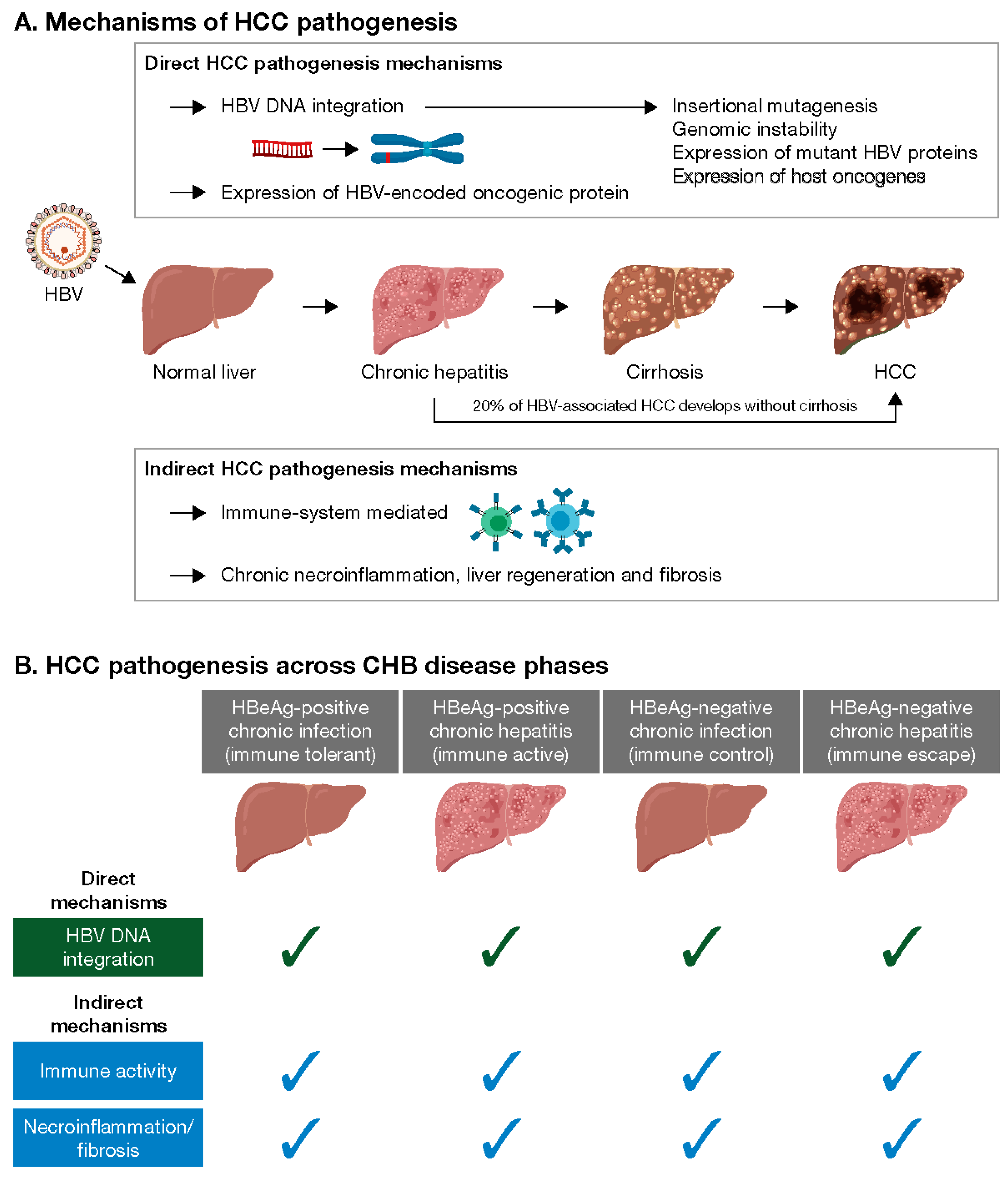
2. Mechanisms of Hepatocarcinogenesis in CHB Patients

During early HBV infection, the immune system is activated as part of the defense mechanism, and in acute cases, this response is beneficial. However, persistent immune activation from chronic infection initiates a series of molecular events [8] including carcinogenesis, driven via direct and indirect mechanisms (Figure 1) [5,9,10].

2.1. Direct Hepatocarcinogenesis

Direct hepatocarcinogenesis results from HBV DNA integration, which occurs prior to observable histological liver damage, and may promote HCC through chromosomal instability (including translocation), insertional mutagenesis, and the expression of mutant HBV genes or host oncogenes [5,9,11,12,13,14]. Integrations are detectable across multiple CHB phases (Figure 1) including the ‘gray zone’ [15,16,17,18], and correlate with levels of viremia [18]. Higher levels of integration have been observed among hepatitis B e antigen (HBeAg)-positive compared with HBeAg-negative patients [16], and among the HBeAg-negative patients, the highest number of integrations has been reported among those with HBV DNA >20,000 IU/mL [17]. Antiviral treatment has been shown to reduce the number of transcriptionally active integrations in patients with CHB [13,18,19].
Clonal expansion of hepatocytes containing integrations has been detected across all CHB phases [20,21], irrespective of HBeAg status or age [19]. It is possible that the immune response may select for hepatocytes with a survival advantage that clonally proliferate and initiate HCC, as although the integrations appear random, some occur in genes regulating cell proliferation and may drive hepatocarcinogenesis [17]. HCC can be polyclonal or monoclonal in origin; monoclonal tumors contain the same HBV DNA integration events, indicating that HBV DNA integration is an early driver in tumor development and remains stable during tumor progression [22].

2.2. Indirect Hepatocarcinogenesis

Indirect hepatocarcinogenesis is driven by chronic HBV-induced necroinflammation, regeneration, and fibrosis and can occur at any stage of disease [9,23,24]. HBV-specific T-cell activity is present in patients in both the IT and IA phases [21,24], and T-cell function has been shown to be similar, irrespective of the disease phase [25]. Analysis of baseline liver biopsies from treated CHB patients similarly found the immune microenvironment, classified as high (i.e., having elevated immune pathways and elevated immune cell signatures corresponding to B cells, T cells and macrophages) or low (not having these), to be independent of HBeAg status and HBV DNA levels [26].

3. Virological Risk Factors for HCC and Impact of Antiviral Therapy

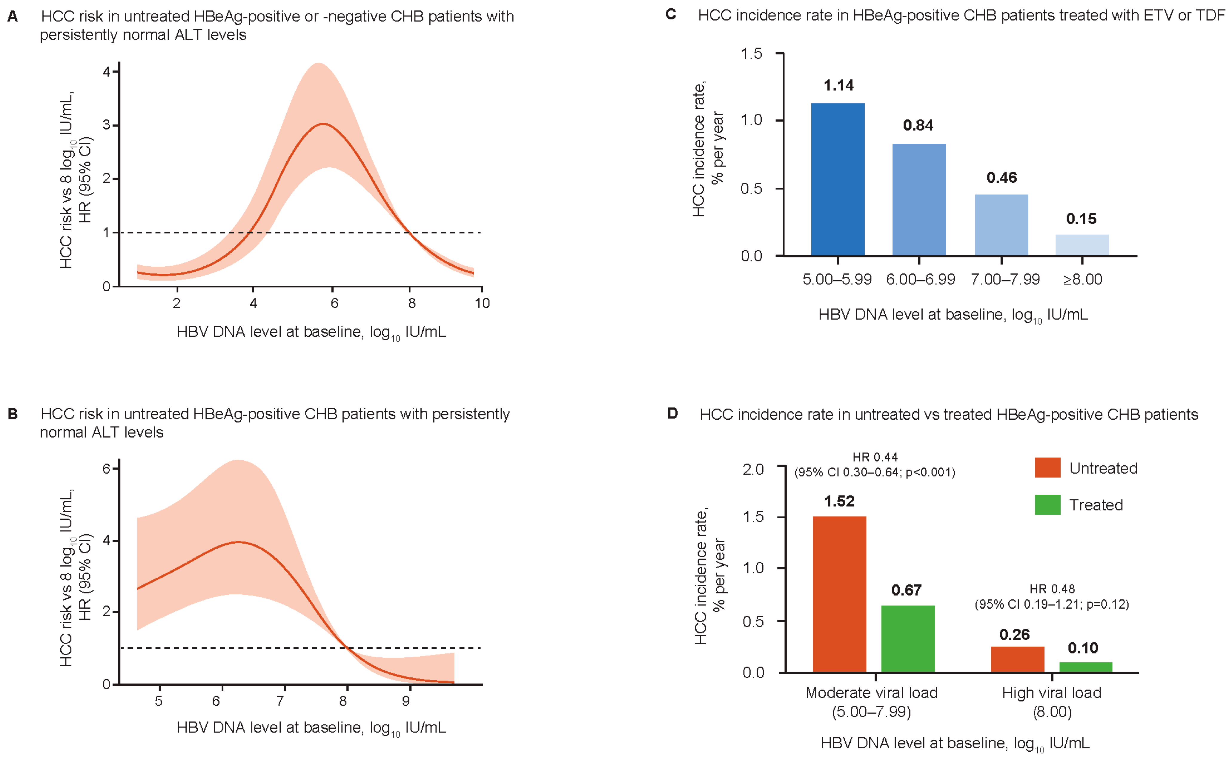
3.1. Reappraisal of the Association between Viral Load and HCC Risk

Elevated HBV DNA is a strong risk factor for HCC and is a key consideration when deciding to initiate therapy in CHB patients [5]. Traditionally, HBeAg status has also been correlated with HCC risk [27,28]. However, as high viral loads can occur in HBeAg-negative patients and test sensitivity may affect its determination [29,30], HBeAg status should always be considered alongside the HBV DNA levels when estimating HCC risk.
Although a linear association between baseline HBV DNA levels and HCC risk, regardless of ALT level or HBeAg status, has been observed with HBV DNA levels up to 6.0 log10 IU/mL [31,32,33], recent analyses suggest a more nuanced relationship [34,35,36]. Analysis of 6949 HBeAg-positive and -negative, non-cirrhotic, treatment-naïve CHB patients with ALT <80 U/L identified a parabolic association between HBV DNA levels and HCC risk (Figure 2A) [34]. Risk was highest among patients with HBV DNA levels of 6.0–7.0 log10 IU/mL and lowest in patients with HBV DNA levels ≤4 log10 IU/mL and >8.0 log10 IU/mL. This association was consistent across age groups, and neither HBeAg status nor ALT levels were predictive of HCC. A subsequent study analyzed the association between pre-treatment HBV DNA levels and HCC risk during treatment with entecavir (ETV) or tenofovir disoproxil fumarate (TDF) in 2073 HBeAg-positive, non-cirrhotic CHB patients [35]. At HBV DNA levels ≥5.0 log10 IU/mL, on-treatment HCC risk increased incrementally with decreasing baseline HBV DNA levels. By multivariable analysis, compared with baseline HBV DNA ≥8.0 log10 IU/mL, the adjusted hazard ratios for HCC risk for baseline HBV DNA 7.00–7.99 log10 IU/mL, 6.00–6.99 log10 IU/mL, and 5.00–5.99 log10 IU/mL were 2.48 (p = 0.03), 3.69 (p = 0.002), and 6.10 (p < 0.001), respectively (Figure 2B). Compared with untreated patients with the same ranges of baseline HBV DNA levels, antiviral treatment significantly reduced HCC risk in patients with moderate viral load (5.00–7.99 log10 IU/mL), but the HCC risk did not decrease to the level of patients who initiated antiviral treatment with a high viral load (≥8.0 log10 IU/mL; Figure 2C,D).
Moderate HBV DNA levels (105–107 IU/mL) are a risk factor for significant inflammation among patients with normal ALT and no significant fibrosis [36]. A fully infected liver can produce 109–1010 virions/mL, which, if the infection is benign in an IT-phase host, would be expected to persist throughout the course of the disease [37]. Most HBeAg-positive CHB patients have very high HBV DNA levels (≥8.0 log10 IU/mL) during the initial phase of infection, and with a parabolic relationship, HCC risk may be relatively low [37]. Low and persistent immune-mediated damage to infected hepatocytes results in a gradual decrease in HBV DNA and progression to the moderate replication phase, which is associated with irreversibly increasing HCC risk. Accordingly, a decrease in viral load in untreated individuals may reflect progressive liver damage and increased HCC risk [37,38].

3.2. Impact of Antiviral Treatment

The current first-line nucleos(t)ide analogs (NAs), ETV, TDF, and tenofovir alafenamide (TAF), have favorable and well-described long-term safety profiles, with minimal to no resistance, even in heavily treatment-experienced patients with pre-existing resistance [3,4,6], and are associated with high adherence rates [39]. While long-term treatment does not eliminate HCC risk, it can reduce liver disease progression, improve necroinflammation and fibrosis, and even reverse cirrhosis [3,7,40,41,42].
However, CHB antiviral treatment indications and guidelines are largely based on evidence from randomized controlled trials (RCTs) that were not designed to demonstrate the impact of these treatments on long-term outcomes such as HCC in a real-world setting. As such, they may fail to address important groups of patients and result in the withholding of therapy in patients who may benefit. Moreover, the current concept of CHB disease phases and corresponding treatment guidelines based on HBeAg, HBV DNA, and ALT are complex and do not represent the whole spectrum of CHB patients observed in clinical practice. Consequently, many patients currently ineligible for NAs according to current guidelines remain at risk of adverse clinical events, particularly HCC. An analysis of treatment-naïve CHB patients showed that among those who developed HCC, 64%, 46%, and 34% did so outside the treatment guideline recommendations from the Asian Pacific Association for the Study of the Liver (APASL), American Association for the Study of Liver Diseases (AASLD), and EASL, respectively [43]. A similar analysis found that 75% of untreated patients who developed HCC were outside the AASLD guidelines [44].

4. Evidence for Risk of Disease Progression across CHB Disease Phases

4.1. Risks in Untreated, HBeAg-Positive CHB Patients in the IT Phase (Normal ALT and High HBV DNA Levels)

Contrary to conventional belief, IT patients may have significant liver injury or fibrosis [45,46]. Among 566 IT patients from 11 studies, 17% and 5% had significant fibrosis or advanced fibrosis, respectively [47]. Studies evaluating long-term outcomes in untreated patients in the IT phase have indicated a significant risk of HCC and adverse liver outcomes (Table 2). A multivariate analysis found that the risk of HCC and death or transplantation was significantly higher in untreated IT patients compared with NA-treated IA patients [48]. However, differences in 5- and 10-year cumulative HCC risk and liver cirrhosis progression were not observed between groups [49]. In another study, 3.7% of 651 IT patients developed HCC during a median 5.2 years of follow-up [50]. The 10-year HCC incidence rate was 2.6% and 20.4% in patients aged <40 years and ≥40 years, respectively. Finally, a retrospective multicenter study of 946 IT patients (mean HBV DNA 108.5 IU/mL) reported a 10-year cumulative HCC risk of 1.7% [51]. Given that age and HBV DNA levels are important determinants of HCC risk, caution is required when interpreting HCC risk in these studies because the patients had a heterogeneous distribution of age and HBV DNA levels. Nonetheless, collectively, these data support the emerging consensus that the IT phase is not always benign, particularly among patients aged ≥40 years [48,49,50,51].

4.2. Risks in Untreated, HBeAg-Negative CHB Patients in the IC Phase (Normal ALT and Low HBV DNA Levels)

While CHB patients in the HBeAg-negative IC phase are generally ineligible for antiviral treatment, evidence suggests they have a higher risk of HCC or liver-related death compared with hepatitis B surface antigen (HBsAg)-negative controls [52]. Moreover, HBeAg-negative CHB patients are a heterogeneous population with respect to the risk of HCC and adverse liver outcomes (Table 2). An individual patient’s HCC risk depends on a combination of direct and indirect hepatocarcinogenesis mechanisms based on their unique disease history. For example, in a prospective observational study of 1192 patients with untreated HBeAg-negative CHB and low viral loads (mean baseline HBV DNA <104 IU/mL), although the HBV DNA level correlated with disease progression, overall disease progression was minimal after 7 years, with no changes in fibrosis or HCC incidence [53]. In contrast, several other studies have reported substantial disease progression in IC patients. One study found an HCC incidence of 5% over a mean follow-up of 63 months among 337 treatment-naïve IC patients [54]. Another analysis of 7977 untreated IC patients found that annual cirrhosis and HCC incidence ranged from 0.3 to 1.3% and 0.04 to 3.8%, respectively [55]. A further study found that HCC occurred in 1.1% of 1014 untreated IC patients over a median follow-up of 42 months [56]. HCC occurred in 1.1% of inactive patients and in 7.7% of the treated patients.
Table 2. HCC risk in CHB patients in HBeAg-positive IT, HBeAg-negative IC, and ‘gray zone’ disease phases.
Author and YearStudy TypePatient PopulationHCC Risk
HBeAg-positive IT disease phase
Kim 2018 [48]Historical cohort studyUntreated IT (n = 413): HBV DNA ≥20,000 IU/mL and ALT < 1× ULN * (mean age 38 years, median HBV DNA 108 IU/mL)
vs. NA-treated IA (n = 1497): HBV DNA ≥20,000 IU/mL and ALT > 2× ULN * (mean age 40 years, median HBV DNA 108 IU/mL)
10-year estimated cumulative HCC incidence:
  • Untreated IT: 12.7%
  • NA-treated IA: 6.1%; p = 0.001
Multivariate analyses showed:
  • Untreated IT group had higher HCC risk vs. NA-treated IA group
    HR = 2.54 (95% CI 1.54–4.18); p < 0.001
  • Older age was independently associated with higher HCC risk
    HR = 1.08 (95% CI 1.06–1.11); p < 0.001
Kwon 2019 [49]Multicenter cohort studyUntreated IT (n = 522): HBV DNA > 106 IU/mL and ALT < 80 U/L (mean age 36 years)
vs. NA-treated IA (n = 609): HBV DNA > 106 IU/mL and ALT > 80 U/L (mean age 41 years)
5- and 10-year cumulative HCC risk
  • Untreated IT: 0.3% and 1.3%
  • NA-treated IA: 0.9% and 3.0%; p = 0.460
Age > 30 years was a significant risk factor in untreated IT group
Seong 2022 [50]Retrospective cohort studyUntreated IT (n = 651): HBV DNA > 107 IU/mL and ALT < 80 U/L (median age 36 years, median HBV DNA 108 IU/mL)After a median follow-up of 5.2 years, 3.7% of patients developed HCC
Patients who developed HCC were significantly older than those who did not (49 years vs. 35 years; p < 0.001)
5-year HCC incidence rate:
  • <40 years: 0%
  • ≥40 years: 3.1%
10-year HCC incidence rate:
  • <40 years: 2.6%
  • ≥40 years: 20.4%
Lee 2020 [51]Multicenter, retrospective cohort studyUntreated IT (n = 946): HBV DNA > 20,000 IU/mL and ALT ≤ 40 U/L (mean age 37 years, mean HBV DNA 109 IU/mL)10-year cumulative HCC risk
  • Untreated IT: 1.7%
Patients who developed HCC were significantly older than those that did not (51 years vs. 37 years; p = 0.001)
HBeAg-negative IC disease phase
Chen 2010 [52]Retrospective analysis of REVEAL-HBV cohortUntreated IC (n = 1932): HBV DNA < 1900 IU/mL and ALT < 45 U/L (mean age 47 years)
vs. uninfected controls (n = 18,137; mean age 46 years)
Annual HCC incidence rate:
  • Untreated IC: 0.06%
  • Uninfected controls: 0.02%
Multivariate analysis showed untreated IC group had higher risk of HCC vs. uninfected controls:
  • HR = 4.6 (95% CI 2.5–8.3)
Older age was a significant HCC risk factor in both groups
Cho 2014 [56]Retrospective studyUntreated IC (n = 1014): HBV DNA < 2000 IU/mL and ALT ≤ 40 IU/mL (mean age 52 years, mean HBV DNA 102 IU/mL)
vs. NA-treated (n = 1378): HBeAg positive, HBV DNA ≥20,000 IU/mL, ALT ≥ 2× ULN  or HBeAg negative, HBV DNA ≥2000 IU/mL, ALT ≥ 2× ULN  or compensated cirrhosis, HBV DNA ≥2000 IU/mL, any ALT or decompensated cirrhosis, any ALT (mean age 48 years, mean HBV DNA 106 IU/mL)
NA-treated patients with HBV DNA < 2000 IU/mL during follow-up were classified as complete responders (CR; n = 1132)
HCC incidence after median follow-up of 42 months:
  • Untreated IC: 1.1%
  • NA-treated: 7.7%
  • NA-treated CR: 6.2%
Annual HCC incidence rate:
  • Untreated IC: 0.3%
  • NA-treated CR: 2.3%
5-year cumulative HCC incidence:
  • Untreated IC: 1.5%
  • NA-treated CR: 11.4%
Seo 2020 [54]Single-center studyUntreated IC (n = 337): HBV DNA < 2000 IU/mL and ALT ≤ 40 U/L (mean age 49 years, mean HBV DNA 309 IU/mL)After a mean follow-up of 63 months, 4.5% of patients developed HCC
Patients who developed HCC were significantly older than those who did not (62 years vs. 56 years; p = 0.035)
Liu 2021 [55]Retrospective analysis of REAL-B registryUntreated IC (n = 7977)
vs. untreated IA (n = 549)
Annual HCC incidence:
  • Untreated IC: 0.04–3.80%
  • Untreated IA: 0.19–6.03%
‘Gray zone’ or indeterminate disease phase
Huang 2022 [57]Retrospective cohort studyNon-cirrhotic, untreated patients (n = 3366) classified by disease phase at baseline (inactive [n = 1370], indeterminate [n = 1303], IA [n = 481], IT [n = 212])By up to 10 years of follow-up, of the 1303 indeterminate patients:
  • 686 (52.7%) remained indeterminate
  • 283 (21.7%) transitioned to the IA phase
  • 314 (24.1%) transitioned to the inactive phase
  • 20 (1.5%) transitioned to the IT phase
Persistently indeterminate vs. persistently inactive patients:
  • 10-year cumulative HCC incidence: 4.6% (95% CI 3.0–7.2) vs. 0.5% (95% CI 0.2–1.3); p < 0.0001
  • Risk of HCC development (multivariate analysis): adjusted HR = 14.1 (95% CI 1.3–153.3); p = 0.03
Tseng 2021 [58]Retrospective analysis of ERADICATE-B cohortPatients (n = 2150) stratified by HBV DNA levels (<2000 IU/mL/2000–<20,000 IU/mL/≥20,000 IU/mL) and ALT levels (≤ULN §/1–2× ULN/≥2× ULN)HCC risk per ‘treatment grey zone’ group compared with HBV DNA
<2000 IU/mL and ALT ≤ ULN group:
  • HBV DNA < 2000 IU/mL and ALT 1–2× ULN: HR = 4.07 (95% CI 1.92–8.64); p < 0.001
  • HBV DNA <2000 IU/mL and ALT ≥ 2× ULN: HR = 5.12 (95% CI 1.97–13.32); p = 0.001
  • HBV DNA 2000–<20,000 IU/mL and ALT ≤ ULN: HR = 2.27 (95% CI 1.16–4.46); p = 0.017
  • HBV DNA 2000–<20,000 IU/mL and ALT 1–2× ULN: HR = 6.69 (95% CI 2.95–15.20); p < 0.001
  • HBV DNA ≥20,000 IU/mL and ALT ≤ULN: HR = 5.18 (95% CI 2.80–9.59); p < 0.001
Choi 2019 [59]Historical cohort studyUntreated inactive phase (n = 3572): HBV DNA < 2000 IU/mL and ALT < ULN (mean age 47 years, median HBV DNA undetectable)
Untreated replicative phase (n = 900): HBV DNA ≥2000 IU/mL and ALT < ULN (mean age 47 years, median HBV DNA 104 IU/mL)
Untreated mildly active phase (n = 396): HBV DNA ≥2000 IU/mL and ALT 1–<2× ULN (mean age 46 years, median HBV DNA 105 IU/mL)
NA-treated active phase (n = 546): HBV DNA ≥2000 IU/mL and ALT ≥ 2× ULN (mean age 47 years, median HBV DNA 107 IU/mL)
HCC cases per 100 patient-years (95% CI):
  • Untreated inactive phase: 0.41 (0.35–0.48)
  • Untreated replicative phase: 0.96 (0.76–1.23)
  • Untreated mildly active phase: 1.94 (1.52–2.48)
  • Treated active phase: 1.32 (1.02–1.71)
Multivariate analysis showed that untreated replicative and mildly active phase groups had higher risk of HCC vs. NA-treated active phase group:
  • Untreated replicative phase: HR = 1.47 (95% CI 1.01–2.15); p = 0.045
Untreated mildly active phase: HR = 2.02 (95% CI 1.41–2.91); p < 0.001
* ULN defined as <30 U/L for males and <19 U/L for females; ULN defined as 40 U/L; Defined according to APASL guidelines; § ULN defined as 35 U/L for males and 25 U/L for females; Defined according to the AASLD guidelines. AASLD, American Association for the Study of Liver Diseases; ALT, alanine aminotransferase; APASL, Asian Pacific Association for the Study of the Liver; CI, confidence interval; CHB, chronic hepatitis B; CR, complete responders; HBeAg, hepatitis B envelope antigen; HBV, hepatitis B virus; HCC, hepatocellular carcinoma; HR, hazard ratio; IA, immune active; IC, immune control; IT, immune tolerant; NA, nucleos(t)ide analog; ULN, upper limit of normal.

4.3. Risks in Untreated ‘Gray Zone’ Patients (HBV DNA ≥2000 IU/mL and Minimally Raised Serum ALT Levels)

Current treatment criteria leave many patients in an untreated ‘gray zone’ or ‘indeterminate’ phase, despite evidence showing them to have increased HCC risk (Table 2). One study of 3366 untreated, non-cirrhotic CHB patients found that of the 38.7% classified as being in an ‘indeterminate’ phase at the baseline, 52.7% remained indeterminate 10 years later [57]. Compared with patients who remained in an inactive phase across the study period, these patients were found to have a 14 times higher risk of developing HCC. Similarly, an analysis of 2150 untreated, HBeAg-negative, non-cirrhotic CHB patients classified as being in the ‘gray zone’ found that these patients had an increased HCC risk [58]. Another study reported HCC in untreated ‘inactive phase’ patients (0.41 cases per 100 patient-years) [59]. This rate is similar to the incidence reported in the REVEAL-HBV study in patients with HBV DNA levels of 2000–20,000 IU/mL (0.30 per 100 patient-years) [31]. Again, caution is needed when comparing these studies as higher baseline HBV DNA levels (up to 106 IU/mL) are associated with increasing HCC risk in HBeAg-negative CHB patients, and patients included in these studies had a heterogeneous distribution of baseline HBV DNA levels.
Collectively, these data provide evidence that patients ineligible for treatment (Table 1) remain at risk for HCC, and that this risk increases with age and increasing HBV DNA levels, regardless of ALT. Serum ALT is commonly used as a surrogate marker of liver injury [60] and plays a role in defining CHB disease phases (Supplementary Figure S1) and indications for treatment (Table 1) [3,4,6,61]. However, natural fluctuations in ALT and limited sensitivity and specificity in reflecting hepatic necroinflammation compromise its use in predicting disease progression, and evidence suggests CHB patients with persistently normal or minimally elevated ALT remain at risk of liver damage (Supplementary Table S1). Given that modern antiviral agents can competently control the low-level viremia seen in ‘gray zone’ patients, initiation of NA treatment may minimize the risk of disease progression and HCC in these patients, who are currently outside treatment indications.

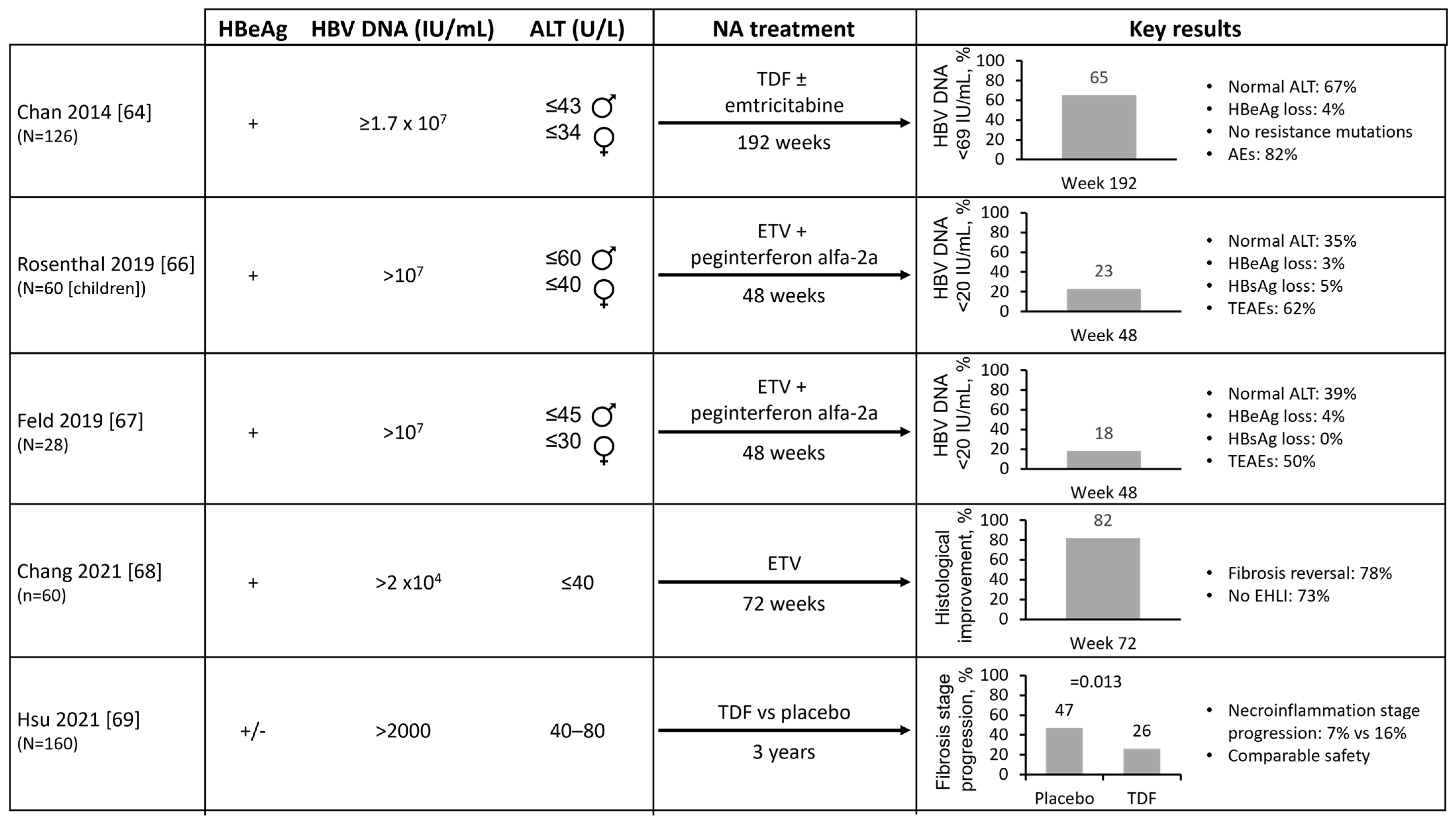
5. Potential Impact of Early HBV Treatment on Hepatocarcinogenesis and Clinical HBV Parameters

5.1. Potential Impact of HBV Treatment on HBV DNA Integration

The intermediary form of HBV prior to integration into the host DNA contains double-stranded linear DNA (dslDNA). As dslDNA is formed via the reverse transcription of HBV RNA [62], NAs should reduce the formation of dslDNA and DNA integration into the host genome. While data on antiviral treatment and HBV DNA integration are limited, some evidence suggests that NA therapy may reduce hepatocarcinogenesis. Analysis of treated and untreated liver biopsies has demonstrated that treatment is associated with reductions in viral load, integrations [13,18], and chromosomal translocations [13]. Further investigation of the effect of antiviral treatment on HBV DNA integrations is required; however, these emerging data provide support for early CHB treatment with respect to HBV DNA integration and reducing HCC risk.

5.2. Potential Impact of Early HBV Treatment on Clinical HBV Parameters

A small number of studies have evaluated the impact of antiviral treatment on the virological, serological, and liver-related outcomes in CHB patients ineligible for treatment under the current guidelines (Figure 3). Data on long-term outcomes including HCC are not available, and RCTs comparing antiviral treatment to no treatment have not been performed. However, a meta-analysis that included two studies of IT patients found moderate-quality evidence for improved intermediate outcomes (viral suppression, HBeAg seroconversion/loss) with antiviral therapy [63]. Additionally, a Phase 2 study of TDF ± emtricitabine treatment in 126 IT patients found that 65% of patients had HBV DNA <69 IU/mL after 192 weeks of treatment, with 42% of patients with a moderate aMAP (age, male, albumin-bilirubin, and platelets) risk score at baseline shifting to the low-risk category with no HCC reported [64,65]. However, only 4% and 0% of patients had HBeAg or HBsAg loss, respectively. Based on these results, the authors concluded that routine NA treatment of patients with IT CHB is not warranted, as reflected in the current guidelines.
A multicenter study investigating ETV + peginterferon alfa-2a treatment in 60 children with IT CHB found that 75% had HBV DNA ≤1000 IU/mL and 23% had HBV DNA <20 IU/mL after 48 weeks of treatment, with HBeAg and HBsAg loss in two patients [66]. Another study of ETV + peginterferon alfa-2a treatment in 28 adult IT patients reported HBV DNA ≤1000 IU/mL in 93% of patients and HBV DNA <20 IU/mL in 18% of patients after 48 weeks [67]. In both studies, HBV DNA levels increased following discontinuation [66,67]. Analysis of 181 treatment-naïve IT CHB patients, where 33% of patients had evident histological liver injury (EHLI) at baseline, reported histological improvement and fibrosis reversal in 82% and 78% of patients with EHLI, respectively, following 72 weeks of ETV treatment, with 73% of patients no longer having EHLI [68].

5.3. Impact of Treatment on ‘Gray Zone’ Patients

Evidence for the treatment of ‘gray zone’ patients was provided by an analysis from the TORCH-B study, a randomized, double-blind, placebo-controlled study examining the treatment of patients with HBV DNA >2000 IU/mL, ALT 40–80 U/L, and no cirrhosis (79% HBeAg negative) [69]. During 3 years of follow-up, the placebo group showed a significantly higher proportion of progression in the fibrosis stage compared with the TDF group (47% vs. 26%; p = 0.013).

5.4. Impact of Early HBV Treatment in Patients with HBV/HIV Co-Infection

A unique group of CHB patients who routinely receive early antiviral treatment are those co-infected with human immunodeficiency virus (HIV). Indeed, provision of antivirals as pre-exposure prophylaxis to individuals at high risk of infection is considered as an important step in HIV control [70]. Many antiretroviral regimens include an NA component (TDF or TAF) and are initiated irrespective of the HBV DNA or ALT levels [3,4]. Consequently, the analysis of HCC risk in these patients can provide data about the potential impact of early antiviral treatment, with the major caveat that these are not RCTs. Among the 3625 HBV/HIV co-infected patients, the HCC incidence remained stable in those on NA treatment, but increased among patients receiving a regimen not including an NA [71]. Results from a study of antiviral-treated HBV mono-infected (n = 53,974) and HBV/HIV co-infected (n = 822) patients demonstrated lower HCC among HBV/HIV co-infected patients vs. HBV mono-infected patients [72]. Similarly, the analysis of claims data found lower HCC rates in HBV/HIV co-infected patients (n = 7764) compared with HBV mono-infected patients (n = 13,964) [73]. Assuming HIV co-infection is a proxy for early HBV antiviral treatment, these data suggest that the universal antiviral treatment of CHB patients may reduce the HCC risk.

6. Cost Effectiveness of Expanded HBV Treatment Strategies

Several studies have estimated that the costs associated with expanded CHB therapy may be offset by reduced expenditure needed for future consequences of disease progression from untreated CHB (Table 3).
A recent Chinese study that modeled the cost effectiveness of 136 expanded treatment strategies found treating all HBsAg-positive patients aged 18–80 years with a treatment coverage of 80% to be the most cost-effective strategy [74]. This strategy was predicted to prevent 82.0% of HBV-related complications by 2050, although a treatment coverage of ≥60% was considered sufficient to achieve the WHO goal of a 65% reduction in CHB-related mortality by 2030. Importantly, lowering the treatment initiation threshold was found to be more effective in preventing CHB-related complications than increasing the treatment coverage. In light of this, the Chinese hepatology and infectious diseases societies have revised their guidelines for the prevention and treatment of CHB, recommending antiviral treatment in all HBsAg-positive patients with detectable serum HBV DNA (i.e., HBV DNA 10–20 IU/mL) over the age of 30 years, regardless of ALT level [75].
Other studies from France and the U.S. have shown that expanding treatment to all CHB patients, regardless of fibrosis level and disease phase, to be the most cost-effective strategy in terms of clinical outcomes [76,77]. Furthermore, an economic impact analysis found that this strategy had a lower overall cost than continuing with the current guidelines, as fewer patients were lost to follow-up or presented with advanced liver disease [78]. Similarly, a Korean study reported that starting treatment in the IT phase was more cost effective than delaying until the IA phase [79]. However, a UK study found that while treating all HBeAg-negative patients was the most cost-effective strategy, for HBeAg-positive patients, it was more cost effective to only treat those with fibrosis stage 2 and above [80].
Studies from Korea have compared the current guidelines with an extended indication (all patients with HBV DNA ≥2000 IU/mL and any ALT). Under this strategy, HCC risk decreased by 1%, 2%, and 6% per 10% increase in the treatment uptake rate under the Korean National Health Insurance, EASL, and extended indications, respectively [81]. This approach was estimated to be highly cost effective and would be most impactful when 70% of patients with HBV DNA ≥2000 IU/mL were treated, regardless of ALT and HBeAg status [82].
However, a study from Saudi Arabia found that the treatment costs needed to be reduced in order for expanded treatment strategies to achieve a positive return on investment (ROI) [83]. Compared with the scenario of no policy change, they reported that the WHO target strategy (diagnose 90% of infections and treat 80% of high viral load patients by 2030) would lead to a 30–35% reduction in HCC and liver-related deaths, while a diagnose-and-treat-all strategy (diagnose and treat all infected patients by 2022) would lead to a 50–55% reduction by 2030. Achieving the WHO targets was estimated to achieve a ROI by 2021; however, the diagnose-and-treat-all strategy would require at least 50% lower treatment costs to achieve a ROI by 2028.
Table 3. Cost effectiveness and clinical impact of the expanded HBV treatment strategies.
Author and YearModel Population and Selected Input ParametersTreatment StrategiesKey Results
Zhang 2023 [74]Chinese model of 136 scenarios based on cross combination of:
  • ALT treatment initiation thresholds (40 U/L, 35 U/L for males and 25 U/L for females, 30 U/L for males and 19 U/L for females, and treating HBsAg-positive individuals regardless of ALT values)
  • Population age groups (18–80, 30–80, and 40–80 years)
  • Implementation durations (2023, 2028, and 2033)
  • Treatment coverages (20%, 40%, 60%, and 80%)
  • Base case: status quo in China
  • 135 treatment-expanding scenarios
  • Treating all HBsAg-positive individuals with 80% coverage was the most effective strategy for reducing HBV-related complications
  • This strategy was also the most cost effective; however, all expanded treatment strategies were cost effective by 2050
Lepers 2020 [76]CHB patients in French ANRS CO22 HEPATHER cohort
  • HBeAg-positive chronic infection: 1%
    F0–F3: 30 years, HCC transition probability = 0.001
    F4: 54 years, HCC transition probability = 0.001
  • HBeAg-positive chronic hepatitis: 6%
    F0–F3: 36 years, HCC transition probability = 0.001
    F4: 54 years, HCC transition probability rate = 0.019
  • HBeAg-negative chronic infection: 56%
    F0–F3: 42 years, HCC transition probability = 0.001
    F4: 54 years, HCC transition probability = 0.017
  • HBeAg-negative chronic hepatitis: 36%
    F0–F3: 42 years, HCC transition probability = 0.001
    F4: 54 years, HCC transition probability = 0.030
  • Follow current treatment recommendations
  • Treat patients in chronic hepatitis disease phase regardless of fibrosis stage
  • Treat patients with ≥F2 fibrosis score regardless of disease phase
  • Treat all patients
  • Treat all patients was the most expensive and cost-effective strategy and the most effective in terms of clinical outcomes
Razavi-Shearer 2021 [77]U.S. model including historical and future impact of immigration using 164 country-specific disease burden and transmission models
  • Base case: current treatment strategy
  • Treat all: treat all HBsAg-positive individuals
  • Disease burden outputs through 2050 compared with base case:
    Treat-all strategy: ↓ 10,000 CHB cases, ↓ 49,000 decompensated cirrhosis cases, ↓ 132,000 HCC cases, ↓ 157,000 deaths
  • Cost effectiveness of treat-all strategy compared with base case:
    Current estimated annual NA price (USD$5400) → cost effective
    Annual NA price USD$2000 → highly cost effective
    Annual NA price USD$750 → cost saving
Razavi 2020 [78]Economic impact analysis
  • Test and treat all HBsAg-positive individuals
  • Treat according to current guidelines
  • Treating according to the currently guidelines was more expensive than test-and-treat-all strategy due to loss to follow-up and presentation with advance liver disease
Kim 2021 [79]Hypothetical CHB patients
  • 35 years of age
  • HBeAg positive
  • Mean HBV DNA 107.6 IU/mL
  • Normal ALT
  • Non-cirrhotic
Annual transition probabilities to HCC:
  • Treat IT: 0.0033
  • Untreated IT: 0.0073
  • Treat IT: start treatment at IT phase
  • Untreated IT: delay treatment until immune-active phase
  • Treat IT strategy was increasingly cost effective compared with untreated IT strategy
  • Inclusion of lost productivity costs showed that treat IT strategy was dominant, with ICER below 0 in most cases
Crossan 2016 [80]Hypothetical CHB patients with suspected fibrosis (n = 1000)
Separate Markov models constructed for HBeAg-positive (starting age 31 years) and -negative (starting age 40 years) patients
Transition probabilities:
  • Moderate fibrosis → HCC: 0.048
  • Cirrhosis → HCC: 0.024
  • Treat all without fibrosis assessment
  • Biopsy all patients and treat those with fibrosis stage ≥2
  • Test all patients with non-invasive test and treat those with fibrosis stage ≥2
  • Treat no patients
  • HBeAg-negative patients: treat all patients was the most cost-effective strategy
  • HBeAg-positive patients: test all patients using FibroScan and treat those with fibrosis stage ≥2 was the most cost-effective strategy
Shim 2016 [81]CHB patients in Korea
  • Mean age: 56 years
  • HBeAg positive: 66%
  • HBV DNA > 104 IU/mL: 48%
  • Mean ALT: 30 U/L
  • Non-cirrhotic
HCC risk predicted using REACH-B score
  • Korean NHI: HBV DNA ≥20,000 IU/mL and ALT ≥80 IU/L
  • EASL: HBV DNA ≥2000 IU/mL and ALT ≥33 IU/L in men and ≥25 IU/L in women
  • New extended indication: HBV DNA ≥2000 IU/mL and any ALT
  • Total 5-year HCC risk:
    Korean NHI: 2.5%
    EASL: 2.1%
    New extended indication: 1.1%
Lim 2021 [82]Virtual CHB cohort based on Korean data
  • Total HBsAg-positive population (2016): 1,478,500
  • Total diagnosed (2020): 1,125,400
  • Newly diagnosed: 19,600
  • On treatment (2019): 262,000
  • Base case: current eligibility requirements maintained through to 2035
  • Treat 70% + cirrhosis: treat 70% of eligible patients if current guidelines extended to include all cirrhotic patients
  • Treat 70% + ULN: treat 70% of eligible patients if current guidelines lowered ALT restriction to ULN
  • Treat 70% + ≥2000 IU/mL: treat 70% of eligible patients if current guidelines removed HBeAg and ALT restrictions and included all those with HBV DNA ≥2000 IU/mL
  • Disease burden outputs by 2035 compared with base case:
    Treat 70% + cirrhosis: ↓ 4300 decompensated cirrhosis cases, ↓ 13,000 HCC cases, ↓ 11,800 deaths
    Treat 70% + ULN: ↓ 7200 decompensated cirrhosis cases, ↓ 26,700 HCC cases, ↓23,000 deaths
    Treat 70% + ≥2000 IU/mL: ↓ 9800 decompensated cirrhosis cases, ↓ 43,300 HCC cases, ↓ 37,000 deaths
  • All scenarios were highly cost effective
Sanai 2020 [83]CHB patients in Saudi Arabia using estimated national prevalence in 2017
  • HBsAg prevalence: 1.7%
  • HCC incidence: 1500 cases
  • 77% of patients aged 35–60 years
  • Base case: current scenario in Saudi Arabia
  • Achieve WHO target: diagnose 90% of infections and treat 80% of high viral load patients by 2030
  • Diagnose and treat all: diagnose and treat all infected patients by 2022
  • Achieve WHO target strategy would cause 30% reduction in HCC and 35% reduction in liver-related deaths and would achieve positive ROI by 2021
  • Diagnose-and-treat-all strategy would cause 50% reduction in HCC and 55% reduction in liver-related deaths and would require ≥50% reduction in treatment costs to achieve positive ROI by 2028
ALT, alanine aminotransferase; CHB, chronic hepatitis B; EASL, European Association for the Study of the Liver; HBeAg, hepatitis B envelope antigen; HBsAg, hepatitis B surface antigen; HBV, hepatitis B virus; HCC, hepatocellular carcinoma; ICER, incremental cost-effectiveness ratio; IT, immune tolerant; NA, nucleos(t)ide analog; NHI, national health insurance; ROI, return on investment; ULN, upper limit of normal; WHO, World Health Organization.

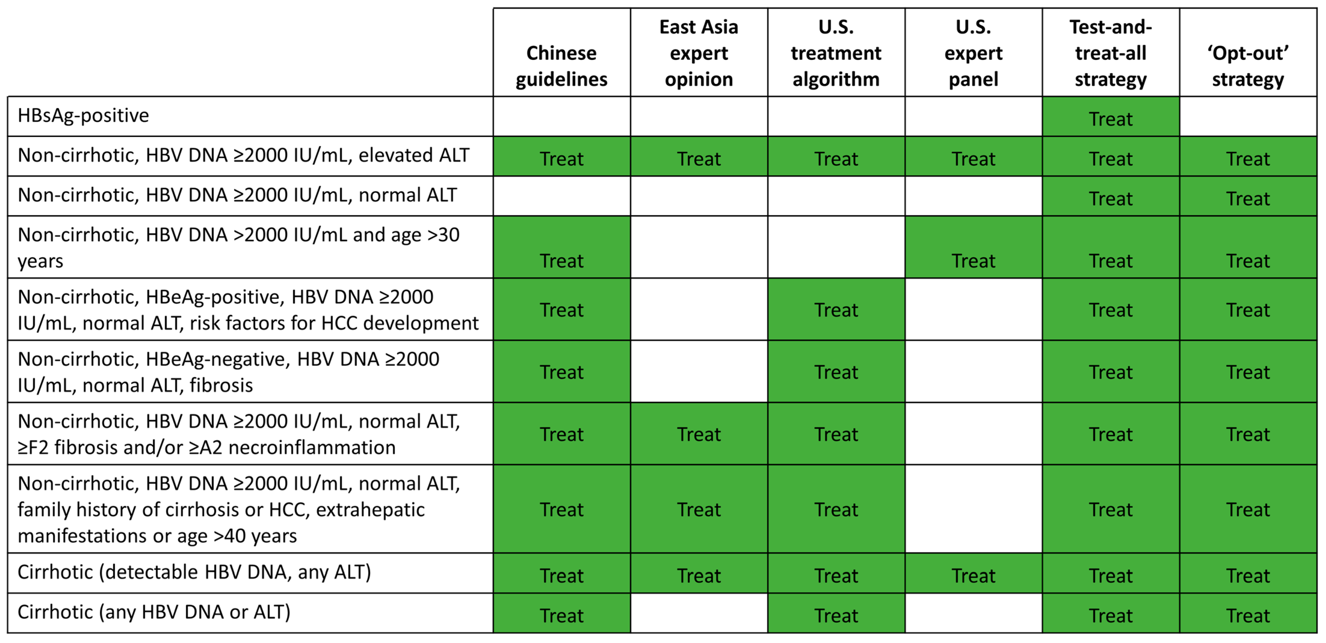
7. Potential Strategies for Expanded HBV Treatment

Current international CHB guidelines are complicated and only recommend antiviral treatment in subsets of patients based on liver damage, serum HBV DNA, and ALT levels (Table 1) [3,4,6]. This is in contrast to hepatitis C or HIV where recent guidelines have promoted the early treatment of all infected patients regardless of end-organ damage [70]. However, evidence from multiple studies, as discussed in this review, demonstrates that patients currently ineligible for antiviral treatment may have liver damage and remain at risk for disease progression and HCC. Consequently, we believe that it is time to consider the adoption of expanded treatment strategies to reduce these risks.
As above-mentioned, the Chinese hepatology and infectious disease societies have recently adopted such an expanded treatment strategy [75]. In their latest guidelines, antiviral treatment is recommended in all HBsAg-positive patients with detectable serum HBV DNA (i.e., HBV DNA > 10–20 IU/mL) over the age of 30 years, regardless of ALT level. Under this strategy, 92% of all patients with HBV in China (75 million individuals) are now considered eligible for treatment. Antiviral treatment is also recommended for HBV DNA seropositive patients younger than 30 years of age with persistently elevated ALT (i.e., >upper limit of normal), those with compensated cirrhosis, and those with a risk factor for disease progression (≥grade 2 inflammation or ≥stage 2 fibrosis, a family history of HBV-related cirrhosis or HCC, or HBV-related extrahepatic manifestations).
There are several other expanded treatment strategies proposed throughout the world including an East Asia expert opinion [61], a U.S. treatment algorithm [84], a test-and-treat-all HBsAg-positive patients strategy, and expert recommendations for the simplification of current guidelines [85] (Figure 4). Both the East Asia expert opinion and the U.S. treatment algorithm propose initiating antiviral treatment in more CHB patients at risk of HCC and adverse liver outcomes than current EASL, AASLD, and APASL treatment guidelines [3,4,6,61,84]. However, they still require the measurement of HBV DNA and ALT levels, and fibrosis assessment prior to treatment decision-making. In contrast, the test-and-treat-all strategy, where all HBsAg-positive patients would be eligible for NA treatment, does not require HBV DNA testing and could reduce the diagnosis costs. However, this approach would require mass screening to diagnose all HBsAg-positive patients, which has major implications for public health policies. A test-and-treat-all protocol for HBV elimination has been successfully implemented in a national program in Uzbekistan [86].
The data that we reviewed collectively provide support for the simplification of treatment initiation strategies that incorporate broader treatment of adult patients with HBV DNA ≥2000 IU/mL, regardless of ALT levels. Furthermore, an ‘opt-out’ strategy to treat all non-cirrhotic patients with HBV DNA ≥2000 IU/mL, regardless of ALT levels, would first define the patients who may remain untreated with minimal long-term risk of disease progression and HCC, namely true ‘inactive carriers’. Compared with traditional guideline recommendations, this strategy would incorporate the treatment of (1) all cirrhotic patients, (2) gray-zone patients with viremia and normal ALT, and (3) IT patients. We submit that the data we have compiled in the preceding sections provide convincing evidence that the benefits of an expanded approach outweigh the costs and any risks associated with early treatment. This simplified approach would only require testing once for HBsAg status and HBV DNA levels, and HBV DNA testing could be omitted in regions with limited access. Optimal CHB treatment may also differ by country or region based on HBV prevalence, the costs of diagnostic testing and NA treatment, and reimbursement policies. An economic impact analysis of HBV in 25 countries suggested that an ‘opt-out’ strategy diagnosing 90% of infections and treating 80% of infected patients would be cost effective or cost saving in all countries [87].
The potential safety implications of expanding the initiation of long-term antiviral treatment should, of course, be considered. However, accumulating clinical experience suggests a minimal risk of side effects associated with current antiviral treatment options, and this must therefore be balanced against the risk of disease progression and HCC in untreated CHB patients. Other potential concerns related to long-term antiviral treatment are resistance and poor adherence. ETV, TDF, and TAF have a high barrier to resistance; no resistance to TDF or TAF has been detected, and resistance to ETV is rare among treatment-naïve patients [3,4]. Furthermore, adherence rates to NA therapies are generally high [39]. Finally, and importantly, the current research to achieve a functional cure for CHB makes us optimistic that any concerns related to long-term NA therapy are likely to be time limited. Although HBV cure may not be anticipated in the immediate future, the bar to initiate antiviral treatment may certainly be lowered.
In conclusion, the available molecular, clinical, and economic data provide a strong rationale for the earlier initiation of antiviral treatment in CHB patients to reduce the risk of disease progression and HCC. Adoption of such a simplified ‘treat to prevent’ approach could save countless lives in a cost-effective manner. In parallel, investment in research efforts into finding a functional cure for CHB should continue to dramatically change the treatment paradigm in the future.

Supplementary Materials

The following supporting information can be downloaded at: https://www.mdpi.com/article/10.3390/v15040997/s1, Figure S1: CHB disease phases; Figure S2: Flow diagram of literature search for clinical review; Table S1: Risk of liver damage in CHB patients with normal or minimally elevated ALT levels; File S1: Phases of CHB disease; File S2: Search strategy for clinical review; File S3: Additional literature.

Author Contributions

All authors contributed to the review of the published data, drafting of the manuscript, and critical revision of the manuscript. All authors have read and agreed to the published version of the manuscript.

Funding

Financial support for third-party medical writing assistance was provided by Gilead Sciences.

Institutional Review Board Statement

Not applicable.

Informed Consent Statement

Not applicable.

Data Availability Statement

No new data were created or analyzed in this study. Data sharing is not applicable to this article.

Acknowledgments

We are grateful to the World Health Organization, in particular Philippa Easterbrook MD, for reviewing this article and providing constructive opinions. Medical writing support was provided by Karen Beckett and Beth Reason of Elements Communications.

Conflicts of Interest

Y.-S.L. has served as a speaker, advisory board member, consultant and/or has received research support from Assembly Biosciences, Bayer, Gilead Sciences, GlaxoSmithKline, OliX Pharmaceuticals, Vaccitech, and Vir Biotechnology, and has received grants from the Patient Centered Clinical Research Project (grant number: HC20C0062) of the National Evidence-Based Healthcare Collaborating Agency and the National R&D Program for Cancer Control through the National Cancer Center (grant number: HA21C010), funded by the Ministry of Health and Welfare of the Republic of Korea. W.R.K. has served as a consultant for Gilead Sciences and received research support from Roche. D.D. has served as a consultant and speaker for Gilead Sciences and received research grants from Assembly Biosciences and Enanta Pharmaceuticals. Professor Kao has nothing to declare. J.F.F. and L.J.Y. are employees of and own stock in Gilead Sciences. L.R.R. has received research grants from Bayer, Boston Scientific, Exact Sciences, Fujifilm Medical Systems, Gilead Sciences, Glycotest Inc., RedHill Biopharma, and TARGET PharmaSolutions, received consulting fees from Global Life Sciences, MedEd Design LLC, Pontifax Venture Capital, and The Lynx Group, served as an advisory board member for AstraZeneca, Bayer, Eisai, Exact Sciences, GRAIL, QED Therapeutics, and Roche, has a patent for the materials and methods for diagnosis, prognosis, monitoring, or recurrence and assessment of therapeutic/prophylactic treatment of pancreaticobiliary cancer (U.S. patent number 9,469,877; issued 18 October 2016), and played an unpaid leadership role for Africa Partners Medical, Hepatitis B Foundation, and West Africa Institute for Liver and Digestive Disease Foundation. L.R.R. has received research grants from Gilead Sciences, the John C Martin Foundation, The Hepatitis Fund, and ZeShan Foundation, has served as an advisory board member for Abbott, AbbVie, Gilead Sciences, Janssen Pharmaceuticals, Merck, Roche, and VBI Vaccines and is an unpaid member of the CDA Foundation Board. Professor Kennedy has received research grants from Gilead Sciences, received consulting fees from Aligos, Antios Therapeutics, Assembly Biosciences, Gilead Sciences, GlaxoSmithKline, Janssen, and Immunocore, and is the BASL HBV special interest group lead.

References

  1. World Health Organization. Hepatitis B Fact Sheet; Published Online 27 July 2021. Available online: https://www.who.int/news-room/fact-sheets/detail/hepatitis-b (accessed on 3 September 2021).
  2. Foreman, K.J.; Marquez, N.; Dolgert, A.; Fukutaki, K.; Fullman, N.; McGaughey, M.; Pletcher, M.A.; Smith, A.E.; Tang, K.; Yuan, C.-W.; et al. Forecasting life expectancy, years of life lost, and all-Cause and cause-Specific mortality for 250 causes of death: Reference and alternative scenarios for 2016-40 for 195 countries and territories. Lancet 2018, 392, 2052–2090. [Google Scholar] [CrossRef] [PubMed]
  3. European Association for the Study of the Liver (EASL). EASL 2017 Clinical Practice Guidelines on the management of hepatitis B virus infection. J. Hepatol. 2017, 67, 370–398. [Google Scholar] [CrossRef]
  4. Sarin, S.K.; Kumar, M.; Lau, G.K.; Abbas, Z.; Chan, H.L.Y.; Chen, C.J.; Chen, D.S.; Chen, H.L.; Chen, P.J.; Chien, R.N.; et al. Asian-Pacific clinical practice guidelines on the management of hepatitis B: A 2015 update. Hepatol. Int. 2016, 10, 1–98. [Google Scholar] [CrossRef]
  5. Lin, C.L.; Kao, J.H. Prevention of hepatitis B virus-related hepatocellular carcinoma. Hepatoma Res. 2021, 7, S303–S309. [Google Scholar] [CrossRef]
  6. Terrault, N.A.; Lok, A.S.F.; McMahon, B.J.; Chang, K.-M.; Hwang, J.P.; Jonas, M.M.; Brown, R.S., Jr.; Bzowej, N.H.; Wong, J.B. Update on prevention, diagnosis, and treatment of chronic hepatitis B: AASLD 2018 hepatitis B guidance. Hepatology 2018, 67, 1560–1599. [Google Scholar] [CrossRef] [PubMed]
  7. Seto, W.K.; Lo, Y.R.; Pawlotsky, J.M.; Yuen, M.F. Chronic hepatitis B virus infection. Lancet 2018, 392, 2313–2324. [Google Scholar] [CrossRef] [PubMed]
  8. Liu, W.B.; Wu, J.F.; Du, Y.; Cao, G.W. Cancer Evolution–Development: Experience of Hepatitis B Virus-Induced Hepatocarcinogenesis. Curr. Oncol. 2016, 23, 49–56. [Google Scholar] [CrossRef]
  9. Ringelhan, M.; McKeating, J.A.; Protzer, U. Viral hepatitis and liver cancer. Philos. Trans. R. Soc. B Biol. Sci. 2017, 372, 20160274. [Google Scholar] [CrossRef]
  10. Chemin, I.; Zoulim, F. Hepatitis B virus induced hepatocellular carcinoma. Cancer Lett. 2009, 286, 52–59. [Google Scholar] [CrossRef]
  11. An, P.; Xu, J.; Yu, Y.; Winkler, C.A. Host and Viral Genetic Variation in HBV-Related Hepatocellular Carcinoma. Front. Genet. 2018, 9, 261. [Google Scholar] [CrossRef]
  12. Ramirez, R.; van Buuren, N.; Gamelin, L.; Soulette, C.; May, L.; Han, D.; Yu, M.; Choy, R.; Cheng, G.; Bhardwaj, N.; et al. Targeted Long-Read Sequencing Reveals Comprehensive Architecture, Burden, and Transcriptional Signatures from Hepatitis B Virus-Associated Integrations and Translocations in Hepatocellular Carcinoma Cell Lines. J. Virol. 2021, 95, e0029921. [Google Scholar] [CrossRef]
  13. Pan, D.; Soulette, C.; Han, D.; Ramirez, R.; Van Buuren, N.; Lin, J.-T.; Tseng, C.-H.; Li, L.; Fletcher, S.; Mo, H.; et al. Decrease in the burden of integrated hepatitis B virus DNA in chronic hepatitis B patients with minimally elevated alanine aminotransferase on tenofovir disoproxil fumarate as revealed by long-read DNA-sequencing. J. Hepatol. 2022, 77, S841–S842. [Google Scholar] [CrossRef]
  14. The Cancer Genome Atlas Research; The Cancer Genome Atlas Research Network. Comprehensive and Integrative Genomic Characterization of Hepatocellular Carcinoma. Cell 2017, 169, 1327–1341. [Google Scholar] [CrossRef] [PubMed]
  15. Larsson, S.B.; Tripodi, G.; Raimondo, G.; Saitta, C.; Norkrans, G.; Pollicino, T.; Lindh, M. Integration of hepatitis B virus DNA in chronically infected patients assessed by Alu-PCR. J. Med. Virol. 2018, 90, 1568–1575. [Google Scholar] [CrossRef] [PubMed]
  16. Erken, R.; Loukachov, V.; van Dort, K.; Hurk, A.V.D.; Takkenberg, R.B.; de Niet, A.; Jansen, L.; Willemse, S.; Reesink, H.W.; Kootstra, N. Quantified integrated hepatitis B virus is related to viral activity in chronic hepatitis B patients. Hepatology 2022, 76, 196–206. [Google Scholar] [CrossRef]
  17. Svicher, V.; Salpini, R.; Piermatteo, L.; Carioti, L.; Battisti, A.; Colagrossi, L.; Scutari, R.; Surdo, M.; Cacciafesta, V.; Nuccitelli, A.; et al. Whole exome HBV DNA integration is independent of the intrahepatic HBV reservoir in HBeAg-negative chronic hepatitis B. Gut 2021, 70, 2337–2348. [Google Scholar] [CrossRef] [PubMed]
  18. Hsu, Y.-C.; Suri, V.; Nguyen, M.H.; Huang, Y.-T.; Chen, C.-Y.; Chang, I.-W.; Tseng, C.-H.; Wu, C.-Y.; Lin, J.-T.; Pan, D.Z.; et al. Inhibition of Viral Replication Reduces Transcriptionally Active Distinct Hepatitis B Virus Integrations with Implications on Host Gene Dysregulation. Gastroenterology 2022, 162, 1160–1170. [Google Scholar] [CrossRef]
  19. Chow, N.; Wong, D.; Lai, C.-L.; Mak, L.-Y.; Fung, J.; Ma, H.-T.; Lei, M.-W.; Seto, W.-K.; Yuen, M.-F. Effect of Antiviral Treatment on Hepatitis B Virus Integration and Hepatocyte Clonal Expansion. Clin. Infect. Dis. 2023, 76, e801–e809. [Google Scholar] [CrossRef]
  20. Tu, T.; Mason, W.S.; Clouston, A.D.; Shackel, N.A.; McCaughan, G.W.; Yeh, M.M.; Schiff, E.R.; Ruszkiewicz, A.R.; Chen, J.W.; Harley, H.A.J.; et al. Clonal expansion of hepatocytes with a selective advantage occurs during all stages of chronic hepatitis B virus infection. J. Viral Hepat. 2015, 22, 737–753. [Google Scholar] [CrossRef]
  21. Mason, W.S.; Gill, U.S.; Litwin, S.; Zhou, Y.; Peri, S.; Pop, O.; Hong, M.L.; Naik, S.; Quaglia, A.; Bertoletti, A.; et al. HBV-DNA Integration and Clonal Hepatocyte Expansion in Chronic Hepatitis B Patients Considered Immune Tolerant. Gastroenterology 2016, 151, 986–998. [Google Scholar] [CrossRef]
  22. Duan, M.; Hao, J.; Cui, S.; Worthley, D.L.; Zhang, S.; Wang, Z.; Shi, J.; Liu, L.; Wang, X.; Ke, A.; et al. Diverse modes of clonal evolution in HBV-related hepatocellular carcinoma revealed by single-cell genome sequencing. Cell Res. 2018, 28, 359–373. [Google Scholar] [CrossRef] [PubMed]
  23. Shin, E.-C.; Sung, P.S.; Park, S.-H. Immune responses and immunopathology in acute and chronic viral hepatitis. Nat. Rev. Immunol. 2016, 16, 509–523. [Google Scholar] [CrossRef]
  24. Kennedy, P.T.F.; Sandalova, E.; Jo, J.; Gill, U.; Ushiro–Lumb, I.; Tan, A.T.; Naik, S.; Foster, G.R.; Bertoletti, A. Preserved T-Cell Function in Children and Young Adults with Immune-Tolerant Chronic Hepatitis B. Gastroenterology 2012, 143, 637–645. [Google Scholar] [CrossRef] [PubMed]
  25. Park, J.-J.; Wong, D.K.; Wahed, A.S.; Lee, W.M.; Feld, J.J.; Terrault, N.; Khalili, M.; Sterling, R.K.; Kowdley, K.V.; Bzowej, N.; et al. Hepatitis B Virus–Specific and Global T-Cell Dysfunction in Chronic Hepatitis B. Gastroenterology 2016, 150, 684–695. [Google Scholar] [CrossRef]
  26. van Buuren, N.; Ramirez, R.; Turner, S.; Chen, D.; Suri, V.; Aggarwal, A.; Moon, C.; Kim, S.; Kornyeyev, D.; Bui, N.; et al. Characterization of the liver immune microenvironment in liver biopsies from patients with chronic HBV infection. JHEP Rep. 2022, 4, 100388. [Google Scholar] [CrossRef]
  27. Yang, H.I.; Lu, S.N.; Liaw, Y.F.; You, S.L.; Sun, C.A.; Wang, L.Y.; Hsiao, C.K.; Chen, P.J.; Chen, D.S.; Chen, C.J.; et al. Hepatitis B e Antigen and the Risk of Hepatocellular Carcinoma. N. Engl. J. Med. 2002, 347, 168–174. [Google Scholar] [CrossRef]
  28. Zhou, T.-C.; Lai, X.; Feng, M.-H.; Tang, Y.; Zhang, L.; Wei, J. Systematic review and meta-analysis: Development of hepatocellular carcinoma in chronic hepatitis B patients with hepatitis e antigen seroconversion. J. Viral Hepat. 2018, 25, 1172–1179. [Google Scholar] [CrossRef] [PubMed]
  29. Wiseman, E.; Fraser, M.A.; Holden, S.; Glass, A.; Rn, B.L.K.; Heron, L.G.; Maley, M.W.; Ayres, A.; Locarnini, S.A.; Levy, M.T. Perinatal transmission of hepatitis B virus: An Australian experience. Med. J. Aust. 2009, 190, 489–492. [Google Scholar] [CrossRef]
  30. Seck, A.; Maylin, S.; Akbar, S.M.F.; Funk, A.L.; Bercion, R.; Mishiro, S.; Ndiaye, B.; Fontanet, A.; Vray, M.; Simon, F.; et al. Poor Sensitivity of Commercial Rapid Diagnostic Tests for Hepatitis B e Antigen in Senegal, West Africa. Am. J. Trop. Med. Hyg. 2018, 99, 428–434. [Google Scholar] [CrossRef] [PubMed]
  31. Chen, C.-J.; Yang, H.-I.; Su, J.; Jen, C.-L.; You, S.-L.; Lu, S.-N.; Huang, G.-T.; Iloeje, U.H.; REVEAL-HBV Study Group. Risk of Hepatocellular Carcinoma Across a Biological Gradient of Serum Hepatitis B Virus DNA Level. JAMA 2006, 295, 65–73. [Google Scholar] [CrossRef]
  32. Iloeje, U.H.; Yang, H.I.; Su, J.; Jen, C.L.; You, S.L.; Chen, C.J. Predicting Cirrhosis Risk Based on the Level of Circulating Hepatitis B Viral Load. Gastroenterology 2006, 130, 678–686. [Google Scholar] [CrossRef]
  33. Iloeje, U.H.; Yang, H.I.; Jen, C.L.; Su, J.; Wang, L.; You, S.; Chen, C. Risk and Predictors of Mortality Associated with Chronic Hepatitis B Infection. Clin. Gastroenterol. Hepatol. 2007, 5, 921–931. [Google Scholar] [CrossRef]
  34. Kim, G.-A.; Han, S.; Choi, G.H.; Choi, J.; Lim, Y.-S. Moderate levels of serum hepatitis B virus DNA are associated with the highest risk of hepatocellular carcinoma in chronic hepatitis B patients. Aliment. Pharmacol. Ther. 2020, 51, 1169–1179. [Google Scholar] [CrossRef]
  35. Choi, W.-M.; Kim, G.-A.; Choi, J.; Han, S.; Lim, Y.-S. Increasing on-treatment hepatocellular carcinoma risk with decreasing baseline viral load in HBeAg-positive chronic hepatitis B. J. Clin. Investig. 2022, 132, e154833. [Google Scholar] [CrossRef] [PubMed]
  36. Liu, J.; Wang, J.; Yan, X.; Xue, R.; Zhan, J.; Jiang, S.; Geng, Y.; Liu, Y.; Mao, M.; Xia, J.; et al. Presence of Liver Inflammation in Asian Patients with Chronic Hepatitis B with Normal ALT and Detectable HBV-DNA in Absence of Liver Fibrosis. Hepatol. Commun. 2021, 6, 855–866. [Google Scholar] [CrossRef]
  37. Zoulim, F.; Mason, W.S. Reasons to consider earlier treatment of chronic HBV infections. Gut 2012, 61, 333–336. [Google Scholar] [CrossRef] [PubMed]
  38. Kim, G.-A.; Lim, Y.-S. Editorial: Hepatitis B DNA thresholds and risk of hepatocellular carcinoma: Different number patterns in HBeAg-positive versus HBeAg-negative patients. Authors’ reply. Aliment. Pharmacol. Ther. 2020, 51, 1440–1441. [Google Scholar] [CrossRef]
  39. Ford, N.; Scourse, R.; Lemoine, M.; Hutin, Y.; Bulterys, M.; Shubber, Z.; Donchuk, D.; Wandeler, G. Adherence to Nucleos(t)ide Analogue Therapies for Chronic Hepatitis B Infection: A Systematic Review and Meta-Analysis. Hepatol. Commun. 2018, 2, 1160–1167. [Google Scholar] [CrossRef]
  40. Liu, K.; Choi, J.; Le, A.; Yip, T.C.-F.; Wong, V.W.-S.; Chan, S.; Chan, H.L.-Y.; Nguyen, M.H.; Lim, Y.-S.; Wong, G.L.-H. Tenofovir disoproxil fumarate reduces hepatocellular carcinoma, decompensation and death in chronic hepatitis B patients with cirrhosis. Aliment. Pharmacol. Ther. 2019, 50, 1037–1048. [Google Scholar] [CrossRef] [PubMed]
  41. Kim, W.R.; Loomba, R.; Berg, T.; Schall, R.E.A.; Yee, L.J.; Dinh, P.V.; Flaherty, J.F.; Martins, E.B.; Therneau, T.M.; Jacobson, I.; et al. Impact of long-term tenofovir disoproxil fumarate on incidence of hepatocellular carcinoma in patients with chronic hepatitis B. Cancer 2015, 121, 3631–3638. [Google Scholar] [CrossRef] [PubMed]
  42. Papatheodoridis, G.V.; Idilman, R.; Dalekos, G.N.; Buti, M.; Chi, H.; van Boemmel, F.; Calleja, J.L.; Sypsa, V.; Goulis, J.; Manolakopoulos, S.; et al. The risk of hepatocellular carcinoma decreases after the first 5 years of entecavir or tenofovir in Caucasians with chronic hepatitis B. Hepatology 2017, 66, 1444–1453. [Google Scholar] [CrossRef]
  43. Sinn, D.H.; Kim, S.E.; Kim, B.K.; Kim, J.H.; Choi, M.S. The risk of hepatocellular carcinoma among chronic hepatitis B virus-infected patients outside current treatment criteria. J. Viral Hepat. 2019, 26, 1465–1472. [Google Scholar] [CrossRef]
  44. Alshuwaykh, O.; Daugherty, T.; Cheung, A.; Goel, A.; Dhanasekaran, R.; Ghaziani, T.T.; Ahmed, A.; Dronamraju, D.; Kumari, R.; Kwong, A.; et al. Incidence of hepatocellular carcinoma in chronic hepatitis B virus infection in those not meeting criteria for antiviral therapy. Hepatol. Commun. 2022, 6, 3052–3061. [Google Scholar] [CrossRef]
  45. Wang, C.Y.; Deng, Y.; Guo, C.; Ji, D. Nomogram for predicting significant liver injury in chronic hepatitis B patients with immune tolerance. J. Hepatol. 2021, 75, S730–S731. [Google Scholar]
  46. Zeng, D.-W.; Huang, Z.-X.; Lin, M.-X.; Kang, N.-L.; Lin, X.; Li, Y.-N.; Zhu, Y.-Y.; Liu, Y.-R. A novel HBsAg-based model for predicting significant liver fibrosis among Chinese patients with immune-tolerant phase chronic hepatitis B: A multicenter retrospective study. Ther. Adv. Gastroenterol. 2021, 14, 17562848211010675. [Google Scholar] [CrossRef]
  47. Lin, M.-H.; Li, H.-Q.; Zhu, L.; Su, H.-Y.; Peng, L.-S.; Wang, C.-Y.; He, C.-P.; Liang, X.-E.; Wang, Y. Liver Fibrosis in the Natural Course of Chronic Hepatitis B Viral Infection: A Systematic Review with Meta-Analysis. Dig. Dis. Sci. 2022, 67, 2608–2626. [Google Scholar] [CrossRef]
  48. Kim, G.-A.; Lim, Y.-S.; Han, S.; Choi, J.; Shim, J.H.; Kim, K.M.; Lee, H.C.; Lee, Y.S. High risk of hepatocellular carcinoma and death in patients with immune-tolerant-phase chronic hepatitis B. Gut 2018, 67, 945–952. [Google Scholar] [CrossRef] [PubMed]
  49. Kwon, J.H.; Lee, S.W.; Jang, J.W.; Yoo, S.H.; Song, D.S.; Kim, H.Y. Low risk of liver disease progression in untreated immune tolerant phase chronic hepatitis B patients compared to treated immune active phase patients: Multi-center cohort study up to 15 years. Hepatology 2019, 71, 574A. [Google Scholar]
  50. Seong, G.; Sinn, D.H.; Kang, W.; Gwak, G.-Y.; Choi, M.S.; Lee, J.H.; Koh, K.C.; Paik, S.W.; Paik, Y.-H. Age and fibrosis index for the prediction of hepatocellular carcinoma risk in patients with high hepatitis B virus DNA but normal alanine aminotransferase. Eur. J. Gastroenterol. Hepatol. 2022, 34, 69–75. [Google Scholar] [CrossRef] [PubMed]
  51. Lee, H.A.; Lee, H.W.; Kim, I.H.; Park, S.Y.; Sinn, D.H.; Yu, J.H.; Seo, Y.S.; Um, S.H.; Lee, J.I.; Lee, K.S.; et al. Extremely low risk of hepatocellular carcinoma development in patients with chronic hepatitis B in immune-tolerant phase. Aliment. Pharmacol. Ther. 2020, 52, 196–204. [Google Scholar] [CrossRef]
  52. Chen, J.D.; Yang, H.I.; Iloeje, U.H.; You, S.-L.; Lu, S.; Wang, L.; Su, J.; Sun, C.; Liaw, Y.; Chen, C. Carriers of Inactive Hepatitis B Virus Are Still at Risk for Hepatocellular Carcinoma and Liver-Related Death. Gastroenterology 2010, 138, 1747–1754. [Google Scholar] [CrossRef]
  53. Graf, C.; Knop, V.; Peiffer, K.H.; Dietz, J.; Berg, T.; Wedemeyer, H.; Cornberg, M.; Petersen, J.; Hueppe, D.; Sprinzl, M.; et al. Clinical outcomes in untreated European patients with chronic hepatitis B virus infection after seven years: A prospective longitudinal study (ALBATROS study). Hepatology 2019, 70, 409A–410A. [Google Scholar]
  54. Seo, S.I.; Kim, H.S.; Yang, B.K.; Kang, J.G.; Shin, W.G.; Lee, J.H.; Kim, H.Y.; Jang, M.K. Predictive factors for risk of hepatocellular carcinoma in immune inactive chronic hepatitis B. Clin. Res. Hepatol. Gastroenterol. 2020, 44, 711–717. [Google Scholar] [CrossRef]
  55. Liu, M.; Tseng, T.-C.; Jun, D.W.; Yeh, M.-L.; Trinh, H.; Wong, G.L.H.; Chen, C.-H.; Peng, C.-Y.; Kim, S.E.; Oh, H.; et al. Transition rates to cirrhosis and liver cancer by age, gender, disease and treatment status in Asian chronic hepatitis B patients. Hepatol. Int. 2021, 15, 71–81. [Google Scholar] [CrossRef] [PubMed]
  56. Cho, J.-Y.; Paik, Y.-H.; Sohn, W.; Cho, H.C.; Gwak, G.-Y.; Choi, M.S.; Lee, J.H.; Koh, K.C.; Paik, S.W.; Yoo, B.C. Patients with chronic hepatitis B treated with oral antiviral therapy retain a higher risk for HCC compared with patients with inactive stage disease. Gut 2014, 63, 1943–1950. [Google Scholar] [CrossRef]
  57. Huang, D.Q.; Li, X.; Le, M.H.; Le, A.K.; Yeo, Y.H.; Trinh, H.N.; Zhang, J.; Li, J.; Wong, C.; Wong, C.; et al. Natural History and Hepatocellular Carcinoma Risk in Untreated Chronic Hepatitis B Patients with Indeterminate Phase. Clin. Gastroenterol. Hepatol. 2022, 20, 1803–1812.e5. [Google Scholar] [CrossRef] [PubMed]
  58. Tseng, T.-C.; Hosaka, T.; Liu, C.-J.; Suzuki, F.; Hong, C.-M.; Kumada, H.; Yang, W.-T.; Hsu, C.-Y.; Su, T.-H.; Yang, H.-C.; et al. Hepatitis B Core-Related Antigen Stratifies the Risk of Liver Cancer in HBeAg-Negative Patients with Indeterminate Phase. Am. J. Gastroenterol. 2022, 117, 748–757. [Google Scholar] [CrossRef]
  59. Choi, G.H.; Kim, G.-A.; Choi, J.; Han, S.; Lim, Y.-S. High risk of clinical events in untreated HBeAg-negative chronic hepatitis B patients with high viral load and no significant ALT elevation. Aliment. Pharmacol. Ther. 2019, 50, 215–226. [Google Scholar] [CrossRef]
  60. McGill, M.R. The past and present of serum aminotransferases and the future of liver injury biomarkers. EXCLI J. 2016, 15, 817–828. [Google Scholar] [CrossRef] [PubMed]
  61. Kao, J.-H.; Hu, T.-H.; Jia, J.; Kurosaki, M.; Lim, Y.-S.; Lin, H.-C.; Sinn, D.H.; Tanaka, Y.; Wong, V.W.-S.; Yuen, M.-F. East Asia expert opinion on treatment initiation for chronic hepatitis B. Aliment. Pharmacol. Ther. 2020, 52, 1540–1550. [Google Scholar] [CrossRef]
  62. Tu, T.; Budzinska, M.A.; Shackel, N.A.; Urban, S. HBV-DNA Integration: Molecular Mechanisms and Clinical Implications. Viruses 2017, 9, 75. [Google Scholar] [CrossRef]
  63. Lok, A.S.F.; McMahon, B.J.; Brown, R.S., Jr.; Wong, J.B.; Ahmed, A.T.; Farah, W.; Almasri, J.; Alahdab, F.; Benkhadra, K.; Mouchli, M.A.; et al. Antiviral therapy for chronic hepatitis B viral infection in adults: A systematic review and meta-analysis. Hepatology 2016, 63, 284–306. [Google Scholar] [CrossRef]
  64. Chan, H.L.Y.; Chan, C.K.; Hui, A.J.; Chan, S.; Poordad, F.; Chang, T.-T.; Mathurin, P.; Flaherty, J.F.; Lin, L.; Corsa, A.; et al. Effects of Tenofovir Disoproxil Fumarate in Hepatitis B e Antigen-Positive Patients with Normal Levels of Alanine Aminotransferase and High Levels of Hepatitis B Virus DNA. Gastroenterology 2014, 146, 1240–1248. [Google Scholar] [CrossRef]
  65. Chan, H.L.Y.; Lim, Y.S.; Hou, J.; Hui, A.J.; Seto, W.K.; Chang, T.T.; Chuang, W.L.; Janssen, H.L.; Fung, S.K.; Ning, Q.; et al. Impact of long-term oral antiviral (OAV) treatment on hepatocellular carcinoma (HCC) risk in immune-tolerant and immune-active chronic hepatitis B patients (CHB) utilizing aMAP, a recently validated risk prediction tool. Hepatology 2021, 74, 487A–488A. [Google Scholar]
  66. Rosenthal, P.; Ling, S.C.; Belle, S.H.; Murray, K.F.; Rodriguez-Baez, N.; Schwarzenberg, S.J.; Teckman, J.; Lin, H.S.; Schwarz, K.B.; Hepatitis B Research Network (HBRN). Combination of Entecavir/Peginterferon Alfa-2a in Children With Hepatitis B e Antigen–Positive Immune Tolerant Chronic Hepatitis B Virus Infection. Hepatology 2019, 69, 2326–2337. [Google Scholar] [CrossRef]
  67. Feld, J.J.; Terrault, N.A.; Lin, H.S.; Belle, S.H.; Chung, R.T.; Tsai, N.; Khalili, M.; Perrillo, R.; Cooper, S.L.; Ghany, M.G.; et al. Entecavir and Peginterferon Alfa-2a in Adults With Hepatitis B e Antigen–Positive Immune-Tolerant Chronic Hepatitis B Virus Infection. Hepatology 2019, 69, 2338–2348. [Google Scholar] [CrossRef] [PubMed]
  68. Chang, X.; Wang, J.; Chen, Y.; Long, Q.; Song, L.; Li, Q.; Liu, H.; Shang, Q.; Yu, Z.; Jiang, L.; et al. A novel nomogram to predict evident histological liver injury in patients with HBeAg-positive chronic hepatitis B virus infection. Ebiomedicine 2021, 67, 103389. [Google Scholar] [CrossRef] [PubMed]
  69. Hsu, Y.-C.; Chen, C.-Y.; Chang, I.-W.; Chang, C.-Y.; Wu, C.-Y.; Lee, T.-Y.; Wu, M.-S.; Bair, M.-J.; Chen, J.-J.; Chen, C.-C.; et al. Once-daily tenofovir disoproxil fumarate in treatment-naive Taiwanese patients with chronic hepatitis B and minimally raised alanine aminotransferase (TORCH-B): A multicentre, double-blind, placebo-controlled, parallel-group, randomised trial. Lancet Infect. Dis. 2021, 21, 823–833. [Google Scholar] [CrossRef] [PubMed]
  70. Leoni, M.C.; Ustianowski, A.; Farooq, H.; Arends, J.E. HIV, HCV and HBV: A Review of Parallels and Differences. Infect. Dis. Ther. 2018, 7, 407–419. [Google Scholar] [CrossRef]
  71. Wandeler, G.; Mauron, E.; Atkinson, A.; Dufour, J.-F.; Kraus, D.; Reiss, P.; Peters, L.; Dabis, F.; Fehr, J.; Bernasconi, E.; et al. Incidence of hepatocellular carcinoma in HIV/HBV-coinfected patients on tenofovir therapy: Relevance for screening strategies. J. Hepatol. 2019, 71, 274–280. [Google Scholar] [CrossRef]
  72. Lui, G.; Yip, T.; Yuen, B.; Sze, A.; Hui, Y.-T.; Tse, Y.-K.; Wong, V.; Wong, G. 358. HIV Infection and the Risk of Hepatocellular Carcinoma in Patients with Hepatitis B Virus (HBV) Co-infection: A Propensity Score-matched Cohort Study. Open Forum Infect. Dis. 2019, 6 (Suppl. 2), S188. [Google Scholar] [CrossRef]
  73. Naggie, S.; Telep, L.; McQuaid, T.; Ramroth, H.; Gaggar, A.; Flaherty, J.F.; Chokkalingam, A.P.; Sulkowski, M.S. Risk of HCC in patients with HBV infection with and without HIV co-infection: A US administrative claims analysis. Hepatology 2020, 72, 494A. [Google Scholar]
  74. Zhang, S.; Wang, C.; Liu, B.; Lu, Q.-B.; Shang, J.; Zhou, Y.; Jia, J.; Xu, X.; Rao, H.; Han, B.; et al. Cost-effectiveness of expanded antiviral treatment for chronic hepatitis B virus infection in China: An economic evaluation. Lancet Reg. Health-West. Pac. 2023, 100738. [Google Scholar] [CrossRef]
  75. Chinese Society of Hepatology; Chinese Medical Association; Chinese Society of Infectious Diseases. Guidelines for the prevention and treatment of chronic hepatitis B (version 2022). Chin. J. Hepatol. 2022, 30, 1309–1331. [Google Scholar] [CrossRef]
  76. Lepers, C.; Fontaine, H.; Carrat, F.; Mathurin, P.; Pol, S.; Zoulim, F.; Yazdanpanah, Y.; Deuffic-Burban, S. Cost-effectiveness of scaling-up treatment with nucleoside analogue (NA) for chronic HBV infection: Towards a simplification of recommendations? (ANRS study). J. Hepatol. 2020, 73, S797–S798. [Google Scholar] [CrossRef]
  77. Razavi-Shearer, D.; Estes, C.; Gamkrelidze, I.; Razavi, H. Cost-effectiveness analysis of treating all HBsAg+ individuals in the United States. Hepatology 2021, 74, 22A. [Google Scholar]
  78. Razavi, H.; Sadirova, S.; Bakieva, S.; Razavi-Shearer, K.; Dunn, R.; Musabaev, E. The case for testing and treating all HBV patients. J. Hepatol. 2020, 73, S807. [Google Scholar] [CrossRef]
  79. Kim, H.-L.; Kim, G.-A.; Park, J.-A.; Kang, H.-R.; Lee, E.-K.; Lim, Y.-S. Cost-effectiveness of antiviral treatment in adult patients with immune-tolerant phase chronic hepatitis B. Gut 2021, 70, 2172–2182. [Google Scholar] [CrossRef]
  80. Crossan, C.; Tsochatzis, E.A.; Longworth, L.; Gurusamy, K.; Papastergiou, V.; Thalassinos, E.; Mantzoukis, K.; Rodriguez-Peralvarez, M.; O’Brien, J.; Noel-Storr, A.; et al. Cost-effectiveness of noninvasive liver fibrosis tests for treatment decisions in patients with chronic hepatitis B in the UK: Systematic review and economic evaluation. J. Viral Hepat. 2016, 23, 139–149. [Google Scholar] [CrossRef]
  81. Shim, J.-J.; Oh, I.H.; Kim, S.B.; Kim, J.W.; Lee, C.K.; Jang, J.Y.; Lee, J.-S.; Kim, B.-H. Predictive Value of Antiviral Effects in the Development of Hepatocellular Carcinoma in the General Korean Population with Chronic Hepatitis B. Gut Liver 2016, 10, 962–968. [Google Scholar] [CrossRef]
  82. Lim, Y.S.; Ahn, S.H.; Shim, J.J.; Razavi, H.; Razavi-Shearer, D.; Sinn, D.H. Impact of expanding antiviral treatment criteria at a population level in the Republic of Korea: A modeling analysis. Hepatol. Int. 2021, 15, S31. [Google Scholar]
  83. Sanai, F.M.; Alghamdi, M.; Dugan, E.; Alalwan, A.; Al-Hamoudi, W.; Abaalkhail, F.; AlMasri, N.; Razavi-Shearer, D.; Razavi, H.; Schmelzer, J.; et al. A tool to measure the economic impact of Hepatitis B elimination: A case study in Saudi Arabia. J. Infect. Public Health 2020, 13, 1715–1723. [Google Scholar] [CrossRef]
  84. Martin, P.; Nguyen, M.H.; Dieterich, D.T.; Lau, D.T.-Y.; Janssen, H.L.; Peters, M.G.; Jacobson, I.M. Treatment Algorithm for Managing Chronic Hepatitis B Virus Infection in the United States: 2021 Update. Clin. Gastroenterol. Hepatol. 2022, 20, 1766–1775. [Google Scholar] [CrossRef]
  85. Dieterich, D.; Graham, C.; Wang, S.; Kwo, P.; Lim, Y.-S.; Liu, C.-J.; Agarwal, K.; Sulkowski, M. It Is Time for a Simplified Approach to Hepatitis B Elimination. Gastro Hep Adv. 2023, 2, 209–218. [Google Scholar] [CrossRef]
  86. Musabaev, E.; Sadirova, S.; Bakieva, S. Simplified test and treat protocols for population level screening and elimination of hepatitis B and hepatitis C in Uzbekistan. J Hepatol. 2021, 75, S642–S643. [Google Scholar]
  87. Razavi, H. Cost-effectiveness analysis of HCV/HBV elimination in the world. In Proceedings of the Global Hepatitis Summit, Virtual Meeting, 18 June 2021. [Google Scholar]
Figure 1. HCC pathogenesis. These schematics summarize some of the key HCC pathogenesis mechanisms in CHB patients (A) and in which CHB disease phases these have been detected (B). (A) HBV infects normal liver hepatocytes and can lead to CHB infection. During HBV infection, chronic hepatitis can develop, leading to cirrhosis and HCC in some patients. Approximately 20% of HBV-associated HCC cases develop in the absence of cirrhosis. HCC pathogenesis mechanisms can be direct or indirect. Direct HCC pathogenesis mechanisms are mediated by HBV and include HBV DNA integration into the host genome and the expression of HBV-encoded oncogenic protein. HBV DNA integration causes changes to the host genome via insertional mutagenesis, promoting genomic instability, and can lead to the expression of mutant HBV proteins. Indirect HCC mechanisms are mediated by the host immune system attacking HBV-infected hepatocytes. This leads to chronic necroinflammation, liver regeneration, and fibrosis, which cause genetic and epigenetic changes within hepatocytes. (B) Several studies have shown that HBV DNA integration into the host genome can be detected in liver samples from CHB patients across disease phases. Immune activity against HBV can also be detected in CHB patients across disease phases, as can necroinflammation and fibrosis in some patients. CHB, chronic hepatitis B; HBeAg, hepatitis B envelope antigen; HBV, hepatitis B virus; HCC, hepatocellular carcinoma.
Viruses 15 00997 g001
Figure 2. Association between the baseline HBV DNA levels and HCC risk in untreated and NA-treated non-cirrhotic adult patients with CHB. (A). In untreated HBeAg-positive and -negative, non-cirrhotic, adult CHB patients with persistently normal ALT levels (n = 6949), HCC risk was the highest with baseline levels of approximately 6 log10 IU/mL. (B). In untreated HBeAg-positive, non-cirrhotic, adult CHB patients with persistently normal ALT levels (n = 2081), HCC risk was the highest with baseline HBV DNA levels of approximately 6 log10 IU/mL. (C). In HBeAg-positive, non-cirrhotic, adult CHB patients treated with ETV or TDF (n = 2073), the on-treatment HCC incidence rate increased incrementally with decreasing baseline HBV DNA levels ≥5 log10 IU/mL. (D). Compared with untreated HBeAg-positive, non-cirrhotic adult CHB patients with normal ALT levels (n = 2643), NA treatment in HBeAg-positive, non-cirrhotic, adult CHB patients (n = 2073) reduced the HCC incidence in patients with moderate baseline viral load (5.00–7.99 log10 IU/mL), but the HCC risk did not decrease to the same extent in patients with a high baseline viral load (≥8.0 log10 IU/mL). ALT, alanine aminotransferase; CHB, chronic hepatitis B; CI, confidence interval; ETV, entecavir; HBeAg, hepatitis B envelope antigen; HBV, hepatitis B virus; HCC, hepatocellular carcinoma; HR, hazard ratio; NA, nucleos(t)ide analog; TDF, tenofovir disoproxil fumarate. All panels in this figure were made by Y-S Lim based on data in Kim G-A et al. Aliment Pharmacol Ther. 2020;51:1169–1179 (panels A and B) and Choi W-M et al. J Clin Invest. 2022;132:e154833 (panels C and D).
Viruses 15 00997 g002
Figure 3. Impact of NA treatment on CHB clinical parameters in patients currently ineligible for antiviral treatment. This schematic summarizes the studies of NA treatment in CHB patients currently ineligible for antiviral treatment including those in the HBeAg-positive chronic infection/immune-tolerant disease phase. Patient numbers are shown in brackets in the first column. Patient baseline characteristics (HBeAg status and HBV DNA and ALT level) are shown in the second column. NA treatment and duration are shown in the third column. Key efficacy and safety results are shown in the fourth column. AE, adverse event; ALT, alanine aminotransferase; CHB, chronic hepatitis B; EHLI, evident histological liver injury; ETV, entecavir; HBeAg, hepatitis B envelope antigen; HBsAg, hepatitis B surface antigen; HBV, hepatitis B virus; NA, nucleos(t)ide analog; TDF, tenofovir disoproxil; TEAE, treatment-emergent adverse event [64,66,67,68,69].
Viruses 15 00997 g003
Figure 4. Potential expanded HBV treatment strategies. This figure summarizes the categories of CHB patients who would be eligible for antiviral treatment initiation under the proposed alternative treatment strategies. The Chinese guidelines [75], East Asian expert opinion [61], U.S. treatment algorithm [84] and U.S. expert panel [85] recommend initiation of antiviral treatment in more CHB patients compared with the current EASL, APASL, or AASLD guidelines, but include different patient subpopulations. The test-and-treat-all strategy would initiate antiviral treatment in all HBsAg-positive patients. The ‘opt-out’ strategy would initiate antiviral treatment in all cirrhotic patients and non-cirrhotic adult patients with HBV DNA ≥2000 IU/mL regardless of HBeAg and ALT status. AASLD, American Association for the Study of Liver Diseases; ALT, alanine aminotransferase; APASL, Asian Pacific Association for the Study of the Liver; CHB, chronic hepatitis B; EASL, European Association for the Study of the Liver; HBeAg, hepatitis B envelope antigen; HBsAg, hepatitis B surface antigen; HBV, hepatitis B virus; HCC, hepatocellular carcinoma.
Viruses 15 00997 g004
Table 1. Antiviral treatment criteria in the international guidelines.
EASL 2017 [3]AASLD 2018 [6]APASL 2016 [4]
No cirrhosisHBeAg-positive/negative AND
HBV DNA > 2000 IU/mL AND
ALT > ULN * and/or moderate liver necroinflammation or fibrosis
HBeAg-positive AND
HBV DNA > 20,000 IU/mL AND
ALT ≥ 2× ULN *
HBeAg-positive AND
HBV DNA > 20,000 IU/mL AND
ALT > 2× ULN *
HBV DNA > 20,000 IU/mL AND
ALT > 2× ULN *
HBeAg-negative AND
HBV DNA ≥2000 IU/mL AND
ALT ≥ 2× ULN *
HBeAg-negative AND
HBV DNA > 2000 IU/mL AND
ALT > 2× ULN *
HBeAg-positive/negative AND
family history of HCC or cirrhosis and extrahepatic manifestations
HBV DNA ≥2000 IU/mL AND
ALT > ULN * AND
significant necroinflammation or fibrosis or age > 40 years
Any HBV DNA or ALT if moderate to severe inflammation or significant fibrosis
CirrhosisDetectable HBV DNA
Any ALT level
HBV DNA < 2000 IU/mL §
Any ALT level
HBV DNA > 2000 IU/mL §
Any ALT level
Please note that the national treatment guidelines may differ from international treatment guidelines. * The EASL and APASL recommended ULN is 40 IU/L, the AASLD recommended ULN is 35 U/L for males and 25 U/L for females; Patients with HBeAg-positive chronic infection may be treated if older than 30 years regardless of the severity of liver histology; ALT > ULN and significant necroinflammation or fibrosis, but HBV DNA < 2000 IU/mL, treat; § For compensated cirrhosis. Patients with decompensated cirrhosis should be treated if there is detectable HBV DNA with any ALT levels. AASLD, American Association for the Study of Liver Diseases; ALT, alanine aminotransferase; APASL, Asian Pacific Association for the Study of the Liver; EASL, European Association for the Study of the Liver; HBeAg, hepatitis B e-antigen; HBV, hepatitis B virus; HCC, hepatocellular carcinoma; ULN, upper limit of normal.
Disclaimer/Publisher’s Note: The statements, opinions and data contained in all publications are solely those of the individual author(s) and contributor(s) and not of MDPI and/or the editor(s). MDPI and/or the editor(s) disclaim responsibility for any injury to people or property resulting from any ideas, methods, instructions or products referred to in the content.

