Brief Report

A Triple Gene-Deleted Pseudorabies Virus-Vectored Subunit PCV2b and CSFV Vaccine Protect Pigs against a Virulent CSFV Challenge

by
Ediane Silva
1,
Elizabeth Medina-Ramirez
1,
Selvaraj Pavulraj
2,
Douglas P. Gladue
1,
Manuel Borca
1 and
Shafiqul I. Chowdhury
2,*
1
US Department of Agricultural, ARS, Plum Island Animal Disease Center, Orient, NY 11957, USA
2
Department of Pathobiological Sciences, School of Veterinary Medicine, Louisiana State University, Baton Rouge, LA 70803, USA
*
Author to whom correspondence should be addressed.
Submission received: 8 September 2023 / Revised: 21 October 2023 / Accepted: 23 October 2023 / Published: 25 October 2023
(This article belongs to the Special Issue Strategies for Preventing Viral Diseases of Domestic Animals)

Abstract

:
Classical swine fever (CSF) remains one of the most economically significant viral diseases affecting domestic pigs and wild boars worldwide. To develop a safe and effective vaccine against CSF, we have constructed a triple gene-deleted pseudorabies virus (PRVtmv)-vectored bivalent subunit vaccine against porcine circovirus type 2b (PCV2b) and CSFV (PRVtmv+). In this study, we determined the protective efficacy of the PRVtmv+ against virulent CSFV challenge in pigs. The results revealed that the sham-vaccinated control group pigs developed severe CSFV-specific clinical signs characterized by pyrexia and diarrhea, and became moribund on or before the seventh day post challenge (dpc). However, the PRVtmv+-vaccinated pigs survived until the day of euthanasia at 21 dpc. A few vaccinated pigs showed transient diarrhea but recovered within a day or two. One pig had a low-grade fever for a day but recovered. The sham-vaccinated control group pigs had a high level of viremia, severe lymphocytopenia, and thrombocytopenia. In contrast, the vaccinated pigs had a low–moderate degree of lymphocytopenia and thrombocytopenia on four dpc, but recovered by seven dpc. Based on the gross pathology, none of the vaccinated pigs had any CSFV-specific lesions. Therefore, our results demonstrated that the PRVtmv+ vaccinated pigs are protected against virulent CSFV challenge.

1. Introduction

Classical swine fever (CSF) is a highly contagious and economically significant viral disease affecting pigs. The disease is endemic in China, Southeast Asian, South and Central American, and East and Central European countries [1,2].
The reintroduction of the virus in disease-free areas can be devastating. In 1997–1998, an outbreak in the Netherlands spread to involve more than 400 herds and cost $2.3 billion to eradicate. Approximately 12 million pigs were killed, some in eradication efforts but most for welfare reasons associated with the epidemic [3,4,5,6]. North America is also at risk of accidentally reintroducing CSF because CSF (CSFV) is still endemic in South and Central America and Caribbean countries [7]. Even though CSFV is mainly transmitted by direct contact with CSFV-infected animals, the indirect transmission of the virus through contaminated feed or swill brought by international travelers to the CSFV-free countries could also be a potential source.
To prevent CSF losses, routine CSFV live modified/attenuated virus (MLV) vaccination is practiced in pigs of many affected regions (e.g., China, Russia, and Southeast Asia) [8]. The most widely used vaccine is the well-known lapinized “Chinese” C-strain-based MLV. The “C”-strain MLV [9] is known for inducing early protection in pigs against the virulent CSFV challenge at 3–5 days post intranasal vaccination [10]. The vaccine has been in use in China for more than two decades. Since the “C”-strain vaccine virus does not have a serological marker, the vaccine origin of the sporadic CSFV outbreaks cannot be ruled out [11]. Due to this limitation, European Union (EU) countries do not permit the vaccination of pigs with the otherwise highly effective MLV “C”-strain or similar MLV vaccines. Further, there is a restriction on trade from countries using the MLV CSFV or that are not yet CSFV-free, which has enormous economic consequences.
Recently, the European Medical Agency approved a chimeric DIVA (differentiating infected from vaccinated animals) marker vaccine (Suvaxyn CSF Marker; Zoetis Belgium SA, Belgium) for use as an emergency vaccine in pigs only in the case of a CSFV outbreak. The chimeric vaccine is based on a bovine viral diarrhea virus (BVDV, cytopathic strain “CP7”) [12] expressing E2 envelope glycoprotein of CSFV strain, Alfort/187 [13]. Although the “CP7_E2alf” vaccine is safe in calves under experimental conditions [14], its safety and stability under field conditions have not been evaluated.
Domestic pigs in North America, the EU, Australia, and New Zealand are pseudorabies virus (PRV)-free [15,16,17,18]. However, the virus remains endemic in feral pigs/wild boars in most PRV-free countries, including North America [19,20,21]. PRV is endemic in the domestic pig populations of Asia, including China, eastern and southeastern Europe, and Latin America [17,18,22]. Therefore, there is always a risk of reintroducing PRV in domestic pigs, even in PRV-free countries, which may cause economic losses.
The co-infection of PRV with other viruses such as CSFV, porcine circovirus type 2 (PCV2), and porcine reproductive and respiratory syndrome virus (PRRSV) are very common in most of the pork-producing countries in eastern Europe and Asia, including China. A study in China revealed that the probability of PRV co-infection with PRRSV, PCV2, and CSFV was 36%, 12.9%, and 1.8%, respectively [23]. Therefore, the complex epidemiological interaction between the circulating PRV with other co-infecting viruses might have resulted in the emergence of the highly neurovirulent PRV strains among the Bartha K61 immunized pigs in China [23,24,25,26].
To develop a safer and more effective virally vectored multivalent vaccine against PRV, CSFV, and PCV2, we constructed a novel triple gene-deleted PRV subunit vaccine vector (PRVtmv), in which PCV2b capsid (Cap) and CSFV envelope proteins E2 and Erns-GMCSF (granulocyte macrophage colony-stimulating factor) are incorporated (PRVtmv+) [27]. Recently, we reported that the PRVtmv+ vaccine was effective against PCV2 challenge in pigs by preventing the viremia and PCV2-mediated leukopenia and immunosuppression [27].
Since we hypothesized that the PRVtmv+ would also be protective against CSFV, in this study, we validated our hypothesis by performing the vaccination challenge experiment in pigs, demonstrating that PRVtmv+ vaccinated pigs are protected against the fatal CSFV clinical disease and gross lesions associated with CSFV by inducing a moderate level of virus-neutralizing antibodies and significantly reducing the viremia.

2. Materials and Methods

2.1. Cells, Medium, Virus, and Titration

The PRVtmv+ vaccine virus was propagated in swine kidney cells (SK) and titrated in Madin-Darby bovine kidney cells (MDBK), as reported earlier [27]. SK and MDBK cells were grown in 10% heat-inactivated fetal bovine serum (FBS; Equa1FETAL, Atlas Biologicals, Fort Collins, CO, USA) and 1× antibiotic-antimycotic solution (34-004-CI, Corning®, Corning, NY, USA) supplemented with Dulbecco’s modified Eagle’s medium (DMEM; #10-017-CV, Corning®, Corning, NY, USA). The CSFV Brescia strain (BICv) was derived from full-length cDNA copies and propagated in SK cells in DMEM medium [28,29]. CSFV titration was performed using SK cells in 96-well plates, as described earlier [30]. Briefly, the viral stock was serially diluted ten-fold in DMEM. A fixed volume of virus-dilution was applied in duplicate into the wells of 96-well cell culture plates over confluent SK cells. The plates were incubated for 96 h at 37 °C. After 4 days in the culture, viral infectivity was determined with an immunoperoxidase assay using the CSFV monoclonal antibody (mAb) WH174 or mAb WH303. The virus titer was calculated using Reed and Muench method [31], and virus titer is expressed as TCID50/mL.

2.2. Viruses

Construction and characterization of PRVtmv+ vaccine vector have been reported earlier [27]. A virus derived from an infectious clone encoding the CSFV Brescia strain (BICv) was used as a challenge virus [28].

2.3. Animals

The animal experiment was performed under biosafety level 3 conditions in the Plum Island Animal Disease Center animal facilities, following a strict protocol approved by the Institutional Animal Care and Use Committee (Number 171.12-21-R, approved on 12-09-21). Further, the LSU Institutional Animal Care and Use Committee endorsed the above-approved protocol.
Based on their vaccination records, all pigs were vaccinated against porcine reproductive and respiratory syndrome virus (PRRSV), PCV2, swine influenza, Hemophilus parasuis, and Mycoplasma hyopneumoniae. Further, the pigs were clinically healthy at the time of immunization. Ten 30–40 lbs. (five-week-old) healthy female Yorkshire crossbred pigs were divided randomly into two groups of five pigs each, and housed in two separate rooms. For randomization, ten pigs were assigned with numbers from 83 to 92. Subsequently, an online tool was used to randomly shuffle the order of numbers and assign the subjects into two groups, viz. CSFV control and PRVtmv+ vaccine group.

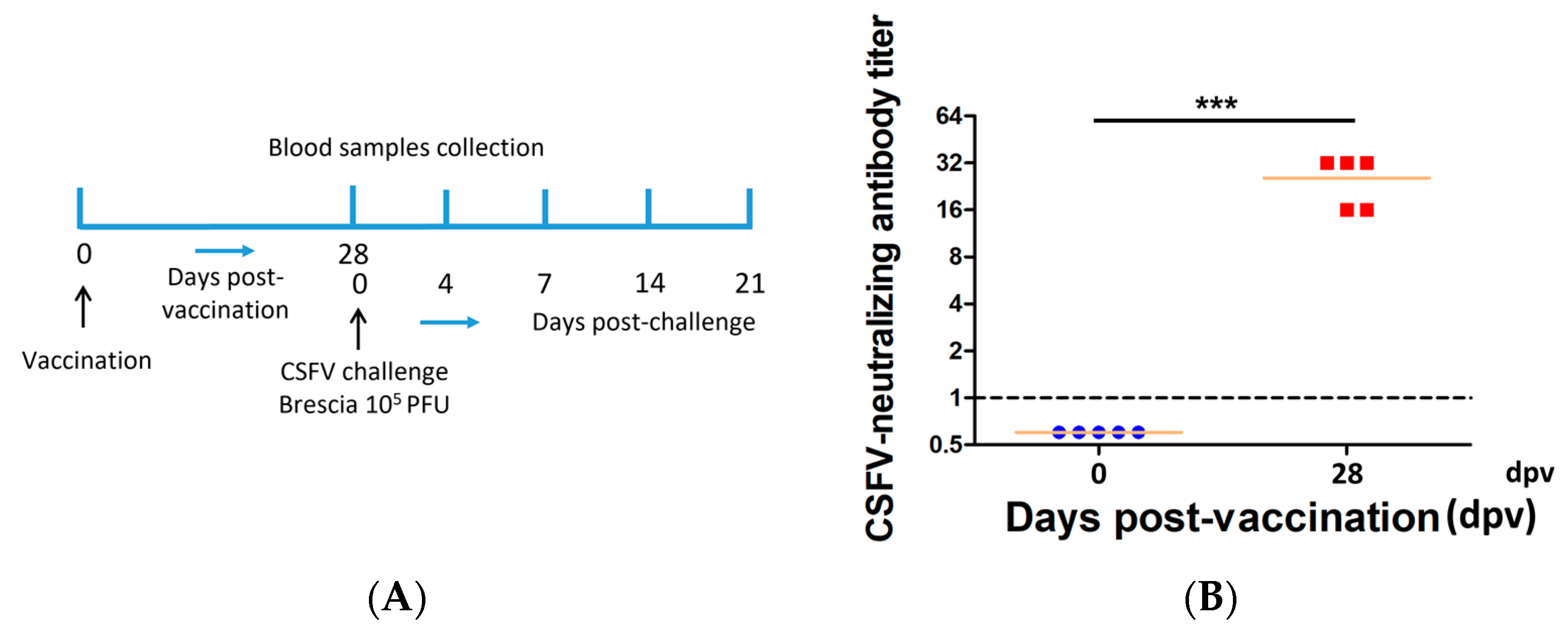
2.4. Vaccination and Challenge

The vaccination, CSFV challenge, and sample collection schemes are shown in Figure 1A. After acclimatizing for seven days, group 1 (control group) and group 2 (PRVtmv+ vaccine group) pigs were sham-vaccinated (cell culture media) or vaccinated with the prototype PRVtmv+ vaccine, using both intranasal (IN) and subcutaneous (SC) vaccination routes, as described earlier [27]. Briefly, 4 × 10⁷ plaque-forming units (PFUs) were used per nostril (total 8 × 10⁷) and filtered (0.2 μm pore size) 4 × 10⁷ PFUs were inoculated intranasally and injected subcutaneously, respectively. At 28 days post vaccination (dpv), all pigs (sham-vaccinated control and vaccinated) were challenged intranasally with 105 TCID50, of the challenge virus BICv.

2.4.1. Clinical Examination and Sample Collection from Pigs Following Vaccination and Challenge

Pigs were monitored daily for 28 days after vaccination and for 21 days following the BICv challenge for any visible clinical signs. Especially after the challenge, clinical signs associated with the CSFV disease, i.e., inappetence, depression, fever, purple skin discoloration, staggering gait, diarrhea, and cough, were recorded. Blood samples were collected from the anterior vena cava for whole blood and serum on 0, 4, 7, 14, and 21 days post challenge (dpc). Pigs showing severe CSF clinical signs were euthanized. All pigs surviving the CSFV challenge were euthanized 21 dpc. A complete necropsy was performed, and gross lesions were recorded.

2.4.2. Total and Differential White Blood Cell Counts

Blood was obtained from the anterior vena cava in ethylenediaminetetraacetic acid (EDTA)-containing tubes (Vacutainer). Total white blood cell, lymphocyte, and platelet counts were obtained using a Beckman Coulter AcT (Beckman, Coulter, CA, USA).

2.5. Detection of CSFV Viremia

To detect viremia, whole EDTA blood was serially diluted ten-fold, and 100 μL from each dilution was inoculated in 96-well plates (Costar, Cambridge, MA, USA), followed by adding SK cells (1 × 10⁴ cells per well). Plates were incubated at 37 °C and 5% CO2 for four days. After four days of incubation, the supernatant was removed from each well, and the cells were fixed with methanol–acetone (50% v/v) solution and air-dried. The presence of virus-infected cells was determined by staining monolayers, as described earlier, with CSFV E2-specific monoclonal antibody WH303 in immunoperoxidase assay using the Vecstatin ABC Kit (Vector Laboratories, Burlingame, CA, USA), following the manufacturer’s instructions [32]. Titers were calculated and expressed as TCID50/mL, as described previously, with a sensitivity of detection of ≥1.8 TCID50/mL [28].
Figure 1. PRVtmv+ immunization in pigs. (A) Schematic showing the PRVtmv+ vaccination, sample collection, and CSFV challenge scheme for the animal experiment. (B) Pigs (n = 5 in each group) were immunized with PRVtmv+ vaccine (for each pig, intranasally—8 × 10⁷ plaque forming units (PFU), and subcutaneously—4 × 10⁷ PFU; 0 days post vaccination (dpv)), or sham-vaccinated and sera samples were collected on 0 and 28 dpv to determine the classical swine fever virus (CSFV)-specific neutralizing antibody titer [33]. The dot plot graph shows each animal’s individual CSFV titer with the mean values of the group (n = 5). *** p < 0.001.
Viruses 15 02143 g001

2.6. Detection of Neutralizing Antibodies

Serum neutralization assays were performed with heat-inactivated serum samples (56 °C for 30 min) [33]. Two-fold serial dilutions of serum were prepared in Dulbecco’s modified Eagle’s medium (DMEM) supplemented with 10% FBS and mixed with equal volumes of BICv containing 102 TCID50. Serum–virus mixtures were incubated for one hour at 37 °C and then transferred to 96-well flat-bottom tissue culture plates (Corning), followed by adding SK cells (1 × 10⁴ cells per well). Plates were incubated at 37 °C and 5% CO2 for 4 days. As mentioned above, after four days, the supernatant was removed, and the cells in 96-well plates were processed for immunohistochemistry using CSFV E2-specific mAb. Neutralizing antibody titers were expressed as the reciprocal of the highest serum dilution (two-fold) that neutralizes BICv, as described elsewhere [31].

2.7. Statistical Analysis

Data were analyzed for statistical significance (p < 0.05) with GraphPad prism 5.04 (San Diego, CA, USA). Comparisons of mean CSFV-specific neutralizing antibody titer and CSFV viremia in blood were performed using a two-way analysis of variance (ANOVA) followed by Bonferroni post-tests when the ANOVA test indicated significant differences.

3. Results

3.1. PRVtmv+ Vaccinated Pigs Remained Clinically Normal and Generated Moderate Levels of CSFV-Specific Neutralizing Antibody Titers

The pigs remained clinically normal after vaccination with PRVtmv+ (intranasal/sub-cutaneous; IN/SQ). On the day of vaccination (0 dpv), mean CSFV-specific neutralizing antibody titers in pigs were less than 1 (negative). On the day of the CSFV challenge (28 dpv), the average CSFV-specific mean neutralizing antibody titers in pigs rose to 25.6 (Figure 1B; Supplementary Table S1), which is more than a 25-fold increase (seroconverted).

3.2. PRVtmv+ Vaccinated Pigs Were Protected against Severe CSFV Clinical Disease

The results revealed that the sham-vaccinated control pigs developed severe CSFV-specific clinical signs, characterized by anorexia, depression, diarrhea, and high fever by 4 dpc, and subsequently became moribund. Therefore, all the sham-vaccinated pigs were euthanized on or before 7 dpc (Figure 2A,B). However, in the PRVtmv+ group, pigs #92 (on 5 dpc) and #84 (on 8 dpc) had a fever of 40.88 °C and 40.22 °C, respectively, for only a day (Figure 2A; Supplementary Table S2). Also, beginning on 7 dpc, three vaccinated pigs (#84, 87, and 88) had diarrhea that lasted for several days but recovered. They were otherwise clinically normal until 21 days post challenge, when all the vaccinated pigs were euthanized.

3.3. PRVtmv+ Vaccinated Pigs Had a Three-Fold Lower CSFV Viremia in Blood

Severe forms of CSF in pigs are associated with high levels of viremia. Therefore, we determined the CSFV viremia in pigs following the challenge. Upon virulent CSFV challenge, all sham-vaccinated control pigs developed moderate-to-severe viremia in the blood on 4 dpc, which persisted until their sacrifice (7 dpc), with a mean CSFV titer of 3.4 and 6.05 log10 TCID50/mL, respectively (Figure 2C; Supplementary Table S3). In contrast, PRVtmv+ vaccine group pigs developed a lower level of viremia, with a mean CSFV titer of 2.45 and 1.84 log10 TCID50/mL on 4 and 7 dpc, respectively. Notably, on 7 dpc, the viremia was reduced significantly compared with that of the sham-vaccinated pigs (16,218-fold reduction) and at 14 and 21 (the 21st day being the day of euthanasia) dpc, all PRVtmv+ vaccinated pigs were negative for viremia, based on a sensitive test for the infectious virus particles, and therefore were clear of the virus.

3.4. PRVtmv+ Vaccinated Pigs Had Significantly Milder Leukopenia and Thrombocytopenia after the CSFV Challenge

Sham-vaccinated control group pigs developed severe hematological changes, including leukopenia, lymphocytopenia, and thrombocytopenia, by 4 dpc (Figure 3A–C; Supplementary Tables S4–S6). The results show that the decrease in leukocyte counts in seven control non-vaccinated pigs was 75% and 80% at 4 and 7 dpc, respectively (Figure 3A; Supplementary Table S4). Similarly, the reduction in the control pigs’ platelet counts was approximately 65% on both 4 and 7 days post challenge (Figure 3C; Supplementary Table S6). However, the corresponding drop in hematological values in the vaccinated pigs was considerably less; the reduction in the leukocyte counts for the PRVtmv+ vaccinated pigs was 35% and 28% on 4 and 7 dpc, respectively, while the decline in platelet counts for the vaccinated pigs was 38% and 30% on, respectively. Thus, the PRVtmv+ vaccination reduced the immunosuppressive effects of CSFV infection by reducing leukopenia, lymphopenia, and thrombocytopenia levels by at least two-fold relative to the control pigs.
PRVtmv+ vaccinated pigs were more likely to have a lack of the fatal CSFV disease, the induction of moderate levels of virus-neutralizing titers, a 3-fold reduced viremia, and reduced immunosuppression. This reflected the protective efficacy of the vaccine.

3.5. PRVtmv+ Vaccinated Pigs Did Not Have Any CSFV-Specific Gross Lesions upon CSFV Challenge

At 21 days post challenge, the PRVtmv+ vaccinated pigs had no clinical signs when they were euthanized. Notably, no CSFV-specific gross lesions were detectable in the spleen, kidney, and tonsils of the vaccinated pigs (Figure 4). All five moribund sham-vaccinated control pigs, were euthanized on days 6 and 7 post challenge. These results demonstrated that the PRVtmv+ vaccine protected the pigs against the most pronounced CSFV-specific lesions, i.e., infarcts, petechiae/hemorrhages, and necrotic ulcers, seen in the spleen, kidney, and tonsils, respectively, of the CSFV-infected pigs [1].

4. Discussion

Previously in this paper, we constructed a PRVtmv+ subunit vaccine vector against PCV2b and CSFV. Further, we determined its efficacy against a PCV2b challenge [27], its ability to reactivate from latency, and nasal shedding property upon latency reactivation [35]. The results revealed the following: (i) the PRVtmv+ vaccination protected pigs against the PCV2b challenge by preventing fecal virus shedding, viremia, leukopenia, and lymphocytopenia [27], and (ii) PRVtmv+ caused abortive infection in the trigeminal ganglia (TG) neurons. Therefore, after the latency reactivation of PRVtmv+ in the TG neurons, the virus did not shed in the nasal secretions. Consequently, unlike the wt PRV or traditional modified live PRV vaccines, which are shed in the nasal secretions and maintained and circulated in the pigs, the latent PRVtmv+ vaccine vector cannot spread in the pig population [35].
In this study, we demonstrate that PRVtmv+ vaccinated pigs showed a moderate level of CSFV-specific neutralizing antibody titers, and they survived the fatal CSFV challenge until euthanasia at 21 days post challenge. On the contrary, the sham-vaccinated control pigs had to be euthanized by 6–7 dpc because of their moribund conditions. Although some PRVtmv+ vaccinated pigs developed transient fever for a day and mild diarrhea, their condition did not linger beyond a day or two. At euthanasia, 21 days post challenge, all pigs, including one pig with mild diarrhea, were clinically normal. None of the PRVtmv+ vaccinated pigs developed any CSFV-specific gross lesions. Together, these results correlated well and demonstrated that the single PRVtmv+ vaccination was sufficient for pigs to be protected from the fatal consequences of CSFV infection.
The “C”-strain vaccine virus is efficacious against CSFV, but lacks a serological marker. Therefore, the vaccinated animals cannot be distinguished from the infected animals during the sporadic CSFV outbreaks [11]. Due to this limitation, EU countries do not permit the vaccination of pigs with the otherwise highly effective MLV “C”-strain or similar MLV vaccines. Further, there are trade restrictions on the countries using the MLV CSFV or that are not yet CSFV-free, which has enormous economic consequences.
The PRVtmv+ can be distinguished serologically from the PRV wt- as well as the CSFV wt-infected pigs (DIVA vaccine). Additionally, as noted above, the PRVtmv+ does not replicate in the TG following reactivation from latency. Therefore, the vaccine virus would not circulate in the pig population. As such, it could be used as a safe and emergency vaccine in PRV-free countries to control a CSFV outbreak, i.e., in EU and North American countries.
The direct and indirect contact of domestic pigs with wild boars has been implicated as a cause of the outbreak of CSFV in Germany Spain, Italy, and the Netherlands [36,37,38]. Thus, CSFV outbreaks in domestic pigs remain a potential threat to the populations of Europe, Central America, and South America, including Brazil and the Caribbean [1,6,8,39]. As noted earlier, routine vaccination against CSFV with the “C”-strain-based vaccination or similar MLVs is not permitted in EU countries because the MLV lacks the DIVA property and cannot be distinguished from the circulating field strain. Instead, a rigorous sero-surveillance of domestic pigs and wild boars has been enforced [40].
Previous studies by other investigators revealed that the pre-colonization of TG with a latent PRV prevented the latent infection in the TG by another superinfecting PRV [41]. Even though we did not validate the latter phenomenon in this study, we believe it would also be applicable to PRVtmv+ infected pigs. As noted above, PRVtmv+ established latency but did not replicate in the post-mitotic TG neurons. Consequently, upon dexamethasone-induced latency reactivation, the vaccine virus was not shed in the nasal secretion [35]. Therefore, PRVtmv+ would be suitable as an alternative subunit vaccine against CSFV, even in countries where PRV has been eradicated from the domestic pig population. The potential advantages of using PRVtmv+ as a live subunit vaccine against CSFV are as follows: (i) the PRVtmv+ is highly attenuated for pigs, yet replicates well enough in the nasal mucosa; (ii) it is highly immunogenic and induces CSFV-specific neutralizing antibodies in pigs; (iii) it has the DIVA property; (iv) it stably expresses inserted chimeric proteins; (v) the vector virus establishes latency in the TG neurons but does not replicate, leading to no nasal virus shedding; (vi) it is a bivalent subunit vaccine and protective against both PCV2 and CSFV challenges; and (vii) if needed, it can likely be administered intranasally as a booster vaccine, since the preexisting antibodies do not interfere with the memory immune response when administered intranasally [42,43], which will be validated soon.

5. Conclusions

Our results demonstrated that the PRVtmv+ vaccinated pigs were protected against fatal CSFV disease and survived until euthanasia at 21 days post challenge. Notably, no gross lesions were detected in the vaccinated pigs at necropsy. The PRVtmv+ has the DIVA property against both wild-type PRV and CSFV, and can be used in countries where PRV and CSF have been eradicated from the domestic pig population.

Supplementary Materials

The following supporting information can be downloaded at: https://0-www-mdpi-com.brum.beds.ac.uk/article/10.3390/v15112143/s1, Table S1: Classical swine fever virus (CSFV)-specific serum neutralizing (SN) antibody titer developed in pigs after PRVtmv+ vaccination; Table S2: Clinical evaluation: body temperature of pigs following the classical swine fever virus (CSFV) challenge; Table S3: Classical swine fever virus (CSFV)-associated viremia in blood following CSFV challenge in pigs; Table S4: Leukocyte count in pigs following virulent classical swine fever virus challenge in pigs; Table S5: Lymphocyte count in pigs following virulent classical swine fever virus challenge in pigs; Table S6: Platelet count in pigs following virulent classical swine fever virus challenge in pigs.

Author Contributions

Conceptualization—S.I.C.; methodology—E.S., E.M.-R., D.P.G. and M.B.; formal analysis—S.I.C., M.B., D.P.G. and S.P.; resources—S.I.C. and M.B.; data curation—E.S., E.M.-R., D.P.G., M.B., S.P. and S.I.C.; writing—original draft preparation—S.I.C.; writing—review and editing—S.I.C., S.P. and M.B.; supervision, project administration and funding acquisition—S.I.C. and M.B. All authors have read and agreed to the published version of the manuscript.

Funding

This work was supported by the United States Department of Agriculture/National Institute of Food and Agriculture grant (USDA/NIFA; 2019-67015-29867) to Shafiqul I. Chowdhury and Manuel Borca.

Institutional Review Board Statement

The study was conducted in accordance with the Institutional Animal Care and Use Committee at the Plum Island Animal Disease Center animal facilities (Number 171.12-21-R, approved on 12 September 2021). Further, the LSU Institutional Animal Care and Use Committee endorsed the above-approved protocol.

Data Availability Statement

Additional relevant data are available in the Supplementary section of the manuscript.

Acknowledgments

We thank the Plum Island Animal Disease Center facility personnel for their excellent technical support with the animal experiment.

Conflicts of Interest

The authors declare no conflict of interest.

References

  1. Blome, S.; Staubach, C.; Henke, J.; Carlson, J.; Beer, M. Classical Swine Fever-An Updated Review. Viruses 2017, 9, 86. [Google Scholar] [CrossRef] [PubMed]
  2. Postel, A.; Austermann-Busch, S.; Petrov, A.; Moennig, V.; Becher, P. Epidemiology, diagnosis and control of classical swine fever: Recent developments and future challenges. Transbound. Emerg. Dis. 2018, 65 (Suppl. S1), 248–261. [Google Scholar] [CrossRef] [PubMed]
  3. Meuwissen, M.P.; Horst, S.H.; Huirne, R.B.; Dijkhuizen, A.A. A model to estimate the financial consequences of classical swine fever outbreaks: Principles and outcomes. Prev. Vet. Med. 1999, 42, 249–270. [Google Scholar] [CrossRef] [PubMed]
  4. Stegeman, A.; Elbers, A.; de Smit, H.; Moser, H.; Smak, J.; Pluimers, F. The 1997-1998 epidemic of classical swine fever in the Netherlands. Vet. Microbiol. 2000, 73, 183–196. [Google Scholar] [CrossRef]
  5. KRAMER-SCHADT, S.; FERNÁNDEZ, N.; THULKE, H.-H. Potential ecological and epidemiological factors affecting the persistence of classical swine fever in wild boar Sus scrofa populations. Mammal. Rev. 2007, 37, 1–20. [Google Scholar] [CrossRef]
  6. Ganges, L.; Crooke, H.R.; Bohorquez, J.A.; Postel, A.; Sakoda, Y.; Becher, P.; Ruggli, N. Classical swine fever virus: The past, present and future. Virus Res. 2020, 289, 198151. [Google Scholar] [CrossRef]
  7. Brown, V.R.; Bevins, S.N. A Review of Classical Swine Fever Virus and Routes of Introduction into the United States and the Potential for Virus Establishment. Front. Vet. Sci. 2018, 5, 31. [Google Scholar] [CrossRef]
  8. Coronado, L.; Perera, C.L.; Rios, L.; Frías, M.T.; Pérez, L.J. A Critical Review about Different Vaccines against Classical Swine Fever Virus and Their Repercussions in Endemic Regions. Vaccines 2021, 9, 154. [Google Scholar] [CrossRef]
  9. Kaden, V.; Lange, E.; Kuster, H.; Muller, T.; Lange, B. An update on safety studies on the attenuated "RIEMSER Schweinepestoralvakzine" for vaccination of wild boar against classical swine fever. Vet. Microbiol. 2010, 143, 133–138. [Google Scholar] [CrossRef]
  10. Rossi, S.; Staubach, C.; Blome, S.; Guberti, V.; Thulke, H.H.; Vos, A.; Koenen, F.; Le Potier, M.F. Controlling of CSFV in European wild boar using oral vaccination: A review. Front. Microbiol. 2015, 6, 1141. [Google Scholar] [CrossRef]
  11. de Smit, A.J.; van Gennip, H.G.; Miedema, G.K.; van Rijn, P.A.; Terpstra, C.; Moormann, R.J. Recombinant classical swine fever (CSF) viruses derived from the Chinese vaccine strain (C-strain) of CSF virus retain their avirulent and immunogenic characteristics. Vaccine 2000, 18, 2351–2358. [Google Scholar] [CrossRef] [PubMed]
  12. Meyers, G.; Tautz, N.; Becher, P.; Thiel, H.J.; Kummerer, B.M. Recovery of cytopathogenic and noncytopathogenic bovine viral diarrhea viruses from cDNA constructs. J. Virol. 1997, 71, 1735. [Google Scholar] [CrossRef] [PubMed]
  13. Reimann, I.; Depner, K.; Trapp, S.; Beer, M. An avirulent chimeric Pestivirus with altered cell tropism protects pigs against lethal infection with classical swine fever virus. Virology 2004, 322, 143–157. [Google Scholar] [CrossRef] [PubMed]
  14. Konig, P.; Blome, S.; Gabriel, C.; Reimann, I.; Beer, M. Innocuousness and safety of classical swine fever marker vaccine candidate CP7_E2alf in non-target and target species. Vaccine 2011, 30, 5–8. [Google Scholar] [CrossRef]
  15. MacDiarmid, S.C. Aujeszky’s disease eradication in New Zealand. Aust. Vet. J. 2000, 78, 470–471. [Google Scholar] [CrossRef]
  16. Muller, T.; Batza, H.J.; Schluter, H.; Conraths, F.J.; Mettenleiter, T.C. Eradication of Aujeszky’s disease in Germany. J. Vet. Med. B Infect. Dis. Vet. Public. Health 2003, 50, 207–213. [Google Scholar] [CrossRef]
  17. OIE. OIE World Animal Health Information System. Available online: https://wahis.woah.org/#/dashboards/country-or-disease-dashboard (accessed on 12 October 2023).
  18. Tan, L.; Yao, J.; Yang, Y.; Luo, W.; Yuan, X.; Yang, L.; Wang, A. Current Status and Challenge of Pseudorabies Virus Infection in China. Virol. Sin. 2021, 36, 588–607. [Google Scholar] [CrossRef]
  19. Liu, A.; Xue, T.; Zhao, X.; Zou, J.; Pu, H.; Hu, X.; Tian, Z. Pseudorabies Virus Associations in Wild Animals: Review of Potential Reservoirs for Cross-Host Transmission. Viruses 2022, 14, 2254. [Google Scholar] [CrossRef]
  20. Pacini, M.I.; Forzan, M.; Cilia, G.; Bernardini, L.; Marzoli, F.; Pedonese, F.; Bandecchi, P.; Fratini, F.; Mazzei, M. Detection of Pseudorabies Virus in Wild Boar Foetus. Animals 2020, 10, 366. [Google Scholar] [CrossRef]
  21. Pedersen, K.; Turnage, C.T.; Gaston, W.D.; Arruda, P.; Alls, S.A.; Gidlewski, T. Pseudorabies detected in hunting dogs in Alabama and Arkansas after close contact with feral swine (Sus scrofa). BMC Vet. Res. 2018, 14, 388. [Google Scholar] [CrossRef]
  22. Zimmerman, J.J. Diseases of Swine, 10th ed.; Wiley-Blackwell: Ames, IA, USA, 2012. [Google Scholar]
  23. Zhou, H.; Pan, Y.; Liu, M.; Han, Z. Prevalence of Porcine Pseudorabies Virus and Its Coinfection Rate in Heilongjiang Province in China from 2013 to 2018. Viral Immunol. 2020, 33, 550–554. [Google Scholar] [CrossRef] [PubMed]
  24. Wu, R.; Bai, C.; Sun, J.; Chang, S.; Zhang, X. Emergence of virulent pseudorabies virus infection in northern China. J. Vet. Sci. 2013, 14, 363–365. [Google Scholar] [CrossRef] [PubMed]
  25. Bo, Z.; Miao, Y.; Xi, R.; Gao, X.; Miao, D.; Chen, H.; Jung, Y.S.; Qian, Y.; Dai, J. Emergence of a novel pathogenic recombinant virus from Bartha vaccine and variant pseudorabies virus in China. Transbound. Emerg. Dis. 2021, 68, 1454–1464. [Google Scholar] [CrossRef] [PubMed]
  26. Delva, J.L.; Nauwynck, H.J.; Mettenleiter, T.C.; Favoreel, H.W. The Attenuated Pseudorabies Virus Vaccine Strain Bartha K61: A Brief Review on the Knowledge Gathered During 60 Years of Research. Pathogens 2020, 9, 897. [Google Scholar] [CrossRef] [PubMed]
  27. Pavulraj, S.; Pannhorst, K.; Stout, R.W.; Paulsen, D.B.; Carossino, M.; Meyer, D.; Becher, P.; Chowdhury, S.I. A Triple Gene-Deleted Pseudorabies Virus-Vectored Subunit PCV2b and CSFV Vaccine Protects Pigs against PCV2b Challenge and Induces Serum Neutralizing Antibody Response against CSFV. Vaccines 2022, 10, 305. [Google Scholar] [CrossRef]
  28. Risatti, G.R.; Holinka, L.G.; Lu, Z.; Kutish, G.F.; Tulman, E.R.; French, R.A.; Sur, J.H.; Rock, D.L.; Borca, M.V. Mutation of E1 glycoprotein of classical swine fever virus affects viral virulence in swine. Virology 2005, 343, 116–127. [Google Scholar] [CrossRef]
  29. Risatti, G.R.; Borca, M.V.; Kutish, G.F.; Lu, Z.; Holinka, L.G.; French, R.A.; Tulman, E.R.; Rock, D.L. The E2 glycoprotein of classical swine fever virus is a virulence determinant in swine. J. Virol. 2005, 79, 3787–3796. [Google Scholar] [CrossRef]
  30. Holinka, L.G.; Fernandez-Sainz, I.; O’Donnell, V.; Prarat, M.V.; Gladue, D.P.; Lu, Z.; Risatti, G.R.; Borca, M.V. Development of a live attenuated antigenic marker classical swine fever vaccine. Virology 2009, 384, 106–113. [Google Scholar] [CrossRef]
  31. Reed, L.J.; Muench, H. A simple method of estimating fifty per cent endpoints12. Am. J. Epidemiol. 1938, 27, 493–497. [Google Scholar] [CrossRef]
  32. Edwards, S.; Moennig, V.; Wensvoort, G. The development of an international reference panel of monoclonal antibodies for the differentiation of hog cholera virus from other pestiviruses. Vet. Microbiol. 1991, 29, 101–108. [Google Scholar] [CrossRef]
  33. OIE. Classical Swine Fever (Infection with Classical Swine Fever Virus). Available online: https://www.woah.org/fileadmin/Home/fr/Health_standards/tahm/3.09.03_CSF.pdf (accessed on 24 March 2023).
  34. Koomkrong, N.; Boonkaewwan, C.; Laenoi, W.; Kayan, A. Blood haematology, muscle pH and serum cortisol changes in pigs with different levels of drip loss. Asian-Australas. J. Anim. Sci. 2017, 30, 1751–1755. [Google Scholar] [CrossRef] [PubMed]
  35. Pavulraj, S.; Stout, R.W.; Paulsen, D.B.; Chowdhury, S.I. Live Triple Gene-Deleted Pseudorabies Virus-Vectored Subunit PCV2b and CSFV Vaccine Undergoes an Abortive Replication Cycle in the TG Neurons following Latency Reactivation. Viruses 2023, 15, 473. [Google Scholar] [CrossRef] [PubMed]
  36. Edwards, S.; Fukusho, A.; Lefevre, P.C.; Lipowski, A.; Pejsak, Z.; Roehe, P.; Westergaard, J. Classical swine fever: The global situation. Vet. Microbiol. 2000, 73, 103–119. [Google Scholar] [CrossRef] [PubMed]
  37. Fritzemeier, J.; Teuffert, J.; Greiser-Wilke, I.; Staubach, C.; Schluter, H.; Moennig, V. Epidemiology of classical swine fever in Germany in the 1990s. Vet Microbiol 2000, 77, 29–41. [Google Scholar] [CrossRef]
  38. Leifer, I.; Hoffmann, B.; Hoper, D.; Bruun Rasmussen, T.; Blome, S.; Strebelow, G.; Horeth-Bontgen, D.; Staubach, C.; Beer, M. Molecular epidemiology of current classical swine fever virus isolates of wild boar in Germany. J. Gen. Virol. 2010, 91, 2687–2697. [Google Scholar] [CrossRef]
  39. de Oliveira, L.G.; Gatto, I.R.H.; Mechler-Dreibi, M.L.; Almeida, H.M.S.; Sonalio, K.; Storino, G.Y. Achievements and Challenges of Classical Swine Fever Eradication in Brazil. Viruses 2020, 12, 1327. [Google Scholar] [CrossRef]
  40. Graham, S.P.; Everett, H.E.; Haines, F.J.; Johns, H.L.; Sosan, O.A.; Salguero, F.J.; Clifford, D.J.; Steinbach, F.; Drew, T.W.; Crooke, H.R. Challenge of Pigs with Classical Swine Fever Viruses after C-Strain Vaccination Reveals Remarkably Rapid Protection and Insights into Early Immunity. PLoS ONE 2012, 7, e29310. [Google Scholar] [CrossRef]
  41. Schang, L.M.; Kutish, G.F.; Osorio, F.A. Correlation between precolonization of trigeminal ganglia by attenuated strains of pseudorabies virus and resistance to wild-type virus latency. J. Virol. 1994, 68, 8470–8476. [Google Scholar] [CrossRef]
  42. Nelson, S.A.; Sant, A.J. Potentiating Lung Mucosal Immunity Through Intranasal Vaccination. Front. Immunol. 2021, 12, 808527. [Google Scholar] [CrossRef]
  43. Zhang, F.; Peng, B.; Chang, H.; Zhang, R.; Lu, F.; Wang, F.; Fang, F.; Chen, Z. Intranasal Immunization of Mice to Avoid Interference of Maternal Antibody against H5N1 Infection. PLoS ONE 2016, 11, e0157041. [Google Scholar] [CrossRef]
Figure 2. Clinical assessment, survival curve, and classical swine fever virus (CSFV)-associated viremia in sham-vaccinated control and PRVtmv+ vaccinated pigs following CSFV challenge. (A) Pigs were challenged with CSFV at 28 days post vaccination (0 days post challenge (dpc)), and rectal temperature was recorded until 21 dpc. Mean rectal temperatures with standard deviation (SD) are given (n = 5). All pigs in the CSFV control group were euthanized on days 6 and 7 dpc. (B) A survival rate is given in terms of percentage in each group (n = 5). (C) Blood samples were collected from pigs on days 0, 4, 7, 14, and 21 post challenge, and the CSFV titer was determined in cell culture. The mean CSFV titer in the blood of each animal from both groups is shown. The dot plot graph represents mean + individual values in each group (n = 5). TCID50—50% tissue culture infectious dose; * p < 0.05 and *** p < 0.001.
Viruses 15 02143 g002
Figure 3. Percent changes in leukocyte, lymphocyte, and platelet count following the classical swine fever virus (CSFV) Brescia strain (BICv) challenge in both groups. Whole blood was collected from pigs on 28 dpv/0 dpc and 49 dpv/21 dpc, (A) leukocyte, (B) lymphocyte, and (C) platelet counts were determined, and their percent changes were calculated. The normal range in pigs: (i) leukocytes—11–22 × 103/μL; (ii) lymphocytes—4.6–12 × 103/μL; (iii) platelets—200–500 × 103/μL [34].
Viruses 15 02143 g003
Figure 4. Photograph of spleen, kidney, and tonsils of PRVtmv+ vaccinated pigs (#84, #87, #88, #91, and #92) euthanized at 21 days post challenge. Spleen (AE), kidney (FJ), and tonsils (KO). The spleen, kidney, and tonsils lack CSFV-specific lesions. The focal areas marked with circles in D most likely represent areas of blood pooling/incomplete extrusion of blood; this is sometimes seen in the spleens of various species during postmortems after euthanasia using barbiturate compounds. Splenic infarctions, even red-type infarcts, are usually sharply demarcated from the rest of the parenchyma.
Viruses 15 02143 g004
Disclaimer/Publisher’s Note: The statements, opinions and data contained in all publications are solely those of the individual author(s) and contributor(s) and not of MDPI and/or the editor(s). MDPI and/or the editor(s) disclaim responsibility for any injury to people or property resulting from any ideas, methods, instructions or products referred to in the content.

