The Synthesis of PbS NPs and Biosorption of Pb(II) by Shinella Zoogloeoides PQ7 in Aqueous Conditions

Zhejiang Provincial Key Laboratory of Organic Pollution Process and Control, Department of Environmental Science, College of Environmental and Resource Sciences, Zhejiang University, Hang Zhou 310000, China
*
Author to whom correspondence should be addressed.
Water 2020, 12(7), 2065; https://0-doi-org.brum.beds.ac.uk/10.3390/w12072065
Submission received: 6 May 2020
/
Revised: 26 May 2020
/
Accepted: 27 May 2020
/
Published: 21 July 2020
(This article belongs to the Section Wastewater Treatment and Reuse)
Abstract
:Increasing heavy metal pollution in water continues to endanger human health. The genus Shinella has potential for heavy metal bioremediation but has rarely been studied. In this study, we report that Shinella zoogloeoides PQ7 turns black in the presence of lead ions. Transmission electron microscopy (TEM), Scanning electron microscopy–energy dispersive X-ray spectroscopy (SEM–EDS), X-ray photoelectron spectroscopy (XPS) and X-ray diffraction (XRD) indicated that PbS nanoparticles (NPs) were synthesized by PQ7. Moreover, PQ7 was used as a biosorbent to remove Pb(II) from aqueous solutions. Biosorption performance was evaluated in terms of contact time, pH, biomass dosage and initial Pb(II) concentration. The equilibrium and kinetic data were consistent with the Freundlich isotherm model (R2 = 0.986) and pseudo-second-order model (R2 = 0.977), respectively. The maximum (qmax) Pb(II) adsorption reached 222.22 mg/g, which was higher than that of other bacteria reported in previous literature. SEM–EDS, XRD and Fourier transform infrared (FTIR) analyses also confirmed the adsorption of Pb(II) by the PQ7 cells. In conclusion, PQ7 is a promising strain in removing and recovering Pb(II) from wastewater.
1. Introduction
Heavy metals are among the most concerning environmental pollutants because of their toxicity and tendency to accumulate in the environment [1,2]. Lead(Pb) is one of most concerning heavy metals [3]. Many studies have reported that Pb(II) can damage human tissues and affect human health. Pb(II) can be toxic by interacting with calcium(II) to replace it in bones or combining with zinc(II) in haem enzymes and metallothioneins [4,5]. Because of these serious effects, it is essential to remove and recover lead ions from wastewater and the environment.
Many methods, including ionic exchange, chemical precipitation and electrolysis, have been used for the removal of lead ions. However, these methods are often not friendly to the environments [6]. Microbes based biotechnology has shown promising future in the bioremediation of heavy metals because of their low cost and high efficiency [2]. Some bacteria can not only survive in the presence of Pb(II) but also form valuable lead compounds. For example, Streptomyces sp. and Pseudomonas vesicularis have been reported to produce red and red–brown pigments Pb3O4 [7]. Pb9(PO4)6, PbSO4, PbS, Pb5(PO4)3Cl, and PbHPO4 are also synthesized by lead-resistant bacteria [8,9,10]. Some bacteria, yeasts and fungi have been reported to synthesize PbS nanoparticles (NPs) [11,12,13,14,15]. PbS is a semiconductor material based on heavy metals. When synthesized at the nanoscale, its chemical, electrical, optical and magnetic properties are superior to those of same-volume semiconductor materials [16]. Compared to physical and chemical, using microorganisms to synthesize PbS nanoparticles is the most economical method [11]. Thus, it is meaningful to find lead-resistant bacteria that not only are efficient absorbents but also form useful lead compounds.
Quite a few species in the genus Shinella had been reported to degrade organic pollutants, such as nicotine, pyridine and polycyclic aromatic hydrocarbons [17,18,19,20]. However, very few studies have explored their bioremediation in terms of heavy metals. Shinella zoogloeoides PQ7 was isolated from contaminated soil [17]. It can resist quite a few heavy metals, such as zinc(II), copper(II), lead(II) and chromium(IV), and turn black in the presence of lead ions. This study was carried out to explore the reason for this color change in the presence of lead ions and the adsorption of Pb(II). To this end, transmission electron microscopy (TEM), scanning electron microscopy–energy dispersive X-ray spectroscopy (SEM–EDS), X-ray photoelectron spectroscopy (XPS) and X-ray diffraction (XRD) were employed to characterize the black materials. Different experimental factors, kinetic models and isotherm models were used to investigate the adsorption behavior.
2. Materials and Methods
2.1. Bacterial Strain and Culture Conditions
Shinella zoogloeoides PQ7 was obtained from our lab collection in 2013 [17]. It was grown in Luria-Bertani (LB) medium (tryptone 10 g/L, sodium chloride 10 g/L, yeast extract 5 g/L) at 30 °C with 200 rpm shaking speed.
2.2. Growth of Shinella Zoogloeoides PQ7 in the Presence of Lead Ions
One milliliter seed culture of PQ7 (OD600 = 1) was inoculated into 100 mL LB liquid medium with lead ions (5 mM). Then, the cultures were cultivated at 30 °C with a shaking speed of 200 rpm. The growth of the cells was monitored daily. LB medium without Pb(II) served as controls. Each experiment was carried out in triplicate.
2.3. Transmission Electron Microscopy (TEM) and Scanning Electron Microscopy–Energy Dispersive X-ray Spectroscopy (SEM–EDS) Analysis
After four days cultivation, when Shinella zoogloeoides PQ7 turned black in the presence of Pb(II) ions, the black bacterial cells and control cells were collected by centrifuging at 10,000 rpm for 15 min and washed 3 times with sterile distilled water for the next steps.
The samples were fixed in 2.5% glutaraldehyde overnight at 4 °C and then washed three times with 0.1 M phosphate buffer. After fixation with osmium acid for 2 h, the bacteria were gradually dehydrated in a gradient series of solutions containing different ethanol concentrations. Then, samples were prepared following standard procedures according to a previous study [1]. Finally, the bacteria were analyzed by SEM–EDS (SU-8010, Hitachi, Tokyo, Japan).
After being dried in a graded sequence of ethanol, the samples for TEM were placed in the mixture of alcohol and isoamylacetate (v:v = 1:1) for 0.5 h, and subsequently transferred to isoamylacetate for 12 h. After that, the samples were washed and dried in liquid CO2 (Hitachi Model HCP-C criticalpoint dryer). Desiccated samples were coated with gold-palladium (Hitachi Model E-1010 ion sputter) and then observed in TEM (Hitachi H7650) [1].
2.4. X-ray Diffraction (XRD) and X-ray Photoelectron Spectroscopy (XPS) Analysis
The black cells were characterized by XRD (D8 Advance, Germany) and XPS (K-Alpha+, Thermo Scientific, Waltham, MA, USA) to analyze the black materials formed by Shinella zoogloeoides PQ7. The samples were treated with a series of gradient concentrations of alcohol and acetone to dehydrate them. After being freeze-dried, the dried materials were mashed and characterized by XRD and XPS.
2.5. Biosorption Experiments
One milliliter seed culture of PQ7 (OD600 = 1) was inoculated into 100 mL LB medium at 30 °C with 200 rpm shaking speed until the stationary phase was reached. The cells were collected by centrifugation at 10,000 rpm for 15 min and washed three times with deionized water [21]. Then, the samples were used for biosorption experiments.
To fully evaluate the adsorption characteristics of lead ions by the strain, several parameters, such as pH, contact time, biomass dosage and initial Pb(II) concentration, were changed. The pH of the 100 mL lead solution was changed using 0.1 M NaOH or HCl solutions. Dry bacterial samples (0.1 g) were suspended in 100 mL lead solutions and then agitated at 30 °C with 200 rpm shaking speed. At certain times, the residual Pb(II) was detected by inductively coupled plasma–mass spectrometry (ICP–MS, 7700X, Agilent, Santa Clare, CA, USA). All biosorption experiments had three biological replicates, and the mean values were also calculated.
Equations (1) and (2) were used to determine the biosorption rate and capacity of lead ions [22]:
where C0 and Ce (mg/L) represent the beginning and end concentrations of lead ions, respectively; qe (mg/g) is the biosorption capacity at equilibrium; and M (g/L) corresponds to the biomass concentration.
2.6. Kinetic and Biosorption Isotherm Studies
The adsorption reaction rate and capacity of an adsorbent could be described by adsorption kinetics. In this study, pseudo-first-order and pseudo-second-order Equations (3) and (4) were used to investigate the kinetics of Pb2+ biosorption [23,24]:
where qt (mg/g) is the concentration of Pb(II) adsorbed at time t (min); qe (mg/g) is the amount of adsorbed Pb2+ ions at equilibrium; and K1 (L/min) and K2 (g/mg/min) are the rate constants for the first-order and second-order processes, respectively.
qt = qe1(1 − exp(−k1t))
Two theoretical adsorption isotherm models were used to describe the equilibrium between the cell surfaces and Pb2+ ions: the Langmuir and Freundlich isotherm models [25]. The Langmuir model assumes that the adsorbent is a homogeneous surface with monolayer formation. In contrast, the Freundlich model isotherm describes adsorption occurring on heterogeneous surfaces with multilayered formation. The Langmuir isotherm model is described by Equation (5) [26]:
where Ce (mg/L) is the equilibrium concentration of Pb2+ ions in aqueous solution; Qmax (mg/g) represents the maximum adsorption capacity; and KL (L/mg) is the Langmuir adsorption constant.
The Freundlich model can be represented by Equation (6) [27]:
where KF (L/g) is the Freundlich constant and n represents the affinity constant reflecting the strength of adsorption.
Log10 qe = log10KF + log10 Ce
2.7. SEM–EDS, Fourier Transform Infrared (FTIR) and XRD Analysis
The cells before and after treatment with 200 mg/L Pb(II) solution were collected by centrifuging at 10,000 rpm for 15 min and were washed three times with deionized water. The samples were prepared for SEM–EDS and Fourier transform infrared (FTIR) analysis. The sample preparation steps of SEM–EDS were the same as those described before. Then, the cell morphology and elemental composition were analyzed by SEM–EDS. After being freeze-dried and mashed, the samples were analyzed by XRD and FTIR (Thermo Scientific, Waltham, MA, USA) to explore the functional groups that interacted with Pb2+ ions on the surfaces of PQ7.
2.8. Statistical Analysis
The experimental data were determined in triplicate and the mean values were calculated. The XRD and XPS data were analyzed by Jade 6.5 and Thermo Avantage, respectively. The pictures were drawn using OriginPro 2018 and Microsoft Excel, 2007.
3. Results and Discussion
3.1. Shinella Zoogloeoides PQ7 Turned Black in the Presence of Lead Ions
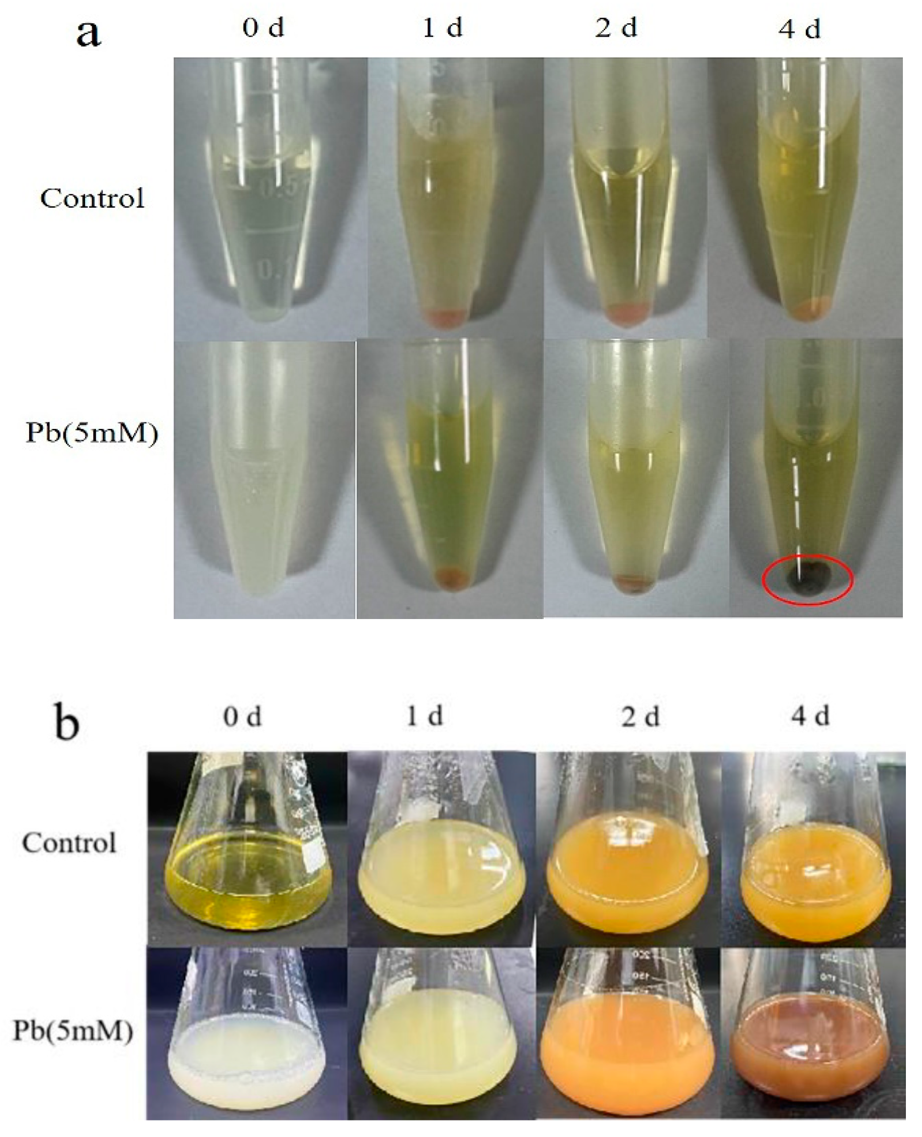
In the liquid medium with lead, we noticed that PQ7 cultures changed color from white to brown compared to the yellowish color on the blank controls. After cultivation for four days, the cells in vibrating liquid medium with Pb(II) turned eventually to black, a black precipitate was visible after centrifugation (Figure 1). The result indicated that PQ7 could produce a substance in the presence of lead ions. Many reports have shown that bacteria can produce nanoparticles with metal ions. For instance, biofilms of bacteria from the family Desulfobacteriaceae were found to produce ZnS nanoparticles [28]. In our study, we deduced that PQ7 might interact with lead ions in a similar way as Desulfobacteriaceae.
3.2. PbS Nanoparticles Were Synthesized by PQ7
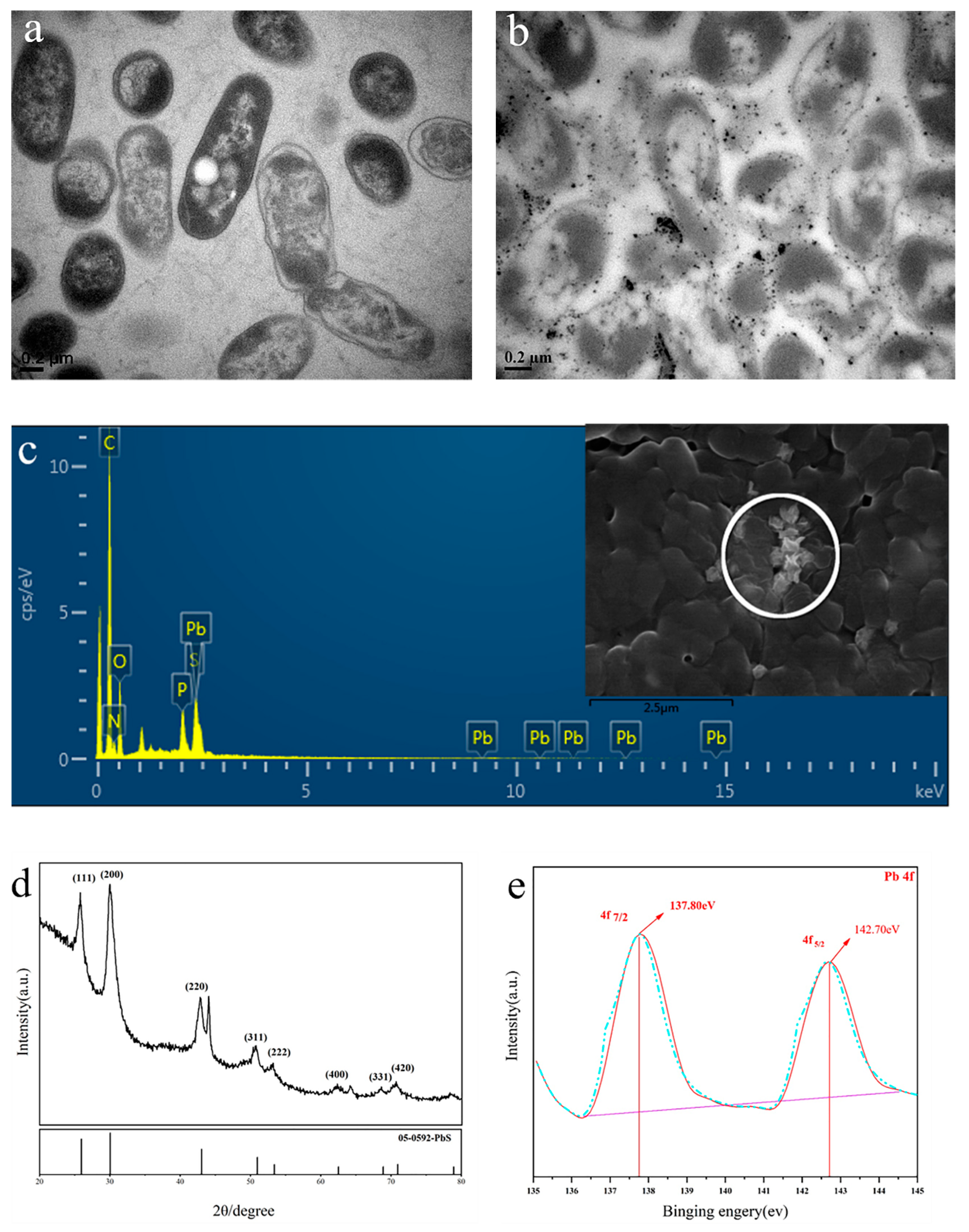
The TEM images indicated that nanoparticles were synthesized by Shinella zoogloeoides PQ7 in contrast to the control (Figure 2a,b). These nanoparticles were distributed outside the cells, and the size of the particles varied widely. It might be that small particles were synthesized and easily aggregated to form larger grains [29]. On the other hand, nanoparticles were also found around the cells in the liquid medium with Pb(II), according to the SEM images (Figure 2c). Based on the EDS elemental analyses, these areas were composed of C, N, P, S and Pb (Figure 2c).
The XRD result for black cells is shown in Figure 2d. From the image, it is clearly observed that the diffraction peaks at approximately 2θ = 25.96°, 30.07°, 43.05°, 50.97°, 53.41°, 62.53°, and 68.88° which correspond to the (111), (200), (220), (311), (222), (400) and (331) reflections, respectively. Based on comparison, these diffraction peaks can be matched with the presence of crystal PbS nanoparticles (according to Joint Committee on Powder Diffraction Standards (JCPDS) No: 05-0592) [30]. The result after fitting the original data by the software Avantage is shown in Figure 2e. Figure 2e displays only two Pb 4f peaks. The Pb 4f7/2 peak at 137.80 eV and Pb 4f5/2 peak at 142.70 eV were assigned to PbS [31]. The TEM, SEM–EDS, XRD and XPS data confirmed that PbS nanoparticles were biosynthesized by Shinella zoogloeoides PQ7.
Sulfate reduction in bacteria is an important mechanism for the formation of sulfide nanoparticles [32]. Sulfate ions are reduced to sulfur ions by this metabolic pathway. Under natural conditions, bacteria such as sulfate-reducing bacteria (SRB) can use reduced sulfur ions to produce hydrogen sulfide by combining with hydrogen ions. When metal ions are present, the hydrogen ions are replaced by metal ions to form insoluble metal sulfide [33,34]. The synthesis of PbS NPs in PQ7 might involve the same steps as described above. Although the synthesis of PbS NPs is not rare for microbes, this is the first report of PbS NPs synthesis by a strain in the genus Shinella.
3.3. Batch Biosorption Experiments
3.3.1. Effect of pH
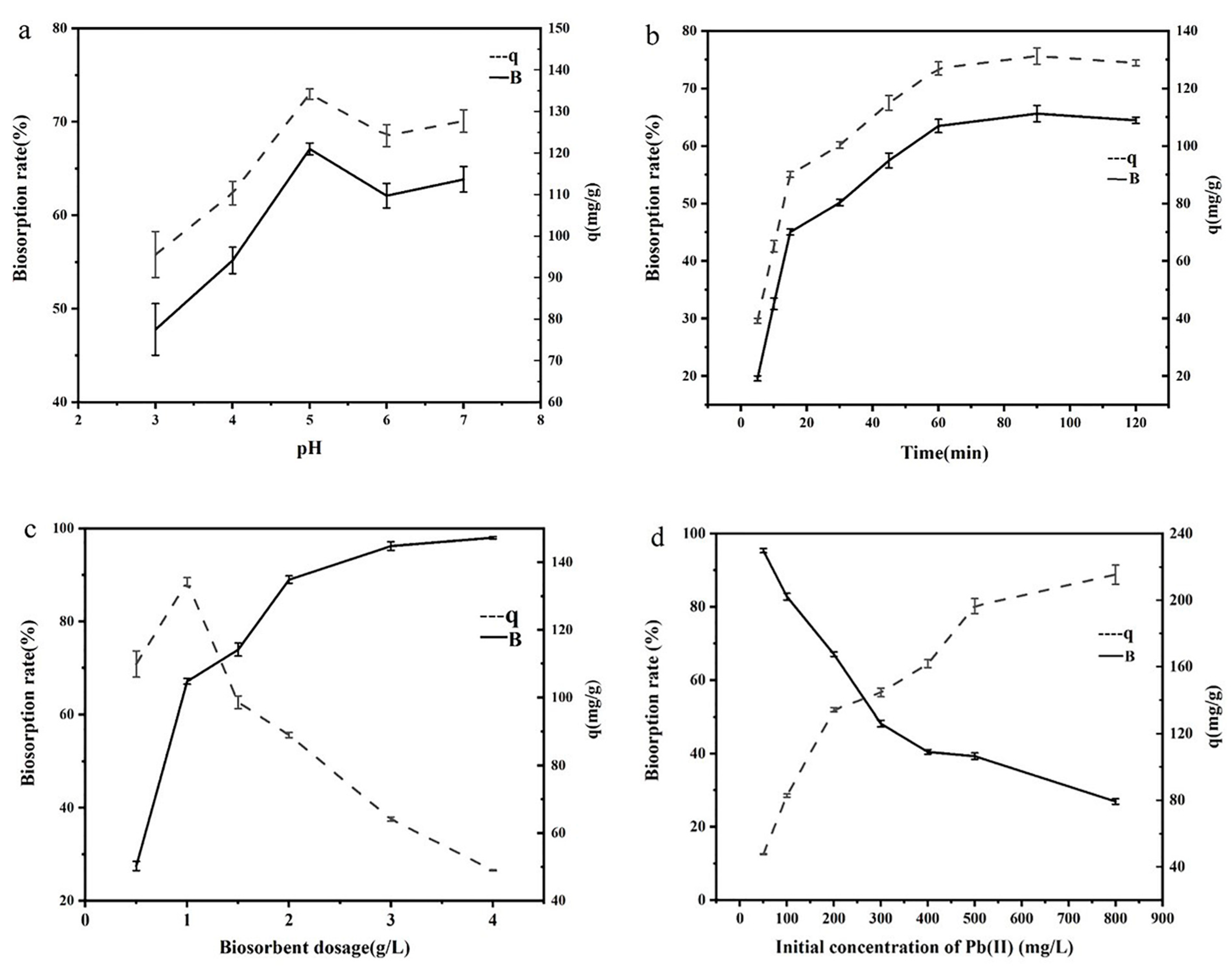
The ability to synthesize PbS NPs suggests that PQ7 might attract lead ions from the environment. To fully explore its adsorption properties, some parameters, such as pH, contact time, biomass dosage and initial Pb(II) concentration, that influence the performance of adsorbents, were investigated. pH can impact the surface chemistry of bacteria to change the sorption efficiency [23]. The biosorption rate and capacity results with respect to pH values from 3.0 to 7.0 are depicted in Figure 3a. The biosorption rate and biosorption capacity both increased as the pH increased from 3 to 5. The maximum biosorption capacity and rate reached 134.22 mg/g and 67.11%, respectively, when the pH reached 5. Then, the rate and capacity of sorption declined when the pH changed from 5 to 7.
These results might be attributed to more carboxyl groups dissociating from the cell surface when the pH was 5, which enhanced the combination of Pb2+ ions with anionic ligands [35]. At pH 3 and 4, functional anionic active sites might be protonated on the cell surface, thus leaving many cationic groups in solution and limiting the biosorption rate and capacity of lead(II) [36]. When the solution is alkaline, precipitation might occur. The hydroxide ions (OH−) in the solution could combine with lead ions (Pb2+) to form precipitation (Pb(OH)2). To distinguish adsorption from precipitation effectively, the pH should not be higher than 7 in adsorption experiments [37]. Therefore, pH 5 is an optimal condition for adsorption experiments of lead ions.
3.3.2. Effect of Contact Time
Figure 3b indicates the effect of contact time on the adsorption of Pb2+ ions by PQ7. In the first 60 min, the adsorption of lead ions was an extremely rapid process. After that, the process slowed and reached equilibrium at 90 min. In the equilibrium stage, the biological adsorption and rate reached 136.27 mg/g and 68.14%, respectively. In the adsorption of lead ions by bacteria, the first stage of the adsorption process of lead ions is the combination of the lead ions in solution with the functional groups in the microbial cell wall, which is fast, reversible and independent of energy metabolism [38]. In the later stage, lead is mainly precipitated or complexed, which is slow [39]. Therefore, the contact time of 90 min served as the most suitable equilibrium time.
3.3.3. Effect of Biosorbent Dosage
The influence of the biomass dosage was investigated by changing the adsorbent dosage from 0.5 g/L to 4.0 g/L. Figure 3c indicates that the biosorption rate increased from 27.48% to 97.99% when the dosage changed from 0.5 g/L to 4.0 g/L. However, the maximum biosorption capacity of 134.21 mg/g was obtained at a dosage of 1 g/L. Moreover, the biosorption capacity declined rapidly as the biosorbent dosage increased from 1.0 g/L to 4.0 g/L. In the case of higher dosages of biological adsorbent, the available exchangeable sites on the cell surface could not be covered completely by the available solute, which led to a low capacity of adsorption. Moreover, the biosorption efficiency was also affected by competition for available metal ion adsorption sites and aggregation or overlapping on the cell surface [40]. Similar results were also indicated in previous studies [41,42]. In this study, 1.0 g/L was the ideal biomass dosage.
3.3.4. Effect of Initial Pb(II) Concentration
The adsorption efficiency and capacity can be affected immensely by the initial metal ion concentration [43]. A single-factor design was used to study the effect of different initial lead ion concentrations (50, 100, 200, 300, 400, 500 and 800 mg/L) at a contact time of 90 min, pH of 5, temperature of 30 °C, biomass dosage of 1 g/L and rotational speed of 200 rpm. As shown in Figure 3d, the sorption rate declined greatly from 95.35% to 26.93% when the initial concentration changed from 50 to 800 mg/L. Interestingly, the sorption capacity increased gradually as the initial concentration increased and reached 215.4 mg/g at a lead concentration of 800 mg/L. This result could be attributed to the solute molecules being less abundant than the effective surface sites at lower concentrations, leading to a much lower sorption capacity. In addition, as the initial concentration increased, the sites on the cell surface would become saturated, and the sorption capacity would become stable [44]. To balance the sorption rate and capacity, 200 mg/L was chosen as a suitable initial concentration; at this value, the sorption rate and capacity were 67.1% and 134.2 mg/g, respectively.
3.4. Isotherm and Kinetic Modeling of the Biosorption of Pb(II)
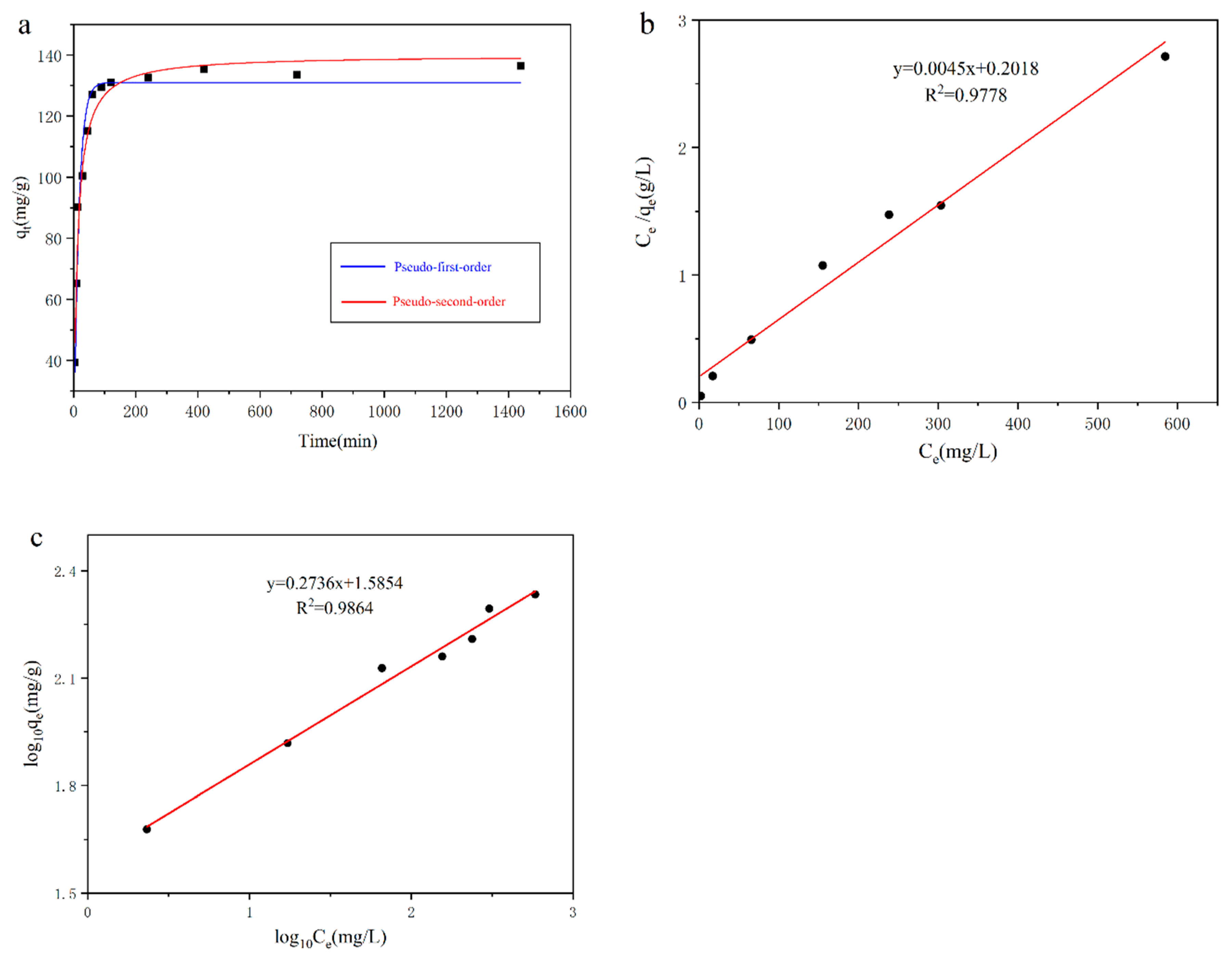
The kinetics were studied to understand the mechanism of adsorption of lead ions. The experimental data were fitted by Equations (3) and (4) to evaluate the parameters of the pseudo-first order and pseudo-second order kinetic models. The data related to the kinetic models are indicated in Table 1 and Figure 4a. The correlation coefficient (R2) of the pseudo-first-order kinetic model (0.962) was lower than that of the pseudo-second-order kinetic model (0.977). Moreover, the experimental data (qexp) were closer to the data (qe2) obtained from the pseudo-second-order kinetic model than those (qe1) calculated from the pseudo-first-order model. This result meant that adsorption occurred not only on the cell surface but also through mass transfer via particle diffusion [45,46]. Thus, the adsorption of lead ions occurred through chemisorption [47].
To further understand the biosorption process of lead ions, the biosorption data were analyzed by the Langmuir and Freundlich isotherm models (Figure 4b,c). The parameters of these two isotherm models are shown in Table 1. From the table and figures, the correlation coefficient (R2) of the Freundlich model (0.986) is better than that of the Langmuir model (0.978). This might be because the process of absorbing lead ions by PQ7 was based on adsorption on different surfaces and because the adsorption sites were not identical to or independent of each other, as reported in previous studies [21,22,23]. Table 2 compares the biosorption of Pb(II) by different bacteria. The maximum Pb(II) adsorption capacity of PQ7 was higher than those obtained in other reports. Thus, we deduced that the metal ion adsorption capacity of these microbes that could synthesize metal sulfide nanoparticles might be higher than that of some metal-resistant bacteria. This result may provide a direction for finding more efficient biosorbents. However, more experiments and data are needed to verify these results.
3.5. SEM–EDS, FTIR and XRD Analyses
It was observed from the SEM images that the morphology of the bacteria changed after treating with lead ions (Figure 5a,b). There were many protuberant particles on the cell surface after adsorption in comparison with the control (Figure 5a,b). The EDS data indicated that Pb(II) was present, and the atomic percentage (at%) was 1.65. The C and O (at%) contents decreased from 71.77 and 18.5 to 69.04 and 15.65, respectively, while the contents of N and P increased from 7.66 and 1.89 to 11.21 and 1.95, respectively, after treating lead ions (Figure 5a,b). These data indicated that C-, N-, P- and O-containing functional groups on the cell surface could combine with Pb(II). It has been reported that functional groups (amino, phosphate, carboxylate and hydroxyl) on the cell surface play an important role in the adsorption process [53,55,56].
FTIR spectra of bacteria before and after treatment with lead ions were obtained to investigate the interactions between Pb and the functional groups present on the cell walls. There were several changed absorption peaks due to the functional groups (Figure 5c). For example, the shift from 1541.77 cm−1 to 1534.87 cm−1 after treatment with Pb(II) corresponded to -NH vibrations [52]. The peak from 1395.72 cm−1 to 1391.18 cm−1 might be attributed to CO32− stretching [57]. In addition, the peak at 1239.42 cm−1 (before contact with Pb(II)) shifted to 1234.42 cm−1 (after contact with Pb(II)), which was caused by C–O (carboxyl) stretching vibrations [58]. Moreover, the peak shift from 1082.91 cm−1 to 1061.24 cm−1 may be due to phosphate stretching vibrations in polysaccharides and nucleic acids [59]. The FTIR data analysis indicated that functional groups such as carboxyl, carbonyl and phosphate groups might participate in lead ion adsorption, which was also shown in the SEM–EDS data.
From the SEM image, Pb compounds might be synthesized on the cell surface after treatment with lead ions. Cells before and after the adsorption of Pb(II) were analyzed by XRD (Figure 5d). On the basis of matching with the standard cards of compounds in the Joint Committee on Powder Diffraction Standards (JCPDS) database, Pb5(PO4)3Cl (JCPDS-01-084-2045) and Pb10(PO4)6(OH)2 (JCPDS-01-087-2477) dominated the cell surface. The peaks of phosphate species were often observed at approximately 30 degrees. Phosphate is one of the important functional groups on the cell surface involved in the interaction between lead ions and cells and makes lead precipitate [60]. Therefore, the higher biosorption capacity of PQ7 might be attributed to the presence of many functional groups, such as phosphate and hydroxyl groups, on the bacterial surface that can induce the precipitation of Pb(II).
4. Conclusions
In this study, we report that Shinella zoogloeoides PQ7 can synthesize PbS NPs in the presence of lead ions. Biosorption experiments with varying contact time, pH, biomass dosage and initial Pb(II) concentration were carried out to explore the adsorption behavior of Pb(II). The equilibrium data followed the Freundlich isotherm model, and the kinetic data fit well with the pseudo second-order model. The maximum (qmax) Pb(II) adsorption reached 222.22 mg/g, which was higher than that of other bacteria reported in previous literature. In summary, Shinella zoogloeoides PQ7 shows excellent potential for heavy metal bioremediation, and to remove Pb(II) from Pb(II)-contaminated wastewater.
Author Contributions
Conceptualization, W.Z. and Y.H.; methodology, W.Z.; investigation W.Z.; resources, Y.H.; data curation, W.Z.; writing—original draft preparation, W.Z.; writing—review and editing, W.Z. and Y.H.; visualization, W.Z.; supervision, Y.H.; funding acquisition, Y.H. All authors have read and agreed to the published version of the manuscript.
Funding
This work was financially supported by Chinese national key research and development plan [2017YFA0207003].
Conflicts of Interest
All authors declare that they have no conflict of interest.
References
- Xiao, X.; Liu, Q.Y.; Lu, X.R.; Li, T.T.; Feng, X.L.; Li, Q.; Liu, Z.Y.; Feng, Y.J. Self-assembly of complex hollow CuS nano/micro shell by an electrochemically active bacterium Shewanella oneidensis MR-1. Int. Biodeterior. Biodegrad. 2017, 116, 10–16. [Google Scholar] [CrossRef]
- Satapute, P.; Paidi, M.K.; Kurjogi, M.; Jogaiah, S. Physiological adaptation and spectral annotation of Arsenic and Cadmium heavy metal-resistant and susceptible strain Pseudomonas taiwanensis. Environ. Pollut. 2019, 251, 555–563. [Google Scholar] [CrossRef] [PubMed]
- Sparks, D.L. Toxic metals in the environment: The role of surfaces. Elements 2005, 1, 193–197. [Google Scholar] [CrossRef]
- Amaral, A.F.S.; Arruda, M.; Cabral, S.; Rodrigues, A.S. Essential and non-essential trace metals in scalp hair of men chronically exposed to volcanogenic metals in the Azores, Portugal. Environ. Int. 2008, 34, 1104–1108. [Google Scholar] [CrossRef] [PubMed] [Green Version]
- Rana, S.V.S. Metals and apoptosis: Recent developments. J. Trace Elem. Med. Biol. 2008, 22, 262–284. [Google Scholar] [CrossRef]
- Farooq, U.; Kozinski, J.A.; Khan, M.A.; Athar, M. Biosorption of heavy metal ions using wheat based biosorbents—A review of the recent literature. Bioresour. Technol. 2010, 101, 5043–5053. [Google Scholar] [CrossRef] [PubMed]
- Zanardini, E.; Andreoni, V.; Borin, S.; Cappitelli, F.; Daffonchio, D.; Talotta, P.; Sorlini, C.; Ranalli, G.; Bruni, S.; Cariati, F. Lead-resistant microorganisms from red stains of marble of the Certosa of Pavia, Italy and use of nucleic acid-based techniques for their detection. Int. Biodeterior. Biodegrad. 1997, 40, 171–182. [Google Scholar] [CrossRef]
- Templeton, A.S.; Trainor, T.P.; Spormann, A.M.; Newville, M.; Sutton, S.R.; Dohnalkova, A.; Gorby, Y.; Brown, G.E. Sorption versus biomineralization of Pb(II) within Burkholderia cepacia biofilms. Environ. Sci. Technol. 2003, 37, 300–307. [Google Scholar] [CrossRef]
- Karnachuk, O.V.; Kurochkina, S.Y.; Tuovinen, O.H. Growth of sulfate-reducing bacteria with solid-phase electron acceptors. Appl. Microbiol. Biotechnol. 2002, 58, 482–486. [Google Scholar] [CrossRef]
- Naik, M.M.; Pandey, A.; Dubey, S.K. Pseudomonas aeruginosa strain WI-1 from Mandovi estuary possesses metallothionein to alleviate lead toxicity and promotes plant growth. Ecotox. Environ. Safe. 2012, 79, 129–133. [Google Scholar] [CrossRef]
- Seshadri, S.; Saranya, K.; Kowshik, M. Green Synthesis of Lead Sulfide Nanoparticles by the Lead Resistant Marine Yeast, Rhodosporidium diobovatum. Biotechnol. Prog. 2011, 27, 1464–1469. [Google Scholar] [CrossRef] [PubMed]
- Priyanka, U.; Gowda, A.K.M.; Elisha, M.G.; Teja, S.B.; Nitish, N.; Mohan, R.B. Biologically synthesized PbS nanoparticles for the detection of arsenic in water. Int. Biodeterior. Biodegrad. 2017, 119, 78–86. [Google Scholar]
- Kowshik, M.; Vogel, W.; Urban, J.; Kulkarni, S.K.; Paknikar, K.M. Microbial synthesis of semiconductor PbS nanocrystallites. Adv. Mater. 2002, 14, 815–818. [Google Scholar] [CrossRef]
- Gong, J.; Zhang, Z.M.; Bai, H.J.; Yang, G.E. Microbiological synthesis of nanophase PbS by Desulfotomaculum sp. Sci. China Ser. E-Technol. Sci. 2007, 50, 302–307. [Google Scholar] [CrossRef]
- Hosseini, M.R.; Sarvi, M.N. Recent achievements in the microbial synthesis of semiconductor metal sulfide nanoparticles. Mater. Sci. Semicond. Process 2015, 40, 293–301. [Google Scholar] [CrossRef]
- Suganya, M.; Balu, A.R. PbS nanopowder—Synthesis, characterization and antimicrobial activity. Mater. Sci. 2017, 35, 322–328. [Google Scholar] [CrossRef] [Green Version]
- Huang, Y.L.; Zeng, Y.H.; Yu, Z.L.; Zhang, J.; Feng, H.; Lin, X.C. In silico and experimental methods revealed highly diverse bacteria with quorum sensing and aromatics biodegradation systems—A potential broad application on bioremediation. Bioresour. Technol. 2013, 148, 311–316. [Google Scholar] [CrossRef]
- Ma, Y.; Wei, Y.; Qiu, J.G.; Wen, R.T.; Hong, J.; Liu, W.P. Isolation, transposon mutagenesis, and characterization of the novel nicotine-degrading strain Shinella sp. HZN7. Appl. Microbiol. Biotechnol. 2014, 98, 2625–2636. [Google Scholar] [CrossRef]
- Biala, S.; Chadha, P.; Saini, H.S. Biodegradation of 4-aminobenzenesulfonate by indigenous isolate Shinella yambaruensis SA1 and its validation by genotoxic analysis. Biotechnol. Bioprocess Eng. 2014, 19, 1034–1041. [Google Scholar] [CrossRef]
- Wu, S.J.; Li, T.F.; Xia, X.; Zhou, Z.J.; Zheng, S.X.; Wang, G.J. Reduction of tellurite in Shinella sp. WSJ-2 and adsorption removal of multiple dyes and metals by biogenic tellurium nanorods. Int. Biodeterior. Biodegrad. 2019, 144, 10. [Google Scholar] [CrossRef]
- Jeong, S.W.; Kim, H.K.; Yang, J.E.; Choi, Y.J. Removal of Pb(II) by Pellicle-Like Biofilm-Producing Methylobacterium hispanicum EM2 Strain from Aqueous Media. Water 2019, 11, 2081. [Google Scholar] [CrossRef] [Green Version]
- Li, H.F.; Lin, Y.B.; Guan, W.M.; Chang, J.L.; Xu, L.; Guo, J.K.; Wei, G.H. Biosorption of Zn(II) by live and dead cells of Streptomyces ciscaucasicus Strain CCNWHX 72-14. J. Hazard. Mater. 2010, 179, 151–159. [Google Scholar] [CrossRef]
- Yuvaraja, G.; Zheng, N.C.; Pang, Y.X.; Su, M.H.; Chen, D.Y.; Kong, L.J.; Mehmood, S.; Subbaiah, M.V.; Wen, J.C. Removal of U(VI) from aqueous and polluted water solutions using magnetic Arachis hypogaea leaves powder impregnated into chitosan macromolecule. Int. J. Biol. Macromol. 2020, 148, 887–897. [Google Scholar] [CrossRef] [PubMed]
- Ho, Y.S.; McKay, G. Pseudo-second order model for sorption processes. Process Biochem. 1999, 34, 451–465. [Google Scholar] [CrossRef]
- Rodriguez-Tirado, V.; Green-Ruiz, C.; Gomez-Gil, B. Cu and Pb biosorption on Bacillus thioparans strain U3 in aqueous solution: Kinetic and equilibrium studies. Chem. Eng. J. 2012, 181, 352–359. [Google Scholar] [CrossRef]
- Langmuir, I. THE ADSORPTION OF GASES ON PLANE SURFACES OF GLASS, MICA AND PLATINUM. J. Am. Chem. Soc. 1918, 40, 1361–1403. [Google Scholar] [CrossRef] [Green Version]
- Freundlich, H. Concerning adsorption in solutions. Zeitschrift Fur Physikalische Chemie--Stochiometrie Und Verwandtschaftslehre 1906, 57, 385–470. [Google Scholar]
- Tanzil, A.H.; Sultana, S.T.; Saunders, S.R.; Shi, L.; Marsili, E.; Beyenal, H. Biological synthesis of nanoparticles in biofilms. Enzyme Microb. Technol. 2016, 95, 4–12. [Google Scholar] [CrossRef]
- Bai, H.J.; Zhang, Z.M.; Guo, Y.; Yang, G.E. Biosynthesis of cadmium sulfide nanoparticles by photosynthetic bacteria Rhodopseudomonas palustris. Colloid Surf. B-Biointerfaces 2009, 70, 142–146. [Google Scholar] [CrossRef]
- Shi, B.Z.; Qi, Y.; Tian, L.C.; Liu, L. The enhanced photoelectrochemical performance of PbS/ZnS quantum dots co-sensitized CdSe nanorods array heterostructure. Mater. Sci. Semicond. Process 2019, 98, 7–12. [Google Scholar] [CrossRef]
- Ettema, A.; Haas, C. AN X-RAY PHOTOEMISSION SPECTROSCOPY STUDY OF INTERLAYER CHARGE-TRANSFER IN SOME MISFIT LAYER COMPOUNDS. J. Phys.-Condes. Matter 1993, 5, 3817–3826. [Google Scholar] [CrossRef]
- Singh, R.; Kumar, A.; Kirrolia, A.; Kumar, R.; Yadav, N.; Bishnoi, N.R.; Lohchab, R.K. Removal of sulphate, COD and Cr(VI) in simulated and real wastewater by sulphate reducing bacteria enrichment in small bioreactor and FTIR study. Bioresour. Technol. 2011, 102, 677–682. [Google Scholar] [CrossRef]
- Da Costa, J.P.; Girao, A.V.; Lourenco, J.P.; Monteiro, O.C.; Trindade, T.; Costa, M.C. Synthesis of nanocrystalline ZnS using biologically generated sulfide. Hydrometallurgy 2012, 117, 57–63. [Google Scholar] [CrossRef]
- Li, X.; Lan, S.M.; Zhu, Z.P.; Zhang, C.; Zeng, G.M.; Livu, Y.G.; Cao, W.C.; Song, B.; Yang, H.; Wang, S.F.; et al. The bioenergetics mechanisms and applications of sulfate-reducing bacteria in remediation of pollutants in drainage: A review. Ecotox. Environ. Safe. 2018, 158, 162–170. [Google Scholar] [CrossRef] [PubMed]
- Jin, Y.; Yu, S.M.; Teng, C.Y.; Song, T.; Dong, L.Y.; Liang, J.S.; Bai, X.; Xu, X.H.; Qu, J.J. Biosorption characteristic of Alcaligenes sp BAPb.1 for removal of lead(II) from aqueous solution. 3 Biotech 2017, 7, 12. [Google Scholar] [CrossRef] [PubMed] [Green Version]
- Rodriguez, C.E.; Quesada, A.; Rodriguez, E. Nickel biosorption by Acinetobacter baumannii and Pseudomonas aeruginosa isolated from industrial wastewater. Braz. J. Microbiol. 2006, 37, 465–467. [Google Scholar] [CrossRef]
- Onundi, Y.B.; Mamun, A.A.; Al Khatib, M.F.; Ahmed, Y.M. Adsorption of copper, nickel and lead ions from synthetic semiconductor industrial wastewater by palm shell activated carbon. Int. J. Environ. Sci. Technol. 2010, 7, 751–758. [Google Scholar] [CrossRef] [Green Version]
- Lan, T.; Ding, C.C.; Liao, J.L.; Li, X.L.; Li, X.L.; Zhang, J.; Zhang, D.; Yang, J.J.; Luo, S.Z.; An, Z.; et al. Biosorption behavior and mechanism of thorium on Bacillus sp dwc-2 isolated from soil. Nucl. Sci. Tech. 2015, 26, 11. [Google Scholar]
- Yetis, U.; Ozcengiz, G.; Dilek, F.B.; Ergen, N.; Dolek, A. Heavy metal biosorption by white-rot fungi. Water Sci. Technol. 1998, 38, 323–330. [Google Scholar] [CrossRef]
- El-Sayed, M.T. Removal of lead(II) by Saccharomyces cerevisiae AUMC 3875. Ann. Microbiol. 2013, 63, 1459–1470. [Google Scholar] [CrossRef]
- Masoudzadeh, N.; Zakeri, F.; Lotfabad, T.B.; Sharafi, H.; Masoomi, F.; Zahiri, H.S.; Ahmadian, G.; Noghabi, K.A. Biosorption of cadmium by Brevundimonas sp ZF12 strain, a novel biosorbent isolated from hot-spring waters in high background radiation areas. J. Hazard. Mater. 2011, 197, 190–198. [Google Scholar] [CrossRef] [PubMed]
- Khadivinia, E.; Sharafi, H.; Hadi, F.; Zahiri, H.S.; Modiri, S.; Tohidi, A.; Mousavi, A.; Salmanian, A.H.; Noghabi, K.A. Cadmium biosorption by a glyphosate-degrading bacterium, a novel biosorbent isolated from pesticide-contaminated agricultural soils. J. Ind. Eng. Chem. 2014, 20, 4304–4310. [Google Scholar] [CrossRef]
- SalehA, T.A.; Gupta, V.K.; Al-Saadi, A.A. Adsorption of lead ions from aqueous solution using porous carbon derived from rubber tires: Experimental and computational study. J. Colloid Interface Sci. 2013, 396, 264–269. [Google Scholar] [CrossRef] [PubMed]
- Vijayaraghavan, K.; Yun, Y.S. Bacterial biosorbents and biosorption. Biotechnol. Adv. 2008, 26, 266–291. [Google Scholar] [CrossRef] [PubMed]
- Sobhanardakani, S.; Jafari, A.; Zandipak, R.; Meidanchi, A. Removal of heavy metal (Hg(II) and Cr(VI)) ions from aqueous solutions using Fe2O3@SiO2 thin films as a novel adsorbent. Process Saf. Environ. Protect. 2018, 120, 348–357. [Google Scholar] [CrossRef]
- Rodrigues, E.; Almeida, O.; Brasil, H.; Moraes, D.; dos Reis, M.A.L. Adsorption of chromium (VI) on hydrotalcite-hydroxyapatite material doped with carbon nanotubes: Equilibrium, kinetic and thermodynamic study. Appl. Clay Sci. 2019, 172, 57–64. [Google Scholar] [CrossRef]
- Zafar, M.N.; Nadeem, R.; Hanif, M.A. Biosorption of nickel from protonated rice bran. J. Hazard. Mater. 2007, 143, 478–485. [Google Scholar] [CrossRef]
- Tunali, S.; Cabuk, A.; Akar, T. Removal of lead and copper ions from aqueous solutions by bacterial strain isolated from soil. Chem. Eng. J. 2006, 115, 203–211. [Google Scholar] [CrossRef]
- Chatterjee, S.K.; Bhattacharjee, I.; Chandra, G. Biosorption of heavy metals from industrial waste water by Geobacillus thermodenitrificans. J. Hazard. Mater. 2010, 175, 117–125. [Google Scholar] [CrossRef]
- Pan, J.H.; Liu, R.X.; Tang, H.X. Surface reaction of Bacillus cereus biomass and its biosorption for lead and copper ions. J. Environ. Sci. 2007, 19, 403–408. [Google Scholar] [CrossRef]
- Wierzba, S.; Latala, A. Biosorption lead(II) and nikel(II) from an aqueous solution by bacterial biomass. Pol. J. Chem. Technol. 2010, 12, 72–78. [Google Scholar] [CrossRef] [Green Version]
- Wang, T.Q.; Yao, J.; Yuan, Z.M.; Zhao, Y.; Wang, F.; Chen, H.L. Isolation of lead-resistant Arthrobactor strain GQ-9 and its biosorption mechanism. Environ. Sci. Pollut. Res. 2018, 25, 3527–3538. [Google Scholar] [CrossRef] [PubMed]
- Ren, G.M.; Jin, Y.; Zhang, C.M.; Gu, H.D.; Qu, J.J. Characteristics of Bacillus sp PZ-1 and its biosorption to Pb(II). Ecotox. Environ. Safe. 2015, 117, 141–148. [Google Scholar] [CrossRef] [PubMed]
- Gupta, V.K.; Rastogi, A. Biosorption of lead from aqueous solutions by green algae Spirogyra species: Kinetics and equilibrium studies. J. Hazard. Mater. 2008, 152, 407–414. [Google Scholar] [CrossRef] [PubMed]
- Lim, M.S.; Yeo, I.W.; Roh, Y.; Lee, K.K.; Jung, M.C. Arsenic reduction and precipitation by shewanella sp.: Batch and column tests. Geosci. J. 2008, 12, 151–157. [Google Scholar] [CrossRef]
- Monteiro, C.M.; Castro, P.M.L.; Malcata, F.X. Biosorption of zinc ions from aqueous solution by the microalga Scenedesmus obliquus. Environ. Chem. Lett. 2011, 9, 169–176. [Google Scholar] [CrossRef] [Green Version]
- Cai, Y.; Li, X.P.; Liu, D.Y.; Xu, C.L.; Ai, Y.W.; Sun, X.M.; Zhang, M.; Gao, Y.; Zhang, Y.C.; Yang, T.; et al. A Novel Pb-Resistant Bacillus subtilis Bacterium Isolate for Co-Biosorption of Hazardous Sb(III) and Pb(II): Thermodynamics and Application Strategy. Int. J. Environ. Res. Public Health 2018, 15, 702. [Google Scholar] [CrossRef] [PubMed] [Green Version]
- Aravindhan, R.; Madhan, B.; Rao, J.R.; Nair, B.U.; Ramasami, T. Bioaccumulation of chromium from tannery wastewater: An approach for chrome recovery and reuse. Environ. Sci. Technol. 2004, 38, 300–306. [Google Scholar] [CrossRef]
- Rodriguez-Sanchez, V.; Guzman-Moreno, J.; Rodriguez-Gonzalez, V.; Flores-de La Torre, J.; Ramirez-Santoyo, R.M.; Vidales-Rodriguez, L.E. Biosorption of lead phosphates by lead-tolerant bacteria as a mechanism for lead immobilization. World J. Microbiol. Biotechnol. 2017, 33, 11. [Google Scholar] [CrossRef]
- Huang, J.; Huang, Z.L.; Zhou, J.X.; Li, C.Z.; Yang, Z.H.; Ruan, M.; Li, H.; Zhang, X.; Wu, Z.J.; Qin, X.L.; et al. Enhancement of heavy metals removal by microbial flocculant produced by Paenibacillus polymyxa combined with an insufficient hydroxide precipitation. Chem. Eng. J. 2019, 374, 880–894. [Google Scholar] [CrossRef]
Figure 1.
(a) The cells of PQ7 turn black in the presence of lead nitrate (5 mM). The red circle highlights the black precipitates. (b) The color change of PQ7 broth with or without lead nitrate (5 mM). The cells were cultivated at 30 °C with 200 rpm shaking speed. At the 4th day, the broth was brown with black precipitate after centrifugation.

Figure 2.
The characterization of the black materials synthesized by Shinella zoogloeoides PQ7. (a) Transmission electron microscope (TEM) image of control cells; (b) TEM image of cells with nanoparticles; (c) Scanning electron Microscopy–energy dispersive X-ray spectroscopy (SEM–EDS) of PQ7 grown with Pb (5 mM); (d) X-ray diffraction (XRD) pattern of PbS nanoparticles synthesized by PQ7; (e) X-ray photoelectron spectroscopy (XPS) image of PbS synthesized by PQ7.

Figure 3.
Some parameters influencing the performance of the adsorbent. (a) pH, (b) contact time, (c) biosorbent dosage and (d) initial Pb(II) concentration. The experimental conditions were as follows: (a) Pb(II) concentration = 200 mg/L, contact time = 90 min, biomass dosage = 1 g/L. (b) pH = 5, Pb(II) concentration = 200 mg/L, biomass dosage = 1 g/L. (c) pH = 5, contact time = 90 min, Pb(II) concentration = 200 mg/L. (d) pH = 5, biomass dosage = 1 g/L, contact time = 90 min. B and q indicate the biosorption rate (%) and biosorption capacity (mg/g), respectively. The values and error bars represent the means ± standard deviation (n = 3).

Figure 4.
Isotherm and kinetic modeling of the biosorption of Pb(II). (a) Biosorption kinetic models; the blue line and red line represent the pseudo-first-order and pseudo-second-order models, respectively. Isotherms of Pb(II) biosorption by PQ7: (b) Langmuir; (c) Freundlich.
Figure 4.
Isotherm and kinetic modeling of the biosorption of Pb(II). (a) Biosorption kinetic models; the blue line and red line represent the pseudo-first-order and pseudo-second-order models, respectively. Isotherms of Pb(II) biosorption by PQ7: (b) Langmuir; (c) Freundlich.

Figure 5.
(a) and (b) were SEM–EDS spectra of PQ7 before and after treatment with lead ions, respectively; (c) FTIR and (d) XRD spectra of PQ7 before and after treatment with lead ions. The conditions were as follows: pH = 5, Pb(II) concentration = 200 mg/L, biomass = 1 g/L, contact time = 90 min.
Figure 5.
(a) and (b) were SEM–EDS spectra of PQ7 before and after treatment with lead ions, respectively; (c) FTIR and (d) XRD spectra of PQ7 before and after treatment with lead ions. The conditions were as follows: pH = 5, Pb(II) concentration = 200 mg/L, biomass = 1 g/L, contact time = 90 min.


Table 1.
Biosorption kinetic and adsorption isotherm parameters for the adsorption of Pb(II).
| Pseudo-First-Order | Pseudo-Second-Order | ||||
| qe1 (mg/g) | K1 (L/min) | R2 | qe2 (mg/g) | K2 (g/mg/min) | R2 |
| 130.98 | 0.065 | 0.962 | 139.91 | 0.000695 | 0.977 |
| Langmuir Isotherm | Freundlich Isotherm | ||||
| Qmax (mg/g) | KL (L/mg) | R2 | KF (L/g) | n | R2 |
| 222.22 | 0.022 | 0.978 | 38.49 | 3.65 | 0.986 |
Table 2.
Comparative analysis of biosorbents for Pb(II) biosorption.
| Bacteria | qmax (mg/g) | Conditions | Reference |
|---|---|---|---|
| Bacillus sp. ATS-1 | 92.3 | pH 5, 25 °C | [48] |
| Geobacillus thermodenitrificans | 32.26 | pH 4.5, 35 °C | [49] |
| Pseudomonas aeruginosa PU21 | 110 | pH 7, 37.5 °C | [48] |
| Alcaligenes sp. BAPb.1 | 66.7 | pH 5, 30 °C | [35] |
| Bacillus cereus | 36.7 | pH 5.5, 30 °C | [50] |
| Pseudomonas fluorescens | 77.6 | pH 6, 30 °C | [51] |
| Bacillus pumilus | 91.4 | pH 6, 30 °C | [51] |
| Arthrobactor GQ-9 | 17.56 | pH 5.5, 35 °C | [52] |
| Geobacillus thermodenitrificans | 32.26 | pH 4.5, 65 °C | [49] |
| Bacillus thioparans U3 | 210.1 | pH 5, 30 °C | [25] |
| Alcaligenes sp. | 56.8 | pH 5, 35 °C | [35] |
| Bacillus sp. PZ-1 | 15.38 | pH 5, 15 °C | [53] |
| Methylobacterium hispanicum EM2 | 79.84 | pH 7, 30 °C | [21] |
| Pseudomonas stutzeri KCCM 34719 | 142 | pH 6, 30 °C | [54] |
| Shinellazoogloeoides PQ7 | 222.22 | pH 5, 30 °C | This study |
© 2020 by the authors. Licensee MDPI, Basel, Switzerland. This article is an open access article distributed under the terms and conditions of the Creative Commons Attribution (CC BY) license (http://creativecommons.org/licenses/by/4.0/).
Share and Cite
MDPI and ACS Style
Zhang, W.; Huang, Y. The Synthesis of PbS NPs and Biosorption of Pb(II) by Shinella Zoogloeoides PQ7 in Aqueous Conditions. Water 2020, 12, 2065. https://0-doi-org.brum.beds.ac.uk/10.3390/w12072065
AMA Style
Zhang W, Huang Y. The Synthesis of PbS NPs and Biosorption of Pb(II) by Shinella Zoogloeoides PQ7 in Aqueous Conditions. Water. 2020; 12(7):2065. https://0-doi-org.brum.beds.ac.uk/10.3390/w12072065
Chicago/Turabian StyleZhang, Wei, and Yili Huang. 2020. "The Synthesis of PbS NPs and Biosorption of Pb(II) by Shinella Zoogloeoides PQ7 in Aqueous Conditions" Water 12, no. 7: 2065. https://0-doi-org.brum.beds.ac.uk/10.3390/w12072065
Note that from the first issue of 2016, this journal uses article numbers instead of page numbers. See further details here.