Article

A Simulation of the Use of High Throughput Sequencing as Pre-Screening Assay to Enhance the Surveillance of Citrus Viruses and Viroids in the EPPO Region

1
Consiglio per la Ricerca in Agricoltura e L’analisi dell’Economia Agraria-Centro di Ricerca Olivicoltura, Frutticoltura e Agrumicoltura (CREA), C. da Li Rocchi, Via Settimio Severo 83, 87036 Rende (CS), Italy
2
Agrobiotech Soc. Coop. z.i. Blocco Palma I, Via V. Lancia 57, 95121 Catania, Italy
3
Formerly, Department of Phytosanitary Science and Technologies, University of Catania, 95123 Catania, Italy
*
Author to whom correspondence should be addressed.
Submission received: 25 March 2021 / Revised: 20 April 2021 / Accepted: 23 April 2021 / Published: 27 April 2021

Abstract

:
Citrus are affected by many viruses and viroids, some globally widespread and some restricted to particular countries or areas. In this study, we simulated the use of high throughput sequencing (HTS) and the bioinformatic analysis of small interfering RNAs (siRNA) as a pre-screening method to guide bioindexing and molecular detection to enhance the surveillance survey of some key or emerging citrus viruses, such as non-European citrus tristeza virus isolates (non-EU CTV), citrus tatter leaf virus, citrus leprosis virus, citrus yellow mosaic virus, and citrus bark cracking viroid, present in the EPPO lists, and the citrus yellow vein clearing virus. The HTS’s ability to detect other citrus viroids was also evaluated. The results demonstrate that HTS provides a comprehensive phytosanitary status of citrus samples either in single and multiple infections of viruses and viroids. It also provides effective information on citrus tristeza virus mixed infections despite not being able to identify the non-EU variants of the virus. Bioindexing checks each single virus infection but does not differentiate viroids on the Etrog citron indicator and is time-consuming. Molecular assays are valuable as confirmation tests of viruses and viroids but many pairs of primers are needed for a full screening and new or non-target pathogens remain undetected. In addition, the genomes of two isolates of the citrus yellow vein clearing virus and the citrus tatter leaf virus, detected in a sample from China, are described.

1. Introduction

The number of viruses and viroids affecting citrus has grown considerably in the last few decades [1]. The different global distribution and the presence of different strains and variants mean that their transborder movement among countries needs to be prevented. In line with the International Plant Protection Convention guidelines for surveillance [2], the main objectives are to monitor pests for quarantine purposes, alien invasive species, pests with a negative economic and/or farming impact, and the natural enemies of pests. In addition, biosecurity reasons may create phytosanitary targets that are not categorized as quarantine pests and which must be included in regular surveillance programs, the aim being the early detection of detrimental viruses [3]. This requires detection and identification methods that are in line with current international standards together with a statistically sound and risk-based pest survey approach [4].
According to the categorization of citrus virus and viroid diseases in the Euro-Mediterranean region, the European and Mediterranean Plant Protection Organization (EPPO) has drawn up two lists of pests whose regulation is relevant for the entire EPPO region or large parts of it [5]. The citrus blight disease, citrus leprosis virus (CiLV), citrus tatter leaf virus (CTLV) and citrus yellow mosaic virus (CYMV), which are not present in the EPPO region, are included in the EPPO A1 List, whereas the citrus bark cracking viroid (CBCVd) and citrus tristeza virus (CTV) non-European isolates (non-EU CTV), and satsuma dwarf virus, are included in the EPPO A2 List. Each of the above viruses and viroids requires different tools and levels of detectability, and complex procedures are needed for reliable results. However, the satsuma dwarf virus affects mainly citrus varieties less relevant in the EPPO region and the causal agent of the citrus blight remains unknown, and no direct test is available [6].
The citrus leprosis syndrome is a disease complex severely affecting leaves, fruits and twigs associated with several known viruses [1]. CTLV (also known as apple stem grooving virus, ASGV) is the causal agent of tatter leaf, a graft incompatibility disease affecting plants grafted on trifoliate orange (Poncirus trifoliata) and its hybrids, now used in the EPPO region to prevent the citrus tristeza decline [7]. CYMV, closely related to the cacao swollen shoot virus [8], causes serious losses in pummelo and sweet orange [9].
For non-EU CTV isolates, in the EPPO A2 List, which are responsible for field stem pitting (SP) on sweet orange regardless of rootstock, and resistance-breaking (RB) of trifoliate orange and its hybrids [10], EU member states are required to plan annual surveys using a statistically sound and risk-based approach, in line with current international standards [4]. CBCVd, the smallest of the citrus viroids, a chimeric recombinant viroid between citrus exocortis viroid (CEVd) and hop stunt viroid (HSVd) [11], induces severe symptoms on trifoliate orange and Carrizo citrange [12,13].
Despite not being reported by the EPPO lists, an additional virus, namely the citrus yellow vein clearing virus (CYVCV) is of current concern worldwide. It is a member of the genus Mandarivirus, which affects lemon production in Turkey and China, as it is easily transmitted by aphids and mechanically [14].
All these viruses and viroids have been fully sequenced by high throughput sequencing (HTS) (Table 1) [15,16,17,18,19,20,21,22,23,24,25,26,27,28] and the potential use of HTS as a routine diagnostic tool for the phytosanitary control of citrus varieties is under evaluation in California [29]. Endemic viruses and viroids of citrus, and new species were confirmed in an Australian living pathogen collection using HTS methods [30]. National-based surveys using HTS have also been launched in Belgium [31]. A recent study has shown that it is possible to detect known and unknown CTV genotypes in mixed infections using an HTS assay [32]. In Italy, HTS has been used to evaluate the genetic structure of the CTV population leading to the discovery of a few infections of T36 strain [33] and the study of variants responsible for seedling yellow (SY) recovery [34]. Moreover, HTS has been used to investigate on a stem pitting isolate from China [35].
HTS has also been used to clarify the genetic differences of old and new citrus viroids [36,37]. All induce leaf bending and curling symptoms on the Etrog citron indicator. However, they are not sufficiently distinctive to be able to be differentiated. Despite the availability of many primers for molecular detection, accurate results are possible only after a bio-amplification on Etrog citron or by collecting specific bands from PolyAcrylamide Gel Electrophoresis (PAGE).
Thanks to the extremely high-throughput data generation, HTS is widely used in many projects dealing with different crops, supported by new bioinformatic instruments for handling the data produced [38]. Exciting results have been achieved in the discovery and characterization of new viruses, the detection of unexpected viral pathogens in plants, the investigation of viral diversity, evolution and spread, and the evaluation of the plant virome [39,40,41].
In this study, we evaluated the use of HTS as a pre-screening method combined with bioindexing and molecular detection in a simulated surveillance survey of citrus virus and viroids regulated by EU phytosanitary measures (Figure 1). The investigation was related to the presence of non-EU CTV isolates, sensu EFSA [4] and to the detection of some virus and viroids in the EPPO A1 and A2 Lists (CTLV, CiLV, CYMV, CBCVd) and to CYVCV. Other citrus viroids and the genetic structure of a local CTV population were also investigated. This involved the sequencing of small interfering RNAs (siRNA), produced in a high quantity by RNA silencing antiviral mechanisms in the plants [40]. The analysis was carried out using siRNA libraries of six field samples of citrus, three of them already analyzed only for the presence of CTV and here re-analyzed for all the viruses and viroids listed above, using a bioinformatic pipeline able to detect mixed virus and viroid infections.
Our results showed that the adoption of HTS for pre-screening can be effectively used for the simultaneous identification of virus and viroids and to detect the prevalent CTV genotypes. However, bioindexing is still necessary to characterize the phenotypic profiles of CTV isolates inducing stem pitting or resistance breaking as well as to confirm the presence of other viruses (i.e., CYVCV and CTLV), supported also by molecular detection. Preliminary results of this multi-disciplinary approach have been presented at the 2019 Joint IOCV XXI and IRCHLB VI [42].

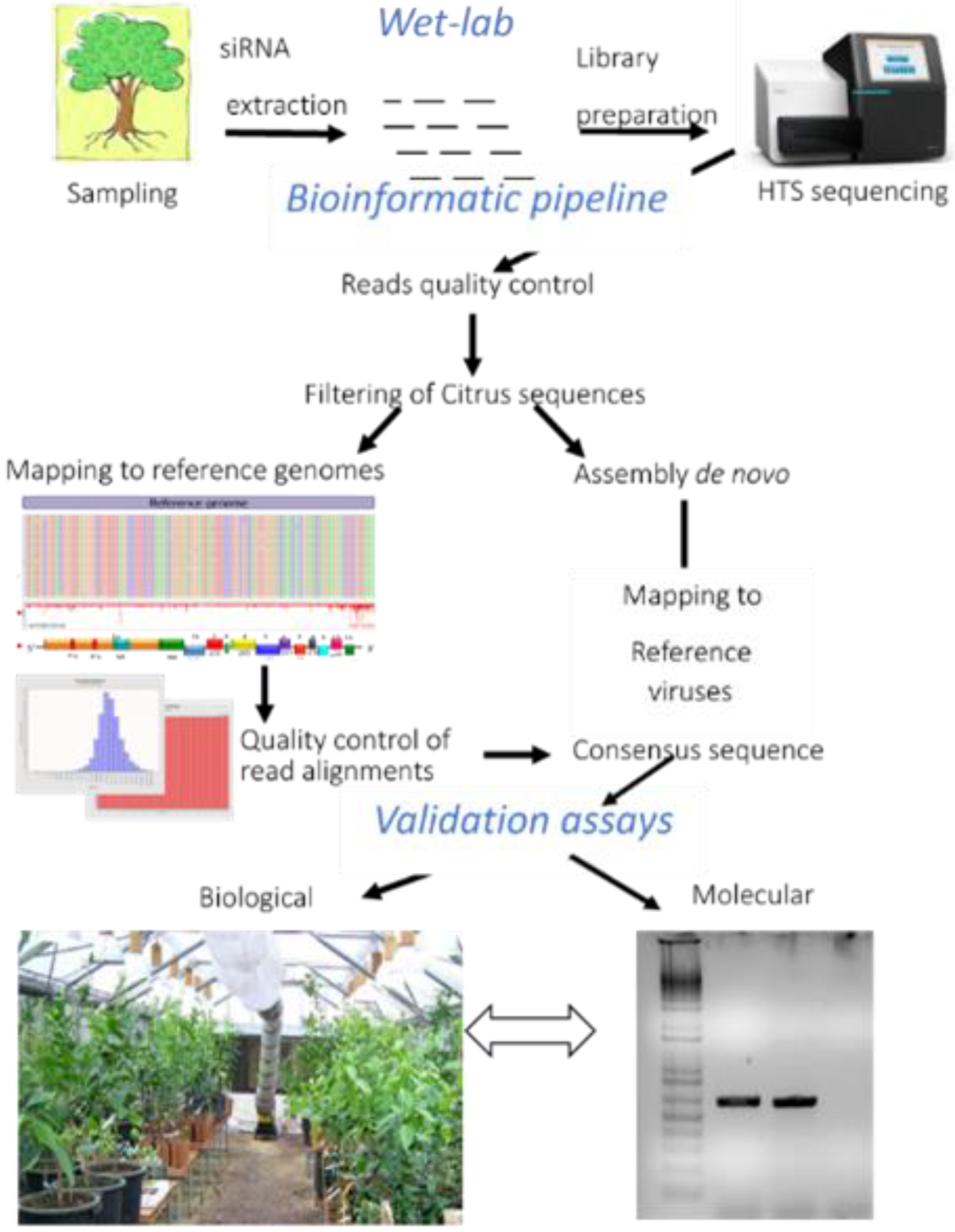
2. Materials and Methods

2.1. Source Plants and Small-RNA Libraries

Six siRNA libraries generated from field sources, tested CTV-positive by enzyme-linked immunosorbent assay (ELISA), were selected for this study. The library of HU-PSTS isolates was generated from a field sweet orange tree in the Hunan province (Chenzou county, China) [35,43] (Table 2). The other libraries were generated from samples collected in a heavily infected CTV focus area in Sicily: Mac25, Mac39, and Mac101 were generated from three ungrafted seedlings of alemow [33,34], whereas the P3R1 and P3R3 libraries were obtained from two different branches of the same tree with different CTV titers (one CTV-ELISA positive and one negative), from a declining Tarocco Sant’Alfio sweet orange grafted on sour orange (Table 2). The Mac25, Mac39, Mac101, and HU-PSTS libraries were previously obtained and partially analyzed only for CTV [33,34,42] and are reanalyzed here with an additional upstream plant host filtering step. P3R1 and P3R3 were fully analyzed for the first time in this study.
The small RNA libraries have been generated according to Licciardello et al., [43] from 200 mg of young bark tissues. The small RNA fraction was extracted using the MirPremier MicroRNA isolation kit (Sigma Aldrich, St. Louis, MO, USA) as instructed by the manufacturer and used as input for library preparation by the NEXT flex Small RNA Sequencing kit (Bioo Scientific, Austin, TX, USA). The resulting libraries were multiplexed, clustered, and sequenced on an Illumina HiSeq 2000 (San Diego, CA, USA) with a single-read 50 cycles sequencing protocol plus indexing. The sequencing runs were analyzed with the Illumina CASAVA pipeline (v1.8.2 San Diego, CA, USA) and small RNA adapters removed using the “Trim sequences” option of the CLC Genomics Workbench (v 6.0.4 CLC Bio, Qiagen, Aarhus, Denmark).

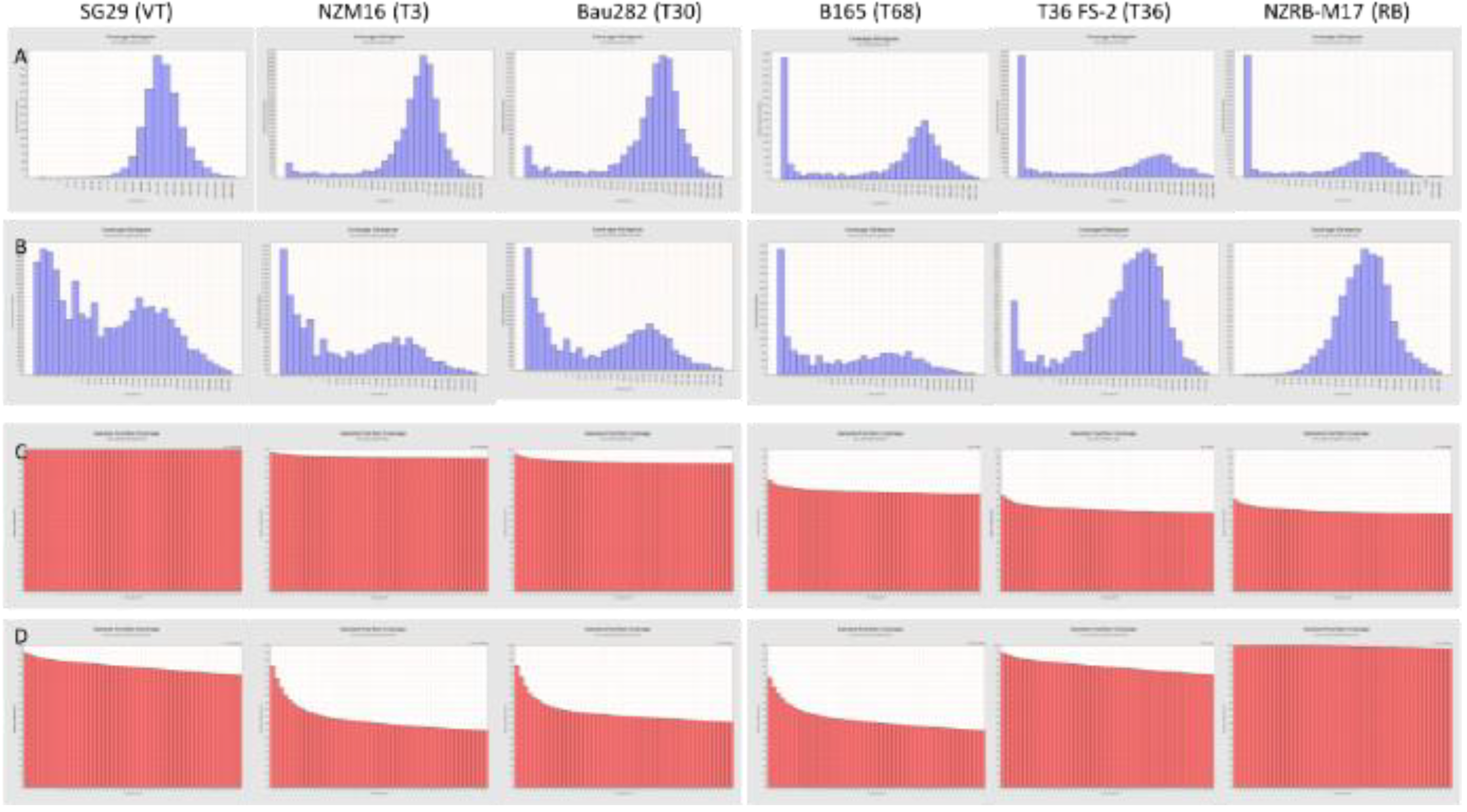
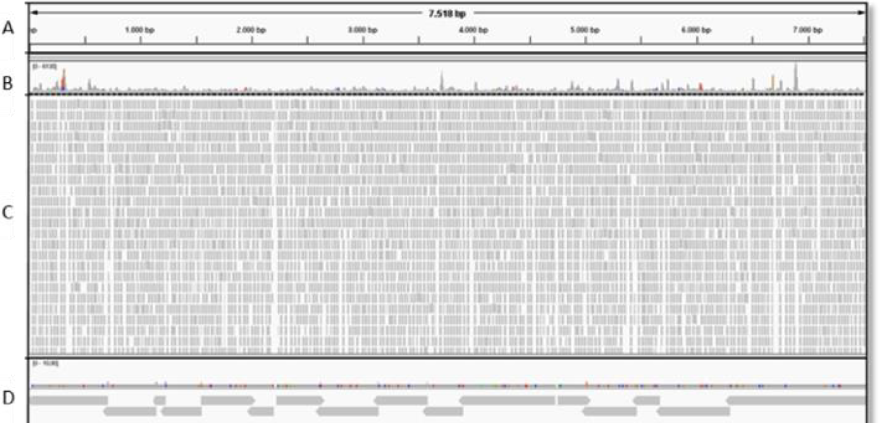
2.2. Data Evaluation by Bioinformatics

For the detection of viral genomes, a “map reads to reference” bioinformatic approach was used to assemble the read sequences to a selected group of known virus and viroid genomic sequences (Table 3). Quality trimmed data were imported into Bowtie2 v. 2.1.0 and a preliminary plant host filtering step was performed after alignment with the chromosomes of Citrus sinensis (NC_023046 to NC_023054), chloroplast (NC_008334) and miRNAs (retrieved from MiRBase database) using the default parameters. For the CTV genotype detection, the unmapped reads were aligned to six reference genomes representative of the six main CTV genotypes [16]. The mapping alignments were further measured by three metrics generated by Qualimap [44] analysis: read counts, percentage of reads count and percentage of genome fraction coverage (GFC) at 50X. Graphical representations showing the quality of the reads aligned against each reference genome, generated by Qualimap, were assessed for a fast evaluation of the unequivocal presence of the virus or viroid in a library.
The consensus sequence was generated by SAM tools 0.1.19 (BSD License, MIT License) and analyzed for similarity against other sequences in the GenBank by BLASTn (NCBI, Bethesda, MD, USA). In addition, a de novo assembly bioinformatic approach has been used for the Mac25 sample and for the CYVCV genome reconstruction in the HU-PSTS using a Velvet (1.2.10, Illumina, San Diego, CA, USA) assembler [45] at different k-mer ranges. Alignment files (.bam) were visualized by Tablet [46] and Integrative Genomics Viewer (IGV) [47].

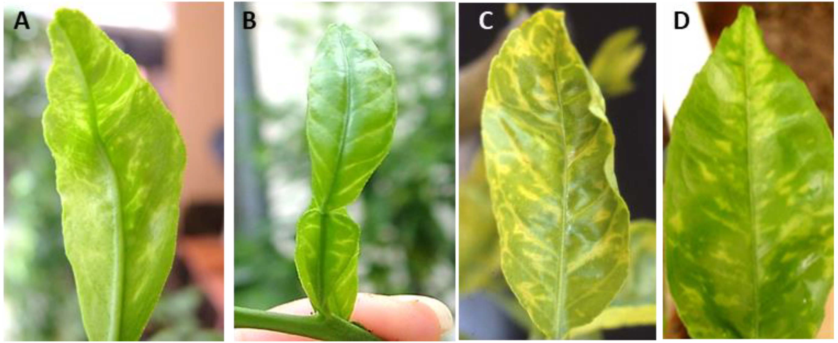
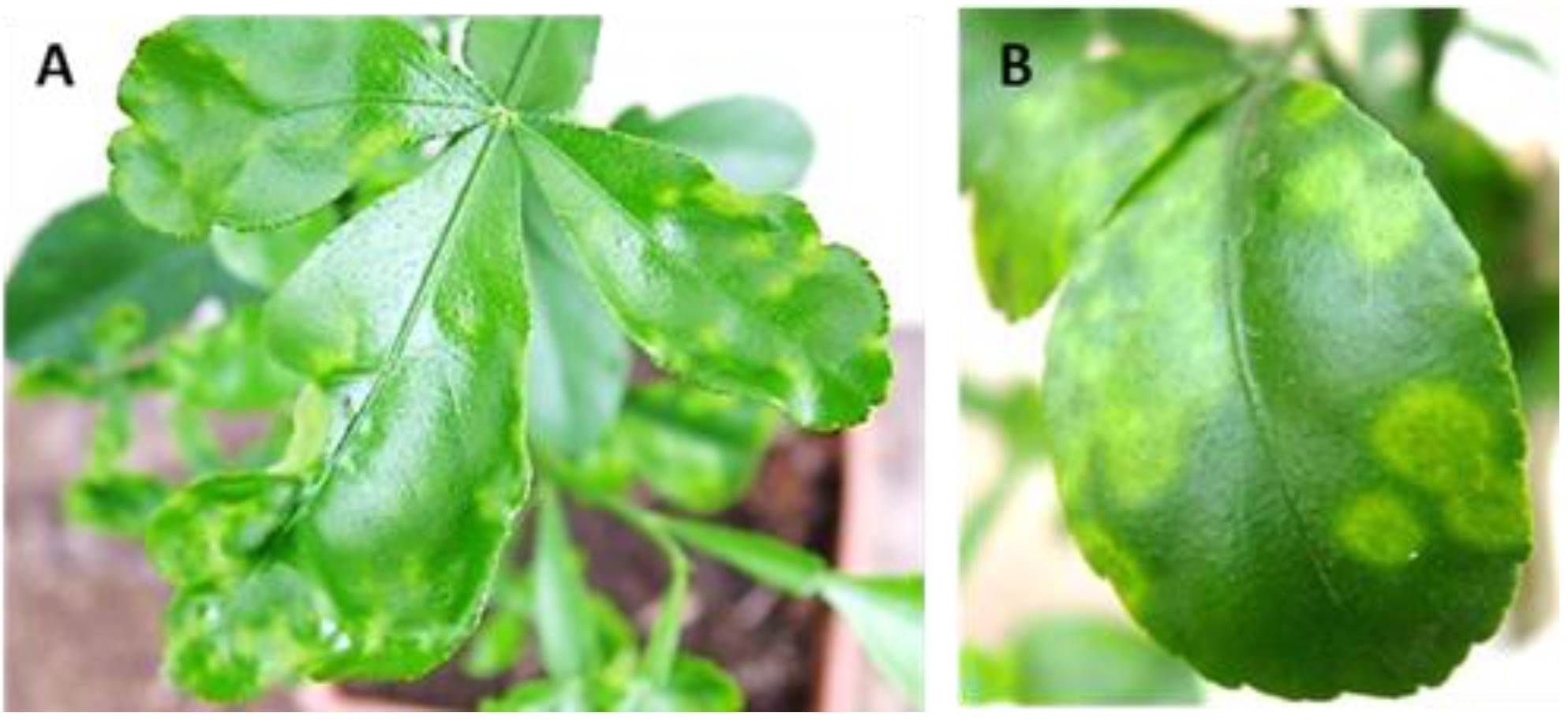
2.3. Biological Indexing

All the trees were indexed by bark inoculation of Mexican lime (Citrus aurantifolia), sour orange (C. aurantium), Duncan and/or Oroblanco grapefruit (C. paradisi), Hamlin sweet orange (C. sinensis) grafted on sour orange, and Etrog citron (C. medica) grafted on C. volkameriana. Supplementary tests were carried out on Carrizo citrange (P. trifoliata x C. sinensis) seedlings for the detection of RB CTV [17] and CTLV searching and on lemon (C. limon) seedlings for detection of CYVCV. At least three plants were inoculated for each indicator and one of each was used as non-inoculated control. The indexing was carried out in a safe greenhouse, with heating and cooling systems, located near Catania (Sicily, Italy, 37°24′ N, 15°03′ E). Symptoms were visually assessed periodically over a 2-year period, according to Garnsey et al. [48].

2.4. RT-PCR and Real-Time RT-PCR

Reverse transcription-polymerase chain reaction(RT-PCR) and real-time RT-PCR were used to validate the bioindexing or HTS results. The total RNA was extracted using Trizol reagent (Invitrogen S.r.l.; Milan, Italy), from 100 mg of bark tissues, previously pulverized with liquid nitrogen, according to the manufacturer’s instructions. For CTV genotype characterization, RT-PCR (for T68 and T3 genotypes, according to Roy et al. [49]) and real-time RT-PCR using specific dual labeled-probes (for VT, T3, T36, T30) were evaluated [50,51]. CYVCV was detected using the primers 1fw and 921rev targeting 5′ region following the PCR conditions previously reported [22]. CTLV was detected using the TL3 and TL4 primer pair and PCR conditions described by Tatineni et al. [52]. For CEVd, HSVd, and citrus dwarfing viroid (CDVd) detection, real-time RT-PCR protocols were used [53,54]. For CBCVd (CVd-IV), the primers and PCR conditions described by Bernard and Duran-Villa [55] were used, whereas CVd-V-R and CVd-V- F primers were used for citrus viroid V (CVd-V) [56].

3. Results

After quality trimming, all the libraries analyzed in this study generated a high number of single reads, respectively, 38.7 M (HU-PSTS), 40.8 M (P3R1), and 29.3 M (P3R3). The total reads were retained for a preliminary host filtering step to remove the majority of the host reads (chromosomes, chloroplasts and miRNAs) which significantly reduced (at least 50%) the number of reads to be analyzed downstream as follows: 19.6 M (HU-PSTS), 8.5 M (P3R1), 4.1 M (P3R3). The unmapped reads were analyzed to search for the presence of non-EU CTV isolates, as well as CLVCV, CTLV, CiLV, CYMV and CBCVd, and other viruses and viroids. From the samples Mac25, Mac39, and Mac101, the total number of reads was 19.6 M, 17.8 M and 9.9 M, respectively. After the host filtering step, the total number of reads was 4.1 M (Mac25), 10.4 M (Mac39), 3.6 M (Mac101), reducing the reads by 20% to 58% of the original siRNA reads.

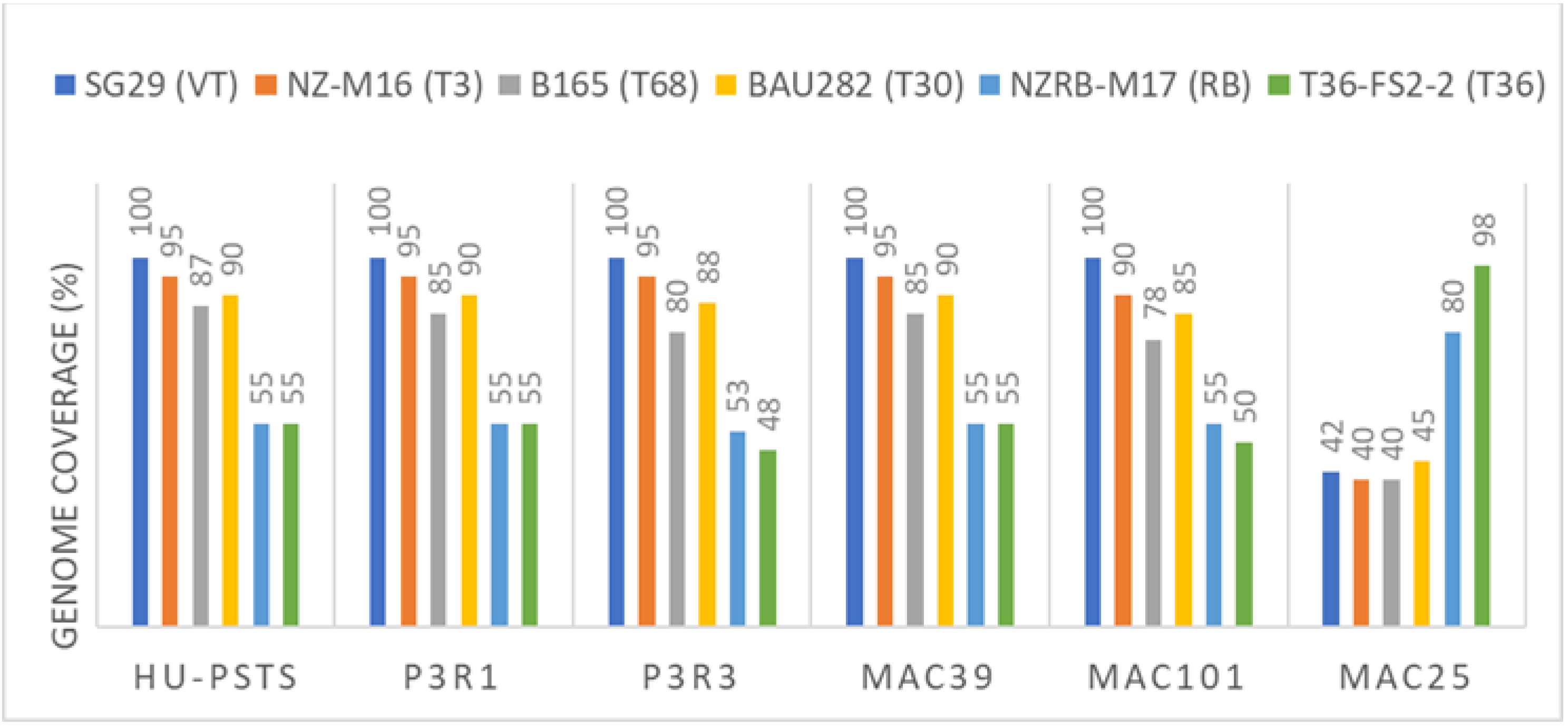
3.1. Searching for Non-EU CTV Isolates

The single reads retained after the host filtering were aligned with reference genomes of six CTV isolates representative of the main six genotypes [17] (Table 3). All the libraries showed a high number of read counts with SG29 (VT) corresponding to 71% of the entire library of P3R1, 58% of P3R3 and 84% in HU-PSTS, and 100% of genome coverage in all these libraries. Alignments with NZM16 (T3), B165 (T68), and Bau282 (T30) gave fewer reads ranging from 66% to 44%, with a coverage of about 95%, 85% and 90%, respectively (Figure 2), whereas alignments with the RB and T36 strains gave less than 35% read counts and a low percentage of coverage (50–45%) (Figure 2). The quality of alignments of the P3R1 library for each reference genome is shown as “genome histograms” and “genome fraction coverage” in Figure 3. Assuming that a 95% of genome coverage can be indicative of the presence of additional CTV strains in mixed infections and a maximum of 92% can be obtained for non-target genomes [32,57], the samples P3R1, P3R3 and HU-PSTS were further characterized by RT-PCR and real-time PCR [49,50,51] for CTV strain identification. P3R1 and P3R3 were positive only for VT, whereas HU-PSTS reacted positively to VT, T3 and T30 probes, and negatively to T36 and T36NS. On the other hand, although the RT-PCR detection for T3 and T68 strains were negative, the high homology of nucleotide sequences between VT and T3 cannot completely exclude the potential presence of T3 in the HU-PSTS.
From a biological point of view, the three sources (P3R1, P3R3, and HU-PSTS) showed a typical seedling yellow (SY) reaction in sour orange (Figure 4), associated with leaf blade malformation and more severe yellowing in HU-PSTS, due to the coinfection of CYVCV (Figure 5). As previously described [35,36,37,38,39,40,41,42,43], the Chinese source induced a severe stem pitting on Duncan grapefruit and a mild stem pitting on Hamlin sweet orange, which makes the isolate compatible with biotype 5 [48]. On the other hand, P3R1 and P3R3 induced stem pitting only on grapefruit compatible with biotype 4. Parallel inoculation of Carrizo citrange gave a negative detection of CTV by ELISA test, revealing an absence of CTV replication, thus excluding the presence of RB isolates in the three sources.
Based on HTS and molecular and biological assays, the results show that P3R1 and P3R3 were infected only by one VT isolate, whereas HU-PSTS was a mixed isolate including at least two genotypes, VT and T30, although the presence of T3 cannot be excluded.
The highest number of reads was generated from the libraries of Mac39 and Mac101 seedlings mapped with SG29 (VT), the prevalent isolate in Sicily [43] after depletion of the host citrus genome. Approximately 9 M (Mac39) and 3.9 M (Mac101) reads were generated (corresponding to 86% and 91% of the entire libraries, respectively), which led to the full genome reconstruction with 100% coverage (Figure 2). The resulting consensus sequences of the assembled genomes were deposited under accession numbers KJ790175 (Mac39) and MW689620 (Mac101). Both genomes showed 99% similarity with SG29.
The Mac25 library, which was reported earlier to contain a T36 CTV isolate (KR263170) [33], was revisited in this study to check the potential presence of CTV isolates biologically non-EU sensu EFSA [4]. When the 4.1 M reads were aligned with CTV reference genomes, the highest number of read counts (18% of the entire library) was obtained after alignment with T36 FS-2 (T36) associated with 98% genome coverage (Figure 2). Alignment with the NZRB-M17 (RB) genome gave 80% coverage, which is not surprising since T36 and RB genotypes are genetically similar (90% nucleotide identity). The percentage coverage with the remaining reference genomes was under 40%. Graphs representing quality alignments for each reference genome are shown in Figure 3. Real-time RT-PCR by Ruiz-Ruiz et al. [50] confirmed a previous analysis by using the protocol of Yokomi et al. [33,51].
Parallel biological indexing showed that the three alemow seedlings had a different SY and SP reaction. Mac25 induced only slight chlorosis on sour orange, whereas Mac39 and Mac101 were asymptomatic (Figure 4). Mac39 and Mac101 caused stem pitting on grapefruit but not on sweet orange, whereas Mac25 did not cause any stem pitting.
HTS screening discriminated the CTV components of the six samples. The results were confirmed by RT-PCR and real-time PCR. Based on biological assays, only the source HU-PSTS met the requirements of a biologic non-EU CTV isolate because of the stem pitting induced on sweet orange [4]. On the other hand, the lack of replication on Carrizo citrange confirms that there was no RB present.

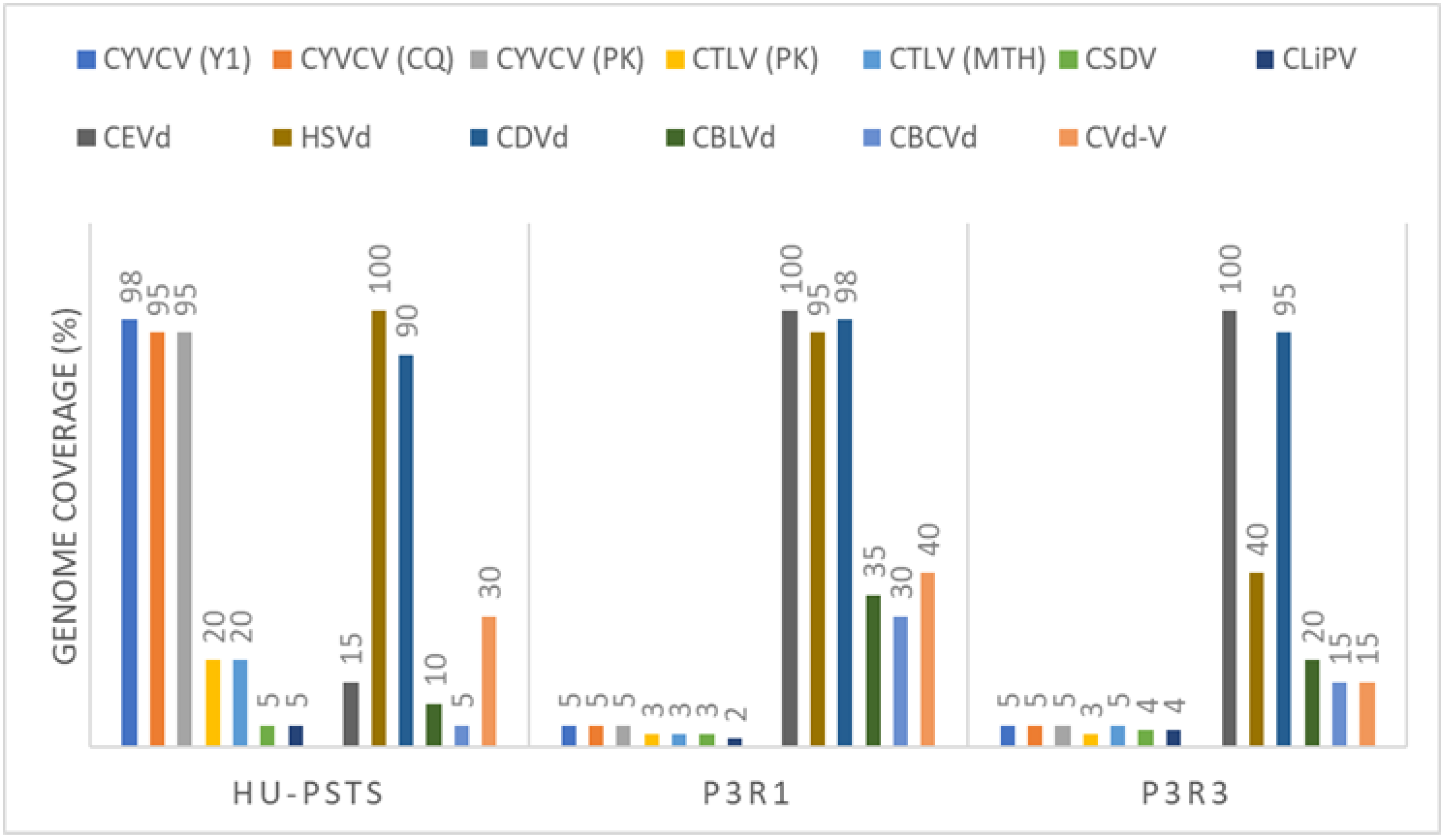
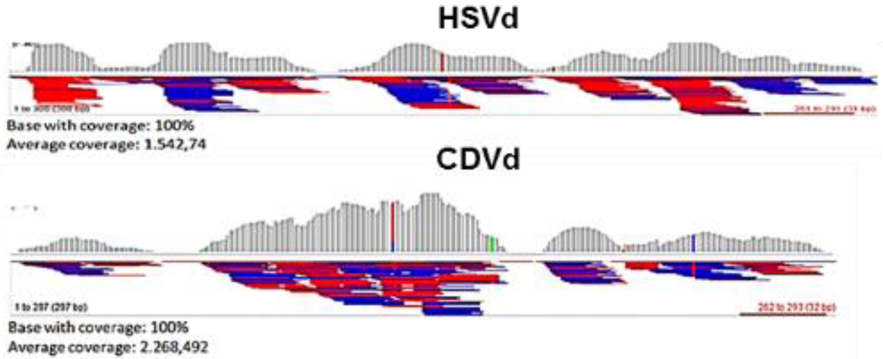
3.2. Searching for Non-EU Virus and Viroids

HTS sequencing of the HU-PSTS source highlighted that the siRNA library contains some viruses and viroids selected for this study. The read alignments with CYVCV isolates from Turkey (Y1) [22], China (CQ), and Pakistan (PK) revealed from 171,807 to 159,685 read counts with 98–95% genome coverage (Figure 5). Moreover, by de novo assembly, a total of 16 contigs, ranging from 821 nt to 1219 nt, were obtained using Kmer-13 and N50. The full genome was reconstructed after mapping the contigs to the CQ reference genome (Figure 6). The resulting consensus sequence of the assembled genome was deposited under accession number KT124646 (HU). BLASTn showed 99% identity with CYVCV CQ (KP313240) and CYVCV GX-GXP (KX156741) isolates from China, 98% with CYVCV RL (KP120977) and CYVCV PK (KP313241) from China and Pakistan, respectively, and 97% with CYVCV Y1 (JX040635). In addition, a 75% identity was found with the closely related Indian citrus ringspot virus (ICRSV) K1 (AF406744) with partial 66% query coverage. This result was also confirmed by read alignments with ICRSV K1 which revealed about 14,000 mapped reads, likely due to the common ancestor [22].
The presence of CYVCV was confirmed by the specific amplicons with the expected size of 921 bp of the 5′ region [22] (Figure S1A). Inoculation of the source HU-PSTS on sour orange and lemon revealed the typical clearing of lateral leaf veins, yellow flecks, and transient ring-spot-like symptoms on spring and autumn flushes, leaf crinkling and warping, and browning of the midribs described for the citrus yellow vein clearing disease [58] (Figure 7).
The HU-PSTS library shared a total of 12,772 read counts with two citrus tatter leaf virus genome references and generated a consensus sequence of almost 6495 nt in length, although with some gaps. However, the detection of CTLV was confirmed by RT-PCR (Figure S1B) and by the inoculation of Carrizo citrange seedlings, which showed typical CTLV symptoms, such as strong yellow mottle or blotch leaf spotting, followed by curling and twisting (i.e., tatter leaf) and a reduction in leaf blade size (Figure 8).
The alignments with the other two citrus viruses listed by EPPO (CiLV and CYMV), investigated in this study, revealed 5900 read counts to CiLV1, 10,500 to CiLV2, but only 4500 to CYMV. The corresponding genome fraction coverage was also low. Other viruses were not explored as they were not of surveillance interest. The libraries of P3R1, P3R3, Mac25, Mac101, and Mac39, showed a low number of read counts and non-significant genome coverage with all the non-EU virus genome references.
The screening for citrus viroids was performed by aligning the siRNA libraries of the six samples against six citrus viroid reference genomes (Table 4). The HU-PSTS source provided a high number of mapped reads with CDVd (32,392) and HSVd (21,939) allowing the full genome reconstruction of these viroids (Table 4, Figure 9). On the other hand, CEVd, CBLVd and CVd-V, had fewer than 5000 reads, suggesting incomplete coverage or the absence of viroids. Real-time RT-PCR analysis confirmed the presence of CDVd and HSVd, and the absence of CEVd, CBLVd and CVdV. Indexing carried out on Etrog citron confirmed the presence of viroids. However, the overlapping symptoms observed, such as leaf epinasty and rugosity, petiole wrinkle and necrosis, midvein necrosis, and browning of the tip of the leaf blade [58,59], mean that it was not possible to differentiate between present and absent viroids.
A considerable number of reads was obtained after alignment of the P3R1 library with CEVd (57,780), CDVd (32,317) and HSVd (15,430), whereas the P3R3 library generated 23,522 reads with CEVd and 17,250 reads with CDVd. An even lower number of reads in the Mac101, Mac39, and Mac25 libraries mapped with viroid genomes references (Table 4). These data, along with those of the P3R1 and P3R3 libraries, were confirmed by real-time PCR and bioindexing in Etrog citron.

4. Discussion

The surveillance of citrus pests is essential to preserve the citriculture worldwide, in accordance with the recommendations of regional phytosanitary services. Conventional diagnostic technologies used to detect all the listed pests include bioindexing and molecular tests. Bioindexing is the most reliable, but it is costly and time-consuming [60]. Although molecular diagnosis is fast and highly specific, many pairs of primers and PCR runs are needed for a full screening. Moreover, new or non-target pathogens remain undetected [40]. Multiplex PCR designed for many viruses and viroids speeds up the lab work, but in some cases may cause problematic interactions.
HTS is the most broad-spectrum diagnostic tool in the field of virus discovery [39,60,61,62]. However, as far as we know, HTS has not been explored for surveillance applications likely due to the costs involved [40].
Given its advantages, we explored sequential HTS pre-screening of samples, complemented by conventional bioindexing and molecular diagnostic technologies, to enhance the surveillance survey of citrus viruses and viroids. A bioinformatic pipeline for the identification of CTV single or mixed genotype infections was designed and used to determine the parameters to discriminate between false positive read mappings and true genotype-specific genome coverage.
According to Bester et al. [32], using simulating data, a genome coverage above 90% was assumed indicative of the presence of a specific genotype, and a genome coverage between 50–90% indicative of the presence of genotype variants not represented in the read mapping reference list. In compliance, 95% genome coverage is indicative of the presence of a genotype when 10,000 single and simulated reads mapped to a specific genotype [57], showing that the genome coverage threshold and depth of the libraries impacted the level of non-target genomes. We used the HTS libraries of six citrus sources to investigate the presence of some virus and viroids of interest for the surveillance according to EPPO A1 List (CiLV and CTLV) and A2 List (non-EU CTV isolates and CBCVd), as well CYVCV, a virus that is currently spreading rapidly in China and Turkey [14]. One source was from China (HU-PSTS), which was assumed to be infected by a non-EU virus, and five were from Sicily, two branches of the same sweet oranges grafted on sour orange (P3R1 and P3R3) and three as ungrafted seedlings (Mac25, Mac39, and Mac101), respectively.
Assuming a genome coverage threshold up to 90%, the read mapping on HU-PSTS, P3R1, and P3R3 sources were indicative of the presence of VT, T3 and T30 CTV genomes. However, only in HU-PSTS the presence of VT and T30 strains was confirmed also by molecular assays, whereas the presence of T3 remained doubtful due to the high homology with VT strains in several parts of the genome [16] and the high non-target genome coverage which can be obtained for the genotypes VT and T3 [32]. Previous results demonstrated that HU-PSTS contained mixed CTV strains [63] and further preliminary HTS analysis led to the conclusion that T3, T68 and T30 could be potentially present in association to the VT strain [35].
On the other hand, in the samples P3R1,P3R3, Mac39, and Mac101, only the VT CTV genotype was confirmed, with a genome coverage of above 99%. The Mac25 source showed the presence of the T36 (98% genome coverage).
HTS was able to identify the genetic differences of the CTV components in each sample but did not explain whether the investigated CTV isolates induce stem-pitting sensu EFSA [4]. However, the subsequent testing with molecular and biological methods helped obtain a more conclusive diagnosis. Real-time RT-PCR and RT-PCR were necessary to support the genetic differences, whereas biological indexing led to a detailed phenotyping of the CTV isolates infecting the six samples. Regarding HU-PSTS, the stem pitting induced in sweet orange and grapefruit enabled us to identify it as a non-EU CTV isolate, biotype 5, whereas the lack of replication in Carrizo citrange meant that RB genotypes were excluded from the components of the isolate. Unfortunately, both biological tests need at least 6–10 months and are costly.
The presence of CYVCV and CTLV in the same source was easily detectable with all three technologies, biological, molecular, and genomic. The data provided by HTS were prompt and detailed, revealing genetic similarity with more than one reference genome, whereas qPCR was faster. The symptomatic detection on sour orange and Carrizo citrange took four and six months after inoculation, respectively.
Mixed viroid infections associated with CTV were detected by HTS in the three field sources (HU-PSTS, P3R1, and P3R3), individually confirmed by molecular testing. A non-conclusive result was obtained for citrus bark cracking viroid, which was highlighted with more than 11,000 reads in the source HU-PSTS. Unfortunately, although the number of reads was much higher than for the other viroids, our attempts to reconstruct the full genome of the viroid were unsuccessful and molecular analysis did not detect it. Moreover, bioindexing on Etrog citron generated undistinguishable and overlapping symptomatic reactions, thus showing that this test was inadequate to identify a specific viroid in mixed infections. Regarding the local field sources (P3R1 and P3R3) and alemow seedlings (Mac25, Mac39, and Mac101), the results of HTS screening were confirmed by biological and molecular tests.
Overall, each of the three detection methods—bioindexing, PCR, and HTS—was helpful in excluding and/or confirming the presence/absence of specific viruses and contributed to the final diagnosis. In most cases they helped to produce additional information. Molecular assays were more suitable than biological indexing regarding the viroid detection, while HTS would have been more adequate if there had been a higher number of reads. The results therefore indicate that the testing strategy needs to be selected based on the pathogen being searched for. The integration of the methods produces faster and more conclusive results.
Our results show that the sequencing depth, host filtering and the selection of reference genomes are crucial for the accurate detection of low titer viruses and viroids. RT-qPCR and HTS and mapping reads had a comparable level of sensitivity for the targeted detection of CTV, CTLV, CYVCV, CDVd, CEVd, and HSVd. They also provided information on the genetic similarity with single strains in GenBank. Similar results obtained for potato viruses demonstrated that a bioinformatics approach improved the sensitivity 10-fold [64].
The HTS analysis was equivalent or superior to the standard bioassay in detecting the viruses and viroids evaluated in the present study, including virus infections at low titers [40,60]. Moreover, our study highlights that it allows to review previous results when a new virus occurs or a different purpose is involved, as in this study. However, despite the stringent conditions used to differentiate genetic variants [57], HTS failed to detect non-EU CTV stem pitting isolates, identified only by bioindexing tests.
Our study shows that in HTS prescreening the number of mapped reads is strictly linked to the titer of the virus or viroid and is related to the total number of reads. For virus and viroid detection in CTV-infected plants, a threshold limit of at least 15,000 reads mapped against the reference with a genomic coverage of 95% is recommended. Variable mapping depths may be acceptable if the presence of the virus or viroid is confirmed by other methods.

5. Conclusions

In conclusion, despite the cost, the use of HTS could provide a comprehensive phytosanitary status of citrus samples, thus reducing the greenhouse footprint, labor, time, and costs. It may enhance the surveillance of citrus viruses and viroids, identify CTV genotypes and enhance phylogenetic studies in mixed infections. Further comparisons, including those aimed at determining limits of detection [65], are evidently needed to clarify the analytical sensitivity required in HTS focused on surveillance surveys and to define specific details of the protocol. This will further contribute to improving the procedure, and the use will support its validation.

Supplementary Materials

The following are available online at https://0-www-mdpi-com.brum.beds.ac.uk/article/10.3390/agriculture11050400/s1, Figure S1. PCR detection of citrus yellow vein clearing virus (A) and citrus tatter leaf virus (B) RNAs isolated from sour orange (lanes 1, 2) and P. trifoliata (lane 3). Positive (+) and negative (−) controls have been added. M1: 100-bp molecular ladder, M2: 1Kb plus molecular ladder (Invitrogen).

Author Contributions

Conceptualization, G.L. and A.F.C.; bioinformatics, R.F.; biological validation, M.R.; molecular validation, G.S.; writing, G.L. and A.F.C. All authors have read and agreed to the published version of the manuscript.

Funding

This work was initially supported by projects funded by MIUR and MISE and by Assessorato delle Risorse Agricole ed Agroalimentari, Regione Siciliana. The work was finalized and developed thanks to resources provided by the project S.I.R.P.A. funded by PO FESR Sicilia 2014–2020, action 1.1.5.

Institutional Review Board Statement

Not applicable.

Informed Consent Statement

Not applicable.

Data Availability Statement

Not applicable.

Conflicts of Interest

The authors declare that they have no conflict of interest.

References

  1. Zhou, C.; Da Graça, J.V.; Freitas-Astúa, J.; Vidalakis, G.; Duran-Vila, N.; Lavagi, I. Citrus viruses and viroids. In The Genus Citrus, 2nd ed.; Talon, M., Caruso, M., Gmitter, F., Jr., Eds.; Woodhead Publishing: Cambridge, UK; Elsevier: Amsterdam, The Netherlands, 2020; pp. 391–410. [Google Scholar]
  2. International Plant Protection Convention (IPPC); Food and Agriculture Organization of the United Nations (FAO). Surveillance ISPM 6; International Plant Protection Convention (IPPC): Rome, Italy; Food and Agriculture Organization of the United Nations (FAO): Rome, Italy, 2018. [Google Scholar]
  3. Ochoa-Corona, F.M. Biosecurity, microbial forensics and plant pathology: Education challenges, overlapping disciplines and research needs. Australas. Plant Pathol. 2011, 40, 335–338. [Google Scholar] [CrossRef]
  4. European Food Safety Authority (EFSA); Schrader, G.; Camilleri, M.; Diakaki, M.; Vos., S. Pest survey card on non-European isolates of citrus tristeza virus. Survey card on non-EU isolates of Citrus tristeza virus. EFSA Support. Publ. 2019, EN-1600. [Google Scholar] [CrossRef] [Green Version]
  5. OEPP/EPPO. EPPO A1 and A2 Lists of Pests Recommended for Regulation as Quarantine Pests (2020). 2019. Available online: https://www.eppo.int/ACTIVITIES/plant_quarantine/A1_list (accessed on 23 March 2020).
  6. EFSA PLH Panel (EFSA Panel on Plant Health). Scientific opinion of pest categorisation of “blight and blight-like” diseases of citrus. EFSA J. 2018, 16, 5248. [Google Scholar]
  7. Tan, S.-H.; Osman, F.; Bodaghi, S.; Dang, T.; Greer, G.; Huang, A.; Hammado, S.; Abu-Hajar, S.; Campos, R.; Vidalakis, G. Full genome characterization of 12 citrus tatter leaf virus isolates for the development of a detection assay. PLoS ONE 2019, 14, e0223958. [Google Scholar] [CrossRef] [PubMed]
  8. Huang, Q.; Hartung, J.S. Cloning and sequence analysis of an infectious clone of citrus yellow mosaic virus that can infect sweet orange via agrobacterium-mediated inoculation. J. Gen. Virol. 2001, 82, 2549–2558. [Google Scholar] [CrossRef]
  9. Ahlawat, Y.S.; Varma, A.; Lockhart, B.E.; Srivastava, M.; Chakraborty, N.K.; Varma, A. Association of a badnavirus with citrus mosaic disease in India. Plant Dis. 1996, 80, 590–592. [Google Scholar] [CrossRef]
  10. EFSA PLH Panel (EFSA Panel on Plant Health). Scientific opinion on the pest categorisation of Citrus tristeza virus (non-European isolates). EFSA J. 2017, 15, 5031. [Google Scholar]
  11. Jakse, J.; Radisek, S.; Pokorn, T.; Matousek, J.; Javornik, B. Deep-Sequencing revealed citrus bark cracking viroid (CBCVd) as a highly aggressive pathogen on hop. Plant Pathol. 2015, 64, 831–842. [Google Scholar] [CrossRef]
  12. Bani Hashemian, S.M.; Serra, P.; Barbosa, C.J.; Juárez, J.; Aleza, P.; Corvera, J.M.; Lluch, A. The effect of a field-source mixture of citrus viroids on the performance of ‘Nules’ clementine and ‘Navelina’ sweet orange trees grafted on carrizo citrange. Plant Dis. 2009, 93, 699–707. [Google Scholar] [CrossRef]
  13. Murcia, N.; Bani Hashemian, S.M.; Serra, P.; Pina, J.A.; Duran-Vila, N. Citrus viroids: Symptom expression and performance of Washington navel sweet orange trees grafted on carrizo citrange. Plant Dis. 2015, 99, 125–136. [Google Scholar] [CrossRef]
  14. Liu, C.; Liu, H.; Hurst, J.; Timko, M.P.; Zhou, C. Recent advances on citrus yellow vein clearing virus in citrus. Hort. Plant J. 2020, 6, 216–222. [Google Scholar] [CrossRef]
  15. Ruiz-Ruiz, S.; Navarro, B.; Gisel, A.; Peña, L.; Navarro, L.; Moreno, P.; Di Serio, F.; Flores, R. Citrus tristeza virus infection induces the accumulation of viral small RNAs (21–24-nt) mapping preferentially at the 3′-terminal region of the genomic RNA and affects the host small RNA profile. Plant Mol. Biol. 2011, 75, 607–619. [Google Scholar] [CrossRef] [PubMed] [Green Version]
  16. Harper, S.J. Citrus tristeza virus: Evolution of complex and varied genotypic groups. Front. Microbiol. 2013, 4, 93. [Google Scholar] [CrossRef] [PubMed] [Green Version]
  17. Yokomi, R.K.; Selvaraj, V.; Maheshwari, Y.; Saponari, M.; Giampetruzzi, A.; Chiumenti, M.; Hajeri, S. Identification and characterization of Citrus tristeza virus isolates breaking resistance in trifoliate orange in California. Phytopathology 2017, 107, 901–908. [Google Scholar] [CrossRef] [Green Version]
  18. Zablocki, O.; Pietersen, G. Characterization of a novel citrus tristeza virus genotype within three cross-protecting source GFMS12 sub-isolates in South Africa by means of illumina sequencing. Arch. Virol. 2014, 159, 2133–2139. [Google Scholar] [CrossRef] [Green Version]
  19. Matsumura, E.E.; Coletta-Filho, H.D.; Nouri, S.; Falk, B.W.; Nerva, L.; Oliveira, T.S.; Dorta, S.O.; Machado, M.A. Deep sequencing analysis of RNAs from citrus plants grown in a citrus sudden death-affected area reveals diverse known and putative novel viruses. Viruses 2017, 9, 92. [Google Scholar] [CrossRef] [Green Version]
  20. Wang, J.; Zhu, D.; Tan, Y.; Zong, X.; Wei, H.; Liu, Q. First report of citrus leaf blotch virus in sweet cherry. Plant Dis. 2015, 100, 1027. [Google Scholar] [CrossRef]
  21. Jo, Y.; Choi, H.; Kim, S.M.; Kim, S.L.; Lee, B.C.; Cho, W.K. Integrated analyses using RNA-seq data reveal viral genomes, single nucleotide variations, the phylogenetic relationship, and recombination for apple stem grooving virus. BMC Genom. 2016, 17, 579. [Google Scholar] [CrossRef] [Green Version]
  22. Loconsole, G.; Onelge, N.; Potere, O.; Giampetruzzi, A.; Bozan, O.; Satar, S.; De Stradis, A.; Savino, V.; Yokomi, R.K.; Saponari, M. Identification and characterization of citrus yellow vein clearing virus, a putative new member of the genus mandarivirus. Phytopathology 2012, 102, 1168–1175. [Google Scholar] [CrossRef] [PubMed] [Green Version]
  23. Loconsole, G.; Saldarelli, P.; Doddapaneni, H.; Savino, V.; Martelli, G.P.; Saponari, M. Identification of a single-stranded DNA virus associated with citrus chlorotic dwarf disease, a new member in the family Geminiviridae. Virology 2012, 432, 162–172. [Google Scholar] [CrossRef] [Green Version]
  24. Vives, M.C.; Velázquez, K.; Pina, J.A.; Moreno, P.; Guerri, J.; Navarro, L. Identification of a new enamovirus associated with citrus vein enation disease by deep sequencing of small RNAs. Phytopathology 2013, 103, 1077–1086. [Google Scholar] [CrossRef] [Green Version]
  25. Roy, A.; Stone, A.; Otero-Colina, G.; Wei, G.; Choudhary, N.; Achor, D.; Shao, J.; Levy, L.; Nakhla, M.K.; Hollingsworth, C.R.; et al. Genome assembly of Citrus leprosis virus nuclear type reveals a close association with Orchid fleck virus. Genome Announc. 2013, 1, e00519-13. [Google Scholar] [CrossRef] [PubMed] [Green Version]
  26. Roy, A.; Shao, J.; Hartung, J.S.; Schneider, W.; Brlansky, R.H. A case study on discovery of novel Citrus leprosis virus cytoplasmic type 2 utilizing small RNA libraries by next generation sequencing and bioinformatic analyses. J. Data Min. Genom. Proteom. 2013, 4, 129. [Google Scholar] [CrossRef]
  27. Navarro, B.L.; Minutolo, M.; Stradis, A.D.; Palmisano, F.; Alioto, D.; Di Serio, F. The first phlebo-like virus infecting plants: A case study on the adaptation of negative-stranded RNA viruses to new hosts. Mol. Plant Pathol. 2018, 19, 1075–1089. [Google Scholar] [CrossRef] [PubMed]
  28. Yan, J.; Zhang, S.; Wu, J.; Yang, F.; Zhou, Y.; Zhou, C.; Cao, M. Complete genome sequences and recombination analysis of three divergent satsuma dwarf virus isolates. Trop. Plant Pathol. 2021, 46, 26–30. [Google Scholar] [CrossRef]
  29. Dang, T.; Menchaca, I.; Bodaghi, S.; Lavagi, I.; Osman, F.; Vidalakis, G. Next generation sequencing as a routine diagnostic tool for citrus variety introduction at the citrus clonal protection program. J. Citrus Pathol. 2019, 6, 6. [Google Scholar]
  30. Chambers, G.; Englezou, A.; Webster, J.; Bogema, D.; Donovan, N. Using next generation sequencing (NGS) to characterize Australia’s living pathogen collection. J. Citrus Pathol. 2019, 6, 4–5. [Google Scholar]
  31. Olmos, A.; Boonham, N.; Candresse, T.; Gentit, P.; Giovani, B.; Kutnjak, D.; Liefting, L.; Maree, H.J.; Minafra, A.; Moreira, A.; et al. High-Throughput sequencing technologies for plant pest diagnosis: Challenges and opportunities. Bull. OEPP EPPO Bull. 2018, 48, 219–224. [Google Scholar] [CrossRef] [Green Version]
  32. Bester, R.; Cook, G.; Maree, H.J. Citrus tristeza virus genotype detection using high-throughput sequencing. Viruses 2021, 13, 168. [Google Scholar] [CrossRef]
  33. Scuderi, G.; Russo, M.; Davino, S.; Ferraro, R.; Catara, A.; Licciardello, G. Occurrence of the T36 genotype of Citrus tristeza virus in citrus orchards in Sicily, Italy. Plant Dis. 2016, 100, 1253. [Google Scholar] [CrossRef]
  34. Bar-Joseph, M.; Catara, A.F.; Licciardello, G. The puzzling phenomenon of seedling yellows recovery and natural spread of asymptomatic infections of citrus tristeza virus: Two sides of the same coin. Hortic. Rev. 2021, 48, 339–362. [Google Scholar]
  35. Licciardello, G.; Scuderi, G.; Ferraro, R.; Russo, M.; Dai, S.M.; Catara, A.; Deng, Z.N. High throughput sequencing of a stem pitting Citrus tristeza virus isolate from Hunan province (P.R. China). J. Citrus Pathol. Under review.
  36. Flores, R.; Delgado, S.; Gas, M.E.; Carbonell, A.; Molina, D.; Gago, S.; De la Pena, M. Viroids: The minimal non-coding RNAs with autonomous replication. FEBS Lett. 2004, 567, 42–48. [Google Scholar] [CrossRef]
  37. Chambers, G.A.; Donovan, N.J.; Bodaghi, S.; Jelinek, S.M.; Vidalakis, G. A novel citrus viroid found in Australia, tentatively named citrus viroid VII. Arch. Virol. 2018, 163, 215–218. [Google Scholar] [CrossRef]
  38. Maree, H.J.; Fox, A.; Al Rwahnih, M.; Boonham, N.; Candresse, T. Application of HTS for routine plant virus diagnostics: State of the art and challenges. Front. Plant Sci. 2018, 9, 1082. [Google Scholar] [CrossRef] [Green Version]
  39. Massart, S.; Chiumenti, M.; De Jonghe, K.; Glover, R.; Haegeman, A.; Koloniuk, I.; Komínek, P.; Kreuze, J.; Kutnjak, D.; Lotos, L.; et al. Virus detection by high-throughput sequencing of small RNAs: Large-scale performance testing of sequence analysis strategies. Phytopathology 2019, 109, 488–497. [Google Scholar] [CrossRef] [PubMed] [Green Version]
  40. Maliogka, V.I.; Minafra, A.; Saldarelli, P.; Ruiz-García, A.B.; Glasa, M.; Katis, N.; Olmos, A. Recent advances on detection and characterization of fruit tree viruses using high-throughput sequencing technologies. Viruses 2018, 10, 436. [Google Scholar] [CrossRef] [PubMed] [Green Version]
  41. Di Serio, F.; Cao, M. Identification of novel viruses and viroids in citrus hosts by high-throughput sequencing: Methodologies, opportunities, challenges and impact. J. Citrus Pathol. 2019, 6, 10–11. [Google Scholar]
  42. Licciardello, G.; Ferraro, R.; Scuderi, G.; Russo, M.; Catara, A. Next generation sequencing of small RNAs from citrus plants with single and mixed infections of viruses and viroids. J. Citrus Pathol. 2019, 6, 5–6. [Google Scholar]
  43. Licciardello, G.; Scuderi, G.; Ferraro, R.; Giampetruzzi, A.; Russo, M.; Lombardo, A.; Raspagliesi, D.; Bar-Joseph, M.; Catara, A. Deep sequencing and analysis of small RNAs in sweet orange grafted on sour orange infected with two Citrus tristeza virus isolates prevalent in Sicily. Arch. Virol. 2015, 160, 2583–2589. [Google Scholar] [CrossRef] [PubMed]
  44. Okonechnikov, K.; Conesa, A.; García-Alcalde, F. Qualimap 2: Advanced multi-sample quality control for high-throughput sequencing data. Bioinformatics 2016, 32, 292–294. [Google Scholar] [CrossRef] [PubMed]
  45. Zerbino, D.R.; Birney, E. Velvet: Algorithms for de novo short read assembly using de Bruijn graphs. Gen. Res. 2008, 18, 821–829. [Google Scholar] [CrossRef] [PubMed] [Green Version]
  46. Li, H.; Handsaker, B.; Wysoker, A.; Fennell, T.; Ruan, J.; Homer, N.; Marth, G.; Abecasis, G.; Durbin, R. The sequence alignment/map format and SAMtools. Bioinformatics 2009, 15, 2078–2079. [Google Scholar] [CrossRef] [Green Version]
  47. Robinson, J.T.; Thorvaldsdóttir, H.; Winckler, W.; Guttman, M.; Lander, E.S.; Getz, G.; Mesirov, J.P. Integrative genomics viewer. Nat. Biotechnol. 2011, 29, 24. [Google Scholar] [CrossRef] [PubMed] [Green Version]
  48. Garnsey, S.M.; Civerolo, E.L.; Gumpf, D.J.; Paul, C.; Hilf, M.E.; Lee, R.F.; Brlansky, R.H.; Yokomi, R.K.; Hartung, J.S. Biological characterization of an international collection of Citrus tristeza virus (CTV) isolates. In Proceedings of the 16th Conference of the International Organization of Citrus Virologist, Monterrey, Mexico, 7–12 November 2004; Hilf, M.E., Duran-Vila, N., Rocha-Peña, M.A., Eds.; International Organization of Citrus Virologist (IOCV): Riverside, CA, USA, 2005; pp. 75–93. [Google Scholar]
  49. Roy, A.; Ananthakrishnan, G.; Hartung, J.S.; Brlansky, R.H. Development and application of a multiplex reverse-transcription polymerase chain reaction assay for screening a global collection of Citrus tristeza virus isolates. Phytopathology 2010, 100, 1077–1088. [Google Scholar] [CrossRef] [PubMed]
  50. Ruiz-Ruiz, S.; Moreno, P.; Guerri, J.; Ambrós, S. Discrimination between mild and severe Citrus tristeza virus isolates with a rapid and highly specific real-time reverse transcription-polymerase chain reaction method using TaqMan LNA probes. Phytopathology 2009, 99, 307–315. [Google Scholar] [CrossRef] [PubMed]
  51. Yokomi, R.K.; Saponari, M.; Sieburth, P.J. Rapid differentiation and identification of potential severe strains of Citrus tristeza virus by real-time reverse transcription-polymerase chain reaction assays. Phytopathology 2010, 100, 319–327. [Google Scholar] [CrossRef] [PubMed] [Green Version]
  52. Tatineni, S.; Afunian, M.R.; Hilf, M.E.; Gowda, S.; Dawson, W.O.; Garnsey, S.M. Molecular characterization of Citrus tatter leaf virus historically associated with meyer lemon trees: Complete genome sequence and development of biologically active in vitro transcripts. Phytopathology 2008, 99, 423–431. [Google Scholar] [CrossRef] [Green Version]
  53. Papayiannis, L.C. Diagnostic real-time RT-PCR for the simultaneous detection of Citrus exocortis viroid and hop stunt viroid. J. Virol. Methods 2014, 96, 93–99. [Google Scholar] [CrossRef]
  54. Rizza, S.; Nobile, G.; Tessitori, M.; Catara, A.; Conte, E. Real time RT-PCR assay for quantitative detection of citrus viroid III in plant tissues. Plant Pathol. 2009, 58, 181–185. [Google Scholar] [CrossRef]
  55. Bernard, L.; Duran-Vila, N. A novel RT-PCR approach for detection and characterization of citrus viroids. Mol. Cell. Probes 2006, 20, 105–113. [Google Scholar] [CrossRef]
  56. Serra, P.; Eiras, M.; Bani-Hashemian, S.M.; Murcia, N.; Kitajima, E.W.; Daros, J.A.; Flores, R.; Duran-Vila, N. Citrus viroid V: Occurrence, host range, diagnosis, and identification of new variants. Phytopathology 2008, 98, 1199–1204. [Google Scholar] [CrossRef] [PubMed] [Green Version]
  57. Ruiz-Garcìa, A.B.; Bester, R.; Olmos, A.; Maree, H.J. Bioinformatic tools and genome analysis of Citrus tristeza virus. In Citrus Tristeza Virus: Methods in Molecular Biology; Catara, A., Bar-Joseph, M., Licciardello, G., Eds.; Humana: New York, NY, USA, 2019; Volume 2015, pp. 163–178. [Google Scholar]
  58. Duran-Vila, N.; Semancik, J. Citrus viroids. In Viroids; Hadidi, A., Flores, R., Randles, J.W., Semancik, J.S., Eds.; CSIRO Publishing: Collingwood, Australia, 2003; pp. 178–194. [Google Scholar]
  59. Duran-Vila, N. Citrus exocortis viroid. In Viroids and Satellites; Hadidi, A., Flores, R., Randles, J.W., Palukaitis, P., Eds.; Academic Press: Cambridge, MA, USA, 2017; pp. 169–179. [Google Scholar]
  60. Al Rwahnih, M.; Daubert, S.; Golino, D.; Islas, C.; Rowhani, A. Comparison of next-generation sequencing versus biological indexing for the optimal detection of viral pathogens of grapevines. Phytopathology 2015, 105, 758–763. [Google Scholar] [CrossRef] [Green Version]
  61. Adams, I.P.; Glover, R.H.; Monger, W.A.; Mumford, R.; Jackeviciene, E.; Navalin-Skiene, M.; Samuitiene, M.; Boonham, N. Next generation sequencing and metagenomic analysis: A universal diagnostic tool in plant virology. Mol. Plant Pathol. 2009, 10, 537–545. [Google Scholar] [CrossRef] [PubMed]
  62. Massart, S.; Olmos, A.; Jijakli, H.; Candresse, T. Current impact and future directions of high throughput sequencing in plant virus diagnostics. Virus Res. 2014, 188, 90–96. [Google Scholar] [CrossRef]
  63. Licciardello, G.; Xiao, C.; Russo, M.; Dai, S.M.; Daden, M.; Deng, Z.N.; Catara, A.F. Genetic structure of Citrus tristeza virus in Hunan province (P.R. China). Acta Hortic. 2015, 1065, 781–790. [Google Scholar] [CrossRef]
  64. Santala, J.; Valkonen, J.P.T. Sensitivity of small RNA-based detection of plant viruses. Front. Microbiol. 2018, 9, 939. [Google Scholar] [CrossRef] [Green Version]
  65. Bukowska-O’sko, I.; Perlejewski, K.; Nakamura, S.; Motooka, D.; Stokowy, T.; Kosinska, J.; Popiel, M.; Płoski, R.; Horban, A.; Lipowski, D. Sensitivity of next-generation sequencing metagenomic analysis for detection of RNA and DNA viruses in cerebrospinal fluid: The confounding effect of background contamination. In Respiratory Treatment and Prevention; Pokorski, M., Ed.; Springer International Publishing: Cham, Switzerland, 2017; pp. 53–62. [Google Scholar]
Figure 1. Schematic workflow of the procedure adopted for the detection of citrus viruses and viroids based on a multi-disciplinary approach in which HTS (including a wet-lab and a bioinformatic phase) is used as pre-screening analysis, whereas biological tests and molecular detection are used as validating assays.
Agriculture 11 00400 g001
Figure 2. Graphical illustration of genome coverage per representative genome reference of six citrus tristeza virus genotypes after read mapping the siRNA library of each sample individually.
Agriculture 11 00400 g002
Figure 3. Quality alignments exemplified as genome histograms (blue) and genome fraction coverage (red) per reference genome representative of six citrus tristeza virus genotypes of P3R1 (VT) (A,B) and Mac25 (T36) (C,D) siRNA libraries, using Qualimap [44].
Agriculture 11 00400 g003
Figure 4. Symptomatic responses of sour orange seedlings inoculated with Mac39 (A), Mac101 (B), Mac25 (C), P3R1 (D), P3R3 (E), 6 months post-inoculation in comparison to non-inoculated control (F).
Agriculture 11 00400 g004
Figure 5. Graphical illustration of genome coverage per representative genome reference of non- EU viruses and viroids after read mapping the siRNA library of each sample individually.
Agriculture 11 00400 g005
Figure 6. Visualization of the consensus alignments of citrus yellow vein clearing HU generated by both “map to reference” alignment of siRNA reads and de novo assembled contigs. (A) CYVCV CQ reference genome; (B) coverage obtained by siRNA reads alignment; (C) siRNA library (171,807 reads) which allowed the CYVCV-HU full genome reconstruction; (D) consensus obtained by alignments of 16 contigs. For the visualization the Integrative genome viewer (IGV) software has been used [47].
Agriculture 11 00400 g006
Figure 7. Leaf symptoms of citrus yellow vein clearing induced by inoculation of HU-PSTS on sour orange (A,B) and lemon (C,D) about 4–5 months post-inoculation.
Agriculture 11 00400 g007
Figure 8. Symptoms of citrus tatter leaf on young (A) and mature (B) leaves of Carrizo citrange induced by bark inoculation of sour orange HU-PSTS infected, about 4–5 months post-inoculation.
Agriculture 11 00400 g008
Figure 9. Genome reconstructions of the hop stunt viroid (HSVd) and citrus dwarfing viroid (CDVd) by alignment of the siRNA reads of HU-PSTS library with reference sequences.
Agriculture 11 00400 g009
Table 1. Complete genome of citrus viruses sequenced by high throughput technologies.
VirusGenusFamilyHostCountryReference
Citrus tristeza virus (CTV)ClosterovirusClosteroviridaeSweet orange, Mexican lime, sour orangeSpain[15]
C. sinensis, C. macrophyllaFlorida[16]
C. sinensis, C. reticulataCalifornia[17]
GrapefruitSouth Africa[18]
Citrus sudden death-associated virus (CSDaV)MarafivirusTymoviridaeSweet orangeBrazil[19]
Citrus leaf blotch virus (CLBV)CitrivirusBetaflexiviridaeSweet CherryChina[20]
Citrus psorosis ophiovirus (CPV)OphiovirusAspiviridaeSweet orangeChina[unpublished]
Citrus tatter leaf virus (CTLV)CapillovirusBetaflexiviridaeAppleChina[21]
Citrus yellow vein clearing virus (CYVCV)MandarivirusAlphaflexiviridaeLemonTurkey[22]
Citrus chlorotic dwarf-associated virus (CCDaV)GeminivirusGeminiviridaeLemonTurkey[23]
Citrus vein enation virus (CVEV)EnamovirusLuteoviridaeEtrog citronSpain[24]
Citrus leprosis virus-N (CiLV-N)DichoravirusRhabdoviridae Mexico[25]
Citrus leprosis virus-C2 (CiLV-2)CilevirusUnassigned Colombia[26]
Citrus concave gum-associated virus (CCGaV)PhlebovirusPhenuiviridaeTarocco sweet orangeItaly[27]
Citrus jingmen-like virus (CJLV)FlavivirusFlaviviridaeValencia sweet orangeBrazil[19]
Citrus virga-like virus (CVLV)VirgavirusVirgaviridaeValencia sweet orangeBrazil[19]
Satsuma dwarf virus (SDV)SadwavirusSecoviridaeSour orange and tangorChina[28]
Table 2. Virus isolate and source plants used to assess the virome by using high-throughput sequencing.
Virus IsolateField PlantOriginAgeTissue Source
HU-PSTSSweet orange/P. trifoliataHunan, China≈30 yearsBark, sour orange
P3R1Sweet orange/sour orangeSicily, Italy≈22 yearsBark, sweet orange
P3R3Sweet orange/sour orangeSicily, Italy≈22 yearsBark, sweet orange
Mac39Alemow seedlingSicily, Italy≈2 yearsBark, sour orange
Mac101Alemow seedlingSicily, Italy≈2 yearsBark, sour orange
Mac25Alemow seedlingSicily, Italy≈2 yearsBark, sour orange
Table 3. List of reference sequences of citrus tristeza virus (one representative genome of each genotype), non-EU viruses and viroids used for genome reconstruction by mapping reads of siRNA libraries.
VirusGenotypeAccessionIsolateOrigin
Citrus tristeza virusVTKC748392SG29Italy
T3EU857538NZ-M16New Zealand
T30KC748391Bau282Italy
T68EU076703B165India
RBFJ525435NZRB-M17New Zealand
T36EU937521T36 FS2-2Florida
Citrus yellow vein clearing JX040635Y1Turkey
NC_026592CQChina
KP313241PKPakistan
Citrus tatter leaf virus KC588948MTHChina
JX416228PKTaiwan
Citrus sudden death-associated virus AY884005 Brazil
Citrus leprosis virus CRNA1NC_008169 Brazil
RNA2NC_008170 Brazil
Citrus yellow mosaic virus DQ875213 India
Viroids
Citrus exocortis viroid AB054599MAJapan
Hop stunt viroid AB211242EX1Japan
Citrus bent leaf viroid NC001651CBLVd-225Israel
Citrus dwarfing viroid isolate EU934031E822California
Citrus viroid V NC010165 Spain
Citrus bark cracking viroid NC003539 Israel
Table 4. Number of read counts obtained after alignments of siRNA libraries with viroids reference genomes. Positive identifications are in bold, considering a threshold limit of 15,000 reads.
Read Count (RC)
ViroidMac39Mac101Mac25P3R1P3R3HU-PSTS
Citrus exocortis viroid2920845957,78023,522602
Hop stunt viroid4249873815,400514621,939
Citrus bent leaf viroid7423747268593600829
Citrus dwarfing viroid17811822832,31717,25032,393
Citrus viroid V191309132749750585374
Citrus bark cracking viroid1611522711,0364932199
Publisher’s Note: MDPI stays neutral with regard to jurisdictional claims in published maps and institutional affiliations.

