Article

Genomic Diversity, Antimicrobial Resistance, Plasmidome, and Virulence Profiles of Salmonella Isolated from Small Specialty Crop Farms Revealed by Whole-Genome Sequencing

1
Center for Food Animal Health, Department of Animal Sciences, College of Food, Agricultural, and Environmental Sciences, The Ohio State University, Wooster, OH 44691, USA
2
Molecular and Cellular Imaging Center, College of Food, Agricultural, and Environmental Sciences, The Ohio State University, Wooster, OH 44691, USA
3
Department of Veterinary Pathobiology, Oklahoma State University, Stillwater, OK 74074, USA
4
Department of Veterinary Science, Martin-Gatton College of Agriculture, Food and Environment, University of Kentucky, Lexington, KY 40546, USA
*
Author to whom correspondence should be addressed.
Submission received: 14 October 2023 / Revised: 10 November 2023 / Accepted: 15 November 2023 / Published: 18 November 2023

Abstract

:
Salmonella is the leading cause of death associated with foodborne illnesses in the USA. Difficulty in treating human salmonellosis is attributed to the development of antimicrobial resistance and the pathogenicity of Salmonella strains. Therefore, it is important to study the genetic landscape of Salmonella, such as the diversity, plasmids, and presence antimicrobial resistance genes (AMRs) and virulence genes. To this end, we isolated Salmonella from environmental samples from small specialty crop farms (SSCFs) in Northeast Ohio from 2016 to 2021; 80 Salmonella isolates from 29 Salmonella-positive samples were subjected to whole-genome sequencing (WGS). In silico serotyping revealed the presence of 15 serotypes. AMR genes were detected in 15% of the samples, with 75% exhibiting phenotypic and genotypic multidrug resistance (MDR). Plasmid analysis demonstrated the presence of nine different types of plasmids, and 75% of AMR genes were located on plasmids. Interestingly, five Salmonella Newport isolates and one Salmonella Dublin isolate carried the ACSSuT gene cassette on a plasmid, which confers resistance to ampicillin, chloramphenicol, streptomycin, sulfonamide, and tetracycline. Overall, our results show that SSCFs are a potential reservoir of Salmonella with MDR genes. Thus, regular monitoring is needed to prevent the transmission of MDR Salmonella from SSCFs to humans.

1. Introduction

Salmonella is a rod-shaped, facultative anaerobic, Gram-negative bacterium in the Enterobacteriaceae family that causes salmonellosis [1]. Two types of Salmonella can be distinguished based on their host adaptability and infectious nature: human typhoidal and non-typhoidal Salmonella (NTS). Typhoidal Salmonella, associated with typhoid fever, is host-restricted and infects humans. It disseminates systematically and colonizes the gallbladder, and infected individuals become chronic carriers. In contrast, NTS, primarily associated with enteric infections, has a broad host range, that includes chickens, cattle, pigs, humans, and other species [2]. NTS is shed intermittently for a short period of time and depends on the environment for its survival and transmission [3]. The clinical presentation of NTS includes gastroenteritis and diarrhea [4]. However, Salmonella can disseminate from the intestine to other systemic organs during invasive infections. Based on surface antigens, there are >2600 serotypes of Salmonella [5], with invasive infections primarily caused by the serotypes S. Typhimurium, S. Enteritidis, and S. Dublin [6]. NTS is the second leading cause of death associated with foodborne illnesses in the USA, with almost 420 deaths reported yearly, causing huge public health impacts [7]. NTS accounted for losses of USD 3.3 billion in terms of treatment and human productivity in the United States due to high morbidity and mortality rates [8].
The difficulty of treating human salmonellosis is associated with the evolution of AMR genes in Salmonella. The extensive use of antibiotics, such as for prophylaxis and metaphylaxis in food-animal production systems, is accelerating the evolution of AMR in Salmonella. For instance, Salmonella in chickens treated with ceftiofur for Salmonella infections has become resistant to ceftiofur [9]. Similarly, the use of high levels of tetracycline and sulfonamide on poultry farms in Nigeria was positively correlated with tetracycline and sulfonamide resistance in Salmonella [10]. Likewise, tetracycline, streptomycin, and nalidixic acid resistance were observed in Salmonella isolated from poultry farms in the southeastern United States [11]. In 2018–2019, azithromycin- and ciprofloxacin-resistant S. Newport was isolated from a dairy farm in the USA [12]. The consumption and improper handling of farm products and direct or indirect contact with the animals on farms pose a risk for the transfer of antibiotic-resistant Salmonella to humans, which can reduce the effectiveness of drugs and increase the rates of hospitalization and mortality in humans [13]. Therefore, the Centers for Disease Control (CDC) and the World Health Organization (WHO) have prioritized the improved monitoring of AMR Salmonella along the food chain, particularly in food-producing animals.
Farm animals infected with Salmonella can repeatedly shed bacteria in their feces, contaminating the farm environment [14,15]. Thus, contaminated farm water and raw manure serve as vehicles for Salmonella transmission to farm produce [16]. Salmonella can be internalized by produce grown in a field irrigated with contaminated water or amended biologically with contaminated manure [17]. Hence, farm environments could be a source of indirect transmission of Salmonella to humans. In previous studies, he prevalence of Salmonella was 15.2% in poultry manure [18], 4.9% in dairy manure [19], and 29.4% in pond water used for irrigation [20]. Once present in the farm environment, Salmonella can form biofilms to persist for a long time [21]. Similarly, Kuang et al. detected S. Typhimurium and S. Enteritidis in farm animals in China, which are the two most important serotypes predominantly reported in human infections [22].
Small specialty crop farms (SSCFs) are an important sector of crop production in the United States [23,24]. The SSCF enrolled in our study are mixed crop-and-animal farm systems where livestock/poultry are grown close to the farm produce in the same facility. Produce from SSCFs is considered fresh, healthy, and safe for consumption [25]. Therefore, it is sold directly to consumers in farmers’ markets, stores, or roadside stands and sometimes to grocery stores [25]. SSCFs are family-owned and have their own practices, that differ between farms. Biological soil amendments of animal origin (BSAAOs), such as poultry and dairy manure, are commonly used as biofertilizers on these SSCFs. Untreated irrigation water is used to irrigate fields. Differences in farming practices, such as the application of BSAAOs and the use of irrigation water potentially contaminated with infected farm animal feces, increase the risk of foodborne pathogens in produce grown on these farms. Similarly, antibiotics are used on SSCFs to enhance growth and prevent and treat diseases in animals [26,27]. The unregulated use of antibiotics can lead to the emergence of multidrug-resistant Salmonella [26,27]. Differences in farming practices, such as the application of BSAAOs, the use of untreated irrigation water, and the indiscriminate use of antibiotics, increase the potential risk of the transmission of antibiotic-resistant foodborne pathogens from SSCFs to humans.
While NTS cause a self-limiting disease, antibiotics are needed to treat invasive infections. Recently, quinolones, fluoroquinolones, and third-generation cephalosporins have been used to treat salmonellosis, with colistin used as a last-resort drug for bacterial infections [28]. The emergence and dissemination of MDR Salmonella, including colistin-resistant isolates from food-animal production farms, are limiting the choice of therapeutic treatment for salmonellosis [29]. Graham et al. reported the possibility of a spillover of AMR genes from SSCFs to humans [27]. Although AMR genes can arise in Salmonella from chromosomal mutations, the primary mechanism to acquire them is through plasmids [21]. Plasmids can additionally harbor virulence genes that enhance the pathogenesis of Salmonella, including mechanisms for host immune evasion, attachment, entry, colonization, and invasive and persistent infection. Several virulence genes, such as cdtB, rck, sodCI, pef, and spv, were detected in Salmonella samples from poultry farms [30].
Although several studies have performed comparative genomic analyses of Salmonella isolated from commercial food-animal production systems, here, we examined the serotypes, resistome, virulome, and plasmidome of Salmonella recovered from SSCFs [31,32], which have not been reported previously. For instance, we characterized the serotypes of Salmonella to detect the serotypes important for human infection, assessed the resistome and virulome to determine AMR genes and virulence genes, and identified the plasmids and their colocalization with AMR and virulence genes to understand the potential for horizontal transmission. With an increasing number of people relying on locally grown produce, it is important to understand the potential public health risks posed by such farms. Therefore, this study utilized whole-genome sequencing (WGS) to assess the genomic diversity among the isolates using pan-genome analysis and phylogenetics to identify ARGs and virulence genes and to characterize the plasmid profiles of Salmonella recovered from SSCFs. In addition, phenotypic assays, such as antimicrobial susceptibility testing and biofilm assays, were performed to associate the results with the genotypic data.

2. Results

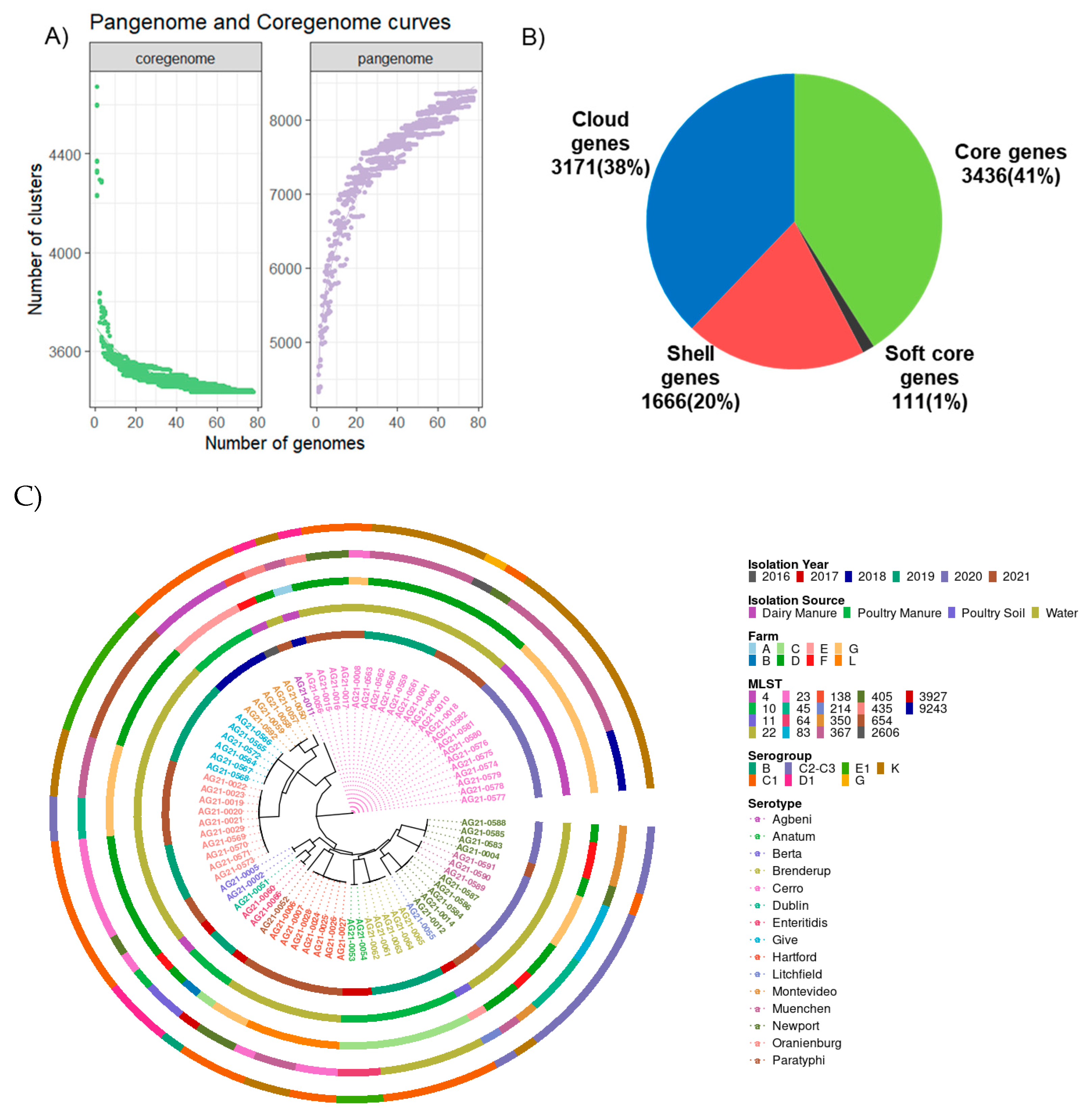
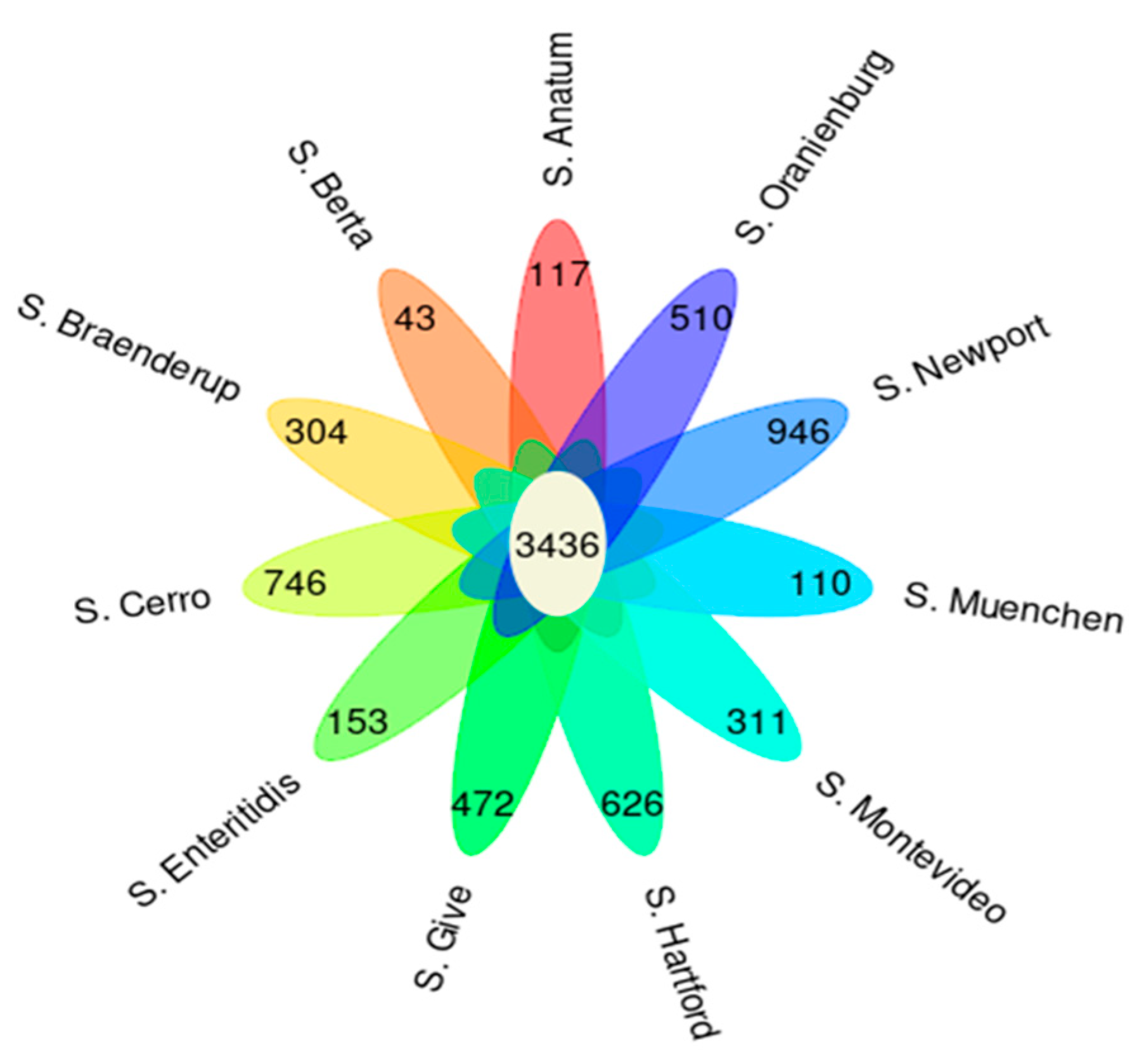
2.1. Genomic Features of Salmonella Revealed 15 Serotypes Belonging to 18 Sequence Types with 3436 Core Genes

In our study, conducted between 2016 and 2021, 2.27% of samples were found to be positive for Salmonella. Salmonella was isolated from four different sources, dairy manure, poultry manure, soil, and water samples, with prevalence of 2.63%, 2.85%, 0.18%, and 8.12%, respectively (Table 1). The number of samples collected from each farm and the frequency of collection during the study period are provided in Table S1. This study is an extension of our earlier published study in which, Salmonella prevalence was further discussed [24]. From these 29 samples, a total of 80 isolates were sequenced. Multiple isolates per samples were sequenced because studies have shown that multiple serotypes can co-exist in the same sample [33]. Two assemblies were discarded due to low genomic coverage, and subsequent analyses were therefore carried out with the remaining 78 isolates. Summary statistics for the genome assemblies are given in Table S2, and serotype averages are provided in Table 2. Overall, the average genomic depth of coverage was 64×, the GC content was 52.18%, the genome size was 4.6 Mbp, and the number of contigs was 43.
Both serotyping and sequence typing were used to classify Salmonella isolated in this study. In silico serotyping using SeqSero2 resulted in the detection of 15 serotypes (Table 3), with S. Cerro being the most common, accounting for 29.4% of genome assemblies. Notably, 8 out of the 15 serotypes reported in our study are among the top 20 serotypes frequently reported in human salmonellosis [34].
We detected 18 unique sequence types (STs) (Table 3 and Table S2). Twelve serotypes were associated with one ST, while three serotypes had two STs: S. Cerro (associated with ST367 and ST9243), S. Montevideo (associated with ST4 and ST138), and S. Newport (associated with ST45 and ST350). To the best of our knowledge, this is the first study to detect ST9243 in S. Cerro.
A pan-genome analysis showed that 41% of genes were core genes, 1% were soft-core genes, 20% were shell genes, and 38% were cloud genes (Figure 1A,B). The accessory genes consist of soft-core genes, shell genes, and cloud genes. Functional annotation of core genes showed that they were mostly involved in metabolic processes such as transcription, translation, cell wall synthesis, energy production, and carbohydrate, amino acid, and lipid transport (Figure S1A). The majority of accessory genes were mobility elements, such as prophages and transposons (Figure S1B). Our finding that core genes represented less than half of all genes was due to the presence of distinct sets of genes among the 15 serotypes of Salmonella and implies relatively high genomic variability among the serotypes.

2.2. A phylogenetic Tree Showed That All Serotypes except S. Newport form Monophyletic Clades

The genetic similarity of the isolates was first computed using average nucleotide identity (ANI) values. The ANI values were between 98 and 100%, which indicates that the isolates were highly similar. Furthermore, a phylogenetic tree was inferred using the core SNPs identified by kSNP3. The optimal k-mer size identified by kSNPS’3 Kchooser was 19 bp (thus, each putative SNP was taken as the central base in a 19-mer). A total of 170,689 SNPs were detected, of which 60,089 were core SNPs. A phylogenetic tree showed that samples assigned to the same serotype formed monophyletic clades, except for S. Newport, which was found across two clades along with S. Muenchen and S. Litchfield (Figure 1C).
Interestingly, six serotypes (S. Cerro, S. Montevideo, S. Enteritidis, S. Oranienburg, S. Hartford, and S. Newport) were found on more than one farm, and multiple serotypes were detected on four farms, suggesting that Salmonella lineages from multiple sources present on one farm also circulate on other farms. To explore the effects of geographical distance on the movement of Salmonella, a multi-dimensional scaling (MDS) plot was constructed to visualize the distances between farms (Figure S2). Our results showed that farms D, F, and G clustered together. S. Cerro was found on farms A, D, F, and G, S. Newport was found on farms D and F, and S. Oranienburg was found on farms D and G. Therefore, due to the close proximity of the three farms D, F, and G, S. Cerro, S. Newport, and S. Oranienburg might have been disseminated from one farm to another. Likewise, the phylogenetic tree showed that five serotypes were recovered from a single farm only (excluding the four serotypes with only one sample): S. Anatum (farm C), S. Braenderup (C), S. Give (D), S. Berta (D), and S. Muenchen (G).

2.3. Pan-Genome-Wide Association Study (Pan-GWAS)

Among 4948 accessory genes, Scoary detected 1172 genes that were significantly associated with a total of eleven serotypes: S. Anatum (117), S. Berta (43), S. Braenderup (304), S. Cerro (746), S. Enteritidis (153), S. Give (472), S. Hartford (626), S. Montevideo (311), S. Muenchen (110), S. Newport (946), and S. Oranienburg (510) (Figure 2). These significantly associated genes included the macrolide export protein in S. Cerro, a mercury resistance protein in S. Newport, and a phosphotransferase protein in S. Give (Table S3). The significance of the associations could not be calculated for the four serotypes containing only one sample per serotype.

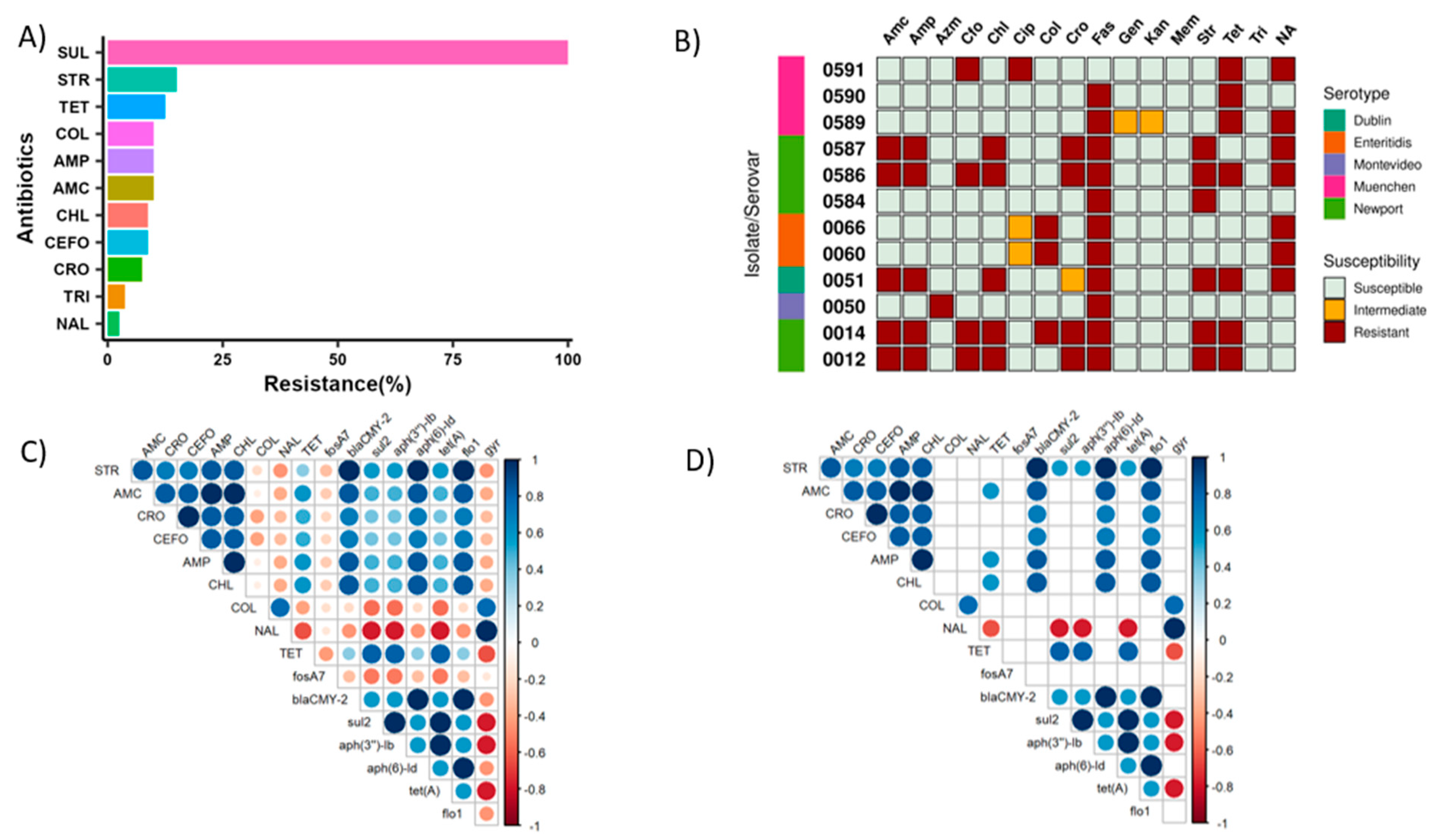
2.4. Antimicrobial Resistance Genes and Detection of the ACSSuT Cassette

The resistome analysis detected the presence of eight unique antimicrobial resistance genes (ARGs) in five serotypes of Salmonella that can confer resistance to drugs within seven antibiotic classes (Table 4). The AMR genes detected were fosA7, blaCMY-2, sul2, aph(3″)-Ib, aph(6)-Id, tet(A), floR, and gyrA_D87Y. Seventy-five percent of isolates with ARGs were MDR. S. Montevideo and S. Enteritidis harbored one ARG, S. Muenchen harbored three ARGs, and S. Dublin and S. Newport harbored five ARGs. Interestingly, S. Dublin and S. Newport harbored the ACSSuT cassette, which confers resistance to ampicillin, chloramphenicol, streptomycin, sulfonamides, and tetracyclines, on a plasmid [35]. The beta-lactamase resistance gene, which confers ampicillin resistance [36], was detected in S. Dublin and S. Newport. Likewise, the SSuT resistance pattern was observed in S. Muenchen. The screening of genomic mutations showed the presence of a single nonsynonymous mutation at the 87th amino acid of the chromosomal quinolone-resistance-determining region (QRDR) in S. Enteritidis. The change from aspartic acid to tyrosine at the 87th position of the gyrase genes confers resistance to quinolones, which is the most detected mutation in nalidixic acid-resistant Salmonella [37].

2.5. Phenotypic Antimicrobial Susceptibility Testing Revealed Colistin Resistance in Some Isolates

The overall resistance percentages according to phenotypic antimicrobial susceptibility testing (AST) for all isolates are shown in Figure 3A. Surprisingly, all 80 isolates were resistant to sulfisoxazole (100%), and resistance to streptomycin was the next most common (15%), followed by tetracycline (12.5%) and colistin (10%). Colistin is a last-resort antibiotic used to treat infections with MDR Gram-negative pathogens. Despite being banned for clinical use and animal production in the USA due to concerns about toxicity, we found the colistin-resistant phenotype in eight isolates [38]. Similarly, we detected resistance to amoxicillin (10% of isolates), ampicillin (10%), trimethoprim–sulfamethoxazole (3.75%), ceftriaxone (7.5%), cefoxitin (8.75%), chloramphenicol (8.75%), and nalidixic acid (2.5%). None of the isolates were resistant to azithromycin, ciprofloxacin, gentamycin, kanamycin, or meropenem. We also found that 26.25% of isolates were resistant to two different classes of antibiotics, while 13.75% were MDR.

2.6. Association between Phenotypic and Genotypic Antibiotic Resistance

A correlation analysis was used to examine the associations between phenotypic and genotypic (eight ARGs) antibiotic resistance (Figure 3C,D). A significant negative correlation was observed between the presence of the tetracycline resistance gene and the nalidixic-resistant phenotype (Figure 3D, p < 0.05). Positive correlations were found between several ARGs and resistance phenotypes (Figure 3D, p < 0.05), such as between the presence of tetracycline gene and tetracycline resistance (Figure 3D, p < 0.05). Similarly, the presence of the beta-lactamase gene was positively correlated with β-lactam amoxicillin resistance, the chloramphenicol gene was positively correlated with chloramphenicol resistance, and the streptomycin resistance genes were positively correlated with streptomycin resistance (Figure 3D, p < 0.05).
We also observed significant correlations between the occurrences of ARGs themselves: for instance, there was a significant negative correlation between the presence of gyrase gene (which confer resistance to quinolones) and tetracycline resistance gene, whereas there was a significant positive correlation between streptomycin resistance genes and amoxicillin, ampicillin, and chloramphenicol resistance genes (Figure 3D, p < 0.05).

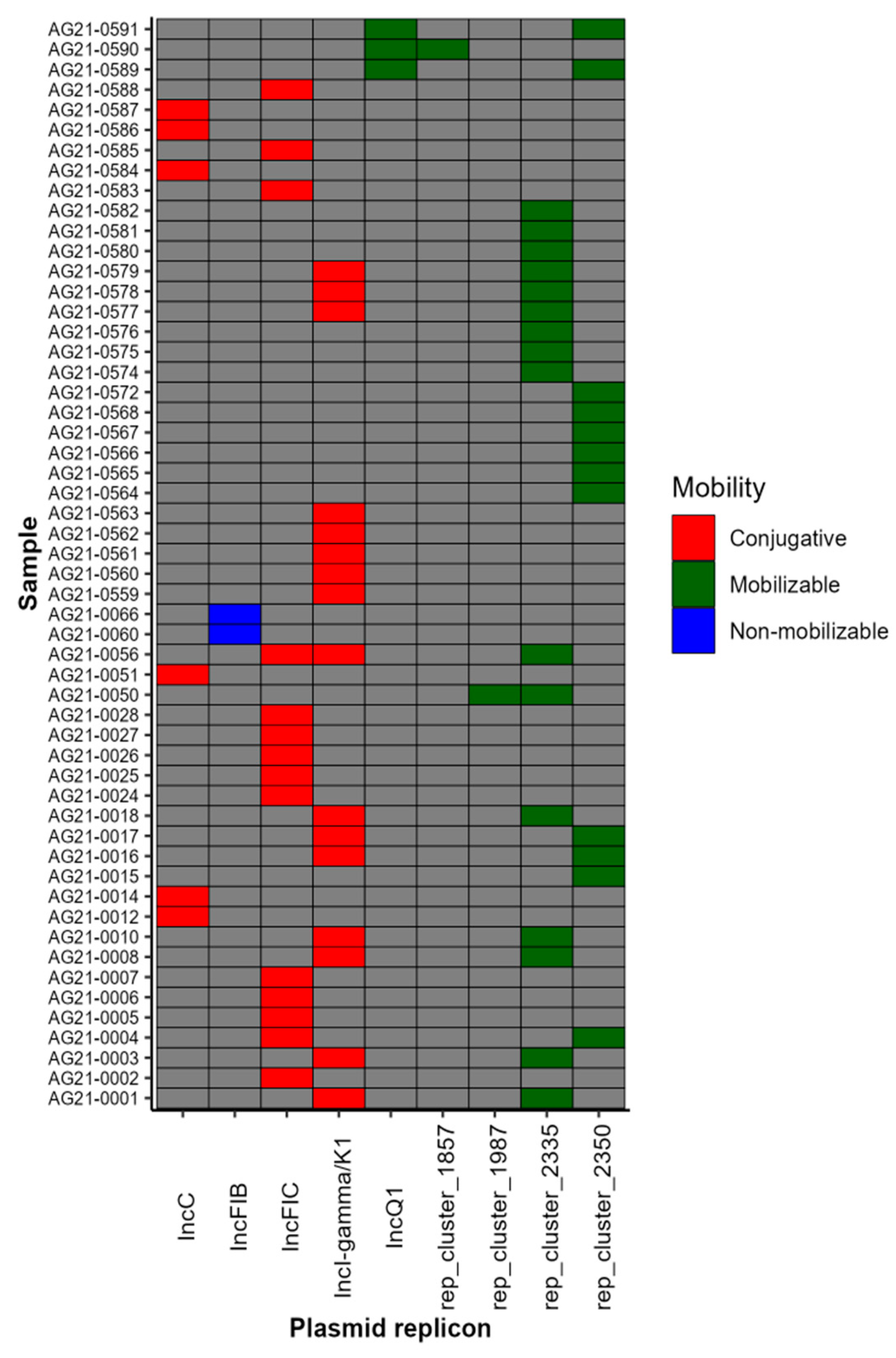
2.7. Plasmid Profile Revealed the Presence of Nine Plasmid Replicons

To understand the genetic basis of ARG dissemination, we investigated the presence of plasmids. A total of nine distinct plasmid replicons were detected, with 69% (54/78) of our isolates containing at least one plasmid (Figure 4). Among isolates with plasmids, different types of incompatibility groups (IncC, IncI-gamma/K1, IncFIB, IncFIC, and IncQ1) were detected in 46% (25/54), rep plasmids were detected in 26% (14/54), and Inc and rep plasmids were detected in 28% (15/54) of the isolates. Additionally, conjugative plasmids were present in 42.6% (23/54), mobilizable plasmids were present in 31.5% (17/54), and non-mobilizable plasmids were present in 3.7% (2/54) of the isolates (Table S4). Some samples harbored more than one plasmid: 22.2% (12/54) contained both conjugative and mobilizable plasmids. Similarly, the occurrence of ARGs on plasmid contigs was evaluated, and we found that 75% of ARGs were assigned to be located on plasmid contigs. Interestingly, the IncC conjugative plasmid replicon was reported on the same contig as the ACSSuT resistance pattern genes in S. Newport and S. Dublin. Likewise, the SSuT resistance pattern in S. Muenchen was detected on the same contig as the IncQ1 mobilizable plasmid replicon.

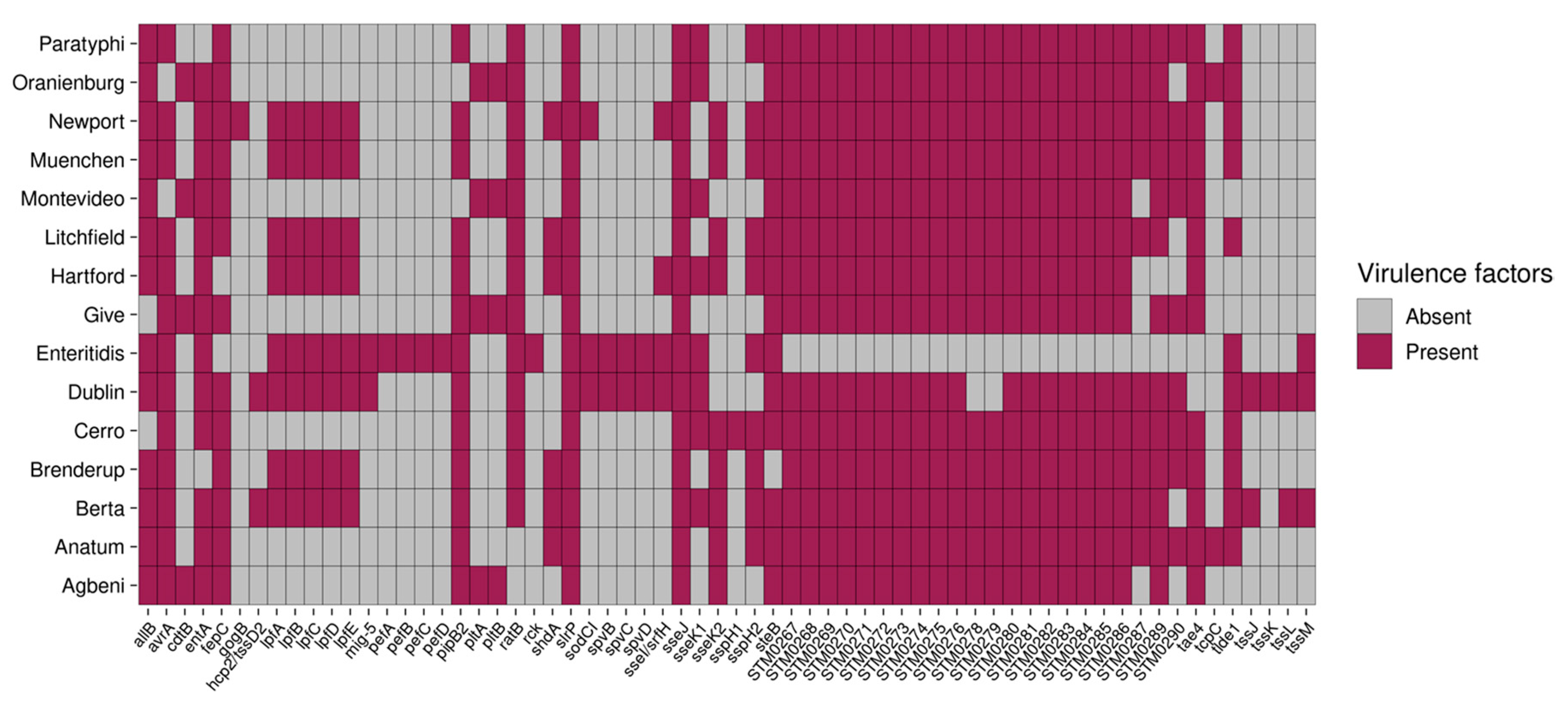
2.8. S. Enteritidis and S. Dublin Have Unique Virulence Genes

Three types of secretion systems (T1SS, T3SS, and T6SS) were located in two Salmonella pathogenicity islands, and one Salmonella centrosome island was observed. A total of 176 unique virulence genes were detected, 112 of which were shared among all isolates (Table S5). These genes represent the core virulome, likely needed for the survival of Salmonella. The remaining 62 genes were not present in all serotypes (variable virulome, Figure 5). Two genes were present in all serotypes but absent in a few individual isolates: slrP was present in 69 isolates, and sseJ was found in 72 isolates.
Virulence genes that were found only in a subset of serotypes included the following. First, the spv genes located on the Salmonella virulence plasmid (pSV), which is involved in survival and systemic infection in host cells, were detected in S. Enteritidis and S. Dublin [39]. The spvABCD genes are arranged in an operon and are positively regulated by the upstream spvR gene [39]. However, spvR, which is needed for the regulation of the spv locus [40], was absent in our isolates. Second, long polar fimbriae genes (lpfABCDE) and plasmid-encoded fimbriae genes (pefABCD) located on the pSV were detected in S. Enteritidis. The serum resistance gene (rck), which increases the invasion of host cells and the resistance to killing mediated by the complement system, was detected in S. Enteritidis. The presence of mig5, rck, spv, and pef genes in S. Enteritidis suggests that these genes play an important role in the pathogenesis of human salmonellosis [41]. Likewise, lpfABCDE was detected in other serotypes, such as S. Braenderup, S. Dublin, S. Muenchen, and S. Newport. Finally, the cdtB gene, which produces the cytolethal distending toxin (CDT), was detected in S. Agbeni, S. Give, S. Oranienburg, and S. Montevideo. However, cdtA and cdtC genes, which are required for the binding of the CDT to target cells, were not detected. Pertussis-like toxins (pltA and pltB), which share structural and functional homology with cdtA and cdtC genes, forming a complex with cdtB and delivering it from the intracellular compartment to target cells, were detected in isolates with cdtB [42].

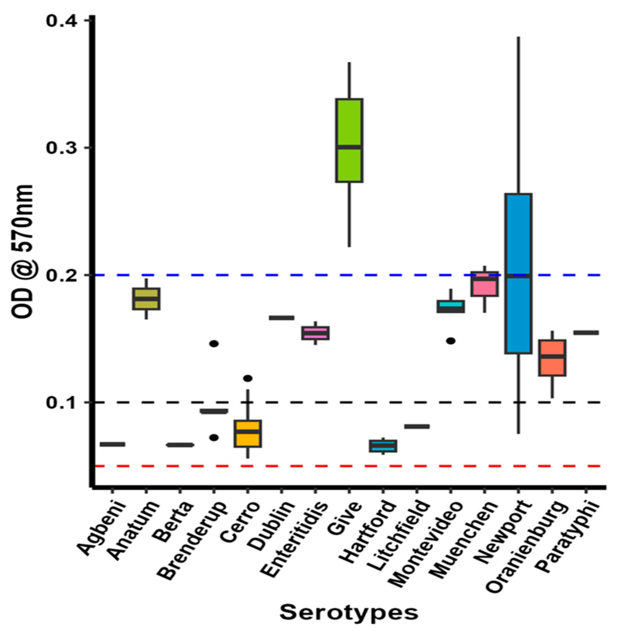
2.9. S. Give Is a Strong Biofilm Producer

The results of the phenotypic biofilm production assay are shown in Figure 6. Five serotypes, S. Agbeni, S. Berta, S. Braenderup, S. Hartford, and S. Litchfield, were low biofilm producers. Most S. Cerro isolates were low biofilm producers, whereas a few were moderate biofilm producers. S. Anatum, S. Dublin, S. Enteritidis, S. Montevideo, S. Muenchen, S. Oranienburg, and S. Paratyphi were moderate biofilm producers. A few S. Newport and S. Give isolates were high biofilm producers, whereas other S. Newport isolates were moderate biofilm producers.

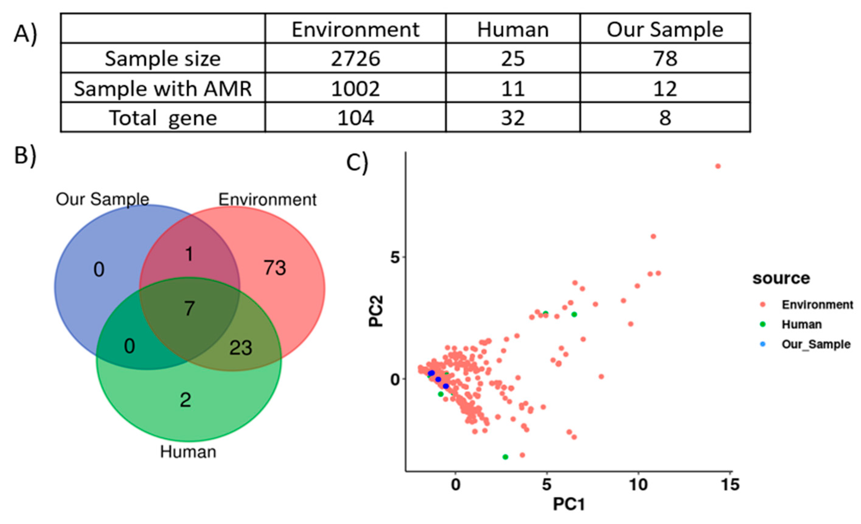
2.10. Comparative Genomic Analysis Revealed SSCFs as Potential Reservoirs for AMR Gene Transmission to Humans

A total of 2751 Salmonella genomes sampled in Ohio were downloaded from NCBI. Among them, AMRFinderPlus detected 104 unique ARGs in 1002 environmental samples and 32 ARGs in 25 human samples (Figure 7A). Among them, seven ARGs were common to humans, the environment, and our Salmonella isolates (Figure 7B). Further, principal component analysis (PCA) showed an overlap of ARG profiles between our isolates and Salmonella isolates reported to be from humans and the environment in Ohio (Figure 7C), suggesting the potential for the transmission of drug-resistant Salmonella from SSCFs to humans.

3. Discussion

Numerous studies have linked salmonellosis outbreaks to food-animal farms [43,44,45]. Clinically and sub-clinically infected farm animals regularly shed Salmonella, thereby contaminating the farm environment. Salmonella can survive outside its host in farm environments such as manure, soil, and water for 184, 332, and 1825 days, respectively [46,47]. The survival or persistence of Salmonella in farm environment poses difficulties in removing Salmonella from the farm due to the possibility of reinfection or reinoculation [48]. BSAAOs are commonly applied as biofertilizers on SSCFs. The application of BSAAO can contaminate the soil because Salmonella can survive in the manure-amended soil for up to 329 days. This increases the risk of Salmonella internalization by the produce grown in the field and transmission to humans from the food production chain. Usegi et al. reported that S. Enteritidis phage type (PT) 30 associated with an outbreak from 2000 to 2001, persisted in an almond orchard for 5 years [49].
Despite evidence of Salmonella outbreaks originating from farms, none of the previous studies have compared Salmonella genomes isolated from SSCFs [50]. Our analysis identified 15 serotypes of Salmonella circulating on eight SSCFs, suggesting the presence of diverse Salmonella in this niche. However, all Salmonella serotypes detected on SSCFs are not equally important for human infection. For instance, S. Cerro is mostly associated with infection in cattle, whereas the serotypes S. Enteritidis and S. Newport are frequently reported in human infections. The most prevalent serotype was S. Cerro ST367, which was recovered from water and dairy manure. This result is consistent with previous studies in which S. Cerro was predominantly isolated from dairy farms [32]; however, the presence of this serotype is not as clinically concerning because S. Cerro is a cattle-adapted serotype that is rarely isolated from humans and is usually limited to gastrointestinal infections [32,51,52]. However, our study also detected two clinically important serotypes, S. Enteritidis and S. Newport, which are the serotypes most frequently reported in human salmonellosis in the USA [34]. The prevalence of clinically important serotypes of Salmonella on SSCFs necessitates the implementation of strict biosecurity measures to reduce their transmission to humans. A correlation analysis was carried out to detect an association between the presence of specific serotypes and specific farm practices, such as the application of either poultry or dairy manure. However, we did not see any statistically significant correlations (p > 0.05) between the presence of certain serotypes and manure application.
The serotypes S. Anatum, S. Berta, S. Braenderup, S. Give, and S. Muenchen were recovered from only a single farm. In contrast, the serotypes S. Cerro, S. Enteritidis, S. Hartford, S. Montevideo, S. Newport, and S. Oranienburg were recovered from multiple farms. The three farms from which S. Cerro, S. Newport, and S. Oranienburg were isolated were in close geographical proximity, suggesting the transmission of serotypes between farms. The presence of the same serotypes on multiple farms was also reported in a previous study [53]. It is likely that these serotypes are circulating via horizontal transfer through the wind, equipment, vehicles, or humans. SSCFs share their farm equipment, between farms, which may be the potential route of Salmonella transmission. The horizontal transmission of Salmonella between dairy farms through tractors was reported in England by Brennan et al. [54].
The analysis of core-genome SNPs is a widely used method for constructing phylogenetic trees and investigating outbreaks [55]. In our phylogenetic analysis, most Salmonella serotypes were monophyletic, except for S. Newport, which was found in two separate clades. An ANI analysis showed no differences between the two strains of S. Newport, with 99–100% similarity between the two S. Newport strains. However, the two S. Newport strains showed differences in their sequence types [56]. One clade was composed of ST45, while the other was made up of ST350. ST45 contained AMR genes and mercury resistance genes, whereas ST350 did not carry any resistance genes. In addition, differences in plasmids content were detected between ST45 and ST350. Since ST45 is more frequently associated with outbreaks of Salmonella in humans, it poses a serious public health concern [57,58]. However, there are no reports on ST350 in outbreaks of S. Newport. Differences in the prevalence of the AMR and heavy-metal-resistance genes in ST45 of S. Newport could also be related to different selection pressures among farms. However, we did not observe any association between the farm identity and the STs of S. Newport.
Our study revealed the presence of eight unique AMR genes in five Salmonella serotypes on fourteen farms. The factors that might contribute to differences in the prevalence of different AMR genes in these farms are unknown. The SSCFs are family-owned and have management practices, that differ between individual farms. We hypothesize that management practices, for example, different uses of antibiotics and other antimicrobials, such as biocides and disinfectants, which co-select AMR genes with antibiotic resistance genes [29], may have contributed to the differences in resistance genes among Salmonella isolates. The majority of Salmonella isolates in our study were resistant to sulfisoxazole (100%), streptomycin (15%), and tetracycline (12.5%), which are the first-line drugs used for the treatment of salmonellosis. Similar to our study, 90% of Salmonella recovered from a hatchery were resistant to sulfisoxazole [59]. In 1990, when Salmonella became resistant to first-line antibiotics such as chloramphenicol, ampicillin, and trimethoprim–sulfamethoxazole, quinolones and fluoroquinolones became the preferred antibiotics for treating Salmonella infections [60]. Resistance to quinolones is attributed to mutations in genes such as gyrA, gyrB, parC, and parE [61]. In our study, we detected a mutation in the A subunit of the gyrase gene of S. Enteritidis that changed the amino acid aspartate to tyrosine at the 87th position, conferring resistance to quinolones in 2.5% of the isolates [62]. In addition, the phenotypic AST data showed that S. Enteritidis was resistant to colistin, a last-resort antibiotic used for treating multidrug-resistant Enterobacteriaceae infections [63]. Moreover, a colistin-resistant phenotype was observed in serogroups B, C2-C3, D, K, and E1, which accounted for 10% of our isolates. The occurrence of colistin resistance in S. Enteritidis was unsurprising because Salmonella serotypes belonging to serogroup D have a higher tendency to develop colistin resistance [29]. The absence of plasmid-mediated mobile colistin resistance (mcr) genes in the isolates reduces the chances of horizontal transmission of colistin resistance genes [64]. However, an examination of the protein sequences of genes that have been reported for colistin resistance in Salmonella (such as pmrAB, LolB, mgrB, and phoPQ) revealed two nonsynonymous mutations in the LolB gene of S. Cerro: P25L and S194N. The presence of these mutations is concerning for public health because colistin-resistant Salmonella can be potentially transmitted to humans from SSCFs. This will reduce the effectiveness of colistin, which is a last-resort antibiotic for bacterial infections. Olaitan et al. reported the horizontal transmission of colistin-resistant E. coli carrying unknown mutation from pigs to humans [65,66]. Likewise, colistin is used at sub-MIC concentrations in combination with other drugs to enhance its effectiveness [65]. However, the synergistic effect will not be observed in the presence of Salmonella carrying mutations in colistin resistance genes. The mutation in LolB was absent in colistin-susceptible S. Cerro. The LolB gene is involved in the transport of lipoproteins to the outer membrane [29]. We speculate that the mutation in the LolB reduces the transport of the lipoprotein to the outer membrane, which decreases the negative charge of the bacteria. Colistin is a positively charged cationic peptide that interacts with the negatively charged bacterial membrane [64]. The modification in the outer membrane of S. Cerro may reduce the interaction of colistin with the bacterial membrane, thereby conferring resistance. The absence of colistin-resistance-conferring mutations and plasmid-mediated colistin resistance genes in other serotypes suggests the presence of a novel colistin resistance mechanism, which needs further study.
An MDR ACSSuT cassette was detected in S. Dublin and S. Newport, while an SSuT cassette was detected in S. Muenchen. Consistent with our study, mobilizable IncQ1 plasmids with the SSuT resistance pattern have been reported in S. Typhimurium [67]. These results suggest that the Salmonella isolates characterized in this study are potential reservoirs for the transmission of MDR ARGs to humans. This finding was further corroborated by PCA analysis, which showed that our isolates clustered with environmental and human isolates (Figure 7C). This implies that Salmonella isolates recovered from humans and environmental sources carried similar sets of ARGs to those detected in our isolates. This represents a serious public health issue because MDR Salmonella can increase the rate of hospitalization, invasive illness, and mortality globally [68].
Plasmids have been reported to play a significant role in the dissemination of AMR genes, virulence genes, and other traits conferring a fitness advantage. Our study detected nine different plasmid replicons, predominantly (97%) belonging to either conjugative or mobilizable plasmids, suggesting that Salmonella frequently acquires genes via the horizontal transfer of plasmids. The IncC conjugative plasmid, which carried the highest number of AMR genes, was detected only in S. Newport and S. Dublin. Consistent with our results, a previous study found the conjugative IncC plasmid in S. Newport [69]. Interestingly, the IncC (broad host) conjugative plasmid replicon was found on the same contig as the ACSSuT cassette in our study. This implies that the IncC plasmid may be disseminating MDR genes within Salmonella and other genera in the Enterobacteriaceae family through conjugation. This is particularly concerning because several studies have detected IncC conjugative plasmids in clinical isolates of pathogenic Salmonella and E. coli [70,71]. The acquisition of the ACSSuT cassette will make Salmonella resistant to multiple antibiotics and limit the treatment options for salmonellosis.
Virulence genes in Salmonella are important for its fitness, such as persistence in a specific niche and successful pathogenesis. In this study, we identified the shdA gene, which encodes the nonfimbrial adherence gene, only in isolates recovered from water, poultry manure, or soil amended with poultry manure. This gene is involved in the persistent colonization and long-term fecal shedding of Salmonella [72]. The shdA gene was detected in six serotypes: S. Anatum, S. Berta, S. Braenderup, S. Hartford, S. Litchfield, and S. Newport. Previous studies have shown the presence of shdA in S. enterica adapted to warm-blooded animals such as mice, poultry, and pigs [72,73,74]. However, the absence of shdA among isolates recovered from the dairy manure in our study needs further exploration, for example, to determine whether this gene is present in cattle-adapted Salmonella. Similarly, multiple virulence genes that enhance the pathogenesis of Salmonella serotypes were detected in our study. The SpvBCD operon, which enhances the intracellular proliferation of Salmonella in human cells and causes invasive infections, was detected in S. Enteritidis and S. Dublin [75]. In S. Enteritidis, it was associated with non-mobilizable IncFIB plasmids, whereas in S. Dublin, it was found on an IncC conjugative plasmid. Consistent with our study, previous studies have shown the presence of the spv operon in S. Dublin and S. Enteritidis [76,77]. Furthermore, the cdtB gene, in association with pltA and pltB genes, which encodes for a typhoid toxin that arrests the cell cycle, was detected in S. Agbeni, S. Give, S. Oranienburg, and S. Montevideo [78]. Earlier studies have reported cdtB genes in all four serotypes [79,80,81]. Although these four serotypes do not represent a concern as MDR bacteria, the virulence genes pose a potential health risk.
To investigate whether certain Salmonella serotypes may be better adapted to farm environments by forming biofilms, we measured the biofilm production capacity of all isolates. Studies have shown that high-biofilm-producing Salmonella serotypes are resistant to antimicrobials, disinfectants, and thermal and environmental stress such as desiccation [82,83]. This resistance allows them to survive in harsh farm environment conditions and cross-contaminate farm produce, which can lead to their transmission to humans through the food production chain. Our results showed that most serotypes were moderate biofilm producers. Notably, S. Give and a few isolates of S. Newport were strong biofilm producers (Figure 6). A correlation analysis was conducted between phenotypic biofilm formation ability and the biofilm-associated genes present in these two serotypes. However, we did not see any significant correlation (p > 0.05) between the phenotypic data and the presence or absence of biofilm-associated genes. Several studies have reported S. Newport as a high biofilm producer and suggested that biofilm formation is a strategy employed by S. Newport to survive in harsh environmental conditions [84,85]. However, here, we also report a high biofilm formation ability in the S. Give serotype, which has not been described previously. Interestingly, the Scoary results demonstrated a significant association between S. Give and several biofilm-related genes (Table S2). For instance, the DNA adenine methylase gene (dam) was significantly associated with S. Give, S. Newport, and S. Montevideo. This gene regulates the expression of the csgD gene, which is involved in the formation of curli and cellulose, important components of biofilms [86]. Furthermore, the Scoary analysis showed a significant association between S. Give and the presence of a multiphosphoryl transfer protein, the phosphoenolpyruvate-carbohydrate phosphotransferase system (PTS) fructose transporter subunit IIB gene, and PTS fructose transporter subunit IIC genes, which facilitate the uptake of extracellular sugars [87]. In S. Give, the sugars transported by PTS might assist in biofilm formation, as reported in Vibrio cholerae [88]. Since the mechanism of biofilm formation is complex and regulated by several genes, we speculate that S. Give uses both dam and PTS genes for high biofilm production.
Our research has a few limitations. Our dataset is derived from samples collected over five years in Northeast Ohio only, and similar data from additional states would help to draw more general conclusions. Likewise, the water samples were collected from 2019 to 2021, while soil and manure samples were collected from 2016 to 2021, which might have affected the results of our study. Additionally, information on antibiotic use was insufficient to conduct any association studies on the use of antibiotics and the Salmonella prevalence. Our future studies are designed to obtain detailed metadata on management practices, including antibiotic, herbicide, and pesticide (type, frequency, dose, etc.) use. This might provide insights into the presence of AMR genes in Salmonella on these farms. Furthermore, data on feed production was not available, making it difficult to rule out the possibility of Salmonella contamination in our sample originating from feed [89]. Therefore, further research to assess the presence of Salmonella in feed and feed crops such as maize and alfa alfa in these farm settings is needed to understand the contribution of feed and feed crops to the dissemination of Salmonella on these farms. The results of such studies may guide the need to conduct larger studies to look at the role of feed and feed crops in the persistence and dissemination of Salmonella and its impact on public health, since feed and ingredients for feed formulation are obtained from different sources.

4. Materials and Methods

4.1. Sample Collection

Soil, poultry manure, dairy manure, pond water, and stream water samples were collected from fourteen SSCFs located in Northeast Ohio from 2016 to 2021 following a previously described protocol [24]. The criteria for selecting the farms were based on their availability for longitudinal sampling throughout the study period. Soil samples were taken from three random sites and mixed in a Nasco Whirl-Pak™ (Fisher Scientific, Waltham, MA, USA). The soil of the farms was amended with either poultry manure (PM) or dairy manure (DM). Dairy heifers housed on a bedded pack during the winter and raised on pasture in the summer were used for the collection of DM. Six to eight fresh manure pats were collected and pooled together in the Whirl-Pak™. Similarly, PM was collected from poultry storage piles at three random sites and pooled. Ten liters of water was collected from either a pond or a stream, based on the source of the water used to irrigate the farm, in a sterile Nalgene gallon. Samples were transferred to the lab in an ice box at 4 °C and processed immediately.

4.2. Sample Processing to Isolate Salmonella

Samples were processed following a previously described protocol [24]. Twenty-five grams of (manure and soil) samples were weighed and resuspended in 225 mL of phosphate-buffered saline (PBS), mixed thoroughly to make a slurry, and ten-fold serially diluted. Then, 100 µL of each dilution was plated on a Xylose-Lysine-Tergitol 4 (XLT-4) plate and incubated at 37 °C for 24 h to isolate Salmonella directly. Similarly, 100 µL of water was plated on an XLT-4 plate without dilution. In addition, water was concentrated by passing it through a Modified Moore Swab (MMS) filter. The filter was aseptically removed and transferred to Whirl-Pak bags, mixed with 250 mL of PBS, serially diluted, and plated on an XLT-4 plate. Apart from direct plating, 10 mL of each sample was enriched in 90 mL of tetrathionate broth (TTB) for 24 h and plated on an XLT-4 plate as described previously. The isolated black colonies typical of Salmonella on XLT-4 plates were stored in BHI broth at −80 °C.

4.3. DNA Extraction

Salmonella DNA was extracted using the boiling method [90]. A loopful of Salmonella culture was resuspended in 100µL of sterile RNase/DNase-free water and boiled at 95 °C for 10 min. The debris was pelleted by centrifugation at 4000× g for 10 min. The supernatant containing the DNA was transferred to new tubes and stored at −20 °C. A total of 2 µL of the lysate was used as a template for PCR for the confirmation of Salmonella using the invasion (invA) gene (final volume of 25 µL and 35 cycles). The 284 bp amplicon of the invA gene was amplified using an initial denaturation at 94 °C for 3 min; denaturation at 94 °C for 1 min, annealing at 53 °C for 2 min, and elongation at 72 °C for 1 min; and a final extension at 72 °C for 7 min [91]. The primers used for PCR were S139F: GTGAAATTATCGCCACGTTCGGGCAA and S141R: TCATCGCACCGTCAAAGGAACC [90]. The size of the PCR product was confirmed by visualizing on a 2% agarose gel, and positive samples were sent for sequencing.
For whole-genome sequencing, DNA was extracted from pure Salmonella isolates (n = 80) using the Wizard Genomic DNA Purification kit (Promega, Madison, WI, USA) following the manufacturer’s instructions.

4.4. Genome Assembly and Annotation

DNA samples were prepared for Illumina MiSeq 2 × 300 bp sequencing using the Nextera DNA Library Preparation Kit (Illumina, South Dakota State University, Brookings, SD, USA). Sequence quality was checked using FastQC v. 0.11.9 [92]. Adapters and poor-quality bases were removed by Trimmomatic v. 0.36 [93] using the quality trimming setting “LEADING: 3 TRAILING: 3 SLIDINGWINDOW:4:20 MINLEN:36”, which removes low-quality bases with a sliding-window approach and subsequently removes reads shorter than 36 bp. Paired-end reads for each isolate were de novo assembled using the SPAdes assembler (v. 3.6.0), specifying k-mer sizes of 21, 33, 55, 77, 99, and 127 [94]. SPAdes uses a de Bruijin graph to assemble reads, in which reads are broken down as k-mers to build the graph. When multiple k-mers are used to make the graphs, multiple graph reduction and correction steps are applied so that the final assembly becomes more complete compared to a single k-mer [95]. Basic assembly statistics, such as the number of contigs, genome size, and GC content, were computed using QUAST v. 5.0.2 [96]. BUSCO v. 5 and CheckM v. 1.2.0 were used to examine the completeness and contamination of the assemblies [97,98,99]. The read coverage was computed by mapping the reads back to the assemblies using BBmap v. 35.49 [100]. Assemblies with >300 contigs and an N50 < 25,000 bp were removed from further analysis [101]. Prokka v. 1.13 [102] was used to annotate each assembly with default settings.

4.5. Pan-Genome Construction

Roary v. 3.13 [103] was used to perform a pan-genome analysis in order to examine shared and unique genes among the isolates. The genes were classified into four different classes: core (present in 99–100% of isolates), soft-core (95–99%), shell (15–95%), and cloud (0–15%) genes. Roary uses MAFFT v7. 508 [104] to align nucleotide sequences.

4.6. Pan-Genome-Wide Association Studies

Scoary v. 1.6.16 was used to find genes whose presence or absence was significantly associated with each serotype [105]. A gene presence–absence file generated by Roary was used as the genotypic data, and the serotype identity was used as the phenotypic data. Genes with a Benjamini–Hochberg-corrected p-value < 0.05 and an odds ratio > 1 were considered significant [106].

4.7. Serotypes, Sequence Type (ST), Average Nucleotide Identity (ANI), and Phylogenetic Structure

Serotyping is the gold standard for the classification of Salmonella. SeqSero2 v. 4.6.1 was used for in silico serotyping using the raw sequence reads [107]. SeqSero2 uses a k-mer-based algorithm to detect the serotype [108]. In SeqSero2, the somatic O-antigen (long-chain lipopolysaccharide located on the outer membrane) is detected by the wzx flippase gene, the wzy polymerase gene, and rfb cluster genes. Likewise, the H-antigen (flagellar antigen) is detected by H1 (fliC) and H2 (fljB) genes [108]. Additionally, the multilocus sequence type (MLST) of each isolate was determined using the command-line tool MLST v. 2.23.0 [109]. To confirm that in silico serotyping was accurate, a phylogenetic tree was constructed using core (i.e., shared) single-nucleotide polymorphisms (SNPs) with the reference-free and alignment-free method implemented in kSNP3 v. 3.1 [110]. This method, which is based on identifying shared short k-mers, was chosen because of its better performance compared to conventional alignment-based [111,112] methods. The kSNP3 K-chooser utility was used to identify the optimal k-mer length, after which kSNP3 was run to identify core SNPs and build a maximum-likelihood phylogeny using Fasttree. The R/Bioconductor package ggtree v. 3.2.1 [113] was used to plot the resulting tree. ANI values were calculated by sourmash v. 4.6.1 to examine the overall genetic similarity among isolates [114].

4.8. In Silico Antimicrobial Resistance Determinant (AMR), Virulence, and Plasmids

AMRFinderPlus v. 3.10.30 was used to detect AMR genes and stress response genes [115] within each assembly. The program detects acquired AMR genes as well as resistance-conferring point mutations within genes. The “Salmonella” organism option was selected, and default coverage (50%) and identity (90%) thresholds were used [114]. ABRicate v. 1.0.1 was used with the Virulence Factor Database (VFDB; “--db vfdb” option) with minimum identity and coverage thresholds of 70% and 50%, respectively [32], to detect virulence genes, including those from secretion systems, and to identify the Salmonella pathogenicity island where the virulence genes were located. Additionally, the MOB-suite v. 3.1.0 [116] utility MOB-typer was used to identify plasmid replicons and predict the mobility of each plasmid. MOB-typer uses three dissemination markers, namely, mobilization protein (relaxase), mating pair formation (MPF) complex, and origin of transfer (oriT) genes, to classify the mobility of the plasmid. A plasmid classified as “conjugative” has both relaxase and MPF, “mobilizable” has either relaxase or oriT but no MPF, and “non-mobilizable” has neither relaxase nor oriT [117,118].

4.9. Phenotypic Antimicrobial Susceptibility Testing (AST)

Antimicrobial susceptibility testing (AST) was performed using 16 different antibiotics belonging to 11 different classes. The broth microdilution method was used following the Clinical and Laboratory Standards Institute (CLSI) guidelines [119]. The minimum inhibitory concentration (MIC) of the antibiotics (μg/mL) tested were as follows: streptomycin (STR: 32–64), kanamycin (KAN: 16–64), gentamycin (GEN: 4–16), amoxicillin (AMC: 8–32), ceftriaxone (CRO: 1–4), cefoxitin (CEFO: 8–32), sulfisoxazole (SUL: 256–512), trimethoprim–sulfamethoxazole (TRI: 2–4), azithromycin (AZM: 16–32), meropenem (MEM: 1–4), ampicillin (AMP: 8–32), chloramphenicol (CHL: 8–32), colistin (COL: 2–2), ciprofloxacin (CIP: 0.06–1), nalidixic acid (NAL: 16–32), and tetracycline (TET: 4–16). To determine MIC, Salmonella was grown overnight, the optical density (OD) was adjusted to make a final concentration of 5 × 10⁶ CFU/mL, and 10 µL of the culture was added to a well of a 96-well plate containing 90 µL of antibiotics. The plate was incubated at 37 °C for 12 h, and the growth of isolates was visually inspected. The MIC results and CLSI breakpoint were used to classify them as “susceptible”, “intermediate”, or “resistant”. In addition, the intermediate isolates were classified as susceptible isolates to make a correlation plot. Isolates that were resistant to three or more different classes of antibiotics were further classified as multidrug-resistant (MDR) Salmonella.

4.10. Biofilm Assay

A biofilm assay was performed following a previously described protocol [120]. Salmonella was grown overnight and adjusted to an OD of 0.05, and 200 µL was added to 96-well plates to allow for biofilm formation. The plate was incubated at 37 °C for 24 h and washed 3 times to remove non-adherent Salmonella. The biofilm was stained with 100 µL crystal violet (0.4% w/v) for 20 min and washed (3 times) with water, and 100 µL of 33%(v/v) (V/V) glacial acetic acid was added to resuspend the biofilm. The OD was measured at 660 nm. The strain was classified according to the amount of biofilm produced based on the following criteria: non-biofilm producer (ODS ≤ ODC), weak biofilm producer (ODC < ODS ≤ 2 × ODC), moderate biofilm producer (2 × ODC < ODS ≤ 4 × ODC), and high biofilm producer (4 × ODC < ODS), where ODC = OD of negative control, and ODS = OD of samples.

4.11. Comparative Genomic Analysis of AMR Genes Using Additional Salmonella Genomes

Comparative genomic analysis was performed to check whether our isolates could be potential reservoirs for AMR gene transmission to humans. A total of 2829 Salmonella genomes from Ohio were available at NCBI as of 1/31/2023 31 January 2023. Out of those, 51 isolates represented our own samples, and for 77 others, the assembly ID was missing. Therefore, we downloaded a total of 2701 Salmonella genome assemblies from NCBI using the NCBI datasets tool (https://github.com/ncbi/datasets (accessed on 31 January 2023). We downloaded the following number of samples for these sample types: 2674 environmental, 23 clinical human, 3 clinical animal, and 1 clinical “missing”. All assemblies were annotated using Prokka v. 1.13, and ARGs were detected using AMRFinderPlus v.3.10.30. Using ARG presence–absence data from these annotations, a PCA was performed using the prcomp function in R to visualize the overall similarity between ARGs in our sample and those from other Ohio samples.

4.12. Statistical Analyses

A metric multi-dimensional scale (MDS) plot was constructed in R using the “vegan” package to visualize the geographic distances between farms. The distance matrix was constructed using the geographical distance between farms in miles. The correlation of genotype with phenotype data for antibiotic susceptibility was conducted by converting them to binary variables: phenotypic sensitivity to an antibiotic was represented as 0, and resistance was represented as 1. Similarly, the presence or absence of a specific AMR gene was represented as 1 and 0, respectively. In R, the “cor” function was used to calculate the correlation, “cor.test” was used to measure the significance of the correlation, and the “corrplot” function (from the package corrplot, v. 0.92) was used to visualize the correlation plot. A one-way ANOVA followed by post hoc tests was used to test whether biofilm formation ability differed between serotypes.

5. Conclusions

In conclusion, our study identified 15 different serotypes of Salmonella circulating on SSCFs in Ohio. Although the prevalence of Salmonella is relatively low on SSCFs compared to commercial farms, the presence of MDR genetic determinants, especially the ACSSuT cassette, emphasizes the need for strict biosecurity measures and intervention strategies. Overall, our results highlight the potential risks associated with the transmission of MDR Salmonella from SSCFs to humans. This study provides valuable information that can be used to prevent Salmonella outbreaks originating from SSCFs and to inform the development of policies aimed at reducing the transmission of MDR Salmonella from farm to fork.

Supplementary Materials

The following supporting information can be downloaded at https://0-www-mdpi-com.brum.beds.ac.uk/article/10.3390/antibiotics12111637/s1: Figure S1: Functional annotation of the core (A) and accessory (B) genes using the cluster of orthologous genes (COG); Figure S2: Multi-dimensional scaling (MDS) plot constructed using the geographical location of the farms; Table S1: The frequency of samples collected from each farm; Table S2: The genome statistics of 78 isolates; Table S3: The genes significantly associated with each serotype by Scoary; Table S4: List of all detected plasmids; Table S5: List of all detected virulence genes.

Author Contributions

Conceptualization, M.B. and G.R.; methodology, M.K., M.B., J.S. and G.R.; software, M.B. and J.W.P.; validation, M.B. and G.R.; formal analysis, M.B. and J.W.P.; investigation, M.B., M.K., J.W.P. and G.R.; data curation, M.B., M.K., J.W.P. and G.R.; writing—original draft preparation, M.B. and J.W.P.; writing—review and editing, M.B., J.W.P., J.S., M.K., G.R., B.V. and Y.A.H.; supervision, G.R.; funding acquisition, G.R. All authors have read and agreed to the published version of the manuscript.

Funding

This research was supported by state and federal funds appropriated to the Ohio Agricultural Research and Development Center, The Ohio State University.

Institutional Review Board Statement

Not applicable.

Informed Consent Statement

Not applicable.

Data Availability Statement

The DNA sequencing reads from this study were deposited into the NCBI SRA: SRP083283 under PRJNA338674 and PRJNA280335.

Acknowledgments

We thank Katie Dodson for technical support.

Conflicts of Interest

The authors declare no conflict of interest.

References

  1. Nair, S.; Patel, V.; Hickey, T.; Maguire, C.; Greig, D.R.; Lee, W.; Godbole, G.; Grant, K.; Chattaway, M.A. Real-Time PCR Assay for Differentiation of Typhoidal and Nontyphoidal Salmonella. J. Clin. Microbiol. 2019, 57, e00167-19. [Google Scholar] [CrossRef]
  2. Feasey, N.A.; Dougan, G.; Kingsley, R.A.; Heyderman, R.S.; Gordon, M.A. Invasive non-typhoidal salmonella disease: An emerging and neglected tropical disease in Africa. Lancet 2012, 379, 2489–2499. [Google Scholar] [CrossRef]
  3. Waldner, L.L.; MacKenzie, K.D.; Köster, W.; White, A.P. From exit to entry: Long-term survival and transmission of Salmonella. Pathogens 2012, 1, 128–155. [Google Scholar] [CrossRef]
  4. Rodriguez-Rivera, L.; Wright, E.; Siler, J.; Elton, M.; Cummings, K.; Warnick, L.; Wiedmann, M. Subtype analysis of Salmonella isolated from subclinically infected dairy cattle and dairy farm environments reveals the presence of both human-and bovine-associated subtypes. Vet. Microbiol. 2014, 170, 307–316. [Google Scholar] [CrossRef]
  5. Guibourdenche, M.; Roggentin, P.; Mikoleit, M.; Fields, P.I.; Bockemühl, J.; Grimont, P.A.; Weill, F.-X. Supplement 2003–2007 (No. 47) to the white-Kauffmann-Le minor scheme. Res. Microbiol. 2010, 161, 26–29. [Google Scholar] [CrossRef]
  6. Balasubramanian, R.; Im, J.; Lee, J.-S.; Jeon, H.J.; Mogeni, O.D.; Kim, J.H.; Rakotozandrindrainy, R.; Baker, S.; Marks, F. The global burden and epidemiology of invasive non-typhoidal Salmonella infections. Hum. Vaccines Immunother. 2019, 15, 1421–1426. [Google Scholar] [CrossRef]
  7. Ray, L.C.; Collins, J.P.; Griffin, P.M.; Shah, H.J.; Boyle, M.M.; Cieslak, P.R.; Dunn, J.; Lathrop, S.; McGuire, S.; Rissman, T. Decreased Incidence of Infections Caused by Pathogens Transmitted Commonly Through Food During the COVID-19 Pandemic—Foodborne Diseases Active Surveillance Network, 10 US Sites, 2017–2020. Morb. Mortal. Wkly. Rep. 2021, 70, 1332. [Google Scholar] [CrossRef]
  8. Hoffmann, S.; Batz, M.B.; Morris, J.G., Jr. Annual cost of illness and quality-adjusted life year losses in the United States due to 14 foodborne pathogens. J. Food Prot. 2012, 75, 1292–1302. [Google Scholar] [CrossRef]
  9. Dutil, L.; Irwin, R.; Finley, R.; Ng, L.K.; Avery, B.; Boerlin, P.; Bourgault, A.-M.; Cole, L.; Daignault, D.; Desruisseau, A. Ceftiofur resistance in Salmonella enterica serovar Heidelberg from chicken meat and humans, Canada. Emerg. Infect. Dis. 2010, 16, 48. [Google Scholar] [CrossRef]
  10. Jibril, A.H.; Okeke, I.N.; Dalsgaard, A.; Kudirkiene, E.; Akinlabi, O.C.; Bello, M.B.; Olsen, J.E. Prevalence and risk factors of Salmonella in commercial poultry farms in Nigeria. PLoS ONE 2021, 15, e0238190. [Google Scholar] [CrossRef]
  11. Velasquez, C.G.; Macklin, K.S.; Kumar, S.; Bailey, M.; Ebner, P.E.; Oliver, H.F.; Martin-Gonzalez, F.S.; Singh, M. Prevalence and antimicrobial resistance patterns of Salmonella isolated from poultry farms in southeastern United States. Poult. Sci. 2018, 97, 2144–2152. [Google Scholar] [CrossRef]
  12. Plumb, I.D.; Schwensohn, C.A.; Gieraltowski, L.; Tecle, S.; Schneider, Z.D.; Freiman, J.; Cote, A.; Noveroske, D.; Kolsin, J.; Brandenburg, J.; et al. Outbreak of Salmonella Newport infections with decreased susceptibility to azithromycin linked to beef obtained in the United States and soft cheese obtained in Mexico—United States, 2018–2019. Morb. Mortal. Wkly. Rep. 2019, 68, 713. [Google Scholar] [CrossRef]
  13. Algarni, S.; Ricke, S.C.; Foley, S.L.; Han, J. The dynamics of the antimicrobial resistance mobilome of Salmonella enterica and related enteric bacteria. Front. Microbiol. 2022, 13, 859854. [Google Scholar] [CrossRef]
  14. Huston, C.L.; Wittum, T.E.; Love, B.C.; Keen, J.E. Prevalence of fecal shedding of Salmonella spp in dairy herds. J. Am. Vet. Med. Assoc. 2002, 220, 645–649. [Google Scholar] [CrossRef]
  15. Chousalkar, K.K.; Willson, N.-L. Nontyphoidal Salmonella infections acquired from poultry. Curr. Opin. Infect. Dis. 2022, 35, 431–435. [Google Scholar] [CrossRef]
  16. Pornsukarom, S.; Thakur, S. Assessing the impact of manure application in commercial swine farms on the transmission of antimicrobial resistant Salmonella in the environment. PLoS ONE 2016, 11, e0164621. [Google Scholar] [CrossRef]
  17. Greene, S.; Daly, E.; Talbot, E.; Demma, L.; Holzbauer, S.; Patel, N.; Hill, T.; Walderhaug, M.; Hoekstra, R.; Lynch, M. Recurrent multistate outbreak of Salmonella Newport associated with tomatoes from contaminated fields, 2005. Epidemiol. Infect. 2008, 136, 157–165. [Google Scholar] [CrossRef]
  18. Rothrock Jr, M.J.; Guard, J.Y.; Oladeinde, A. Salmonella diversity along the farm-to-fork continuum of pastured poultry flocks in the southeastern United States. Front. Anim. Sci. 2021, 2, 761930. [Google Scholar] [CrossRef]
  19. Fossler, C.P.; Wells, S.J.; Kaneene, J.B.; Ruegg, P.L.; Warnick, L.D.; Bender, J.B.; Eberly, L.E.; Godden, S.M.; Halbert, L.W. Herd-level factors associated with isolation of Salmonella in a multi-state study of conventional and organic dairy farms: I. Salmonella shedding in cows. Prev. Vet. Med. 2005, 70, 257–277. [Google Scholar] [CrossRef]
  20. Li, B.; Vellidis, G.; Liu, H.; Jay-Russell, M.; Zhao, S.; Hu, Z.; Wright, A.; Elkins, C.A. Diversity and antimicrobial resistance of Salmonella enterica isolates from surface water in southeastern United States. Appl. Environ. Microbiol. 2014, 80, 6355–6365. [Google Scholar] [CrossRef]
  21. Li, C.; Tyson, G.H.; Hsu, C.H.; Harrison, L.; Strain, E.; Tran, T.T.; Tillman, G.E.; Dessai, U.; McDermott, P.F.; Zhao, S. Long-read sequencing reveals evolution and acquisition of antimicrobial resistance and virulence genes in Salmonella enterica. Front. Microbiol. 2021, 12, 777817. [Google Scholar] [CrossRef]
  22. Kuang, X.; Hao, H.; Dai, M.; Wang, Y.; Ahmad, I.; Liu, Z.; Zonghui, Y. Serotypes and antimicrobial susceptibility of Salmonella spp. isolated from farm animals in China. Front. Microbiol. 2015, 6, 602. [Google Scholar] [CrossRef]
  23. Deblais, L.; Jang, H.; Kauffman, M.; Gangiredla, J.; Sawyer, M.; Basa, S.; Poelstra, J.W.; Babu, U.S.; Harrison, L.M.; Hiett, K.L. Whole genome characterization of thermophilic Campylobacter species isolated from dairy manure in small specialty crop farms of Northeast Ohio. Front. Microbiol. 2023, 14, 1074548. [Google Scholar] [CrossRef]
  24. Hailu, W.; Helmy, Y.A.; Carney-Knisely, G.; Kauffman, M.; Fraga, D.; Rajashekara, G. Prevalence and antimicrobial resistance profiles of foodborne pathogens isolated from dairy cattle and poultry manure amended farms in northeastern Ohio, the United States. Antibiotics 2021, 10, 1450. [Google Scholar] [CrossRef]
  25. Pires, A.F.; Kukielka, E.A.; Haghani, V.; Stover, J.K.; Ramos, T.D.M.; Kim, J.V.S.; Jay-Russell, M.T. Survey of farmers market managers in California: Food safety perspectives. J. Ext. 2020, 58, 17. [Google Scholar] [CrossRef]
  26. Andoh, L.A.; Dalsgaard, A.; Obiri-Danso, K.; Newman, M.J.; Barco, L.; Olsen, J.E. Prevalence and antimicrobial resistance of Salmonella serovars isolated from poultry in Ghana. Epidemiol. Infect. 2016, 144, 3288–3299. [Google Scholar] [CrossRef]
  27. Graham, J.P.; Eisenberg, J.N.; Trueba, G.; Zhang, L.; Johnson, T.J. Small-scale food animal production and antimicrobial resistance: Mountain, molehill, or something in-between? Environ. Health Perspect. 2017, 125, 104501. [Google Scholar] [CrossRef]
  28. Chen, J.; Ed-Dra, A.; Zhou, H.; Wu, B.; Zhang, Y.; Yue, M. Antimicrobial resistance and genomic investigation of non-typhoidal Salmonella isolated from outpatients in Shaoxing city, China. Front. Public Health 2022, 10, 988317. [Google Scholar] [CrossRef]
  29. Fortini, D.; Owczarek, S.; Dionisi, A.M.; Lucarelli, C.; Arena, S.; Carattoli, A.; Enter-Net Italia Colistin Resistance Study Group; Villa, L.; García-Fernández, A. Colistin Resistance Mechanisms in Human Salmonella enterica Strains Isolated by the National Surveillance Enter-Net Italia (2016–2018). Antibiotics 2022, 11, 102. [Google Scholar] [CrossRef]
  30. Li, Y.; Kang, X.; Ed-Dra, A.; Zhou, X.; Jia, C.; Müller, A.; Liu, Y.; Kehrenberg, C.; Yue, M. Genome-based assessment of antimicrobial resistance and virulence potential of isolates of non-pullorum/gallinarum Salmonella Serovars recovered from dead poultry in China. Microbiol. Spectr. 2022, 10, e00965-22. [Google Scholar] [CrossRef]
  31. Cummings, K.J.; Warnick, L.D.; Davis, M.A.; Eckmann, K.; Gröhn, Y.T.; Hoelzer, K.; MacDonald, K.; Root, T.P.; Siler, J.D.; McGuire, S.M. Farm animal contact as risk factor for transmission of bovine-associated Salmonella subtypes. Emerg. Infect. Dis. 2012, 18, 1929. [Google Scholar] [CrossRef]
  32. Carroll, L.M.; Buehler, A.J.; Gaballa, A.; Siler, J.D.; Cummings, K.J.; Cheng, R.A.; Wiedmann, M. Monitoring the Microevolution of Salmonella enterica in Healthy Dairy Cattle Populations at the Individual Farm Level Using Whole-Genome Sequencing. Front. Microbiol. 2021, 12, 763669. [Google Scholar] [CrossRef]
  33. Micallef, S.A.; Goldstein, R.E.R.; George, A.; Kleinfelter, L.; Boyer, M.S.; McLaughlin, C.R.; Estrin, A.; Ewing, L.; Beaubrun, J.J.-G.; Hanes, D.E. Occurrence and antibiotic resistance of multiple Salmonella serotypes recovered from water, sediment and soil on mid-Atlantic tomato farms. Environ. Res. 2012, 114, 31–39. [Google Scholar] [CrossRef]
  34. Tack, D.M.; Ray, L.; Griffin, P.M.; Cieslak, P.R.; Dunn, J.; Rissman, T.; Jervis, R.; Lathrop, S.; Muse, A.; Duwell, M. Preliminary incidence and trends of infections with pathogens transmitted commonly through food—Foodborne Diseases Active Surveillance Network, 10 US Sites, 2016–2019. Morb. Mortal. Wkly. Rep. 2020, 69, 509. [Google Scholar] [CrossRef]
  35. Boyd, D.; Cloeckaert, A.; Chaslus-Dancla, E.; Mulvey, M.R. Characterization of variant Salmonella genomic island 1 multidrug resistance regions from serovars Typhimurium DT104 and Agona. Antimicrob. Agents Chemother. 2002, 46, 1714–1722. [Google Scholar] [CrossRef]
  36. Chen, S.; Zhao, S.; White, D.G.; Schroeder, C.M.; Lu, R.; Yang, H.; McDermott, P.F.; Ayers, S.; Meng, J. Characterization of multiple-antimicrobial-resistant Salmonella serovars isolated from retail meats. Appl. Environ. Microbiol. 2004, 70, 1–7. [Google Scholar] [CrossRef]
  37. Cloeckaert, A.; Chaslus-Dancla, E. Mechanisms of quinolone resistance in Salmonella. Vet. Res. 2001, 32, 291–300. [Google Scholar] [CrossRef]
  38. Gharaibeh, M.H.; Shatnawi, S.Q. An overview of colistin resistance, mobilized colistin resistance genes dissemination, global responses, and the alternatives to colistin: A review. Vet. World 2019, 12, 1735. [Google Scholar] [CrossRef]
  39. Silva, C.; Puente, J.L.; Calva, E. Salmonella virulence plasmid: Pathogenesis and ecology. Pathog. Dis. 2017, 75, ftx070. [Google Scholar] [CrossRef]
  40. Guiney, D.G.; Fierer, J. The role of the spv genes in Salmonella pathogenesis. Front. Microbiol. 2011, 2, 129. [Google Scholar] [CrossRef]
  41. Kuijpers, A.F.; Bonacic Marinovic, A.A.; Wijnands, L.M.; Delfgou-van Asch, E.H.; van Hoek, A.H.; Franz, E.; Pielaat, A. Phenotypic prediction: Linking in vitro virulence to the genomics of 59 Salmonella enterica strains. Front. Microbiol. 2019, 9, 3182. [Google Scholar] [CrossRef]
  42. Miller, R.; Wiedmann, M. Dynamic duo—The Salmonella cytolethal distending toxin combines ADP-ribosyltransferase and nuclease activities in a novel form of the cytolethal distending toxin. Toxins 2016, 8, 121. [Google Scholar] [CrossRef]
  43. Wang, Y.-T.; Lei, C.-W.; Liu, S.-Y.; Chen, X.; Gao, Y.-F.; Zhang, Y.; Tang, Y.; Zhang, A.; Yang, X.; Wang, H.-N. Tracking Salmonella enterica by whole genome sequencing of isolates recovered from broiler chickens in a poultry production system. Int. J. Food Microbiol. 2021, 350, 109246. [Google Scholar] [CrossRef]
  44. Grinnell, M.; Provo, G.; Marsden-Haug, N.; Stigi, K.A.; DeBess, E.; Kissler, B.; Crarey, E.; Tate, H.; Pringle, J.; Grass, J. Outbreak of Salmonella Heidelberg infections linked to a single poultry producer—13 states, 2012–2013. Morb. Mortal. Wkly. Rep. 2013, 62, 553. [Google Scholar]
  45. Rajagopal, R.; Mini, M. Outbreaks of salmonellosis in three different poultry farms of Kerala, India. Asian Pac. J. Trop. Biomed. 2013, 3, 496–500. [Google Scholar] [CrossRef]
  46. You, Y.; Rankin, S.C.; Aceto, H.W.; Benson, C.E.; Toth, J.D.; Dou, Z. Survival of Salmonella enterica serovar Newport in manure and manure-amended soils. Appl. Environ. Microbiol. 2006, 72, 5777–5783. [Google Scholar] [CrossRef]
  47. Liao, C.H.; Shollenberger, L. Survivability and long-term preservation of bacteria in water and in phosphate-buffered saline. Lett. Appl. Microbiol. 2003, 37, 45–50. [Google Scholar] [CrossRef]
  48. Toth, J.D.; Aceto, H.W.; Rankin, S.C.; Dou, Z. Survival characteristics of Salmonella enterica serovar Newport in the dairy farm environment. J. Dairy Sci. 2011, 94, 5238–5246. [Google Scholar] [CrossRef]
  49. Uesugi, A.R.; Danyluk, M.D.; Mandrell, R.E.; Harris, L.J. Isolation of Salmonella Enteritidis phage type 30 from a single almond orchard over a 5-year period. J. Food Prot. 2007, 70, 1784–1789. [Google Scholar] [CrossRef]
  50. Wilson, A.; Fox, E.M.; Fegan, N.; Kurtböke, D.Í. Comparative genomics and phenotypic investigations into antibiotic, heavy metal, and disinfectant susceptibilities of Salmonella enterica strains isolated in Australia. Front. Microbiol. 2019, 10, 1620. [Google Scholar] [CrossRef]
  51. Cummings, K.J.; Warnick, L.D.; Elton, M.; Rodriguez-Rivera, L.D.; Siler, J.D.; Wright, E.M.; Gröhn, Y.T.; Wiedmann, M. Salmonella enterica serotype Cerro among dairy cattle in New York: An emerging pathogen? Foodborne Pathog. Dis. 2010, 7, 659–665. [Google Scholar] [CrossRef]
  52. Cohn, A.R.; Orsi, R.H.; Carroll, L.M.; Liao, J.; Wiedmann, M.; Cheng, R.A. Salmonella enterica serovar Cerro displays a phylogenetic structure and genomic features consistent with virulence attenuation and adaptation to cattle. Front. Microbiol. 2022, 13, 4725. [Google Scholar] [CrossRef]
  53. Tang, B.; Elbediwi, M.; Nambiar, R.B.; Yang, H.; Lin, J.; Yue, M. Genomic Characterization of Antimicrobial-Resistant Salmonella enterica in Duck, Chicken, and Pig Farms and Retail Markets in Eastern China. Microbiol. Spectr. 2022, 10, e01257-22. [Google Scholar] [CrossRef]
  54. Brennan, M.L.; Kemp, R.; Christley, R.M. Direct and indirect contacts between cattle farms in north-west England. Prev. Vet. Med. 2008, 84, 242–260. [Google Scholar] [CrossRef]
  55. Ford, L.; Carter, G.P.; Wang, Q.; Seemann, T.; Sintchenko, V.; Glass, K.; Williamson, D.A.; Howard, P.; Valcanis, M.; Castillo, C.F.S. Incorporating whole-genome sequencing into public health surveillance: Lessons from prospective sequencing of Salmonella Typhimurium in Australia. Foodborne Pathog. Dis. 2018, 15, 161–167. [Google Scholar] [CrossRef]
  56. Sangal, V.; Harbottle, H.; Mazzoni, C.J.; Helmuth, R.; Guerra, B.; Didelot, X.; Paglietti, B.; Rabsch, W.; Brisse, S.; Weill, F.-X. Evolution and population structure of Salmonella enterica serovar Newport. J. Bacteriol. 2010, 192, 6465–6476. [Google Scholar] [CrossRef]
  57. Saidenberg, A.B.; Stegger, M.; Semmler, T.; Rocha, V.G.; Cunha, M.P.; Souza, V.A.; Cristina Menão, M.; Milanelo, L.; Petri, B.S.S.; Knöbl, T. Salmonella Newport outbreak in Brazilian parrots: Confiscated birds from the illegal pet trade as possible zoonotic sources. Environ. Microbiol. Rep. 2021, 13, 702–707. [Google Scholar] [CrossRef]
  58. Paudyal, N.; Pan, H.; Wu, B.; Zhou, X.; Zhou, X.; Chai, W.; Wu, Q.; Li, S.; Li, F.; Gu, G. Persistent asymptomatic human infections by Salmonella enterica serovar Newport in China. Msphere 2020, 5, 10–1128. [Google Scholar] [CrossRef]
  59. Hur, J.; Kim, J.H.; Park, J.H.; Lee, Y.-J.; Lee, J.H. Molecular and virulence characteristics of multi-drug resistant Salmonella Enteritidis strains isolated from poultry. Vet. J. 2011, 189, 306–311. [Google Scholar] [CrossRef]
  60. Aarestrup, F.M.; Wiuff, C.; Mølbak, K.; Threlfall, E.J. Is it time to change fluoroquinolone breakpoints for Salmonella spp.? Antimicrob. Agents Chemother. 2003, 47, 827–829. [Google Scholar] [CrossRef]
  61. Baucheron, S.; Le Hello, S.; Doublet, B.; Giraud, E.; Weill, F.-X.; Cloeckaert, A. ramR mutations affecting fluoroquinolone susceptibility in epidemic multidrug-resistant Salmonella enterica serovar Kentucky ST198. Front. Microbiol. 2013, 4, 213. [Google Scholar] [CrossRef] [PubMed]
  62. Koide, K.; San, L.L.; Pachanon, R.; Park, J.-H.; Ouchi, Y.; Kongsoi, S.; Utrarachkij, F.; Nakajima, C.; Suzuki, Y. Amino acid substitution Ser83Ile in GyrA of DNA gyrases confers high-level quinolone resistance to nontyphoidal Salmonella without loss of supercoiling activity. Microb. Drug Resist. 2021, 27, 1397–1404. [Google Scholar] [CrossRef] [PubMed]
  63. Elbediwi, M.; Li, Y.; Paudyal, N.; Pan, H.; Li, X.; Xie, S.; Rajkovic, A.; Feng, Y.; Fang, W.; Rankin, S.C. Global burden of colistin-resistant bacteria: Mobilized colistin resistance genes study (1980–2018). Microorganisms 2019, 7, 461. [Google Scholar] [CrossRef] [PubMed]
  64. Jovčić, B.; Novović, K.; Filipić, B.; Velhner, M.; Todorović, D.; Matović, K.; Rašić, Z.; Nikolić, S.; Kiškarolj, F.; Kojić, M. Genomic characteristics of colistin-resistant Salmonella enterica subsp. enterica Serovar infantis from poultry farms in the Republic of Serbia. Antibiotics 2020, 9, 886. [Google Scholar] [CrossRef] [PubMed]
  65. Olaitan, A.O.; Dia, N.M.; Gautret, P.; Benkouiten, S.; Belhouchat, K.; Drali, T.; Parola, P.; Brouqui, P.; Memish, Z.; Raoult, D.; et al. Acquisition of extended-spectrum cephalosporin-and colistin-resistant Salmonella enterica subsp. enterica serotype Newport by pilgrims during Hajj. Int. J. Antimicrob. Agents 2015, 45, 600–604. [Google Scholar] [CrossRef]
  66. Saleh, N.M.; Hesham, M.S.; Amin, M.A.; Samir Mohamed, R. Acquisition of colistin resistance links cell membrane thickness alteration with a point mutation in the lpxD gene in Acinetobacter baumannii. Antibiotics 2020, 9, 164. [Google Scholar] [CrossRef]
  67. Oliva, M.; Monno, R.; D’Addabbo, P.; Pesole, G.; Dionisi, A.; Scrascia, M.; Chiara, M.; Horner, D.; Manzari, C.; Luzzi, I. A novel group of IncQ1 plasmids conferring multidrug resistance. Plasmid 2017, 89, 22–26. [Google Scholar] [CrossRef]
  68. Solghan, S.M.; Dumas, N.B.; Root, T.P.; Quinlan, T.M.; Armstrong, L.R.; Spina, N.L.; Zansky, S.M. Multidrug-resistant nontyphoidal Salmonella in New York state’s foodborne diseases active surveillance network counties. Foodborne Pathog. Dis. 2010, 7, 167–173. [Google Scholar] [CrossRef]
  69. Cao, G.; Allard, M.; Hoffmann, M.; Muruvanda, T.; Luo, Y.; Payne, J.; Meng, K.; Zhao, S.; McDermott, P.; Brown, E. Sequence analysis of IncA/C and IncI1 plasmids isolated from multidrug-resistant Salmonella Newport using single-molecule real-time sequencing. Foodborne Pathog. Dis. 2018, 15, 361–371. [Google Scholar] [CrossRef]
  70. Carraro, N.; Durand, R.; Rivard, N.; Anquetil, C.; Barrette, C.; Humbert, M.; Burrus, V. Salmonella genomic island 1 (SGI1) reshapes the mating apparatus of IncC conjugative plasmids to promote self-propagation. PLoS Genet. 2017, 13, e1006705. [Google Scholar] [CrossRef]
  71. Fernández-Alarcón, C.; Singer, R.S.; Johnson, T.J. Comparative genomics of multidrug resistance-encoding IncA/C plasmids from commensal and pathogenic Escherichia coli from multiple animal sources. PLoS ONE 2011, 6, e23415. [Google Scholar] [CrossRef] [PubMed]
  72. Kingsley, R.A.; Santos, R.L.; Keestra, A.M.; Adams, L.G.; Bäumler, A.J. Salmonella enterica serotype Typhimurium ShdA is an outer membrane fibronectin-binding protein that is expressed in the intestine. Mol. Microbiol. 2002, 43, 895–905. [Google Scholar] [CrossRef] [PubMed]
  73. Boyen, F.; Pasmans, F.; Donne, E.; Van Immerseel, F.; Morgan, E.; Adriaensen, C.; Hernalsteens, J.-P.; Wallis, T.; Ducatelle, R.; Haesebrouck, F. The fibronectin binding protein ShdA is not a prerequisite for long term faecal shedding of Salmonella typhimurium in pigs. Vet. Microbiol. 2006, 115, 284–290. [Google Scholar] [CrossRef] [PubMed]
  74. Zhang, D.; Zhuang, L.; Wang, C.; Zhang, P.; Zhang, T.; Shao, H.; Han, X.; Gong, J. Virulence gene distribution of Salmonella Pullorum isolates recovered from chickens in China (1953–2015). Avian Dis. 2018, 62, 431–436. [Google Scholar] [CrossRef]
  75. Fierer, J. Extra-intestinal Salmonella infections: The significance of spv genes. Clin. Infect. Dis. 2001, 32, 519–520. [Google Scholar] [CrossRef]
  76. Hsu, C.-H.; Li, C.; Hoffmann, M.; McDermott, P.; Abbott, J.; Ayers, S.; Tyson, G.H.; Tate, H.; Yao, K.; Allard, M. Comparative genomic analysis of virulence, antimicrobial resistance, and plasmid profiles of Salmonella Dublin isolated from sick cattle, retail beef, and humans in the United States. Microb. Drug Resist. 2019, 25, 1238–1249. [Google Scholar] [CrossRef]
  77. Cao, G.; Zhao, S.; Kuang, D.; Hsu, C.-H.; Yin, L.; Luo, Y.; Chen, Z.; Xu, X.; Strain, E.; McDermott, P. Geography shapes the genomics and antimicrobial resistance of Salmonella enterica Serovar Enteritidis isolated from humans. Sci. Rep. 2023, 13, 1331. [Google Scholar] [CrossRef]
  78. Figueiredo, R.; Card, R.; Nunes, C.; AbuOun, M.; Bagnall, M.C.; Nunez, J.; Mendonça, N.; Anjum, M.F.; da Silva, G.J. Virulence characterization of Salmonella enterica by a new microarray: Detection and evaluation of the cytolethal distending toxin gene activity in the unusual host S. Typhimurium. PLoS ONE 2015, 10, e0135010. [Google Scholar] [CrossRef]
  79. Miller, R.A.; Wiedmann, M. The cytolethal distending toxin produced by nontyphoidal Salmonella serotypes Javiana, Montevideo, Oranienburg, and Mississippi induces DNA damage in a manner similar to that of serotype Typhi. MBio 2016, 7, e02109-16. [Google Scholar] [CrossRef]
  80. Rodriguez-Rivera, L.D.; Bowen, B.M.; den Bakker, H.C.; Duhamel, G.E.; Wiedmann, M. Characterization of the cytolethal distending toxin (typhoid toxin) in non-typhoidal Salmonella serovars. Gut Pathog. 2015, 7, 19. [Google Scholar] [CrossRef]
  81. Villa-Rojas, R.; Zhu, M.J.; Paul, N.C.; Gray, P.; Xu, J.; Shah, D.H.; Tang, J. Biofilm forming Salmonella strains exhibit enhanced thermal resistance in wheat flour. Food Control 2017, 73, 689–695. [Google Scholar] [CrossRef]
  82. Steenackers, H.; Hermans, K.; Vanderleyden, J.; De Keersmaecker, S.C. Salmonella biofilms: An overview on occurrence, structure, regulation and eradication. Food Res. Int. 2012, 45, 502–531. [Google Scholar] [CrossRef]
  83. Den Bakker, H.C.; Moreno Switt, A.I.; Govoni, G.; Cummings, C.A.; Ranieri, M.L.; Degoricija, L.; Hoelzer, K.; Rodriguez-Rivera, L.D.; Brown, S.; Bolchacova, E. Genome sequencing reveals diversification of virulence factor content and possible host adaptation in distinct subpopulations of Salmonella enterica. BMC Genom. 2011, 12, 1–11. [Google Scholar] [CrossRef] [PubMed]
  84. Avila-Novoa, M.-G.; Guerrero-Medina, P.-J.; Navarrete-Sahagún, V.; Gómez-Olmos, I.; Velázquez-Suárez, N.-Y.; De la Cruz-Color, L.; Gutiérrez-Lomelí, M. Biofilm formation by multidrug-resistant serotypes of Salmonella isolated from fresh products: Effects of nutritional and environmental conditions. Appl. Sci. 2021, 11, 3581. [Google Scholar] [CrossRef]
  85. Moraes, J.O.; Cruz, E.A.; Souza, E.G.; Oliveira, T.C.; Alvarenga, V.O.; Peña, W.E.; Sant’Ana, A.S.; Magnani, M. Predicting adhesion and biofilm formation boundaries on stainless steel surfaces by five Salmonella enterica strains belonging to different serovars as a function of pH, temperature and NaCl concentration. Int. J. Food Microbiol. 2018, 281, 90–100. [Google Scholar] [CrossRef]
  86. Castañeda, M.d.R.A.; Sarnacki, S.H.; Llana, M.N.; Guerra, A.G.L.; Giacomodonato, M.N.; Cerquetti, M.C. Dam methylation is required for efficient biofilm production in Salmonella enterica serovar Enteritidis. Int. J. Food Microbiol. 2015, 193, 15–22. [Google Scholar] [CrossRef]
  87. Baek, J.; Yoon, H. Cyclic di-GMP Modulates a Metabolic Flux for Carbon Utilization in Salmonella enterica Serovar Typhimurium. Microbiol. Spectr. 2023, 11, e03685-22. [Google Scholar] [CrossRef]
  88. Houot, L.; Chang, S.; Pickering, B.S.; Absalon, C.; Watnick, P.I. The phosphoenolpyruvate phosphotransferase system regulates Vibrio cholerae biofilm formation through multiple independent pathways. J. Bacteriol. 2010, 192, 3055–3067. [Google Scholar] [CrossRef]
  89. Davies, R.; Wales, A. Investigations into Salmonella contamination in poultry feedmills in the United Kingdom. J. Appl. Microbiol. 2010, 109, 1430–1440. [Google Scholar] [CrossRef]
  90. Freschi, C.R.; Carvalho, L.F.d.O.; Oliveira, C.J.B.d. Comparison of DNA-extraction methods and selective enrichment broths on the detection of Salmonella Typhimurium in swine feces by polymerase chain reaction (PCR). Braz. J. Microbiol. 2005, 36, 363–367. [Google Scholar] [CrossRef]
  91. Rahn, K.; De Grandis, S.; Clarke, R.; McEwen, S.; Galan, J.; Ginocchio, C.; Curtiss Iii, R.; Gyles, C. Amplification of an invA gene sequence of Salmonella typhimurium by polymerase chain reaction as a specific method of detection of Salmonella. Mol. Cell. Probes 1992, 6, 271–279. [Google Scholar] [CrossRef]
  92. Andrews, S. FastQC: A Quality Control Tool for High Throughput Sequence Data; Babraham Bioinformatics, Babraham Institute: Cambridge, UK, 2010. [Google Scholar]
  93. Bolger, A.M.; Lohse, M.; Usadel, B. Trimmomatic: A flexible trimmer for Illumina sequence data. Bioinformatics 2014, 30, 2114–2120. [Google Scholar] [CrossRef]
  94. Bankevich, A.; Nurk, S.; Antipov, D.; Gurevich, A.A.; Dvorkin, M.; Kulikov, A.S.; Lesin, V.M.; Nikolenko, S.I.; Pham, S.; Prjibelski, A.D. SPAdes: A new genome assembly algorithm and its applications to single-cell sequencing. J. Comput. Biol. 2012, 19, 455–477. [Google Scholar] [CrossRef]
  95. Ribeiro, P.G.; Torres Jiménez, M.F.; Andermann, T.; Antonelli, A.; Bacon, C.D.; Matos-Maraví, P. A bioinformatic platform to integrate target capture and whole genome sequences of various read depths for phylogenomics. Mol. Ecol. 2021, 30, 6021–6035. [Google Scholar] [CrossRef]
  96. Gurevich, A.; Saveliev, V.; Vyahhi, N.; Tesler, G. QUAST: Quality assessment tool for genome assemblies. Bioinformatics 2013, 29, 1072–1075. [Google Scholar] [CrossRef]
  97. Parks, D.H.; Imelfort, M.; Skennerton, C.T.; Hugenholtz, P.; Tyson, G.W. CheckM: Assessing the quality of microbial genomes recovered from isolates, single cells, and metagenomes. Genome Res. 2015, 25, 1043–1055. [Google Scholar] [CrossRef]
  98. Seppey, M.; Manni, M.; Zdobnov, E.M. BUSCO: Assessing genome assembly and annotation completeness. In Gene Prediction; Springer: Berlin/Heidelberg, Germany, 2019; pp. 227–245. [Google Scholar]
  99. Simão, F.A.; Waterhouse, R.M.; Ioannidis, P.; Kriventseva, E.V.; Zdobnov, E.M. BUSCO: Assessing genome assembly and annotation completeness with single-copy orthologs. Bioinformatics 2015, 31, 3210–3212. [Google Scholar] [CrossRef]
  100. Bushnell, B. BBMap: A Fast, Accurate, Splice-Aware Aligner; Lawrence Berkeley National Lab. (LBNL): Berkeley, CA, USA, 2014.
  101. Fenske, G.J.; Thachil, A.; McDonough, P.L.; Glaser, A.; Scaria, J. Geography shapes the population genomics of Salmonella enterica Dublin. Genome Biol. Evol. 2019, 11, 2220–2231. [Google Scholar] [CrossRef]
  102. Seemann, T. Prokka: Rapid prokaryotic genome annotation. Bioinformatics 2014, 30, 2068–2069. [Google Scholar] [CrossRef]
  103. Page, A.J.; Cummins, C.A.; Hunt, M.; Wong, V.K.; Reuter, S.; Holden, M.T.; Fookes, M.; Falush, D.; Keane, J.A.; Parkhill, J. Roary: Rapid large-scale prokaryote pan genome analysis. Bioinformatics 2015, 31, 3691–3693. [Google Scholar] [CrossRef]
  104. Katoh, K.; Misawa, K.; Kuma, K.-I.; Miyata, T. MAFFT: A novel method for rapid multiple sequence alignment based on fast Fourier transform. Nucleic Acids Res. 2002, 30, 3059–3066. [Google Scholar] [CrossRef]
  105. Brynildsrud, O.; Bohlin, J.; Scheffer, L.; Eldholm, V. Rapid scoring of genes in microbial pan-genome-wide association studies with Scoary. Genome Biol. 2016, 17, 238. [Google Scholar] [CrossRef]
  106. Greening, S.S.; Zhang, J.; Midwinter, A.C.; Wilkinson, D.A.; McDougall, S.; Gates, M.C.; French, N.P. The genetic relatedness and antimicrobial resistance patterns of mastitis-causing Staphylococcus aureus strains isolated from New Zealand dairy cattle. Vet. Sci. 2021, 8, 287. [Google Scholar] [CrossRef]
  107. Zhang, S.; den Bakker, H.C.; Li, S.; Chen, J.; Dinsmore, B.A.; Lane, C.; Lauer, A.; Fields, P.I.; Deng, X. SeqSero2: Rapid and improved Salmonella serotype determination using whole-genome sequencing data. Appl. Environ. Microbiol. 2019, 85, e01746-19. [Google Scholar] [CrossRef]
  108. Uelze, L.; Grützke, J.; Borowiak, M.; Hammerl, J.A.; Juraschek, K.; Deneke, C.; Tausch, S.H.; Malorny, B. Typing methods based on whole genome sequencing data. One Health Outlook 2020, 2, 3. [Google Scholar] [CrossRef]
  109. Jolley, K.A.; Maiden, M.C. BIGSdb: Scalable analysis of bacterial genome variation at the population level. BMC Bioinform. 2010, 11, 595. [Google Scholar] [CrossRef]
  110. Gardner, S.N.; Slezak, T.; Hall, B.G. kSNP3. 0: SNP detection and phylogenetic analysis of genomes without genome alignment or reference genome. Bioinformatics 2015, 31, 2877–2878. [Google Scholar] [CrossRef]
  111. Bernard, G.; Greenfield, P.; Ragan, M.A.; Chan, C.X. k-mer similarity, networks of microbial genomes, and taxonomic rank. Msystems 2018, 3, e00257-18. [Google Scholar] [CrossRef]
  112. Bernard, G.; Chan, C.X.; Ragan, M.A. Alignment-free microbial phylogenomics under scenarios of sequence divergence, genome rearrangement and lateral genetic transfer. Sci. Rep. 2016, 6, 28970. [Google Scholar] [CrossRef]
  113. Yu, G.; Smith, D.K.; Zhu, H.; Guan, Y.; Lam, T.T.Y. ggtree: An R package for visualization and annotation of phylogenetic trees with their covariates and other associated data. Methods Ecol. Evol. 2017, 8, 28–36. [Google Scholar] [CrossRef]
  114. Brown, C.T.; Irber, L. sourmash: A library for MinHash sketching of DNA. J. Open Source Softw. 2016, 1, 27. [Google Scholar] [CrossRef]
  115. Feldgarden, M.; Brover, V.; Gonzalez-Escalona, N.; Frye, J.G.; Haendiges, J.; Haft, D.H.; Hoffmann, M.; Pettengill, J.B.; Prasad, A.B.; Tillman, G.E. AMRFinderPlus and the Reference Gene Catalog facilitate examination of the genomic links among antimicrobial resistance, stress response, and virulence. Sci. Rep. 2021, 11, 12728. [Google Scholar] [CrossRef]
  116. Robertson, J.; Nash, J.H. MOB-suite: Software tools for clustering, reconstruction and typing of plasmids from draft assemblies. Microb. Genom. 2018, 4, e000206. [Google Scholar] [CrossRef]
  117. Matlock, W.; Chau, K.K.; AbuOun, M.; Stubberfield, E.; Barker, L.; Kavanagh, J.; Pickford, H.; Gilson, D.; Smith, R.P.; Gweon, H.S. Genomic network analysis of environmental and livestock F-type plasmid populations. ISME J. 2021, 15, 2322–2335. [Google Scholar] [CrossRef]
  118. Douarre, P.-E.; Mallet, L.; Radomski, N.; Felten, A.; Mistou, M.-Y. Analysis of COMPASS, a new comprehensive plasmid database revealed prevalence of multireplicon and extensive diversity of IncF plasmids. Front. Microbiol. 2020, 11, 483. [Google Scholar] [CrossRef]
  119. Patel, J.B.; Cockerill, F.; Bradford, P.A. Performance Standards for Antimicrobial Susceptibility Testing: Twenty-Fifth Informational Supplement; Clinical and Laboratory Standards Institute: Wayne, PA, USA, 2015. [Google Scholar]
  120. El Hag, M.; Feng, Z.; Su, Y.; Wang, X.; Yassin, A.; Chen, S.; Peng, D.; Liu, X. Contribution of the csgA and bcsA genes to Salmonella enterica serovar Pullorum biofilm formation and virulence. Avian Pathol. 2017, 46, 541–547. [Google Scholar] [CrossRef]
Figure 1. (A) Rarefaction curves for core (3436) and pan-genes (8384) of the 78 isolates. The x-axis represents the number of genomes. (B) Pie chart showing the pan-genome composition of the isolates. Core genes are present in 100% of isolates, with soft-core genes in 95–99%, shell genes in 15–99%, and cloud genes in <15% of isolates. (C) Whole-genome core SNP-based phylogeny of Salmonella enterica isolates (n = 78), with samples (tips) colored by serotype. Each isolate is color-coded differently in five different circles based on the isolation year, isolation source, farm type, MLST, and serogroup.
Antibiotics 12 01637 g001
Figure 2. Floral Venn diagram showing the number of orthologous genes present in all isolates in the center of the flower as core genes (3436). The petals represent positively associated genes with serotype based on Scoary results.
Antibiotics 12 01637 g002
Figure 3. (A) Summary of overall resistance percentages of Salmonella isolates (n = 80) to 16 different types of antibiotics determined by broth microdilution method. The data shown in the bar graph are averages across two independent experiments. The minimum inhibitory concentrations (MICs) of the antibiotics (μg/mL) tested were as follows: streptomycin (STR: 32–64), kanamycin (KAN: 16–64), gentamycin (GEN: 4–16), amoxicillin (AMC: 8–32), ceftriaxone (CRO: 1–4), cefoxitin (CEFO: 8–32), sulfisoxazole (SUL: 256–512), trimethoprim-sulfamethoxazole (TRI: 2–4), azithromycin (AZM: 16–32), meropenem (MEM: 1–4), ampicillin (AMP: 8–32), chloramphenicol (CHL: 8–32), colistin (COL: 2–2), ciprofloxacin (CIP: 0.06–1), nalidixic acid (NAL: 16–32), and tetracycline (TET: 4–16). (B) Phenotypic AST results of the isolates possessing AMR genes. (C,D) Correlation matrix of phenotypic (antibiotic resistance) and genotypic (antibiotic resistance genes) features showing all correlations (C) and significant (p < 0.05) correlations (D). Circles are only shown for significant correlations (D), where circle size indicates the strength of the correlation, and circle color indicates the sign of the correlation: blue circles indicate a positive correlation, and red ones represent a negative correlation. Antibiotics (phenotypic resistance) are listed first with capitalized names, and ARGs (genotypic resistance) are listed next and have all-lowercase names.
Antibiotics 12 01637 g003
Figure 4. Heatmap showing the presence of conjugative, mobilizable, and non-mobilizable plasmids among the isolates. The MOB-suite package was used to determine the plasmid replicon, plasmid mobility, and size of the plasmid (Table S4).
Antibiotics 12 01637 g004
Figure 5. Variable virulence genes identified and detected by ABRicate and the Virulence Factor Database (VFDB).
Antibiotics 12 01637 g005
Figure 6. Biofilm formation ability of the isolates, as determined by crystal violet staining. Isolates between the red (OD = 0.05) and the black (OD = 0.10) lines are weak biofilm producers, those between the black and the blue (OD = 0.20) lines are moderate biofilm producers, and those above the blue line are high biofilm producers.
Antibiotics 12 01637 g006
Figure 7. A comparison of AMR gene profiles of Salmonella isolates from this study with those deposited in NCBI. (A) Table with the details of the number of the isolates downloaded and their AMR genes, including our isolates. The downloaded isolates from Ohio were classified as either “environment” or “human” based on the source of their isolation. (B) Venn diagram showing the number of AMR genes shared between our samples and human and environmental isolates. (C) PCA of presence/absence of antibiotic resistance genes, with each dot representing an individual isolate.
Antibiotics 12 01637 g007
Table 1. List of Salmonella collected between 2016 and 2021 from four different isolation sources.
YearSalmonella-Positive
Samples
Farm
20161Dairy manure
20172Poultry manure
20171Dairy manure
20171Soil
20181Poultry manure
20181Dairy manure
20193Poultry manure
20193Water
20203Water
20203Dairy manure
202110Water
Total29
Table 2. Assembly statistics of the Salmonella enterica serotypes isolated from SSCFs. For serotypes with more than one isolate, each row represents the average calculated from different isolates per serotype.
SerotypeCoverageSize (Mb)ContigsGC %BUSCON50CompletenessContaminationGene
S. Agbeni35.44.54252.298.65382,607100.00.34292
S. Anatum66.94.62452.298.65562,293100.00.04338
S. Berta69.44.74152.298.65289,914100.00.04465
S. Brenderup76.04.72752.298.65497,120100.00.14419
S. Cerro69.34.66152.298.65174,62799.90.14431
S. Dublin55.94.93752.198.65406,02499.70.34824
S. Enteritidis64.94.632.552.198.65440,347100.00.24444
S. Give78.74.52952.298.65454,909100.00.04378
S. Hartford36.84.86052.198.65236,09599.40.14592
S. Litchfield76.34.45252.298.65252,267100.00.04391
S. Montevideo71.34.631.852.398.65433,986100.00.24348
S. Muenchen78.24.62952.298.65521,499100.00.04345
S. Newport62.74.83652.198.65563,829100.00.14691
S. Oranienburg53.24.63352.198.65542,191100.00.04335
S. Paratyphi82.44.62952.398.65369,141100.00.54309
Table 3. Serotype, serogroup, and sequence type classification of Salmonella assemblies.
SerotypeNumberSerogroupMLST
S. Anatum2E164
S. Agbeni1G2606
S. Braenderup5C122
S. Berta2D1435
S. Cerro23K367, 9243
S. Dublin1D110
S. Enteritidis2D111
S. Give6E1654
S. Hartford7C1405
S. Litchfield1C2–C3214
S. Montevideo5C14, 138
S. Muenchen3C2–C383
S. Oranienburg10C123
S. Newport9C2–C3 45, 350
S. Paratyphi B var. L(+) tartrate+1B3927
Table 4. Seven antimicrobial-resistant genes detected in 12 samples and 5 serotypes.
Sample_IdSerotypesGeneAntibiotic Class
AG21-0051DublinblaCMY-2, sul2, aph(3″)-Ib, aph(6)-Id, tet(A), floRCephalosporin, sulfonamide, streptomycin, streptomycin, tetracycline, chloramphenicol/florfenicol
AG21-0060EnteritidisgyrA_D87YQuinolone
AG21-0066EnteritidisgyrA_D87YQuinolone
AG21-0012NewportfloR, tet(A), aph(6)-Id, aph(3″)-Ib, sul2, blaCMY-2Chloramphenicol/florfenicol, tetracycline, streptomycin, streptomycin, sulfonamide, cephalosporin
AG21-0014NewportfloR, tet(A), aph(6)-Id, aph(3″)-Ib, sul2, blaCMY-2Chloramphenicol/florfenicol, tetracycline, streptomycin, streptomycin, sulfonamide, cephalosporin
AG21-0584NewportfloR, tet(A), aph(6)-Id, aph(3″)-Ib, sul2, blaCMY-2Chloramphenicol/florfenicol, tetracycline, streptomycin, streptomycin, sulfonamide, cephalosporin
AG21-0586NewportfloR, tet(A), aph(6)-Id, aph(3″)-Ib, sul2, blaCMY-2Chloramphenicol/florfenicol, tetracycline, streptomycin, streptomycin, sulfonamide, cephalosporin
AG21-0587NewportfloR, tet(A), aph(6)-Id, aph(3″)-Ib, sul2, blaCMY-2Chloramphenicol/florfenicol, tetracycline, streptomycin, streptomycin, sulfonamide, cephalosporin
AG21-0050MontevideofosA7Fosfomycin
AG21-0589Muenchenaph(3″)-Ib, sul2, tet(A)Streptomycin, sulfonamide, tetracycline
AG21-0590Muenchenaph(3″)-Ib, sul2, tet(A)Streptomycin, sulfonamide, tetracycline
AG21-0591Muenchenaph(3″)-Ib, sul2, tet(A)Streptomycin, sulfonamide, tetracycline
Disclaimer/Publisher’s Note: The statements, opinions and data contained in all publications are solely those of the individual author(s) and contributor(s) and not of MDPI and/or the editor(s). MDPI and/or the editor(s) disclaim responsibility for any injury to people or property resulting from any ideas, methods, instructions or products referred to in the content.

