Article

Antiadhesive Properties of Imidazolium Ionic Liquids Based on (−)-Menthol Against Candida spp.

by
Jakub Suchodolski
1,*,
Joanna Feder-Kubis
2 and
Anna Krasowska
1
1
Department of Biotransformation, Faculty of Biotechnology, University of Wrocław, 50-383 Wrocław, Poland
2
Faculty of Chemistry, Wrocław University of Science and Technology, Wybrzeże Wyspiańskiego 27, 50-370 Wrocław, Poland
*
Author to whom correspondence should be addressed.
Int. J. Mol. Sci. 2021, 22(14), 7543; https://0-doi-org.brum.beds.ac.uk/10.3390/ijms22147543
Submission received: 29 April 2021 / Revised: 25 June 2021 / Accepted: 12 July 2021 / Published: 14 July 2021
(This article belongs to the Special Issue Microbial Biofilms and Antibiofilm Agents 2.0)

Abstract

:
Infections with Candida spp. are commonly found in long-time denture wearers, and when under immunosuppression can lead to stomatitis. Imidazolium ionic liquids with an alkyl or alkyloxymethyl chain and a natural (1R,2S,5R)-(−)-menthol substituent possess high antifungal and antiadhesive properties towards C. albicans, C. parapsilosis, C. glabrata and C. krusei. We tested three compounds and found they disturbed fungal plasma membranes, with no significant hemolytic properties. In the smallest hemolytic concentrations, all compounds inhibited C. albicans biofilm formation on acrylic, and partially on porcelain and alloy dentures. Biofilm eradication may result from hyphae inhibition (for alkyl derivatives) or cell wall lysis and reduction of adhesins level (for alkyloxymethyl derivative). Thus, we propose the compounds presented herein as potential anti-fungal denture cleaners or denture fixatives, especially due to their low toxicity towards mammalian erythrocytes after short-term exposure.

1. Introduction

Fungi of the Candida spp. form microbial communities on human skin, and in the oral and genitourinary niches, with the posterior tongue and oral mucosa being the primary residence [1]. Within these niches, the commensal existence of Candida spp. varies by up to 75% in a healthy human population [2]. Under immunosuppression, Candida spp. are responsible for opportunistic infections, including life-threatening systemic fungaemias [3]. Chronic oral stomatitis associated with Candida spp. is common among patients with HIV, cancer or type 2 diabetes mellitus [4,5,6,7,8,9], although 50–70% of healthy denture wearers also suffer from Candida-associated denture stomatitis (CADS) [4,10,11]. CADS is predominantly caused by C. albicans; however C. parapsilosis, C. glabrata, C. krusei, C. tropicalis and C. dubliniensis have also been isolated from diseased tissues [12].
As the infection spreads, a biofilm grows progressively over the denture surface, leading to inflammation of the denture-exposed palatal mucosa and severe pain [11]. Fungal biofilms are encapsulated within a matrix of exopolymeric substance and have been shown to resist antimicrobials and immune host response [13,14]. Inhibition of Candida biofilm formation on denture materials is the first step towards the prevention of CADS [15].
The treatment of CADS focuses on sanitization of an existing, or fabrication of a new denture [16]. Oral antifungal agents such as amphotericin B, nystatin, miconazole, ketoconazole, itraconazole and chlorhexidine have been used for this purpose [13,15,17], but with limited to no effect [15]. Azoles are not effective in denture biofilm eradication [17,18] and once administered, the intraoral concentrations of antifungal drugs tend to be transient due to a diluent effect of saliva and cleansing effect of the oral muscles [19]. Prophylaxis with antifungal agents leads to the emergence of resistant strains [12,20], especially to azole antifungals [21].
New strategies that can either prevent fungal adhesion, or dislodge previously formed biofilms are increasingly needed [22]. Ionic liquids (ILs), such as those containing ammonium, pyridinium or imidazolium cations are effective as anti-microbial agents against bacterial rods, cocci and bacilli, and also fungi [23,24,25,26]. Recently, ILs based on the imidazolium cation have been patented and reported to act as anti-fungal mouthwashes, denture cleaners and denture fixatives. Furthermore, they can be used to prepare antimicrobial ion delivery systems to clean dentures and dentistry equipment, and can be added to dental tissue conditioner to improve its antifungal activity [27,28,29].
The presence of (1R,2S,5R)-(−)-menthol, a natural monoterpene alcohol, in the structure of ILs may significantly enhance their antimicrobial properties [30]. Previously, we have described strong antifungal and anti-adhesive properties of ammonium ILs based on (1R,2S,5R)-(−)-menthol towards C. albicans [31]. Taking into account the positive results of imidazolium ILs in dentistry [27,28,29], and our previous microbial tests highlighting strong antifungal and anti-adhesive properties of ammonium ILs with (−)-menthol moieties [31], we found it was justified to test ionic compounds containing a combination of the imidazolium cation with the monoterpene discussed in here.
Herein, we selected alkylimidazolium and alkoxymethylimidazolium chlorides, another group of (1R,2S,5R)-(−)-menthol-based ILs, in which the cationic region is based on the heterocyclic imidazole ring (all structures information are given in methodology section, Section 5.2). Our aim was to evaluate their antifungal and anti-adhesive properties on pathogenic Candida spp. and investigate their influence on C. albicans biofilm development on dentures.

2. Results

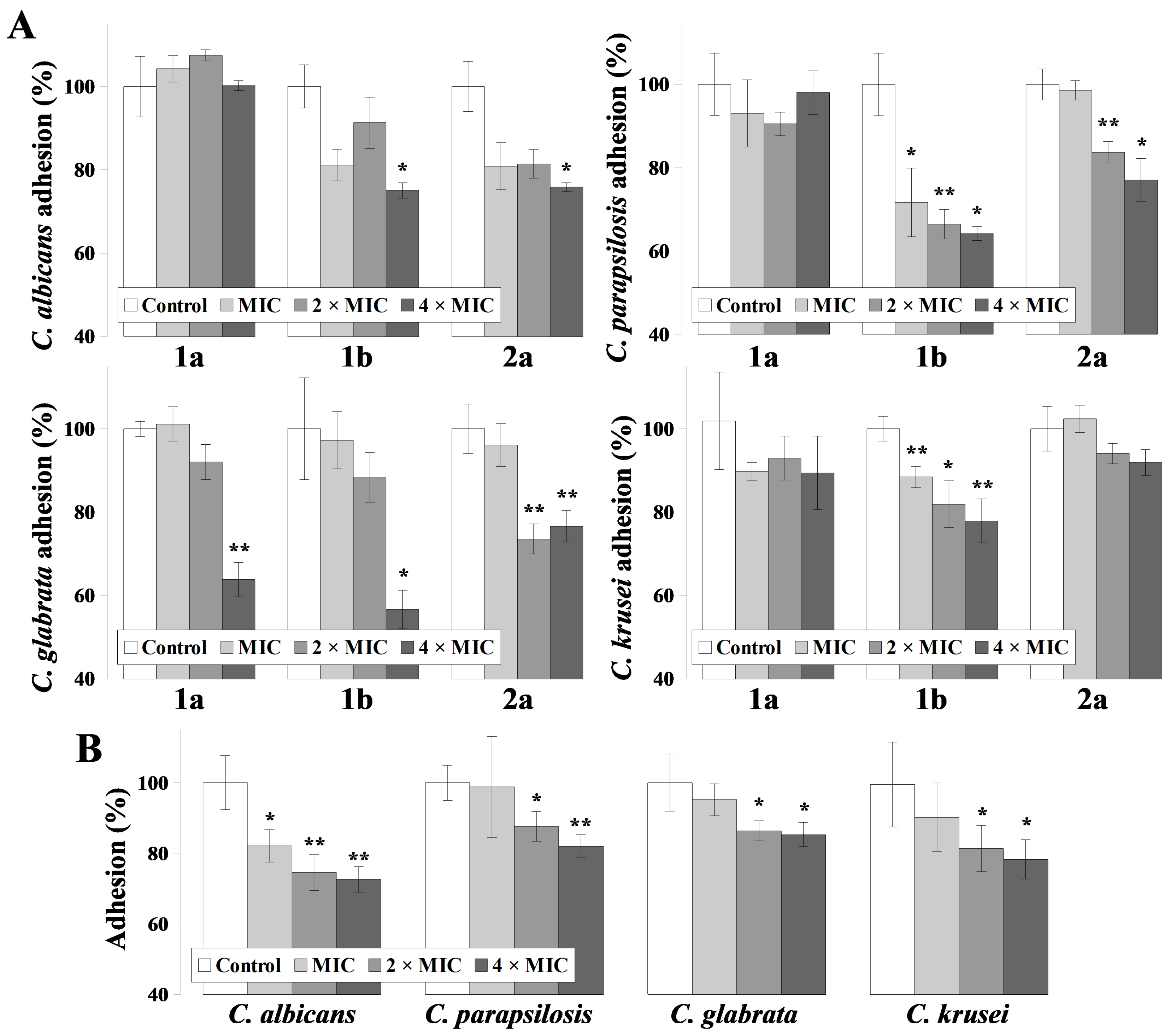
2.1. Antifungal and Antiadhesive Properties of ILs

Table 1 summarizes the minimal inhibitory and fungicidal concentration (MIC90 and MFC, respectively) values of ILs tested against Candida spp. (for ILs structures see Section 5.2. No effect of the alkyl chain length (1a and 1b) was observed to influence any inhibitory activity against all tested fungi. However, MFC values were two-fold lower for the compound with longer alkyl chain (1b) in each case. The compound with alkyloxymethyl substituent (2a) displayed reduced inhibitory activity when compared with salt structures containing alkyl residues. No differences were seen for MIC90 values of 1a and 1b against C. albicans, C. parapsilosis and C. glabrata, which seems justified as those compounds differ by only one CH2 group in the alkyl chain. Both 1a and 1b displayed an MIC90 two-fold lower against C. krusei. Salts containing a nonyloxymethyl substituent (2a) displayed an identical MIC90 value against all tested fungi. The MFC value of ILs with nonyl substituent (1a) is two- (C. glabrata) or four- (C. albicans; C. parapsilosis; C. krusei) fold higher than that of the MIC90 towards corresponding fungi. The MFC value of ILs with decyl substituent (1b) is identical (C. parapsilosis; C. glabrata; C. krusei) or two-fold higher (C. albicans) than that for the MIC90 towards corresponding fungi. Finally, the MFC value of 2a is identical (C. glabrata) or two-fold (C. albicans; C. parapsilosis; C. krusei) higher than that of the MIC90 value (Table 1). The tested ILs were ranked as less active than a known antifungal drug, amphotericin B (AMB). No differences were seen for both MIC90 and MFC values (0.25 and 0.5 µM, respectively) of AMB against C. albicans, C. parapsilosis and C. glabrata. In case of C. krusei, both MIC90 and MFC values were two-fold higher than for the other Candida spp.
Cells from adherent Candida spp. exhibit increased resistance towards antimycotics and the host immune system than planktonic cells. Thus, for the evaluation of ILs on fungal adhesion, higher concentrations were used, corresponding to 1, 2 and 4 times the MIC90 values (Figure 1).
IL with nonyl substituent (1a) did not influence the adherent properties of C. albicans or C. parapsilosis, but in the highest concentrations reduced C. glabrata adhesion by ~40% (p = 0.0012). Furthermore, C. krusei adhesion was reduced by approximately 10%, regardless of 1a concentrations, although these changes were not significant (p = 0.21; 0.32; 0.22 for MIC, 2 × MIC and 4 × MIC, respectively). IL with alkoxymethyl chain (2a) reduced the adhesion of C. albicans in ~20–30% (p = 0.047 for 4 × MIC conc.); C. parapsilosis in ~30–40% (p = 0.012; 0.007; 0.011 for MIC, 2 x MIC and 4 × MIC, respectively); C. glabrata adhesion was reduced by approximately 45% when present at highest concentrations (p = 0.016), and C. krusei by approximately 10–20% (p = 0.007; 0.015; 0.007 for MIC, 2 × MIC and 4 × MIC, respectively). IL with decyl substituent (1b) did not significantly reduce C. krusei adhesion and displayed lower activity than 1a and 2a against C. glabrata ~20% reduction after treatment with 2 × MIC and 4 × MIC conc. (p = 0.005 and 0.007, respectively). Chloride salt 1b displayed a higher than 1a and lower than 2a activity in C. parapsilosis adhesion reduction (~20–25%, p = 0.005 and 0.03 for 2 × MIC and 4 × MIC, respectively) and similarly to 2a reduced C. albicans adhesion in ~20–25% (p = 0.04 for highest concentration). The activity of IL 2a was comparable to the properties of AMB in case of C. albicans (~27% reduction in highest concentration, p = 0.002), C. parapsilosis (~20% reduction in highest concentration, p = 0.3) and C. krusei (~20% reduction in highest concentration, p = 0.03). However, IL 2a displayed higher activity towards C. glabrata than AMB (~15% reduction in highest concentration, p = 0.03).

2.2. The Activity of ILs in Permeabilization of Fungal and Erythroid Membranes

We tested ILs for their activity in permeabilizing fungi plasma membranes (PMs), by treating cells with concentrations of ILs corresponding to their MIC90 values (Table 2). Chloride salt with nonyl substituent (1a) displayed high activity in PM permeabilization of C. krusei (p = 0.03), moderate activity for C. albicans (however, not statistically significant, p = 0.14) or C. glabrata (p = 9.9 × 10⁻⁴) and no activity towards C. parapsilosis. Chloride salt with nonyloxymethyl substituent (2a) displayed no activity (C. parapsilosis), moderate activity (C. albicans, p = 3.8 × 10⁻⁴; C. krusei, p = 0.016) and high activity (C. glabrata, p = 0.007) for PM permeabilization. No permeabilization was detected after treating C. albicans cells with chloride salt with decyl substituent (1b), statistically significant (p = 0.046) permeabilization was observed for C. krusei and moderate activity for C. parapsilosis and C. glabrata (p = 0.011 and 0.02, respectively). The tested ILs were ranked as less active in fungal PM permeabilization than AMB. The treatment with AMB resulted in ~40% permeabilization in case of C. albicans (p = 2.3 × 10⁻⁴), C. glabrata (p = 1.1 × 10⁻⁴) and C. krusei (p = 0.026), or ~30% in case of C. parapsilosis (p = 6.7 × 10⁻⁵).
To examine any possible toxic effect towards animal cells, ILs were tested for their lytic activity on sheep erythroid PMs, in comparison to AMB (Table 3). For this part of study, higher concentrations were selected, corresponding to MFC values of tested fungi (12.5; 25 and 50 µM in case of ILs, and 0.25; 0.5 and 1 µM in case of AMB, Table 1). The compounds were ranked in terms of hemolytic activity as follows: 1a > 1b > 2a. IL with alkoxymethyl chain (2a) produced <5% hemolysis, regardless of the concentration used. Compounds with alkyl substituents (1a and 1b) were more hemolytic in 12.5 than in 25 µM concentrations, and in the highest concentration (50 µM), both compounds produced >10% and >5% hemolysis, respectively. AMB, on the other hand, displayed high toxicity towards erythrocytes, resulting in almost 90% hemolysis in the lowest concentration, and total hemolysis in higher concentrations (Table 3).

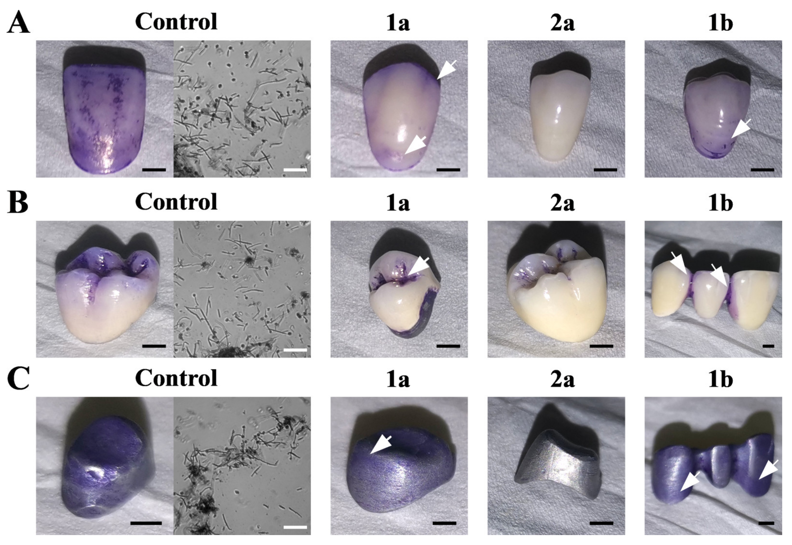
2.3. ILs Prevent C. Albicans Biofilm Formation on Dentures

We evaluated the ability of ILs to eradicate C. albicans biofilm formation on a number of dentures, namely acrylic dental crowns, PFM dental crowns and dental alloy substructures (Figure 2). This approach has the following disadvantage: impossibility of reliable biofilm quantification. However, during in vivo Candida spp. infections and biofilm formation, the fungi attach themselves into porous structure of dentures, and thus we selected them for the studies over commonly used discs. Here, an IL concentration of 25 µM was selected for this investigation. In each condition, a C. albicans biofilm was formed after 72 h incubation, and the collected biomass was microscopically evaluated for the presence of hyphae, pseudohyphae and blastoconidia characteristic of C. albicans (Figure 2). Next, the materials were treated with ILs for 2 h and stained with crystal violet (CV). In the presence of salts with alkyl chains (1a and 1b), residual CV staining was observed on each of the tested dentures (Figure 2). IL with alkyloxymethyl substituent (2a) protected acrylic dental crowns and dental alloy substructure from biofilm formation, but did not fully protect those made from PFM. Microscopic observations of the stained biomass from dentures exposed to both 1a and 1b (Figure 2, white arrows) confirmed the presence of C. albicans hyphae and blastoconidia (data not shown).
Fungal adhesion to biomedical surfaces is strongly related to the structure of the cell wall, and the presence of adhesins (Als proteins) on the surface of the cell. In turn, the progression to biofilm formation is dependent on the ability of C. albicans to form hyphae. In conditions similar to those above (ILs conc. = 25 µM), we examined C. albicans cell wall integrity, its ability to form hyphae, and the level of ALS genes expression, encoding Als1 and Als5 adhesins (Figure 3). Calcofluor white staining of C. albicans cells treated with 1a and 1b reveals well-defined, intense chitin fluoresce with clearly visible bud scars, comparable with the control staining (Figure 3A). After exposure to IL 2a, C. albicans cells revealed low fluorescence intensity and thinner calcofluor white-stained chitin layer surrounding cells than the controls. In the presence of compounds with alkyl substituents (1a and 1b), hyphae formation by C. albicans cells was completely inhibited (Figure 3B), and the percentage of cells forming hyphae was <1%. After treatment with IL 2a, 78.2 ± 17.2% cells formed hyphae, significantly less when compared to the control conditions (94 ± 0.6% cell forming hyphae). Despite some reduction, the result was not statistically significant (p = 0.12). On the other hand, after the treatment with IL 2a, the expression of ALS1 and ALS5 genes was reduced by 50 or 55%, respectively (Figure 3C). The results were statistically significant in both cases (p = 0.04 for either ALS gene). However, in case of compounds with alkyl substituents (1a and 1b), the reduction of gene expression was to a lesser degree. The treatment with IL 1a resulted in 10 or 20% reduction in case of ALS5 or ALS1 gene expression, respectively. It was observed that IL with longer alkyl substituent 1b resulted in 40 or 45% reduction in case of ALS5 or ALS1 gene expression, respectively. Despite observing a chain-length dependence, the results were not statistically significant (p = 0.07 for both ALS gene after treatment with 1b).

3. Discussion

In this research, three imidazolium-based ILs were chosen for investigations based on previous reports of strong antimicrobial properties of those compounds [23,24]. Moreover, imidazolium cation containing ILs are already used for denture cleaning applications [27,28,29]. The incorporation of natural terpene alcohol, (1R,2S,5R)-(−)-menthol in the structure of ILs follows the trend of researching more natural products in CADS treatment (such as: apple cider, probiotics, curcumin, tea tree oil or fulvic acid) [14,32,33].
No correlation was found between the alkyl chain length of the compounds and fungicidal activity for any of the tested fungi (Table 1); a similar observation was found in our previous studies on ammonium ILs based on (1R,2S,5R)-(−)-menthol towards the C. albicans SC5314 strain [31]. Here however, a positive correlation between the alkyl/alkoxymethyl chain length and fungicidal activity of the compounds was found (Table 1), which was not previously observed in the case of ammonium ILs [31]. Interestingly, imidazolium ILs are less active towards C. albicans than our previously tested ammonium ILs, with the alkyloxymethyl substituted compound being the least active in the current report (2a, Table 1). The cationic structures of the ILs salts with chloride anions and commonly used benzalkonium chloride (BAC) were previously ranked in terms of antimicrobial activity as follows: ammonium CILs > alkylimidazolium CILs > alkoxymethylimidazolium CILs > BAC > pyridinium CILs [30]. However, interspecies differences towards ILs were observed, especially with C. glabrata and C. krusei that were twice as vulnerable towards the fungicidal effects as C. albicans (MFCs, Table 2). C. glabrata is known to be the second most prevalent Candida species, being isolated from acrylic denture surfaces and the palatal mucosa [34]. Since C. glabrata is more resistant to commercial denture cleaners and sodium hypochlorite (NaOCl) solutions than other Candida spp. [35], our result is especially promising.
ILs are generally considered cytotoxic, especially imidazolium derivatives [36], and thus should not be administered intravenously. However, here we evaluated the potential of the compounds as short-term eradicators of adherent cells or biofilm of Candida spp., in addition to their short-term safety towards mammalian erythrocytes, which are generally very vulnerable.
An essential step in the initiation of CADS and biofilm formation is the adhesion of Candida spp. to denture material [12]. Microbial adhesion on biomedical surfaces depends on the surface structure and composition of biomaterials, microbial cell surface hydrophobicity and specific interactions between cell surface structures and the surfaces of biomaterials [37,38]. For a tentative comparison of ILs activity in their ability to detach adherent Candida spp. cells, we examined their influence on cells adherent to polystyrene surface (Figure 1). Imidazolium (Figure 1) and ammonium [31] ILs reduced C. albicans adhesion in a comparable manner (20–30% reduction). Kanjanamekanant et al. [28] also demonstrated that IL-incorporated tissue conditioner reduces C. albicans adhesion to a similar degree (~25%). Despite the least fungistatic and fungicidal activities (Table 1), the alkyloxymethyl substituted compound (2a) was generally the most active against adherent cells (Figure 1). The results are especially promising since 2a was the least hemolytic among the tested compounds (Table 3) and most (C. albicans and C. glabrata) or second most active (C. krusei) in PM permeabilization (Table 2).
The tested ILs displayed lower fungicidal, higher permeabilization and similar antiadhesive activities when compared to AMB—one of the “gold standards” during antifungal treatment (Figure 1, Table 1 and Table 2). The results represent in vitro situation, using common laboratory Candida spp. strains, and laboratory medium. However, during in vivo treatment of CADS, common antifungal agents such as AMB are not effective [15]. Additionally, AMB as a polyene drug binds to ergosterol in the fungal plasma membrane (PM), inducing permeabilization and cytoplasm leakage [39]. Polyenes also display specificity towards mammalian cholesterol, which results in high cytoxicity of those drugs, and the treatment is associated with a number of adverse effects among patients [39]. Here, this effect of AMB is represented by high hemolytic activity, which is not the case of the tested ILs (Table 3). Thus, despite lower in vitro activity of ILs towards Candida spp. than AMB, the usage of ILS in further studies seems justified—especially due to their activity towards clinical C. albicans isolates (Supplementary Materials). We have found that fluconazole (FLC)-resistant isolates are vulnerable towards the ILs in comparable manner as FLC-sensitive strains in either planktonic (Table S1) or adherent (Figure S1) form.
Similarly to previously examined ammonium ILs [31], a positive correlation was found between the alkyl chain length of the compounds (1a and 1b) and antiadhesive activities towards C. albicans and C. parapsilosis (Figure 1). However, an opposite correlation was found in the hemolytic activity, as IL containing nonyl substituent (1a) was more hemolytic than the one with decyl substituent (1b) (Table 4). Previously we have demonstrated a similar feature of ammonium IL with decyl substituent and (−)-menthol moiety being more hemolytic than ammonium IL with dodecyl substituent [31]. The hemolytic properties of amphipathic molecules do not always correspond linearly to increasing alkyl chain length, as previously reported for quaternary ammonium salts [40]. The hemolytic activities depend not only on the hydrophobic moiety of the amphiphiles, but also on the size of the hydrophilic group, CMC and symmetry of the entire compound [41]. In yeast cells, intraspecies differences in PM composition affect resistance towards amphiphiles. As shown herein, ILs with nonyl substituent (1a) is more active in PM permeabilization of C. albicans and C. krusei, and 1b is more active towards C. parapsilosis and glabrata (Table 2).
To be able to apply ILs in the protection of dentures from biofilm formation, only C. albicans strain was chosen, as this is the predominant cause of CADS and general microbial stomatitis [10]. In in vitro studies, C. albicans was shown to form a biofilm on a number of different denture biomaterials, including polymethyl methacrylate (PMMA, known as acrylic) and polyamide resins [13,15,35,42], resilient denture liners [17], polyvinyl chloride (PVC), polyurethane and teflon [43]. Here, we were able to observe the formation of C. albicans’ biofilm on acrylic and PFM dental crowns, and dental alloy substructures (Figure 2). The concentration of ILs was chosen as 25 μM, since under such conditions ILs were the least hemolytic to mammalian erythrocytes (Table 3). All tested compounds visually eradicated biofilm that had formed on dentures (Figure 2). Similar results were observed for clinical, FLC-resistant isolates (Figure S2, Supplementary Materials). However, contrasting 1a and 1b, 2a did not fully protect acrylic and alloy substructures, probably due to the highest antiadhesive properties for C. albicans (Figure 1). This is likely to occur through the ability to inhibit hyphae development (Figure 3). In vivo, C. albicans biofilms consists of networks of yeast cells and hyphae, embedded into cracks and imperfections of the dentures [13,14]. Similarly, we observed long hyphaes in biomass samples from the dentures (Figure 2, controls). Previously, we have identified high activities of alkyl substituted ammonium ILs in inhibiting C. albicans hyphae development [31]. However, alkyloxymethyl substituted ILs (2a) did not inhibit filamentation (Figure 2B), but displayed activity in cell wall lysis (Figure 2A). We hypothesize, that this ability of IL 2a is also connected to reducing the presence of Als proteins on the cell surface of the fungi. Here, we observed that treatment of C. albicans cells with IL 2a significantly reduced gene expression of two ALS genes (namely, ALS1 and ALS5, Figure 3C). Both genes encode cell-surface glycoproteins, commonly known as adhesins, which are crucial for aggregative effects, and the attachment of the fungus to biotic and abiotic surfaces [44,45]. Thus, most likely due to this ability IL 2a was the most effective in biofilm eradication from dentures (Figure 2).
Some strategies have been described to combat Candida biofilm in dentures, such as soaking in NaOCl, microwave irradiation, the application of effervescent cleansing tabs [18,46], preservatives and disinfectants [15] or treating surfaces with plasma [47]. However, harsh treatments may alter the surfaces of the biomaterials [15], and efficient treatment of CADS relies not only on the treatment of the infected tissues, but also on preserving the denture [48]. In our case, no alterations in denture structures were observed after treatment with ILs (Figure 2).
In the human oral cavity, Candida spp. form mixed biofilms with streptococci and staphylococci bacteria [10,49]. The potential application of the presented compounds in situations resembling in vivo stomatitis (e.g., their activity against mixed Candida-bacteria biofilms) is promising, as all tested compounds were previously described to possess strong antibacterial properties [23,24]. However further development in this area is needed to examine their potential against biofilms of mixed Candida spp. and other bacterial species.

4. Conclusions

The results from this study demonstrated high antifungal and anti-adhesive (especially 2a and 1b) properties of imidazolium-based ILs with (1R,2S,5R)-(−)-menthol towards pathogenic Candida spp. Additionally, presented ILs displayed low hemolytic activities, when compared to antifungal gold-standard, amphotericin B. For further studies, we were experimenting with C. albicans fungus. The tested ILs visually eradicated C. albicans biofilm formed on dentures made from different materials, partially in case of 1a and 1b and totally in case of compound 2a. Biofilm eradication in case of 1a and 1b was correlated with inhibiting hyphae formation. Additionally, 1a seem not to affect the adhesion phenotypically or ALS genes expression, whereas 1b affects both processes. In case of 2a the effect of eradicating C. albicans biofilm on dentures was connected to fungal cell wall lysis and reduction of adhesins (2a). Based on this research, it can be concluded that the compounds presented here may possess a potential as anti-fungal denture cleaners or denture fixatives (especially 2a, a strong biofilm eradicator), due to their high antifungal activities, and low toxicity towards mammalian cells in short-term exposure.

5. Materials and Methods

5.1. Chemicals

Chemicals and reagents used in this study were purchased from the following sources: sodium dodecyl sulfate (SDS), crystal violet (CV), phosphate-buffered saline (PBS), and calcofluor white (CFW) (Sigma-Aldrich, Poznań, Poland); d-glucose, bacteriological agar, propidium iodide (PI), and fetal bovine serum (FBS) (manufacturer: Bioshop and distributor: Lab Empire, Rzeszów, Poland); yeast extract (YE) and peptone (manufacturer: BD and distributor: Diagmed, Warszawa, Poland). All chemicals were analytical grade purity.

5.2. Preparation of ILs

The syntheses of 3-alkyl-1-[(1R,2S,5R)-(−)-menthoxymethyl]imidazolium chlorides (1a and 1b) abbreviated [Cn-Im-C1OMen][Cl], where n = 9 or 10, were described in our previous publication [23]. 1-(1R,2S,5R)-(−)-Menthoxymethyl-3-nonyloxymethyl imidazolium chloride (2a), abbreviated as [C1OC9-Im-C1OMen][Cl] was published by us previously [24]. The synthesized quaternary imidazolium chlorides were characterized by nuclear magnetic resonance (1H NMR and 13C NMR). The results were comparable with those presented previously by our group [23,24]. Elemental analyses were performed for the obtained chlorides (1a,1b,2a), and the results are given in Table 4.

5.3. Strains and Growth Conditions

C. albicans SC5314 was a kind gift of Prof. D. Sanglard (Lausanne, Switzerland) [50]. C. parapsilosis CBS10947 was a kind gift of Dr K. Góralska (Łódź, Poland) while C. glabrata ATCC90030 and C. krusei ATCC6258 were kind gifts of Dr M. Dyląg (Wrocław, Poland). Clinical C. albicans isolates (description in Supplementary Materials) were kind gifts of Prof. S. Milewski (Gdańsk, Poland) and prof. J. Morschhäuser (Wurzburg, Germany). Fungal strains were routinely grown at 28 °C on YPD medium (2% glucose, 1% peptone, 1% YE) with agitation (120 rpm). Agar (2% final concentration) was used for solidification of the medium.

5.4. Determination of Minimal Inhibitory and Fungicidal Concentrations (MICs and MFCs)

Experiments were performed according to the Clinical and Laboratory Standards Institute (2008), 3rd ed. M27-A3 [51], with modifications described previously [31]. Briefly, MICs were determined by serially diluting ILs in YPD medium in sterile 96-well plates (Sarstedt) and inoculating with fungal suspensions (final OD600 per well = 0.01). After incubation (48 h; 28 °C), the OD600 was measured (ASYS UVM 340 Biogenet). The concentrations of ILs, which resulted in ≥90% growth inhibition, were determined as MICs. Each well was then replated on YPD agar and incubated for 5 days at 28 °C. The lowest concentrations of ILs, which resulted in a complete absence of fungal growth were determined as MFCs.

5.5. Antiadhesive Properties of ILs

Post-adherence treatment with ILs was performed in a similar manner to that described previously by our team [31,52]. Candida spp. suspensions were prepared by centrifuging YPD-grown cultures and resuspending fungal mass in PBS to an OD600 = 0.6. Sterile 96-well plates were inoculated with the prepared suspensions and incubated for 2 h at 37 °C to induce fungal adhesion. Afterwards, adherent cells were washed with PBS to remove planktonic cells and treated with ILs (concentrations equivalent to 1, 2 or 4 × MIC90 for each fungi) for 2 h at 37 °C. Then, each well was washed with PBS and adherent cells were fixed with 0.1% CV solution for 5 min, and washed with PBS. Cells were then treated with a solution of isopropanol 0.04 N HCL with 0.1% SDS to permeabilize adherent cells and release the CV dye. The absorbance of CV was measured at 590 nm (ASYS UVM 340 Biogenet). The percentage of cells that were removed was determined in comparison to adherent cells treated with PBS only (designated 100% adhesion).

5.6. Propidium Iodide (PI) Staining

The assessment of PM permeability was performed as described previously [53], with modifications. Briefly, 3 mL of Candida spp. cell suspensions (OD600 = 0.1) in PBS were mixed with ILs (concentrations equivalent to MIC90 against each fungi). Samples were incubated for 2 h at 37 °C, washed with PBS and stained with PI to the final dye concentration of 6 × 10⁻⁶ M for 5 min. Then, cell suspensions were washed with PBS and observed under the Zeiss Axio Imager A2 microscope equipped with the Zeiss Axiocam 503 mono microscope camera and Zeiss HBO100 mercury lamp. The percentage of permeabilized cells was evaluated by counting PI positive cells out of one hundred in three independent repetitions for each experiment.

5.7. Hemolysis Assay

ILs were tested for hemolytic activity, as described previously [31]. Briefly, 5 mL of fresh sheep blood was centrifuged (2000 rpm, 15 min), washed with PBS and resuspended in PBS to achieve hematocrit level of 50%. Red blood cells (RBCs) were treated with ILs (concentrations = 12.5, 25 and 50 µM) for 2 h at 37 °C. Afterwards, RBCs were centrifuged (2000 rpm, 5 min) and the absorbance of the supernatant was measured at 540 nm (ASYS UVM 340 Biogenet). The percentage of hemolysis was determined in comparison to the hemoglobin absorbance released by a 1% SDS solution (100% hemolysis).

5.8. Biofilm Formation on Dentures

To assess the influence of ILs on biofilm formation by C. albicans strain SC5314 on dentures, the following materials were used: acrylic dental crowns, porcelain-fused-to-metal (PFMs) dental crowns and dental alloy substructures. All dentures were kind gifts from Prof. M. Łukaszewicz (Wrocław, Poland). Prior to the experiments, the dentures were sterilized by 1 h incubation in 70% pure ethanol (EtOH) solution, washed with sterile water to remove EtOH residues and dried under aseptic conditions. The experiments were performed by aseptically placing dentures in YPD, supplemented with FBS, and inoculating them with YPD-grown C. albicans suspension (0.01% Vol/Vol). After 72 h of incubation at 37 °C, the dentures were treated for 2 h with the addition of ILs (conc. = 25 µM), washed with PBS and fixed with 0.1% CV solution for 10 min, then washed with PBS to remove the stain residue. The dentures were then dried and photographed. Microscopic preparations were made of biofilm samples which were imaged using the Zeiss Axio Imager A2 microscope equipped with Zeiss Axiocam 503 mono microscope camera.

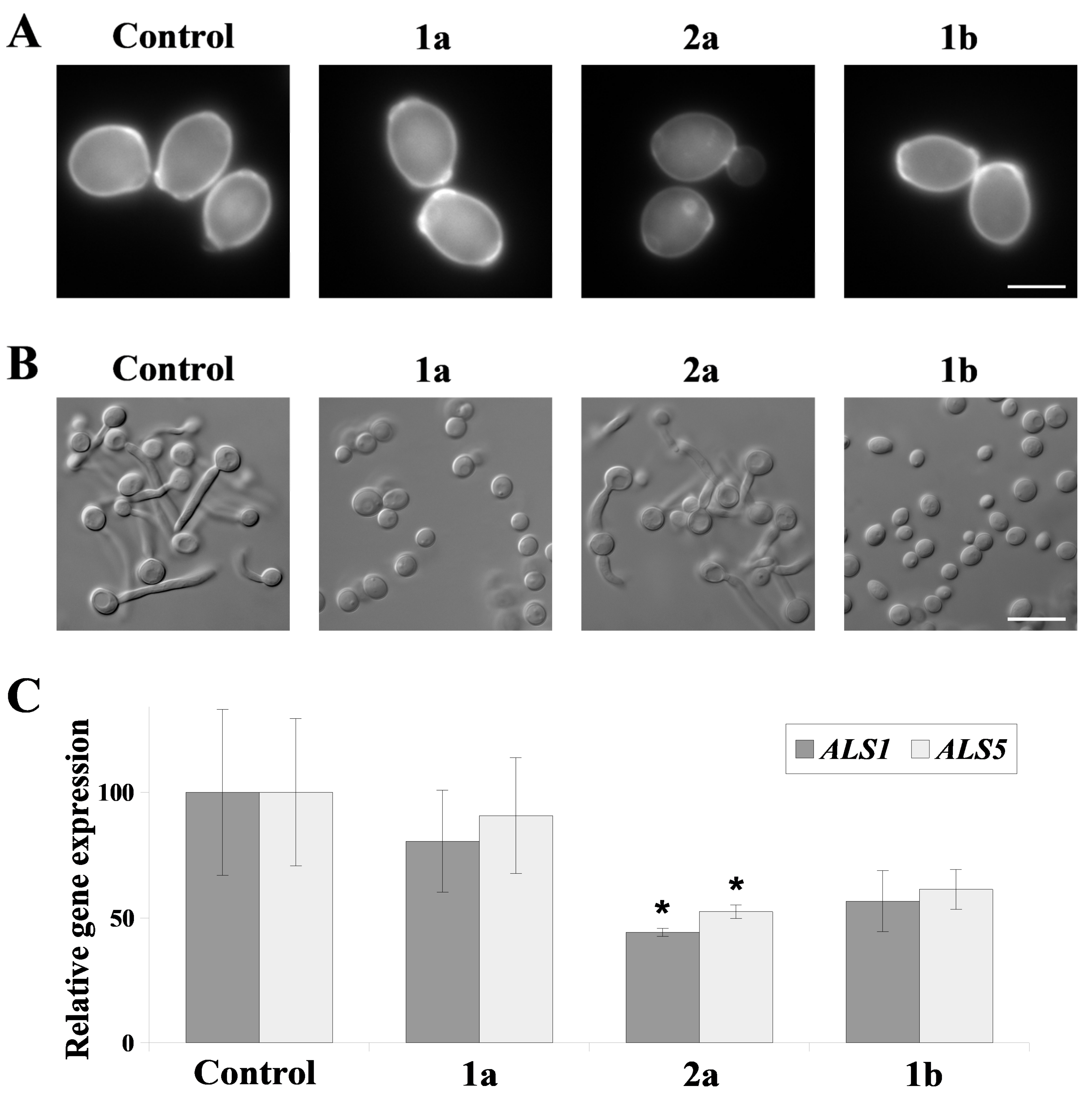
5.9. The Impact of ILs on Yeast-to-Hyphae Transition

Experiments were performed as described before [54], with modifications. YPD-grown C. albicans were pelleted, washed with fresh YPD medium and resuspended in YPD medium (OD600 = 0.4). The suspensions were treated with ILs (conc. = 25 µM) and FBS (final conc. = 10%) for 2 h at 37 °C. The samples were observed under the Zeiss Axio Imager A2 microscope equipped with Canon PowerShot G10 camera for assessment of cell morphology. For quantitative estimation, cells of each assay were counted in three independent repetitions determining the percentage of hyphae forming cells.

5.10. Calcofluor White Staining

Calcofluor white was used to assess cell wall integrity in C. albicans, following our protocol [55]. YPD-grown C. albicans cells were pelleted, washed with PBS and treated with ILs (conc. = 25 µM) for 2 h at 37 °C. Afterwards, the samples were washed with PBS, stained with calcofluor white (final conc. = 25 µM) for 5 min. Then, cell suspensions were washed with PBS, pelleted and observed under the Zeiss (Poznań, Poland) Axio Imager A2 microscope equipped with Zeiss Axiocam 503 mono microscope camera and the Zeiss HBO100 mercury lamp.

5.11. Real Time Quantitive Polymerase Chain Reaction

YPD-grown C. albicans cells were pelleted, washed with PBS and treated with ILs (conc. = 25 µM) for 2 h at 37 °C. Next, the cells were pelleted, and concentrated (PBS; OD600 = 20). RNA was isolated using the Total RNA Mini Kit (A&A Biotechnology, Gdańsk, Poland). Synthesis of cDNA and calculation of gene expression levels were performed as previously described [56]. The following gene-specific primers were used: ACT1_F (5′-TCC AGC TTT CTA CGT TTC CA-3′), ACT1_R (5′-GTC AAG TCT CTA CCA GCC AA-3′), ALS1_F (5′-TGT TGG TGT GAC TAC TTC CT-3′), ALS1_R (5′-TGT ACC ACT GTG TCA AT-3′), ALS5_F (5′-CTC CAC TAG TTA TGG GGA TGT-3′) and ALS5_R (5′-TGA GGG AGA AAT ATA AGC GTC A-3′).

5.12. Statistical Analysis

Unless stated otherwise, data represent the mean ± standard errors from at least three biological replicates. Microscopic observations were performed for at least two independent replicates, of which the representative images were included in the figures. When indicated, statistical significance was determined by a Student’s t-test (binomial, unpaired).

Supplementary Materials

The following are available online at https://0-www-mdpi-com.brum.beds.ac.uk/article/10.3390/ijms22147543/s1, Table S1: Minimal inhibitory concentrations (MIC90; µM) and minimal fungicidal concentrations (MFC; µM) of ionic liquids (ILs) or fluconazole (FLC) towards clinical C. albicans isolates.; Figure S1: Detachment of adherent C. albicans clinical isolates after 2 h incubation on polystyrene surfaces by ionic liquids (ILs) (means ± SD; n = 3). Results are presented as percentage of adherent cells relative to untreated controls (100% adhesion). Statistical analysis of detachment at each concentration was performed towards corresponding control experiments (isolates untreated by ILs = 100% adhesion) (* p < 0.05; ** p < 0.01 and *** p < 0.001).; Figure S2: C. albicans clinical isolates (B4 and Gu5) biofilm formation visualized by crystal violet (CV) dye on acrylic dental crowns. Scale bar = 2 mm. Biofilm mass formed on control probes was observed under microscope (40x), scale bar = 50 µm. Samples were treated with ILs (1a; 2a; 1b) for 2 h to formed C. albicans biofilm.

Author Contributions

Conceptualization, J.S. and A.K.; methodology, investigation, J.S. and J.F.-K.; writing—original draft preparation, J.S.; and writing—review and editing, A.K. and J.F.-K.; supervision, A.K. All authors have read and agreed to the published version of the manuscript.

Funding

This work was supported by Wrocław Centre of Biotechnology, programme The Leading National Research Centre (KNOW) for years 2014–2018 (J.S. and A.K.). J.F.-K. is grateful for the financial support given from the Polish Ministry of Science and Higher Education by subvention activity for the Faculty of Chemistry of Wrocław University of Science and Technology. Publication of this article in open access was financially supported by the Excellence Initiative—Research University (IDUB) programme for the University of Wrocław.

Institutional Review Board Statement

Not applicable.

Informed Consent Statement

Not applicable.

Data Availability Statement

The data presented in this study are available on request from the corresponding author (J.S.).

Acknowledgments

We gratefully acknowledge Daria Derkacz (Faculty of Biotechnology, University of Wrocław) for her support with Real Time-qPCR analyses.

Conflicts of Interest

The authors declare no conflict of interest.

References

  1. Salerno, C.; Pascale, M.; Contaldo, M.; Esposito, V.; Busciolano, M.; Milillo, L.; Guida, A.; Petruzzi, M.; Serpico, R. Candida-associated denture stomatitis. Med. Oral Patol. Oral Cir. Bucal. 2011, 16, e139–43. [Google Scholar] [CrossRef]
  2. Swidergall, M. Candida albicans at Host Barrier Sites: Pattern Recognition Receptors and Beyond. Pathogens 2019, 8, 40. [Google Scholar] [CrossRef] [Green Version]
  3. Ksiezopolska, E.; Gabaldón, T. Evolutionary emergence of drug resistance in Candida opportunistic pathogens. Genes 2018, 9, 461. [Google Scholar] [CrossRef] [Green Version]
  4. Offenbacher, S.; Barros, S.P.; Bencharit, S.; Yu, N.; Preisser, J.; Moss, K.; Loewy, Z.G. Differential Mucosal Gene Expression Patterns in Candida-Associated, Chronic Oral Denture Stomatitis. J. Prosthodont. 2019, 28, 202–208. [Google Scholar] [CrossRef] [PubMed]
  5. Dorocka-Bobkowska, B.; Zozulinska-Ziolkiewicz, D.; Wierusz-Wysocka, B.; Hedzelek, W.; Szumala-Kakol, A.; Budtz-Jörgensen, E. Candida-associated denture stomatitis in type 2 diabetes mellitus. Diabetes Res. Clin. Pract. 2010, 90, 81–86. [Google Scholar] [CrossRef] [PubMed]
  6. Motta-Silva, A.C.; Aleva, N.A.; Chavasco, J.K.; Armond, M.C.; França, J.P.; Pereira, L.J. Erythematous oral candidiasis in patients with controlled type II diabetes mellitus and complete dentures. Mycopathologia 2010, 169, 215–223. [Google Scholar] [CrossRef] [PubMed]
  7. Brito, G.N.B.; Inocêncio, A.C.; Querido, S.M.R.; Jorge, A.O.C.; Koga-Ito, C.Y. In vitro antifungal susceptibility of Candida spp. oral isolates from HIV positive patients and control individuals. Braz. Oral Res. 2011, 25, 28–33. [Google Scholar] [CrossRef] [PubMed] [Green Version]
  8. Patel, P.K.; Erlandsen, J.E.; Kirkpatrick, W.R.; Berg, D.K.; Westbrook, S.D.; Louden, C.; Cornell, J.E.; Thompson, G.R.; Vallor, A.C.; Wickes, B.L.; et al. The changing epidemiology of oropharyngeal candidiasis in patients with HIV/AIDS in the era of antiretroviral therapy. AIDS Res. Treat. 2012, 2012, 262471. [Google Scholar] [CrossRef] [Green Version]
  9. Schelenz, S.; Abdallah, S.; Gray, G.; Stubbings, H.; Gow, I.; Baker, P.; Hunter, P.R. Epidemiology of oral yeast colonization and infection in patients with hematological malignancies, head neck and solid tumors. J. Oral Pathol. Med. 2011, 40, 83–89. [Google Scholar] [CrossRef] [PubMed]
  10. Cavalcanti, I.M.G.; Nobbs, A.H.; Ricomini-Filho, A.P.; Jenkinson, H.F.; Del Bel Cury, A.A. Interkingdom cooperation between Candida albicans, Streptococcus oralis and Actinomyces oris modulates early biofilm development on denture material. Pathog. Dis. 2016, 74, ftw002. [Google Scholar] [CrossRef] [Green Version]
  11. Nett, J.E.; Marchillo, K.; Spiegel, C.A.; Andes, D.R. Development and validation of an in vivo Candida albicans biofilm denture model. Infect. Immun. 2010, 78, 3650–3659. [Google Scholar] [CrossRef] [Green Version]
  12. Li, Z.; Sun, J.; Lan, J.; Qi, Q. Effect of a denture base acrylic resin containing silver nanoparticles on Candida albicans adhesion and biofilm formation. Gerodontology 2016, 33, 209–216. [Google Scholar] [CrossRef] [PubMed]
  13. Redding, S.; Bhatt, B.; Rawls, H.R.; Siegel, G.; Scott, K.; Lopez-Ribot, J. Inhibition of Candida albicans biofilm formation on denture material. Oral Surg. Oral Med. Oral Pathol. Oral Radiol. Endodontol. 2009, 107, 669–672. [Google Scholar] [CrossRef]
  14. Alalwan, H.; Rajendran, R.; Lappin, D.F.; Combet, E.; Shahzad, M.; Robertson, D.; Nile, C.J.; Williams, C.; Ramage, G. The anti-adhesive effect of curcumin on Candida albicans biofilms on denture materials. Front. Microbiol. 2017, 8, 659. [Google Scholar] [CrossRef] [PubMed]
  15. Tsutsumi, C.; Takakuda, K.; Wakabayashi, N. Reduction of Candida biofilm adhesion by incorporation of prereacted glass ionomer filler in denture base resin. J. Dent. 2016, 44, 37–43. [Google Scholar] [CrossRef] [PubMed]
  16. Altarawneh, S.; Bencharit, S.; Mendoza, L.; Curran, A.; Barrow, D.; Barros, S.; Preisser, J.; Loewy, Z.G.; Gendreau, L.; Offenbacher, S. Clinical and Histological Findings of Denture Stomatitis as Related to Intraoral Colonization Patterns of Candida albicans, Salivary Flow, and Dry Mouth. J. Prosthodont. 2013, 22, 13–22. [Google Scholar] [CrossRef] [Green Version]
  17. Bueno, M.G.; Urban, V.M.; Barbério, G.S.; da Silva, W.J.; Porto, V.C.; Pinto, L.; Neppelenbroek, K.H. Effect of antimicrobial agents incorporated into resilient denture relines on the Candida albicans biofilm. Oral Dis. 2015, 21, 57–65. [Google Scholar] [CrossRef] [PubMed]
  18. Neppelenbroek, K.H.; Pavarina, A.C.; Palomari Spolidorio, D.M.; Sgavioli Massucato, E.M.; Spolidorio, L.C.; Vergani, C.E. Effectiveness of microwave disinfection of complete dentures on the treatment of Candida-related denture stomatitis. J. Oral Rehabil. 2008, 35, 836–846. [Google Scholar] [CrossRef] [PubMed]
  19. Ellepola, A.N.B.; Samaranayake, L.P.; Khan, Z.U. Extracellular phospholipase production of oral Candida albicans isolates from smokers, diabetics, asthmatics, denture wearers and healthy individuals following brief exposure to polyene, echinocandin and azole antimycotics. Braz. J. Microbiol. 2016, 47, 911–916. [Google Scholar] [CrossRef] [Green Version]
  20. Suchodolski, J.; Krasowska, A. Fructose Induces Fluconazole Resistance in Candida albicans through Activation of Mdr1 and Cdr1 Transporters. Int. J. Mol. Sci. 2021, 22, 2127. [Google Scholar] [CrossRef]
  21. De Almeida, R.F.M.; Santos, F.C.; Marycz, K.; Alicka, M.; Krasowska, A.; Suchodolski, J.; Panek, J.J.; Jezierska, A.; Starosta, R. New diphenylphosphane derivatives of ketoconazole are promising antifungal agents. Sci. Rep. 2019, 9, 16214. [Google Scholar] [CrossRef] [Green Version]
  22. Ruiz-Ruigomez, M.; Badiola, J.; Schmidt-Malan, S.M.; Greenwood-Quaintance, K.; Karau, M.J.; Brinkman, C.L.; Mandrekar, J.N.; Patel, R. Direct Electrical Current Reduces Bacterial and Yeast Biofilm Formation. Int. J. Bacteriol. 2016, 2016, 9727810. [Google Scholar] [CrossRef] [PubMed] [Green Version]
  23. Pernak, J.; Feder-Kubis, J.; Cieniecka-Rosłonkiewicz, A.; Fischmeister, C.; Griffin, S.T.; Rogers, R.D. Synthesis and properties of chiral imidazolium ionic liquids with a (1R,2S,5R)-(-)-menthoxymethyl substituent. New J. Chem. 2007, 31, 879–892. [Google Scholar] [CrossRef]
  24. Feder-Kubis, J.; Kubicki, M.; Pernak, J. 3-Alkoxymethyl-1-(1R,2S,5R)-(-)-menthoxymethylimidazolium salts-based chiral ionic liquids. Tetrahedron Asymmetry 2010, 21, 2709–2718. [Google Scholar] [CrossRef]
  25. Pernak, J.; Feder-Kubis, J. Synthesis and properties of chiral ammonium-based ionic liquids. Chem. Eur. J. 2005, 11, 4441–4449. [Google Scholar] [CrossRef]
  26. Pernak, J.; Feder-Kubis, J. Chiral pyridinium-based ionic liquids containing the (1R,2S,5R)-(-)-menthyl group. Tetrahedron Asymmetry 2006, 17, 1728–1737. [Google Scholar] [CrossRef]
  27. Elder, S.T.; Preuss, A.; Schoening, K.-U.; Muehlbauer, K. Anti-Microbial Compositions. U.S. Patent 20080070966, 31 July 2012. [Google Scholar]
  28. Kanjanamekanant, K.; Limpuangthip, N.; Arksornnukit, M. Physical and Mechanical Properties of Antifungal Ionic Liquid-Incorporated Dental Tissue Conditioner. Mater. Sci. Appl. 2017, 8, 376–388. [Google Scholar] [CrossRef] [Green Version]
  29. Bergamo, V.Z.; Donato, R.K.; Nemitz, M.C.; Acasigua, G.A.X.; Selukar, B.S.; Lopes, W.; Dalla Lana, D.F.; Teixeira, M.L.; Teixeira, H.F.; Schrekker, H.S.; et al. Assessing an imidazolium salt’s performance as antifungal agent on a mouthwash formulation. J. Appl. Microbiol. 2016, 121, 1558–1567. [Google Scholar] [CrossRef]
  30. Feder-Kubis, J.; Tomczuk, K. The effect of the cationic structures of chiral ionic liquids on their antimicrobial activities. Tetrahedron 2013, 69, 4190–4198. [Google Scholar] [CrossRef]
  31. Suchodolski, J.; Feder-Kubis, J.; Krasowska, A. Antifungal activity of ionic liquids based on (−)-menthol: A mechanism study. Microbiol. Res. 2017, 197, 56–64. [Google Scholar] [CrossRef]
  32. Mota, A.C.L.G.; de Castro, R.D.; de Araújo Oliveira, J.; de Oliveira Lima, E. Antifungal Activity of Apple Cider Vinegar on Candida Species Involved in Denture Stomatitis. J. Prosthodont. 2015, 24, 296–302. [Google Scholar] [CrossRef]
  33. Ishikawa, K.H.; Mayer, M.P.A.; Miyazima, T.Y.; Matsubara, V.H.; Silva, E.G.; Paula, C.R.; Campos, T.T.; Nakamae, A.E.M. A multispecies probiotic reduces oral candida colonization in denture wearers. J. Prosthodont. 2015, 24, 194–199. [Google Scholar] [CrossRef]
  34. Zomorodian, K.; Haghighi, N.N.; Rajaee, N.; Pakshir, K.; Tarazooie, B.; Vojdani, M.; Sedaghat, F.; Vosoghi, M. Assessment of Candida species colonization and denture-related stomatitis in complete denture wearers. Med. Mycol. 2011, 49, 208–211. [Google Scholar] [CrossRef] [Green Version]
  35. De Freitas Fernandes, F.S.; Pereira-Cenci, T.; Da Silva, W.J.; Filho, A.P.R.; Straioto, F.G.; Del Bel Cury, A.A. Efficacy of denture cleansers on Candida spp. biofilm formed on polyamide and polymethyl methacrylate resins. J. Prosthet. Dent. 2011, 105, 51–58. [Google Scholar] [CrossRef]
  36. García-Lorenzo, A.; Tojo, E.; Tojo, J.; Teijeira, M.; Rodríguez-Berrocal, F.J.; González, M.P.; Martínez-Zorzano, V.S. Cytotoxicity of selected imidazolium-derived ionic liquids in the human Caco-2 cell line. Sub-structural toxicological interpretation through a QSAR study. Green Chem. 2008, 10, 508–551. [Google Scholar] [CrossRef]
  37. Krasowska, A.; Sigler, K. How microorganisms use hydrophobicity and what does this mean for human needs? Front. Cell. Infect. Microbiol. 2014, 4, 112. [Google Scholar] [CrossRef] [PubMed] [Green Version]
  38. Suchodolski, J.; Muraszko, J.; Korba, A.; Bernat, P.; Krasowska, A. Lipid composition and cell surface hydrophobicity of Candida albicans influence the efficacy of fluconazole-gentamicin treatment. Yeast 2020, 37, 117–129. [Google Scholar] [CrossRef]
  39. Suchodolski, J.; Derkacz, D.; Bernat, P.; Krasowska, A. Capric acid secreted by Saccharomyces boulardii influences the susceptibility of Candida albicans to fluconazole and amphotericin B. Sci. Rep. 2021, 11, 6519. [Google Scholar] [CrossRef]
  40. King, A.; Chakrabarty, S.; Zhang, W.; Zeng, X.; Ohman, D.E.; Wood, L.F.; Abraham, S.; Rao, R.; Wynne, K.J. High antimicrobial effectiveness with low hemolytic and cytotoxic activity for PEG/quaternary copolyoxetanes. Biomacromolecules 2014, 15, 456–467. [Google Scholar] [CrossRef] [PubMed]
  41. Vinardell, M.P.; Infante, M.R. The relationship between the chain length of non-ionic surfactants and their hemolytic action on human erythrocytes. Comp. Biochem. Physiol. C Pharmacol. Toxicol. Endocrinol. 1999, 124, 117–120. [Google Scholar] [CrossRef]
  42. Ramage, G.; Tomsett, K.; Wickes, B.L.; López-Ribot, J.L.; Redding, S.W. Denture stomatitis: A role for Candida biofilms. Oral Surg. Oral Med. Oral Pathol. Oral Radiol. Endodontol. 2004, 98, 53–59. [Google Scholar] [CrossRef] [PubMed]
  43. Estivill, D.; Arias, A.; Torres-Lana, A.; Carrillo-Muñoz, A.J.; Arévalo, M.P. Biofilm formation by five species of Candida on three clinical materials. J. Microbiol. Methods 2011, 86, 238–242. [Google Scholar] [CrossRef]
  44. Hoyer, L.L.; Green, C.B.; Oh, S.H.; Zhao, X. Discovering the secrets of the Candida albicans agglutinin-like sequence (ALS) gene family—A sticky pursuit. Med. Mycol. 2008, 46, 1–15. [Google Scholar] [CrossRef] [PubMed] [Green Version]
  45. Hoyer, L.L.; Cota, E. Candida albicans agglutinin-like sequence (Als) family vignettes: A review of als protein structure and function. Front. Microbiol. 2016, 7, 280. [Google Scholar] [CrossRef] [PubMed] [Green Version]
  46. Buergers, R.; Rosentritt, M.; Schneider-Brachert, W.; Behr, M.; Handel, G.; Hahnel, S. Efficacy of denture disinfection methods in controlling Candida albicans colonization in vitro. Acta Odontol. Scand. 2008, 66, 174–180. [Google Scholar] [CrossRef] [PubMed]
  47. Zamperini, C.A.; de Lima Carneiro, H.; Rangel, E.C.; Cruz, N.C.; Vergani, C.E.; Machado, A.L. In vitro adhesion of Candida glabrata to denture base acrylic resin modified by glow-discharge plasma treatment. Mycoses 2013, 56, 134–144. [Google Scholar] [CrossRef]
  48. Garaicoa, J.L.; Fischer, C.L.; Bates, A.M.; Holloway, J.; Avila-Ortiz, G.; Guthmiller, J.M.; Johnson, G.K.; Stanford, C.; Brogden, K.A. Promise of Combining Antifungal Agents in Denture Adhesives to Fight Candida Species Infections. J. Prosthodont. 2018, 27, 755–762. [Google Scholar] [CrossRef]
  49. Johnson, C.C.; Yu, A.; Lee, H.; Fidel, P.L.; Noverr, M.C. Development of a contemporary animal model of candida albicans-associated denture stomatitis using a novel intraoral denture system. Infect. Immun. 2012, 80, 1736–1743. [Google Scholar] [CrossRef] [Green Version]
  50. Fonzi, W.A.; Irwin, M.Y. Isogenic Strain Construction and Gene Mapping in Candida albicans. Genetis 1993, 134, 717–728. [Google Scholar] [CrossRef]
  51. CLSI. Reference method for broth dilutionantifungal susceptibility testing of yeast. In Approved Standard: M27-A3 28, 3rd ed.; Clinical and Laboratory Standards Institute: Wayne, NJ, USA, 2008; p. 604. [Google Scholar]
  52. Biniarz, P.; Baranowska, G.; Feder-Kubis, J.; Krasowska, A. The lipopeptides pseudofactin II and surfactin effectively decrease Candida albicans adhesion and hydrophobicity. Antonie Leeuwenhoek Int. J. Gen. Mol. Microbiol. 2015, 108, 343–353. [Google Scholar] [CrossRef] [Green Version]
  53. Suchodolski, J.; Krasowska, A. Plasma Membrane Potential of Candida albicans Measured by Di-4-ANEPPS Fluorescence Depends on Growth Phase and Regulatory Factors. Microorganisms 2019, 7, 110. [Google Scholar] [CrossRef] [Green Version]
  54. Sztafrowski, D.; Suchodolski, J.; Muraszko, J.; Sigler, K.; Krasowska, A. The influence of N and S poles of static magnetic field (SMF) on Candida albicans hyphal formation and antifungal activity of amphotericin B. Folia Microbiol. 2019, 64, 727–734. [Google Scholar] [CrossRef] [PubMed] [Green Version]
  55. Suchodolski, J.; Derkacz, D.; Muraszko, J.; Panek, J.J.; Jezierska, A. Fluconazole and Lipopeptide Surfactin Interplay During Candida albicans Plasma Membrane and Cell Wall Remodeling Increases Fungal Immune System Exposure. Pharmaceutics 2020, 12, 314. [Google Scholar] [CrossRef] [PubMed] [Green Version]
  56. Szczepaniak, J.; Łukaszewicz, M.; Krasowska, A. Estimation of Candida albicans ABC transporter behavior in real-time via fluorescence. Front. Microbiol. 2015, 6, E1382. [Google Scholar] [CrossRef] [PubMed] [Green Version]
Figure 1. Detachment of adherent Candida spp. cells after 2 h incubation on polystyrene surfaces by ionic liquids (ILs) (A) or amphotericin B (AMB) (B) (means ± SD; n = 3). Results are presented as percentage of adherent cells relative to untreated controls (100% adhesion). Statistical analysis of detachment at each concentration was performed towards corresponding control experiments (fungi untreated by ILs or AMB = 100% adhesion) (*, p < 0.05 and **, p < 0.01).
Ijms 22 07543 g001
Figure 2. C. albicans biofilm formation visualized by crystal violet (CV) dye on. (A) acrylic dental crowns. (B) PFM dental crowns and (C) dental alloy substructures. Scale bar = 2 mm. Biofilm mass formed on control probes was observed under microscope (40×), scale bar = 50 µm. Samples were treated with ILs (1a,2a,1b) for 2 h to formed C. albicans biofilm. Arrows indicate partial biofilm formed on materials treated with ILs (1a) and (1b).
Ijms 22 07543 g002
Figure 3. (A) C. albicans calcofluor white staining following a 2 h treatment with 1a,2a,1b ILs (conc. = 25 µM). Control experiment represents untreated calcofluor white-stained cells. Scale bar = 2.5 µm. (B) FBS-induced hyphae formation after 2 h treatment with 1a,2a,1b ILs (conc. = 25 µM). Control condition was C. albicans treated with FBS only. Scale bar = 10 µm. (C) Relative ALS1 and ALS5 gene expression in C. albicans following a 2 h treatment with 1a,2a,1b ILs (conc. = 25 µM). Gene expression levels are reported as means of 2−∆∆CT values (n = 6) ± SD; normalized to 100% for untreated cells (control). Statistical analyses were performed by comparing expression under treatment with each IL to untreated cells. * p < 0.05.
Ijms 22 07543 g003
Table 1. Minimal inhibitory concentrations (MIC90; µM) and minimal fungicidal concentrations (MFC; µM) of ionic liquids (ILs) or amphotericin B (AMB) towards C. albicans SC5314, C. parapsilosis CBS10947, C. glabrata ATCC90030 and C. krusei ATCC6258.
Compound C. albicansC. parapsilosisC. glabrataC. krusei
1aMIC12.512.512.56.25
MFC50502525
1bMIC25252525
MFC50252525
2aMIC12.512.512.56.25
MFC252512.512.5
AMBMIC0.250.250.250.5
MFC0.50.50.51
Table 2. Percentage of cells with permeabilized PMs after 2 h treatment with ILs or AMB (mean ± SD; n = 3). Statistical analysis was performed by comparing experiments of non-treated fungal cells (control conditions) with those treated with ILs or AMB. * p < 0.05; ** p < 0.01 and *** p < 0.001.
Condition/CompoundC. albicansC. parapsilosisC. glabrataC. krusei
Control3.12 ± 1.72.02 ± 0.721.43 ± 1.10.99 ± 0.67
1a12.42 ± 6.882.74 ± 0.9212.34 ± 0.46 ***36.28 ± 11.54 *
1b22.48 ± 2.19 ***3.86 ± 0.9942.15 ± 6.12 **16.45 ± 7.84 *
2a4.42 ± 0.9221.29 ± 8.47 *23.8 ± 5.92 *9.6 ± 3.49 *
AMB37.3 ± 4.17 ***32.38 ± 3.15 ***43.2 ± 2.15 ***38.2 ± 10.7 *
Table 3. Hemolytic activity of ILs or AMB (%). The positive control of 1% SDS was designated as 100% hemolysis (mean ± SD; n = 3). Statistical analysis was performed by comparing experiments performed by spontaneous hemolysis in PBS to IL- or AMB-treated erythrocytes. * p < 0.05; ** p < 0.01 and *** p < 0.001.
ILs Conc. (µM)
Compound0 (Control)12.52550
1a1.09 ± 0.014.81 ± 0.02 **1.51 ± 0.5311.56 ± 2.13
1b1.11 ± 0.022.1 ± 0.363.01 ± 0.393.23 ± 0.6
2a1.07 ± 0.014.55 ± 0.821.78 ± 0.446.09 ± 0.48 *
AMB Conc. (µM)
0 (Control)0.250.51
AMB1.34 ± 0.0486.7 ± 0.43 ***98.6 ± 1.53 ***100 ± 0.73 ***
Table 4. Structure, name, abbreviation, empirical formula, yield, elementary analysis, both calculated and observed for tested ionic liquids with a natural (1R,2S,5R)-(−)-menthol moiety (1a,1b,2a).
Structure, Name, Abbreviation, Empirical FormulaYield
[%]
Elementary Analysis
[%]
CalculationFound
1a Ijms 22 07543 i001
1-[(1R,2S,5R)-(−)-menthoxymethyl]-3-nonylimidazolium chloride
[C9-Im-C1OMen][Cl]
C23H43ClN2O
98.0 aC 69.22
H 10.86
N 7.02
C 69.31
H 10.92
N 6.90
1b Ijms 22 07543 i002
3-decyl-1-[(1R,2S,5R)-(−)-menthoxymethyl]imidazolium chloride
[C10-Im-C1OMen][Cl]
C24H45ClN2O
97.5 bC 69.78
H 10.98
N 6.78
C 69.69
H 11.08
N 6.82
2a Ijms 22 07543 i003
1-(1R,2S,5R)-(−)-menthoxymethyl-3-nonyloxymethylimidazolium chloride
[C1OC9-Im-C1OMen][Cl]
C24H45ClN2O2
96.0 cC 67.18
H 10.57
N 6.53
C 67.24
H 10.66
N 6.41
a literature data yield for 1a = 97% [23]; b literature data yield for 1b = 97% [23]; c literature data yield for 2a = 94.5% [24].
Publisher’s Note: MDPI stays neutral with regard to jurisdictional claims in published maps and institutional affiliations.

