Article

circSMARCA5 Is an Upstream Regulator of the Expression of miR-126-3p, miR-515-5p, and Their mRNA Targets, Insulin-like Growth Factor Binding Protein 2 (IGFBP2) and NRAS Proto-Oncogene, GTPase (NRAS) in Glioblastoma

by
Aurora Eliana Merulla
1,
Michele Stella
1,
Cristina Barbagallo
1,
Rosalia Battaglia
1,
Angela Caponnetto
1,
Giuseppe Broggi
2,
Roberto Altieri
3,4,
Francesco Certo
3,4,
Rosario Caltabiano
2,
Marco Ragusa
1,
Giuseppe Maria Vincenzo Barbagallo
3,4,
Cinzia Di Pietro
1,
Michele Purrello
1,3,† and
Davide Barbagallo
1,3,*,†
1
Department of Biomedical and Biotechnological Sciences, Section of Biology and Genetics “Giovanni Sichel”, University of Catania, Via Santa Sofia 89, 95123 Catania, Italy
2
Department of Medical, Surgical Sciences and Advanced Technologies “G.F. Ingrassia”, Section of Anatomic Pathology, University of Catania, Via Santa Sofia 87, 95123 Catania, Italy
3
Interdisciplinary Research Centre on the Diagnosis and Therapy of Brain Tumors, University of Catania, Via Santa Sofia 78, 95123 Catania, Italy
4
Department of Medical, Surgical Sciences and Advanced Technologies “G.F. Ingrassia”, Neurological Surgery, Policlinico Rodolico-San Marco University Hospital, University of Catania, Via Santa Sofia 78, 95123 Catania, Italy
*
Author to whom correspondence should be addressed.
These authors contributed equally to this work.
Int. J. Mol. Sci. 2022, 23(22), 13676; https://0-doi-org.brum.beds.ac.uk/10.3390/ijms232213676
Submission received: 1 October 2022 / Revised: 3 November 2022 / Accepted: 6 November 2022 / Published: 8 November 2022
(This article belongs to the Special Issue Non-coding RNAs in Glioblastoma Multiforme)

Abstract

:
The involvement of non-coding RNAs (ncRNAs) in glioblastoma multiforme (GBM) pathogenesis and progression has been ascertained but their cross-talk within GBM cells remains elusive. We previously demonstrated the role of circSMARCA5 as a tumor suppressor (TS) in GBM. In this paper, we explore the involvement of circSMARCA5 in the control of microRNA (miRNA) expression in GBM. By using TaqMan® low-density arrays, the expression of 748 miRNAs was assayed in U87MG overexpressing circSMARCA5. Differentially expressed (DE) miRNAs were validated through single TaqMan® assays in: (i) U87MG overexpressing circSMARCA5; (ii) four additional GBM cell lines (A172; CAS-1; SNB-19; U251MG); (iii) thirty-eight GBM biopsies; (iv) twenty biopsies of unaffected brain parenchyma (UC). Validated targets of DE miRNAs were selected from the databases TarBase and miRTarbase, and the literature; their expression was inferred from the GBM TCGA dataset. Expression was assayed in U87MG overexpressing circSMARCA5, GBM cell lines, and biopsies through real-time PCR. TS miRNAs 126-3p and 515-5p were upregulated following circSMARCA5 overexpression in U87MG and their expression was positively correlated with that of circSMARCA5 (r-values = 0.49 and 0.50, p-values = 9 × 10⁻⁵ and 7 × 10⁻⁵, respectively) in GBM biopsies. Among targets, IGFBP2 (target of miR-126-3p) and NRAS (target of miR-515-5p) mRNAs were positively correlated (r-value = 0.46, p-value = 0.00027), while their expression was negatively correlated with that of circSMARCA5 (r-values = −0.58 and −0.30, p-values = 0 and 0.019, respectively), miR-126-3p (r-value = −0.36, p-value = 0.0066), and miR-515-5p (r-value = −0.34, p-value = 0.010), respectively. Our data identified a new GBM subnetwork controlled by circSMARCA5, which regulates downstream miRNAs 126-3p and 515-5p, and their mRNA targets IGFBP2 and NRAS.

1. Introduction

Glioblastoma multiforme (GBM) is the most common and aggressive malignant tumor among those affecting the central nervous system (CNS), with an average annual age-adjusted incidence rate of 3.23 per 100,000 individuals per year and a median overall survival of 14 months following diagnosis, according to the 2021 Central Brain Tumor Registry of the United States (CBTRUS) statistics [1]. The classification and diagnostic criteria of tumors of the CNS, published by the World Health Organization (WHO) in 2021, state that molecular signatures such as isocitrate dehydrogenase (IDH) mutational status, epidermal growth factor receptor (EGFR) gene amplification, telomerase reverse transcriptase (TERT) promoter mutation, and duplications and deletions occurring in chromosomes 7 and 10 need to be associated with specific histological and immunohistochemical features for a correct diagnosis of GBM [2]. Genomic features of GBM have been largely studied thanks to the data collected within The Cancer Genome Atlas (TCGA) [3]; accordingly, GBM can be classified into four molecular subtypes (classical, mesenchymal, proneural, and neural), based on specific gene expression profiles [4]. Non-coding RNA, especially microRNA (miRNA) expression profiles have also been used to classify GBM into different subtypes [5]. Intra- and inter-GBM tumor heterogeneity at different levels (cellular, molecular, and microenvironmental) defeat the effectiveness of therapeutical approaches currently used, leading to high rates of chemoresistance and relapse [6]. Therefore, GBM is still an incurable malignant tumor with less than 5% of individuals surviving 5 years after diagnosis.
Circular RNAs (circRNAs) are a recently discovered class of RNAs, mostly non-coding, characterized by covalently bound 5′ and 3′ termini [7,8]. The main biogenetic mechanism of circRNAs is back-splicing, occurring during pre-mRNA maturation [9]. CircRNAs are synthesized by almost all living organisms (apart from eubacteria) and by many viruses [10,11], following tissue- and developmental-specific patterns of expression, and appear particularly abundant in the brain [12]. The peculiar structure of circRNAs allows them to be more resistant to exonuclease-mediated digestion than their linear counterparts [13]. CircRNAs have been detected also in biological fluids, inside extracellular vesicles (EVs) [14,15,16], and, for these reasons, they may be used as stable non-invasive diagnostic, prognostic, or response-to-therapy biomarkers for several diseases, including GBM [17,18]. CircRNAs perform their function mainly by: (i) sponging miRNAs, being part of the competitive endogenous RNA (ceRNA) networks; (ii) interacting with RNA binding proteins (RBPs), defining their subcellular localization and modulating their activity; (iii) working as scaffold for transcription factors during the assembly of the pre-initiation complex in the first phase of RNA transcription [19]. Altered expression of different intra- and extra-cellular oncogenic or tumor-suppressive circRNAs has been linked to the pathogenesis and progression of several types of cancer [20].
CircSMARCA5 has been recently characterized as a downregulated tumor suppressor (TS) in GBM and additional tumors by ourselves and other researchers [21,22,23,24,25,26,27,28]; it also has been detected in serum-derived EVs, together with circHIPK3, and has been described as a good diagnostic candidate biomarker of GBM [17]. We also found that circSMARCA5 elicits its function at least in part by acting as a decoy for the pleiotropic serine- and arginine-rich splicing factor 1 (SRSF1) protein, therefore regulating its splicing activity [23].
MiRNAs belong to a large group of short (about 20-nucleotides long) non-coding RNAs, which act as post-transcriptional negative regulators of gene expression and whose sequences have been conserved during evolution [29]. The canonical pathway of miRNA biogenesis consists of the synthesis of large precursor molecules (pri-miRNA), followed by processing mediated by Drosha and Dicer enzymes that leads to the production of pre-miRNAs and mature miRNAs, respectively [30]. Similar to circRNAs, miRNAs are expressed according to tissue- and developmental-specific patterns and are involved in the regulation of many biological functions, including physiological and pathological conditions such as cancer [31,32,33,34,35,36,37]. The altered expression and function of miRNAs has been ascertained to play a critical role in the pathogenesis and progression of GBM [38,39,40,41,42,43,44].
To expand our knowledge of the pathways regulated by circSMARCA5, in this study we investigated its potential role as an upstream regulator of the microRNAome (miRNAome) in GBM cells.

2. Results

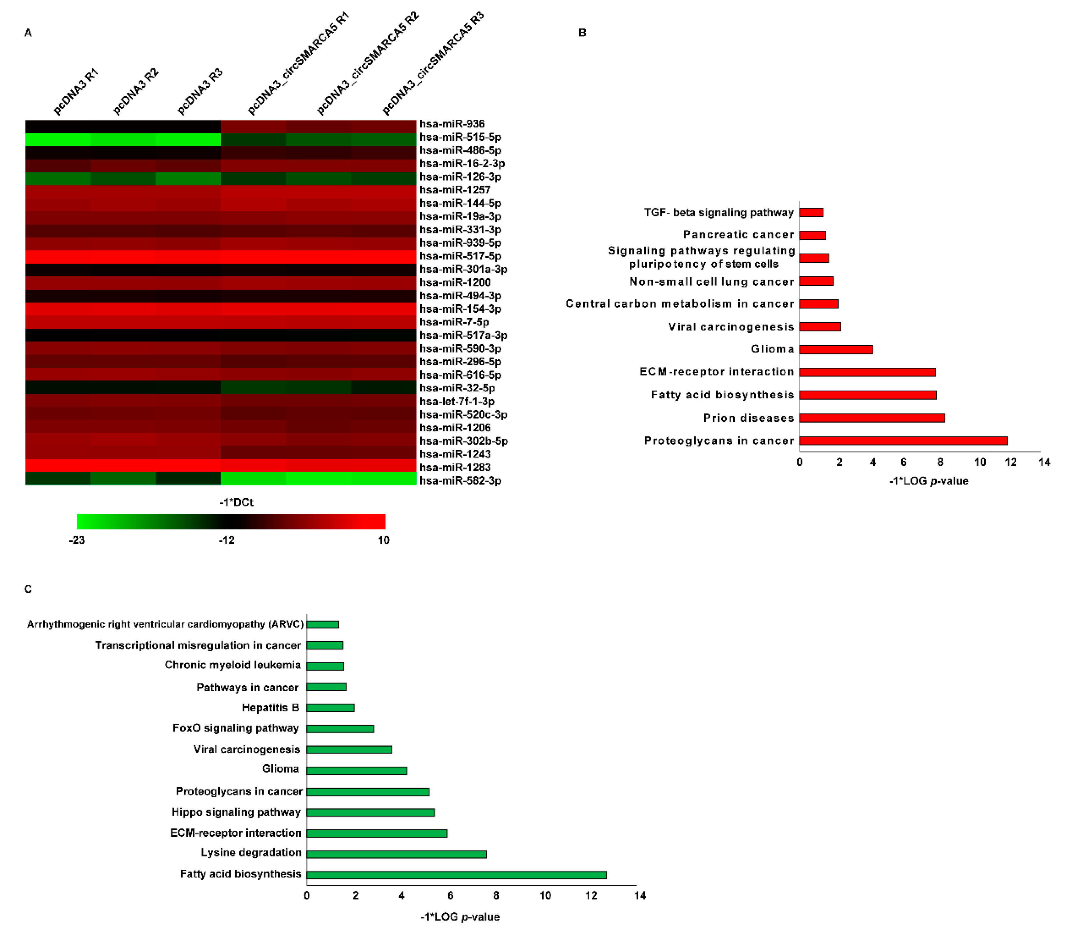
2.1. The miRNAome Expression Profile Is Dysregulated upon circSMARCA5 Overexpression in U87MG

To check for circSMARCA5-mediated regulation of the microRNAome in GBM cells, the expression of 748 miRNAs was assayed in U87MG transfected for 24 h with the plasmid vector expressing circSMARCA5 or with an empty pcDNA3.1 vector (NC) through real-time PCR, by using two different sets of TaqMan® Array MicroRNA Cards (A and B) (see Section 4). As shown in Figure 1A and Figure S1, a total of 11 and 17 miRNAs were differentially expressed (DE) between U87MG overexpressing circSMARCA5 and NCs, in Cards A and B, respectively. Among DE miRNAs, 15 were upregulated and 13 downregulated in U87MG overexpressing circSMARCA5 as compared to NCs. Analysis performed with DIANA miRPath showed an involvement of DE miRNAs in biological processes (BPs) and pathways related to glioma (Figure 1B,C; Table S1).

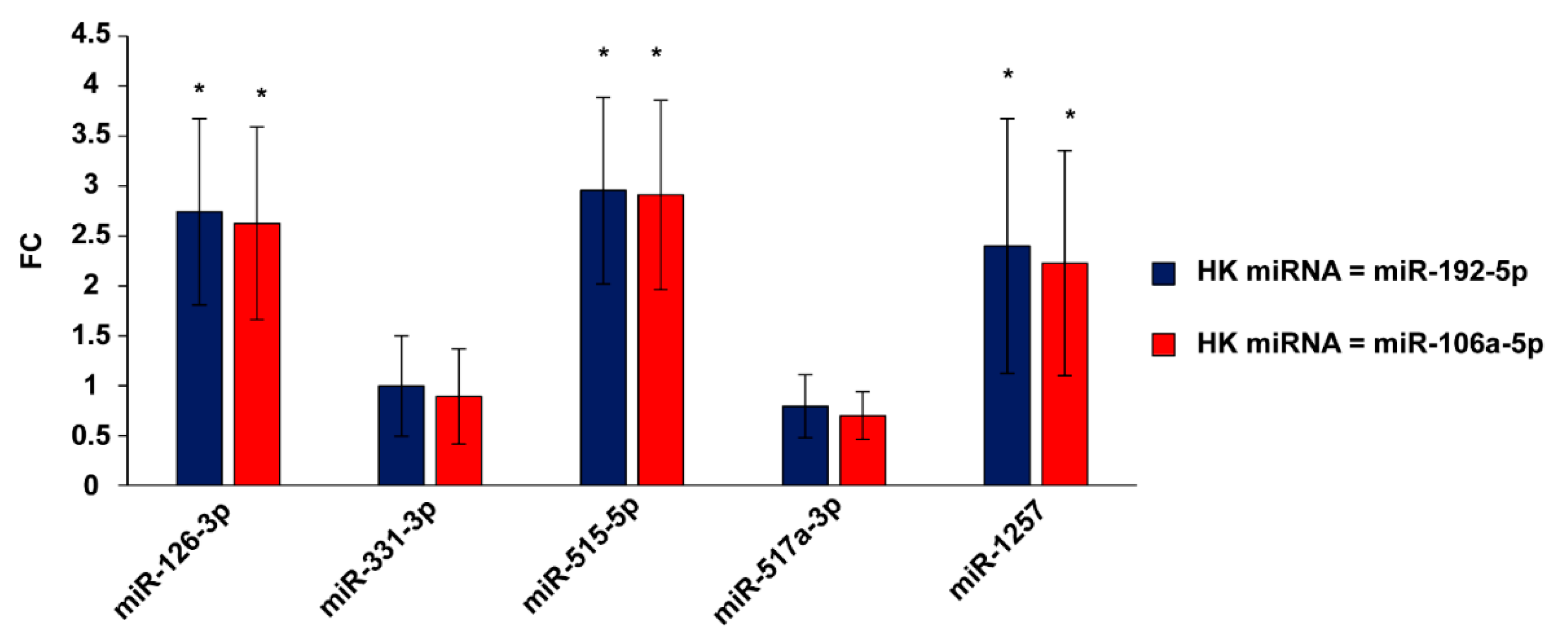
2.2. miRNAs 126-3p, 515-5p, and 1257 Are Upregulated upon circSMARCA5 Overexpression in U87MG

Based on our previous characterization of circSMARCA5 as TS in GBM, we focused on DE miRNAs that showed, upon circSMARC5 overexpression: (i) a fold-change (FC) of at least 1.5 and (ii) an upregulation or downregulation associated with a known TS or oncogenic function in GBM (or other neoplasia), respectively. These criteria allowed us to filter six DE miRNAs (five upregulated TS RNAs (miR-126-3p, miR-144-5p, miR-331-3p, miR-515-5p, and miR-1257) and one downregulated onco-miRNA (miR-517a-3p)) to be further analysed (Table 1).
Upregulation of miRNAs 126-3p, 515-5p, and 1257 in U87MG overexpressing circSMARCA5 was validated through single TaqMan® assays; miR-144-5p was not detected in any sample following real-time PCR; expression of miR-331-3p and miR-517a-3p did not significantly change in U87MG overexpressing circSMARCA5 as compared to NCs (Figure 2).

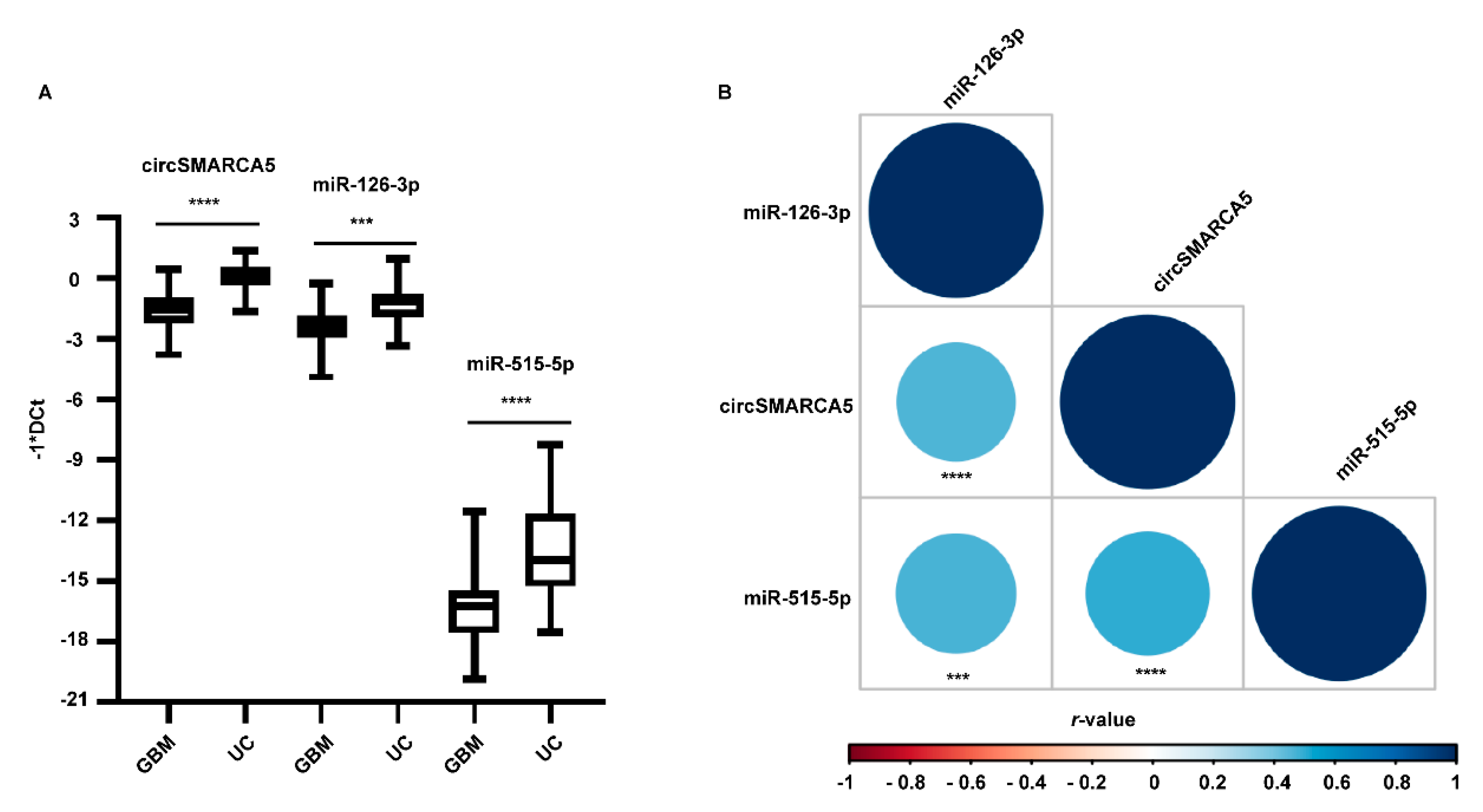
2.3. miRNAs 126-3p and 515-5p Are Downregulated in GBM Biopsies and Their Expression Positively Correlates with That of circSMARCA5

To further analyse the correlation between circSMARCA5 and miRNAs 126-3p, 515-5p, and 1257, their expression was initially assayed in a training set cohort made up of five GBM and five UC samples. As shown in Figure S2, miRNAs 126-3p and 515-5p were significantly downregulated, while miR-1257 was upregulated in GBM vs. unaffected brain parenchyma (UC). The expression of miR-126-3p, miR-515-5p, and circSMARCA5 was then assayed in a validation set cohort made up of 38 GBM and 21 UC samples: circSMARCA5, miR-126-3p, and miR-515-5p were confirmed as downregulated in GBM biopsies, as compared to UCs (FC = −2.32, −1.59, and −2.82, p-values = 9.34 × 10⁻⁹, 7.9 × 10⁻⁴ and 2.97 × 10⁻⁶, Student’s t-test, respectively) (Figure 3A). MiR-126-3p and miR-515-5p were also positively correlated with circSMARCA5 (r-values = 0.49 and 0.50, p-values = 9 × 10⁻⁵ and 7 × 10⁻⁵, Spearman’s correlation test, respectively) (Figure 3B) and downregulated in all the GBM cell lines used in this study, as compared to brain cells from healthy donors (Figure S3).

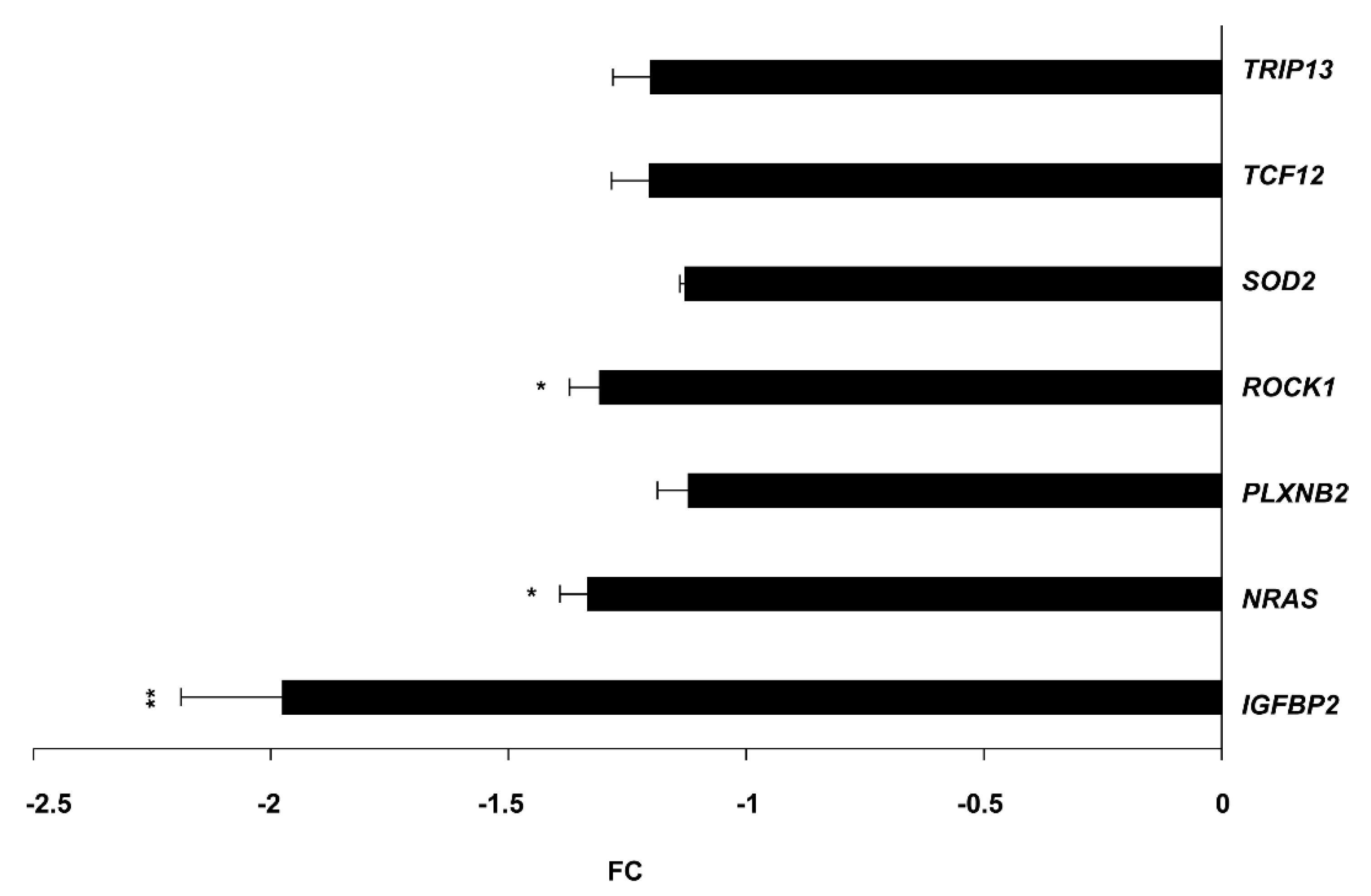
2.4. IGFBP2 (Target of miR-126-3p), NRAS and ROCK1 (Targets of miR-515-5p) mRNAs Are Downregulated upon circSMARCA5 Overexpression in U87MG

To widen our knowledge of the identified circSMARCA5/miR-126-3p/miR-515-5p axis, we searched for the targets of the two candidate miRNAs. A manual literature search allowed us to initially select 17 validated targets (9 of miR-126-3p and 8 of miR-515-5p) in GBM or other neoplasias (Table S2). This first selection was followed by the analysis of the Tumor Cancer Gene Atlas (TCGA), whose data were retrieved from the University of Alabama Cancer Database (UALCAN) to search for upregulated targets in GBM as compared to UCs. Based on TCGA data, we focused on eight mRNA targets (IGFBP2, NRAS, PLXNB2, ROCK1, SOD2, TCF12, TRIP13, and VCAM1) (Table 2), whose expression was first assayed in U87MG overexpressing circSMARCA5. Among the assayed targets, IGFBP2, NRAS, and ROCK1 mRNAs were significantly downregulated (FC IGFBP2, NRAS, ROCK1 = −1.97, −1.33, and −1.31, respectively) (Figure 4).

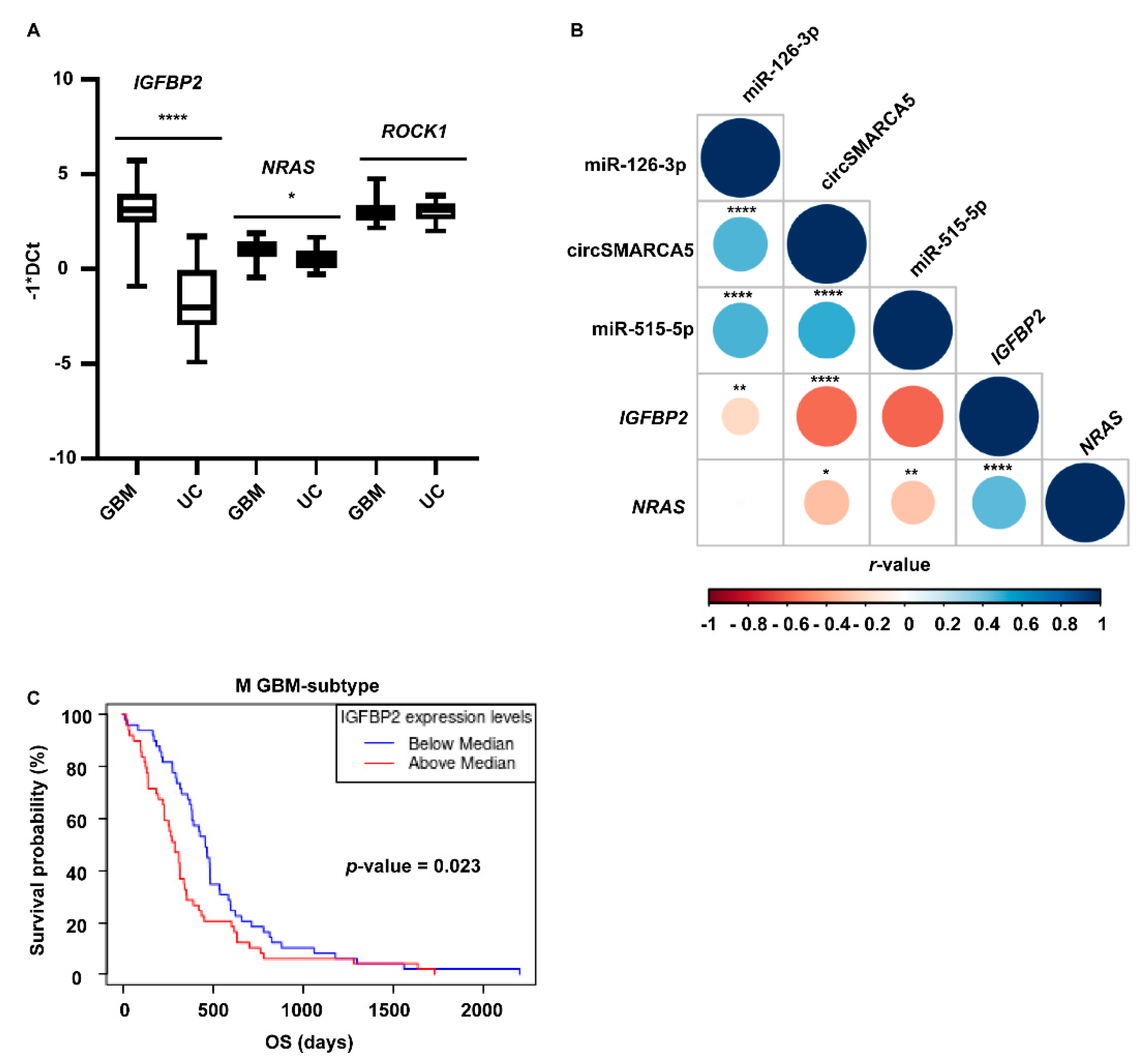
2.5. IGFBP2 and NRAS mRNAs Are Upregulated in GBM Biopsies and Cell Lines with Respect to UCs and Their Expression Negatively Correlates with That of circSMARCA5

The expression of IGFBP2, NRAS, and ROCK1 mRNAs was then assayed in the same validation set cohort (made of 38 GBM and 21 UC samples) used to verify the differential expression of candidate DE miRNAs. IGFBP2 and NRAS were upregulated in GBM biopsies and cell lines with respect to UCs (Figure 5A and Figure S4) and their expression negatively correlated with that of circSMARCA5 (r-values = −0.58 and −0.30, p-values = 0 and 0.019, Spearman’s correlation test, respectively), and with that of their negative regulators, miR-126-3p and miR-515-5p, respectively (r-values = −0.36 and −0.34, p-values = 0.0066 and 0.010, Spearman’s correlation test, respectively) (Figure 5B). IGFBP2 mRNA and protein were significantly upregulated in classical (C) and mesenchymal (M) GBM subtypes, when compared to the proneural (P) and neural (N) ones, based on TCGA data (Figures S5 and S6); NRAS mRNA was, instead, significantly upregulated in the N GBM subtype when compared to the other subtypes (Figure S7). These data are in agreement with the worst prognosis of M-subtype patients showing a higher IGFBP2 mRNA expression (p-value = 0.023, Kaplan–Meier survival curve comparison) (Figure 5C). IGFBP2 and NRAS mRNAs were positively correlated based on our dataset (r-value = 0.46, p-value = 0.00027) (Figure 5B), and GBM TCGA and normal brain cortex GTEx gene expression data (r-value = 0.76, p-value = 4.5 × 10−52) (Figure S8). To extend the pathway downstream to IGFBP2, we assayed the expression of the vascular endothelial growth factor A (VEGFA) mRNA (whose transcription is known to be activated by IGFBP2 protein that acts as an enhancer on its promoter—see Section 3) in (i) U87MG overexpressing circSMARCA5 and (ii) in the same validation set cohort used to verify the differential expression of IGBP2 mRNA. VEGFA mRNA was significantly downregulated in U87MG upon circSMARCA5 overexpression and its expression positively correlated with that of IGFBP2 in the validation set cohort used in this study (Figure S9).

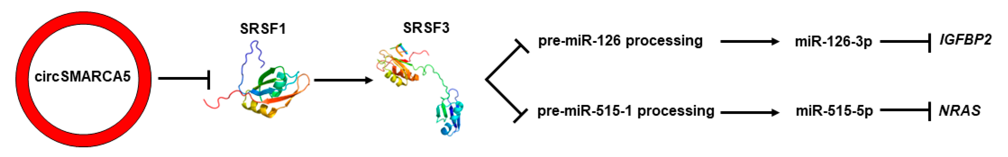
2.6. Serine- and Arginine-Rich Splicing Factor 3 (SRSF3) Is Predicted to Bind Pre-miRNAs 126 and 515-1

To search for RNA motifs common to pre-miRNAs 126 (precursor of miR-126-3p) and 515-1 (precursor of miR-515-5p), their sequences were given as input to the MEME tool. Two motifs, CUUCAA and CUCCAA, were identified within pre-miR-126 and pre-mir-515-1 sequences, respectively (p-values = 4.72 × 10⁻⁴ and 2.12 × 10⁻⁴, respectively). Both motifs have been validated by other researchers to be bound by SRSF3, a known post-transcriptional regulator of miRNA processing (Table 3), and occur in the apical loop and the stem regions of pre-miRNAs 126 and 515-1 hairpins, respectively, according to the RNAStructure tool’s prediction (Figure S10).

3. Discussion

The pathway leading to the expression of miRNAs is very complex and tightly regulated by several factors at different steps [64]. Altered expression of the microRNAome in GBM has been extensively studied, although the molecular mechanisms steering specific miRNA dysregulation have been elucidated only in a few cases [65,66,67]. Numerous RBPs play a crucial role in the post-transcriptional processing of pri-miRNAs and pre-miRNAs [68]: among them, the splicing factor SRSF1 has been demonstrated to regulate the expression of several mature miRNAs by a cross-talk with the enzymes involved in the processing of their precursors, through mechanisms that have been only partially explained to date [69]. The interplay between circRNAs and miRNAs has been mainly described as a ceRNA network, in which circRNAs, including circSMARCA5, act as sponges for miRNAs [24,25,26,27,70,71,72,73,74]; however, circRNAs have not been yet reported as upstream regulators of miRNA expression, to the best of our knowledge. Because of their role as decoys for several RBPs, here we hypothesize that circRNAs may act as upstream epigenetic regulators of the miRNAome inside cells. In this work, we specifically investigated the circSMARCA5-mediated regulation of the miRNAome in GBM cells. We previously characterized circSMARCA5 as a TS circRNA in GBM and we demonstrated that it performs its function by sponging the RBP SRSF1 [22,23,75]. Our data ascertained that circSMARCA5 plays a role in the control of the miRNA expression inside GBM cells and that several dysregulated miRNAs upon circSMARCA5 overexpression are involved in glioma pathways. Further investigation led us to focus on miRNAs 126-3p and 515-5p: (i) both were upregulated in U87MG upon circSMARCA5 overexpression; (ii) their expression was positively correlated with that of circSMARCA5; (iii) they have been characterized as TS in GBM and additional cancers by other scholars [45,46,47,48,49,54,55,56,76,77,78,79,80,81,82,83,84,85,86,87]. CircSMARCA5-mediated upstream control of miRNAs 126-3p and 515-5p is also supported by: (i) the observed downregulation of their two selected mRNA targets, IGFBP2 and NRAS, upon circSMARCA5 overexpression; (ii) positive correlation between the expression of IGFBP2 and NRAS mRNAs; (iii) negative correlation between the expression of IGFBP2 and NRAS mRNAs and circSMARCA5. CircSMARCA5 is also functionally linked to miRNAs 126-3p, 515-5p and their targets; indeed, similar to circSMARCA5 [21,22,23], miR-126-3p and its target IGFBP2 are involved in GBM progression, by regulating cell migration, invasion [45,88,89,90], and angiogenesis [91,92]. To deepen the knowledge of the latter molecular axis, we also investigated the expression of VEGFA mRNA, both in U87MG overexpressing circSMARCA5 and in the validation cohort of GBM and UC biopsies. We previously showed that circSMARCA5 affects the ratio between pro- and anti-angiogenic isoforms of VEGFA mRNA in GBM by regulating alternative splicing of VEGFA pre-mRNA, tethering the splicing factor SRSF1 [22]. Here we showed that the amount of pan-VEGFA mRNA decreased in U87MG upon circSMARCA5 overexpression and that VEGFA and IGFBP2 mRNAs were positively correlated. Unless here we focused on an axis involving (non-coding and coding) RNA molecules, data obtained on the expression of VEGFA mRNA suggest that IGFBP2 may be upstream regulated by circSMARCA5 also at the protein level: indeed, IGFBP2 was described as an enhancer for the transcription of VEGFA in neuroblastoma cells [93] and IGFBP2 and VEGFA were shown to be positively correlated at the protein level in GBM tissues [94]. MiR-126-3p can be also carried in biological fluids through EVs [95,96]: it would be interesting to investigate if and how the delivery of this molecule to cells at different sites from the bulk tumor play a role in the cancer progression and resistance to the current therapies. MiR-515-5p and its target NRAS are also known to be involved in GBM progression by regulating cell migration, growth [53,54,55,56,97], and angiogenesis [98,99]. In an attempt to find a link between the upstream regulator circSMARCA5 and the downstream-regulated intronic miRNAs 126-3p and 515-5p, we also searched for RBPs that may commonly bind and, potentially, regulate pre-miR-126 and pre-miR-515 processing. Our prediction allowed us to identify the splicing factor SRSF3 as an RBP that potentially binds both pre-miRNAs. We previously found that SRSF3 splicing is regulated by SRSF1 and, indirectly, by circSMARCA5 in GBM cells, where the pro-oncogenic full-length functional SRSF3 mRNA isoform is overexpressed when compared to the truncated non-functional one [21]. SRSF3 has been described as a direct or indirect positive regulator of the processing of several pri-miRNAs such as pri-miRNAs 30a, 142, and miR-132/212, by interacting with a CNNC motif, recruiting DROSHA to the cleavage site, and enhancing the Microprocessor activity [100,101]. Based on our prediction, SRSF3 would interact with different motifs other than CNNC on pre-miRNA 126 and 515 sequences, paving the way to alternative mechanisms of SRSF3-mediated pri- and, eventually, pre-miRNA processing. Most specifically, based on our data, we speculate that in a GBM cell context and in particular for pre-miRNAs 126 and 515-1 processing, SRSF3 may function as a negative regulator (Figure 6). As previously reported for the RBP RNA binding fox-1 homolog 3 (RBFOX3), the same RBP can stimulate or block the processing of individual pri- or pre-miRNAs depending on the cell context and the specific miRNA precursor structure [102].
Collectively, our data suggest circSMARCA5 as an upstream regulator of the expression of TS miRNAs 126-3p and 515-5p and their downstream targets IGFBP2 and NRAS mRNA in GBM cells, extending our knowledge on the disrupted tumor suppressive pathways mediated by circSMARCA5 in GBM cells. Prospectively, these pathways may be considered for targeted molecular therapeutic approaches, especially by using recently discovered genomic editing techniques [103].

4. Materials and Methods

4.1. Cell Lines and Biopsies

GBM cell lines A172, CAS-1, SNB-19, U251MG, and U87MG were cultured as described in the supplementary materials and methods. All cell lines were purchased from the Interlab Cell Line Collection (ICLC), located at the IRCCS Ospedale Policlinico San Martino, Genova, Italy. Cell lines were used between the 5th and 10th passage and their viability was assessed through the Trypan Blue Exclusion Test (ThermoFisher Scientific, Waltham, MA, USA) before each experiment, according to the protocol described by W Strober [104]. In total, 38 GBM and 21 UC biopsies were obtained, characterized by pathologists, and stored until their processing, as previously described [22]. Informed consent was signed by the patients before surgery. Demographic data of the patients enrolled in this study are summarized in Table S3. The entire study was performed according to the Declaration of Helsinki and approved by the local ethical Committee of the Azienda Ospedaliero-Universitaria “Policlinico-Vittorio Emanuele”, Catania, Italy (project identification code: 166/2015/PO, 17 December 2015).

4.2. Cell Transfection

U87MG cells were transfected by using lipofectamine 2000 (Thermofisher Scientific, Waltham, MA, USA), as previously described [21]. Briefly, 5 × 10⁴ cells were seeded in a 24-well plate, cultured for 24 h, and transfected with 500 ng of NC or the vector expressing circSMARCA5 (pcDNA3.1_circSMARCA5) for 24 h, according to the manufacturer’s instructions. Three replicates for each experimental condition were carried out and analysed accordingly. Data on circSMARCA5 overexpression upon transfection of U87MG are reported in Figure S11.

4.3. RNA Extraction

Total RNA was isolated through Trizol™ (Thermofisher Scientific, Waltham, MA, USA) in accordance with the manufacturer’s instructions and quantified by spectrophotometry, as previously described [105]. FirstChoice® Human Brain Reference RNA (Ambion, Austin, TX, USA) was used as an additional UC.

4.4. microRNA TaqMan® Arrays

MicroRNA TaqMan® Arrays (Thermofisher Scientific, Waltham, MA, USA) were performed as previously described [106]. Briefly, 300 ng of total RNA isolated from each of the three biological replicates of U87MG, transfected for 24 h with the vector pcDNA3.1_circSMARCA5 or with NC, were reverse-transcribed into specific cDNAs of 748 microRNAs using the TaqMan™ MicroRNA Reverse Transcription Kit (ThermoFisher Scientific, Waltham, MA, USA) and the Megaplex™ RT Primers Human Pool A v 2.1 and Pool B v 3.0 (ThermoFisher Scientific, Waltham, MA, USA). The products of Megaplex™ reactions were then pre-amplified using the following kits: Megaplex™ PreAmp Primers, Human Pool A v. 2.1, and Human Pool B v. 3.0 (ThermoFisher Scientific, Waltham, MA, USA), according to the manufacturer’s instructions. The Megaplex™ PreAmp product of each sample was then loaded into an independent TaqMan® Array MicroRNA Card A (in the case of samples reverse-transcribed and pre-amplified with pool A primers) or B (with samples reverse-transcribed and pre-amplified with pool B primers) (ThermoFisher Scientific, Waltham, MA, USA). PCR was performed using a QuantStudio™ 5 Real-Time PCR System (ThermoFisher Scientific, Waltham, MA, USA) (Figure S12).

4.5. Array Data Analysis

EDS files generated by the run of microRNA TaqMan® Arrays were imported into the dashboard of a ThermoFisher cloud (https://apps.thermofisher.com/apps/spa/#/dashboard, accessed on November 2021) and then analysed through relative quantification application. Cycle thresholds (Cts) were then calculated by the software and exported in a CSV file. Data from Cards A and B were analysed independently. Briefly, miRNAs that showed Cts > 35 in all the experimental conditions were considered too late and filtered out from data analysis. Correlations between the mean or median Ct value of each card and the Ct value of each miRNA were calculated to select candidate housekeeping (HK) miRNAs. A selection of 15 and 3 candidate HK miRNAs, among those showing the strongest correlation with the mean and median Ct values, were given as input to RefFinder (http://blooge.cn/RefFinder/?type=reference, accessed on November 2021) to select the best reference miRNAs within Cards A and B, respectively (Table S4). Reference miRNAs (miR-192-5p and miR-106a-5p for Card A; miR-452-3p and miR-19a-5p for Card B) were used to obtain DCts, (Ct of the transcript of interest—Ct of the reference transcript). DCts of 190 and 58 miRNAs were given as input to the MeV (Multiple Experiment Viewer) tool v. 4.7.1 to retrieve significant DE miRNAs within Cards A and B, respectively. A graphical representation of microRNA TaqMan® Arrays’ data analysis is reported in Figure S12.

4.6. Real-Time PCR

DE miRNAs were validated through single TaqMan™ microRNA assays. Briefly, 30 ng of total RNA were reverse transcribed through the TaqMan™ microRNA Reverse Transcription Kit (ThermoFisher Scientific, Waltham, MA, USA) by using miRNA-specific primers and then amplified through the TaqMan™ Universal Master Mix II (ThermoFisher Scientific, Waltham, MA, USA) by using specific TaqMan™ assays, according to the manufacturer’s instructions. Messenger RNAs of candidate miRNA targets were amplified by using the Power SYBR™ Green RNA-to-Ct™ 1-Step Kit (ThermoFisher Scientific, Waltham, MA, USA). PCRs were run in a QuantStudio™ 5 Real-Time PCR System (ThermoFisher Scientific, Waltham, MA, USA). Real-time PCR data were represented as −1*DCts, FC, or log FC within the text (see supplementary methods for further explanation). The list of TaqMan™ assays and primers used in this study is shown in Table S5.

4.7. In Silico Analyses

BPs and pathways regulated by DE miRNAs were retrieved through DIANA miRPath 3.0 [107]: validated targets stored in TarBase v. 7.0 were selected to calculate BP and pathway enrichment. The expression of miRNA targets from the GBM TCGA dataset was retrieved through the UALCAN, GBM-BioDP, and Gene Expression Profiling Interactive Analysis (GEPIA) databases [108,109,110]. Multiple Em for Motif Elicitation (MEME) suite v. 5.4.1 [111] was used to retrieve RNA motifs within pre-miRNA sequences, using default parameters. ATtRACT database v. 0.99β [112] identified RBPs validated to bind specific RNA motifs. The RNA Structure tool [113] was used to calculate and visualize the secondary structures common to pre-miRNA sequences.

4.8. Statistical Analyses

Pearson’s and Spearman’s correlation tests were used to calculate correlations between the mean or median Ct value of each card and the Ct value of each miRNA, in order to select candidate housekeeping (HK) miRNAs. Only miRNAs showing correlation coefficients (r-values) ≥ 0.8 and p-value < 0.05 were considered as candidate DE miRNAs to be given as input to RefFinder. DE miRNAs were calculated through the Significance Analysis of Microarray (SAM) method within MeV tool v. 4.7.1 [114]. Only miRNAs reporting q-values = 0 were considered DE. Correlation tests and statistical significance were performed and calculated through GraphPad Prism v. 8.0.2. Student’s t-test was used to identify DE miRNAs and targets after single real-time PCR assays; for this, p-values < 0.05 were considered significant.

Supplementary Materials

The following supporting information can be downloaded at: https://0-www-mdpi-com.brum.beds.ac.uk/article/10.3390/ijms232213676/s1. References [47,53,54,55,56,115,116,117,118,119,120,121,122] are cited in the supplementary materials.

Author Contributions

Conceptualization, D.B.; methodology, D.B., A.E.M. and M.S.; validation, C.B., R.B. and A.C.; formal analysis, D.B., A.E.M. and M.S.; investigation, D.B. and A.E.M.; resources, G.M.V.B., R.A., F.C., R.C. and G.B.; data curation, D.B., M.R. and C.D.P.; writing—original draft preparation, D.B. and M.P.; writing—review and editing, D.B. and M.P.; visualization, D.B.; supervision, M.P.; funding acquisition, D.B. and M.P. All authors have read and agreed to the published version of the manuscript.

Funding

This research has been partly funded by “PIAno di inCEntivi per la Ricerca (PIA.CE.RI.) di Ateneo 2020/2022”—“linea di intervento 3, STARTING GRANT” (Project: “Multifaceted Epigenetic Landscape of CircSMARCA5 in Glioblastoma Multiforme” (EpiCGli)) from the University of Catania (Prot. n. 0306492, 29 March 2021) and by “Fondi di ateneo 2020/2022, Università di Catania, linea Open Access”.

Institutional Review Board Statement

The study was conducted in accordance with the Declaration of Helsinki, and approved by the local ethical Committee of the Azienda Ospedaliero-Universitaria “Policlini-co-Vittorio Emanuele”, Catania, Italy (project identification code: 166/2015/PO, 17 December 2015).

Informed Consent Statement

Informed consent was obtained from all subjects involved in the study. Written informed consent was obtained from the patients to publish this paper.

Data Availability Statement

The datasets used and/or analysed during the current study are available from the corresponding author upon reasonable request.

Acknowledgments

We thank Thomas Birkballe Hansen for hosting Davide Barbagallo and Angela Caponnetto in his lab and for sharing with us the pcDNA3_circSMARCA5 expression vector. We acknowledge the interesting discussions with Massimo Romani and his Collaborators at IST, Genova, and we thank Noemi Zuccaro and Andrea Giuseppe Toscano (two students of the bachelor’s degree course in Biotechnology, at the University of Catania) for their assistance in this work. This study was supported by “PIAno di inCEntivi per la Ricerca (PIA.CE.RI.) di Ateneo 2020/2022”—“linea di intervento 3, STARTING GRANT” (Project: “Multifaceted Epigenetic Landscape of CircSMARCA5 in Glioblastoma Multiforme” (EpiCGli)) from the University of Catania and by “Fondi di ateneo 2020/2022, Università di Catania, linea Open Access”.

Conflicts of Interest

The authors declare no conflict of interest. The funders had no role in the design of the study; the collection, analyses, or interpretation of data; the writing of the manuscript; or the decision to publish the results.

References

  1. Ostrom, Q.T.; Cioffi, G.; Waite, K.; Kruchko, C.; Barnholtz-Sloan, J.S. CBTRUS Statistical Report: Primary Brain and Other Central Nervous System Tumors Diagnosed in the United States in 2014–2018. Neuro-Oncology 2021, 23, iii1–iii105. [Google Scholar] [CrossRef] [PubMed]
  2. Louis, D.N.; Perry, A.; Wesseling, P.; Brat, D.J.; Cree, I.A.; Figarella-Branger, D.; Hawkins, C.; Ng, H.K.; Pfister, S.M.; Reifenberger, G.; et al. The 2021 WHO Classification of Tumors of the Central Nervous System: A summary. Neuro-Oncology 2021, 23, 1231–1251. [Google Scholar] [CrossRef] [PubMed]
  3. The Cancer Genome Atlas Research Network. Comprehensive genomic characterization defines human glioblastoma genes and core pathways. Nature 2008, 455, 1061–1068. [CrossRef] [PubMed] [Green Version]
  4. Verhaak, R.G.; Hoadley, K.A.; Purdom, E.; Wang, V.; Qi, Y.; Wilkerson, M.D.; Miller, C.R.; Ding, L.; Golub, T.; Mesirov, J.P.; et al. Integrated genomic analysis identifies clinically relevant subtypes of glioblastoma characterized by abnormalities in PDGFRA, IDH1, EGFR, and NF1. Cancer Cell 2010, 17, 98–110. [Google Scholar] [CrossRef] [Green Version]
  5. Kim, T.M.; Huang, W.; Park, R.; Park, P.J.; Johnson, M.D. A developmental taxonomy of glioblastoma defined and maintained by MicroRNAs. Cancer Res. 2011, 71, 3387–3399. [Google Scholar] [CrossRef] [Green Version]
  6. Altieri, R.; Barbagallo, D.; Certo, F.; Broggi, G.; Ragusa, M.; Di Pietro, C.; Caltabiano, R.; Magro, G.; Peschillo, S.; Purrello, M.; et al. Peritumoral Microenvironment in High-Grade Gliomas: From FLAIRectomy to Microglia-Glioma Cross-Talk. Brain Sci. 2021, 11, 200. [Google Scholar] [CrossRef]
  7. Hansen, T.B.; Jensen, T.I.; Clausen, B.H.; Bramsen, J.B.; Finsen, B.; Damgaard, C.K.; Kjems, J. Natural RNA circles function as efficient microRNA sponges. Nature 2013, 495, 384–388. [Google Scholar] [CrossRef]
  8. Memczak, S.; Jens, M.; Elefsinioti, A.; Torti, F.; Krueger, J.; Rybak, A.; Maier, L.; Mackowiak, S.D.; Gregersen, L.H.; Munschauer, M.; et al. Circular RNAs are a large class of animal RNAs with regulatory potency. Nature 2013, 495, 333–338. [Google Scholar] [CrossRef]
  9. Kristensen, L.S.; Andersen, M.S.; Stagsted, L.V.W.; Ebbesen, K.K.; Hansen, T.B.; Kjems, J. The biogenesis, biology and characterization of circular RNAs. Nature reviews. Genetics 2019, 20, 675–691. [Google Scholar] [CrossRef]
  10. Wang, P.L.; Bao, Y.; Yee, M.C.; Barrett, S.P.; Hogan, G.J.; Olsen, M.N.; Dinneny, J.R.; Brown, P.O.; Salzman, J. Circular RNA is expressed across the eukaryotic tree of life. PLoS ONE 2014, 9, e90859. [Google Scholar] [CrossRef]
  11. Danan, M.; Schwartz, S.; Edelheit, S.; Sorek, R. Transcriptome-wide discovery of circular RNAs in Archaea. Nucleic Acids Res. 2012, 40, 3131–3142. [Google Scholar] [CrossRef] [PubMed]
  12. Rybak-Wolf, A.; Stottmeister, C.; Glažar, P.; Jens, M.; Pino, N.; Giusti, S.; Hanan, M.; Behm, M.; Bartok, O.; Ashwal-Fluss, R.; et al. Circular RNAs in the Mammalian Brain Are Highly Abundant, Conserved, and Dynamically Expressed. Mol. Cell 2015, 58, 870–885. [Google Scholar] [CrossRef] [PubMed] [Green Version]
  13. Suzuki, H.; Tsukahara, T. A view of pre-mRNA splicing from RNase R resistant RNAs. Int. J. Mol. Sci. 2014, 15, 9331–9342. [Google Scholar] [CrossRef] [PubMed] [Green Version]
  14. Li, Y.; Zheng, Q.; Bao, C.; Li, S.; Guo, W.; Zhao, J.; Chen, D.; Gu, J.; He, X.; Huang, S. Circular RNA is enriched and stable in exosomes: A promising biomarker for cancer diagnosis. Cell Res. 2015, 25, 981–984. [Google Scholar] [CrossRef] [PubMed] [Green Version]
  15. Li, Y.; Feng, W.; Kong, M.; Liu, R.; Wu, A.; Shen, L.; Tang, Z.; Wang, F. Exosomal circRNAs: A new star in cancer. Life Sci. 2021, 269, 119039. [Google Scholar] [CrossRef]
  16. Li, S.; Li, Y.; Chen, B.; Zhao, J.; Yu, S.; Tang, Y.; Zheng, Q.; Li, Y.; Wang, P.; He, X.; et al. exoRBase: A database of circRNA, lncRNA and mRNA in human blood exosomes. Nucleic Acids Res. 2018, 46, D106–D112. [Google Scholar] [CrossRef] [PubMed] [Green Version]
  17. Stella, M.; Falzone, L.; Caponnetto, A.; Gattuso, G.; Barbagallo, C.; Battaglia, R.; Mirabella, F.; Broggi, G.; Altieri, R.; Certo, F.; et al. Serum Extracellular Vesicle-Derived circHIPK3 and circSMARCA5 Are Two Novel Diagnostic Biomarkers for Glioblastoma Multiforme. Pharmaceuticals 2021, 14, 618. [Google Scholar] [CrossRef]
  18. Wang, S.; Zhang, K.; Tan, S.; Xin, J.; Yuan, Q.; Xu, H.; Xu, X.; Liang, Q.; Christiani, D.C.; Wang, M.; et al. Circular RNAs in body fluids as cancer biomarkers: The new frontier of liquid biopsies. Mol. Cancer 2021, 20, 13. [Google Scholar] [CrossRef]
  19. Ebbesen, K.K.; Kjems, J.; Hansen, T.B. Circular RNAs: Identification, biogenesis and function. Biochim. Biophys. Acta 2016, 1859, 163–168. [Google Scholar] [CrossRef]
  20. Kristensen, L.S.; Jakobsen, T.; Hager, H.; Kjems, J. The emerging roles of circRNAs in cancer and oncology. Nat. Rev. Clin. Oncol. 2022, 19, 188–206. [Google Scholar] [CrossRef]
  21. Barbagallo, D.; Caponnetto, A.; Cirnigliaro, M.; Brex, D.; Barbagallo, C.; D’Angeli, F.; Morrone, A.; Caltabiano, R.; Barbagallo, G.M.; Ragusa, M.; et al. CircSMARCA5 Inhibits Migration of Glioblastoma Multiforme Cells by Regulating a Molecular Axis Involving Splicing Factors SRSF1/SRSF3/PTB. Int. J. Mol. Sci. 2018, 19, 480. [Google Scholar] [CrossRef] [PubMed] [Green Version]
  22. Barbagallo, D.; Caponnetto, A.; Brex, D.; Mirabella, F.; Barbagallo, C.; Lauretta, G.; Morrone, A.; Certo, F.; Broggi, G.; Caltabiano, R.; et al. CircSMARCA5 Regulates VEGFA mRNA Splicing and Angiogenesis in Glioblastoma Multiforme Through the Binding of SRSF1. Cancers 2019, 11, 194. [Google Scholar] [CrossRef] [PubMed] [Green Version]
  23. Barbagallo, D.; Caponnetto, A.; Barbagallo, C.; Battaglia, R.; Mirabella, F.; Brex, D.; Stella, M.; Broggi, G.; Altieri, R.; Certo, F.; et al. The GAUGAA Motif Is Responsible for the Binding between circSMARCA5 and SRSF1 and Related Downstream Effects on Glioblastoma Multiforme Cell Migration and Angiogenic Potential. Int. J. Mol. Sci. 2021, 22, 1678. [Google Scholar] [CrossRef] [PubMed]
  24. Yang, S.; Gao, S.; Liu, T.; Liu, J.; Zheng, X.; Li, Z. Circular RNA SMARCA5 functions as an anti-tumor candidate in colon cancer by sponging microRNA-552. Cell Cycle 2021, 20, 689–701. [Google Scholar] [CrossRef] [PubMed]
  25. Li, Q.; Tang, H.; Hu, F.; Qin, C. Circular RNA SMARCA5 inhibits gastric cancer progression through targeting the miR-346/ FBXL2 axis. RSC Adv. 2019, 9, 18277–18284. [Google Scholar] [CrossRef] [Green Version]
  26. Liu, H.; Wu, Y.; Wang, S.; Jiang, J.; Zhang, C.; Jiang, Y.; Wang, X.; Hong, L.; Huang, H. Circ-SMARCA5 suppresses progression of multiple myeloma by targeting miR-767-5p. BMC Cancer 2019, 19, 937. [Google Scholar] [CrossRef] [Green Version]
  27. Miao, X.; Xi, Z.; Zhang, Y.; Li, Z.; Huang, L.; Xin, T.; Shen, R.; Wang, T. Circ-SMARCA5 suppresses colorectal cancer progression via downregulating miR-39-3p and upregulating ARID4B. Dig. Liver Dis. 2020, 52, 1494–1502. [Google Scholar] [CrossRef]
  28. Tan, Y.; Zhang, T.; Liang, C. Circular RNA SMARCA5 is overexpressed and promotes cell proliferation, migration as well as invasion while inhibits cell apoptosis in bladder cancer. Transl. Cancer Res. 2019, 8, 1663–1671. [Google Scholar] [CrossRef]
  29. Lee, R.C.; Feinbaum, R.L.; Ambros, V. The C. elegans heterochronic gene lin-4 encodes small RNAs with antisense complementarity to lin-14. Cell 1993, 75, 843–854. [Google Scholar] [CrossRef]
  30. Krol, J.; Loedige, I.; Filipowicz, W. The widespread regulation of microRNA biogenesis, function and decay. Nat. Rev. Genet. 2010, 11, 597–610. [Google Scholar] [CrossRef]
  31. Maugeri, M.; Barbagallo, D.; Barbagallo, C.; Banelli, B.; Di Mauro, S.; Purrello, F.; Magro, G.; Ragusa, M.; Di Pietro, C.; Romani, M.; et al. Altered expression of miRNAs and methylation of their promoters are correlated in neuroblastoma. Oncotarget 2016, 7, 83330–83341. [Google Scholar] [CrossRef] [PubMed] [Green Version]
  32. Barbagallo, C.; Caltabiano, R.; Broggi, G.; Russo, A.; Puzzo, L.; Avitabile, T.; Longo, A.; Reibaldi, M.; Barbagallo, D.; Di Pietro, C.; et al. LncRNA LINC00518 Acts as an Oncogene in Uveal Melanoma by Regulating an RNA-Based Network. Cancers 2020, 12, 3867. [Google Scholar] [CrossRef] [PubMed]
  33. Ragusa, M.; Barbagallo, D.; Chioccarelli, T.; Manfrevola, F.; Cobellis, G.; Di Pietro, C.; Brex, D.; Battaglia, R.; Fasano, S.; Ferraro, B.; et al. CircNAPEPLD is expressed in human and murine spermatozoa and physically interacts with oocyte miRNAs. RNA Biol. 2019, 16, 1237–1248. [Google Scholar] [CrossRef] [PubMed]
  34. Cirnigliaro, M.; Barbagallo, C.; Gulisano, M.; Domini, C.N.; Barone, R.; Barbagallo, D.; Ragusa, M.; Di Pietro, C.; Rizzo, R.; Purrello, M. Expression and Regulatory Network Analysis of miR-140-3p, a New Potential Serum Biomarker for Autism Spectrum Disorder. Front. Mol. Neurosci. 2017, 10, 250. [Google Scholar] [CrossRef] [PubMed] [Green Version]
  35. Russo, A.; Ragusa, M.; Barbagallo, C.; Longo, A.; Avitabile, T.; Uva, M.G.; Bonfiglio, V.; Toro, M.D.; Caltabiano, R.; Mariotti, C.; et al. miRNAs in the vitreous humor of patients affected by idiopathic epiretinal membrane and macular hole. PLoS ONE 2017, 12, e0174297. [Google Scholar] [CrossRef]
  36. Parodi, F.; Carosio, R.; Ragusa, M.; Di Pietro, C.; Maugeri, M.; Barbagallo, D.; Sallustio, F.; Allemanni, G.; Pistillo, M.P.; Casciano, I.; et al. Epigenetic dysregulation in neuroblastoma: A tale of miRNAs and DNA methylation. Biochim. Biophys. Acta 2016, 1859, 1502–1514. [Google Scholar] [CrossRef]
  37. Ragusa, M.; Barbagallo, C.; Brex, D.; Caponnetto, A.; Cirnigliaro, M.; Battaglia, R.; Barbagallo, D.; Di Pietro, C.; Purrello, M. Molecular Crosstalking among Noncoding RNAs: A New Network Layer of Genome Regulation in Cancer. Int. J. Genom. 2017, 2017, 4723193. [Google Scholar] [CrossRef]
  38. Ciafrè, S.A.; Galardi, S.; Mangiola, A.; Ferracin, M.; Liu, C.G.; Sabatino, G.; Negrini, M.; Maira, G.; Croce, C.M.; Farace, M.G. Extensive modulation of a set of microRNAs in primary glioblastoma. Biochem. Biophys. Res. Commun. 2005, 334, 1351–1358. [Google Scholar] [CrossRef]
  39. Barbagallo, D.; Condorelli, A.; Ragusa, M.; Salito, L.; Sammito, M.; Banelli, B.; Caltabiano, R.; Barbagallo, G.; Zappalà, A.; Battaglia, R.; et al. Dysregulated miR-671-5p / CDR1-AS / CDR1 / VSNL1 axis is involved in glioblastoma multiforme. Oncotarget 2016, 7, 4746–4759. [Google Scholar] [CrossRef] [Green Version]
  40. Chan, J.A.; Krichevsky, A.M.; Kosik, K.S. MicroRNA-21 is an antiapoptotic factor in human glioblastoma cells. Cancer Res. 2005, 65, 6029–6033. [Google Scholar] [CrossRef]
  41. Gillies, J.K.; Lorimer, I.A. Regulation of p27Kip1 by miRNA 221/222 in glioblastoma. Cell Cycle 2007, 6, 2005–2009. [Google Scholar] [CrossRef] [PubMed] [Green Version]
  42. Silber, J.; Lim, D.A.; Petritsch, C.; Persson, A.I.; Maunakea, A.K.; Yu, M.; Vandenberg, S.R.; Ginzinger, D.G.; James, C.D.; Costello, J.F.; et al. miR-124 and miR-137 inhibit proliferation of glioblastoma multiforme cells and induce differentiation of brain tumor stem cells. BMC Med. 2008, 6, 14. [Google Scholar] [CrossRef] [PubMed]
  43. Kefas, B.; Godlewski, J.; Comeau, L.; Li, Y.; Abounader, R.; Hawkinson, M.; Lee, J.; Fine, H.; Chiocca, E.A.; Lawler, S.; et al. microRNA-7 inhibits the epidermal growth factor receptor and the Akt pathway and is down-regulated in glioblastoma. Cancer Res. 2008, 68, 3566–3572. [Google Scholar] [CrossRef] [Green Version]
  44. Skog, J.; Würdinger, T.; van Rijn, S.; Meijer, D.H.; Gainche, L.; Sena-Esteves, M.; Curry, W.T., Jr.; Carter, B.S.; Krichevsky, A.M.; Breakefield, X.O. Glioblastoma microvesicles transport RNA and proteins that promote tumour growth and provide diagnostic biomarkers. Nat. Cell Biol. 2008, 10, 1470–1476. [Google Scholar] [CrossRef] [PubMed]
  45. Li, Y.; Li, Y.; Ge, P.; Ma, C. miR-126 Regulates the ERK Pathway via Targeting KRAS to Inhibit the Glioma Cell Proliferation and Invasion. Mol. Neurobiol. 2017, 54, 137–145. [Google Scholar] [CrossRef] [PubMed]
  46. Xu, Y.; Xu, W.; Lu, T.; Dai, Y.; Liang, W. miR-126 affects the invasion and migration of glioma cells through GATA4. Artif. Cells Nanomed. Biotechnol. 2017, 45, 1–7. [Google Scholar] [CrossRef] [PubMed] [Green Version]
  47. Chen, S.R.; Cai, W.P.; Dai, X.J.; Guo, A.S.; Chen, H.P.; Lin, G.S.; Lin, R.S. Research on miR-126 in glioma targeted regulation of PTEN/PI3K/Akt and MDM2-p53 pathways. Eur. Rev. Med. Pharmacol. Sci. 2019, 23, 3461–3470. [Google Scholar] [CrossRef]
  48. Luan, Y.; Zuo, L.; Zhang, S.; Wang, G.; Peng, T. microRNA-126 acts as a tumor suppressor in glioma cells by targeting insulin receptor substrate 1 (IRS-1). Int. J. Clin. Exp. Pathol. 2015, 8, 10345–10354. [Google Scholar]
  49. Han, L.; Liu, H.; Wu, J.; Liu, J. miR-126 Suppresses Invasion and Migration of Malignant Glioma by Targeting Mature T Cell Proliferation 1 (MTCP1). Med. Sci. Monit. Int. Med. J. Exp. Clin. Res. 2018, 24, 6630–6637. [Google Scholar] [CrossRef]
  50. Liu, G.M.; Lu, T.C.; Sun, M.L.; Jia, W.Y.; Ji, X.; Luo, Y.G. Ginsenoside Rd Inhibits Glioblastoma Cell Proliferation by Up-Regulating the Expression of miR-144-5p. Biol. Pharm. Bull 2020, 43, 1534–1541. [Google Scholar] [CrossRef]
  51. Epis, M.R.; Giles, K.M.; Candy, P.A.; Webster, R.J.; Leedman, P.J. miR-331-3p regulates expression of neuropilin-2 in glioblastoma. J. Neurooncol. 2014, 116, 67–75. [Google Scholar] [CrossRef] [PubMed]
  52. Chen, H.H.; Zong, J.; Wang, S.J. LncRNA GAPLINC promotes the growth and metastasis of glioblastoma by sponging miR-331-3p. Eur. Rev. Med. Pharmacol. Sci. 2019, 23, 262–270. [Google Scholar] [CrossRef] [PubMed]
  53. Wang, Z.; Han, Y.; Li, Q.; Wang, B.; Ma, J. LncRNA DLGAP1-AS1 accelerates glioblastoma cell proliferation through targeting miR-515-5p/ROCK1/NFE2L1 axis and activating Wnt signaling pathway. Brain Behav. 2021, 11, e2321. [Google Scholar] [CrossRef] [PubMed]
  54. Zheng, K.; Xie, H.; Wu, W.; Wen, X.; Zeng, Z.; Shi, Y. CircRNA PIP5K1A promotes the progression of glioma through upregulation of the TCF12/PI3K/AKT pathway by sponging miR-515-5p. Cancer Cell Int. 2021, 21, 27. [Google Scholar] [CrossRef]
  55. Zhang, Y.; Zhang, Y.; Wang, S.; Li, Q.; Cao, B.; Huang, B.; Wang, T.; Guo, R.; Liu, N. SP1-induced lncRNA ZFPM2 antisense RNA 1 (ZFPM2-AS1) aggravates glioma progression via the miR-515-5p/Superoxide dismutase 2 (SOD2) axis. Bioengineered 2021, 12, 2299–2310. [Google Scholar] [CrossRef]
  56. Pardo, O.E.; Castellano, L.; Munro, C.E.; Hu, Y.; Mauri, F.; Krell, J.; Lara, R.; Pinho, F.G.; Choudhury, T.; Frampton, A.E.; et al. miR-515-5p controls cancer cell migration through MARK4 regulation. EMBO Rep. 2016, 17, 570–584. [Google Scholar] [CrossRef]
  57. Du, C.L.; Peng, F.; Liu, K.Q. miR-517a is up-regulated in glioma and promotes glioma tumorigenesis in vitro and in vivo. Biosci. Rep. 2019, 39, BSR20181196. [Google Scholar] [CrossRef] [Green Version]
  58. Feng, J.; Kim, S.T.; Liu, W.; Kim, J.W.; Zhang, Z.; Zhu, Y.; Berens, M.; Sun, J.; Xu, J. An integrated analysis of germline and somatic, genetic and epigenetic alterations at 9p21.3 in glioblastoma. Cancer 2012, 118, 232–240. [Google Scholar] [CrossRef]
  59. Zhao, C.; Gao, Y.; Guo, R.; Li, H.; Yang, B. Microarray expression profiles and bioinformatics analysis of mRNAs, lncRNAs, and circRNAs in the secondary temozolomide-resistant glioblastoma. Investig. New Drugs 2020, 38, 1227–1235. [Google Scholar] [CrossRef]
  60. Hisaoka, M.; Matsuyama, A.; Nakamoto, M. Aberrant calreticulin expression is involved in the dedifferentiation of dedifferentiated liposarcoma. Am. J. Pathol. 2012, 180, 2076–2083. [Google Scholar] [CrossRef]
  61. Liu, H.X.; Chew, S.L.; Cartegni, L.; Zhang, M.Q.; Krainer, A.R. Exonic splicing enhancer motif recognized by human SC35 under splicing conditions. Mol. Cell. Biol. 2000, 20, 1063–1071. [Google Scholar] [CrossRef] [PubMed]
  62. Cavaloc, Y.; Bourgeois, C.F.; Kister, L.; Stevenin, J. The splicing factors 9G8 and SRp20 transactivate splicing through different and specific enhancers. RNA 1999, 5, 468–483. [Google Scholar] [CrossRef] [PubMed] [Green Version]
  63. Hargous, Y.; Hautbergue, G.M.; Tintaru, A.M.; Skrisovska, L.; Golovanov, A.P.; Stevenin, J.; Lian, L.Y.; Wilson, S.A.; Allain, F.H. Molecular basis of RNA recognition and TAP binding by the SR proteins SRp20 and 9G8. EMBO J. 2006, 25, 5126–5137. [Google Scholar] [CrossRef] [Green Version]
  64. Tran, N.; Hutvagner, G. Biogenesis and the regulation of the maturation of miRNAs. Essays Biochem. 2013, 54, 17–28. [Google Scholar] [CrossRef] [PubMed] [Green Version]
  65. Chen, Q.; Wang, W.; Chen, S.; Chen, X.; Lin, Y. miR-29a sensitizes the response of glioma cells to temozolomide by modulating the P53/MDM2 feedback loop. Cell. Mol. Biol. Lett. 2021, 26, 21. [Google Scholar] [CrossRef] [PubMed]
  66. Palanichamy, J.K.; Rao, D.S. miRNA dysregulation in cancer: Towards a mechanistic understanding. Front. Genet. 2014, 5, 54. [Google Scholar] [CrossRef]
  67. Croce, C.M. Causes and consequences of microRNA dysregulation in cancer. Nat. Rev. Genet. 2009, 10, 704–714. [Google Scholar] [CrossRef]
  68. Michlewski, G.; Cáceres, J.F. Post-transcriptional control of miRNA biogenesis. RNA 2019, 25, 1–16. [Google Scholar] [CrossRef] [Green Version]
  69. Wu, H.; Sun, S.; Tu, K.; Gao, Y.; Xie, B.; Krainer, A.R.; Zhu, J. A splicing-independent function of SF2/ASF in microRNA processing. Mol. Cell 2010, 38, 67–77. [Google Scholar] [CrossRef] [Green Version]
  70. Ebbesen, K.K.; Hansen, T.B.; Kjems, J. Insights into circular RNA biology. RNA Biol. 2017, 14, 1035–1045. [Google Scholar] [CrossRef] [Green Version]
  71. Salmena, L.; Poliseno, L.; Tay, Y.; Kats, L.; Pandolfi, P.P. A ceRNA hypothesis: The Rosetta Stone of a hidden RNA language? Cell 2011, 146, 353–358. [Google Scholar] [CrossRef] [PubMed]
  72. Tay, Y.; Rinn, J.; Pandolfi, P.P. The multilayered complexity of ceRNA crosstalk and competition. Nature 2014, 505, 344–352. [Google Scholar] [CrossRef] [PubMed] [Green Version]
  73. Barbagallo, D.; Palermo, C.I.; Barbagallo, C.; Battaglia, R.; Caponnetto, A.; Spina, V.; Ragusa, M.; Di Pietro, C.; Scalia, G.; Purrello, M. Competing endogenous RNA network mediated by circ_3205 in SARS-CoV-2 infected cells. Cell. Mol. Life Sci. CMLS 2022, 79, 75. [Google Scholar] [CrossRef] [PubMed]
  74. Dong, C.; Fan, B.; Ren, Z.; Liu, B.; Wang, Y. CircSMARCA5 Facilitates the Progression of Prostate Cancer Through miR-432/PDCD10 Axis. Cancer Biother. Radiopharm. 2021, 36, 70–83. [Google Scholar] [CrossRef] [PubMed]
  75. Broggi, G.; Salvatorelli, L.; Barbagallo, D.; Certo, F.; Altieri, R.; Tirro, E.; Massimino, M.; Vigneri, P.; Guadagno, E.; Maugeri, G.; et al. Diagnostic Utility of the Immunohistochemical Expression of Serine and Arginine Rich Splicing Factor 1 (SRSF1) in the Differential Diagnosis of Adult Gliomas. Cancers 2021, 13, 2086. [Google Scholar] [CrossRef]
  76. Zhang, X.; Zhou, J.; Xue, D.; Li, Z.; Liu, Y.; Dong, L. miR-515-5p acts as a tumor suppressor via targeting TRIP13 in prostate cancer. Int. J. Biol. Macromol. 2019, 129, 227–232. [Google Scholar] [CrossRef]
  77. Mobini, K.; Banakar, E.; Tamaddon, G.; Mohammadi-Bardbori, A. 6-Formylindolo[3,2-b]carbazole (FICZ) Enhances The Expression of Tumor Suppressor miRNAs, miR-22, miR-515-5p, and miR-124-3p in MCF-7 Cells. Cell J. 2020, 22, 115–120. [Google Scholar] [CrossRef]
  78. Wen, L.J.; Wang, Y.S.; Tan, P.Y. miR-515-5p inhibits the proliferation, migration and invasion of human breast cancer cells by targeting CBX4. Exp. Ther. Med. 2021, 22, 1328. [Google Scholar] [CrossRef]
  79. Li, J.; Tang, Z.; Wang, H.; Wu, W.; Zhou, F.; Ke, H.; Lu, W.; Zhang, S.; Zhang, Y.; Yang, S.; et al. CXCL6 promotes non-small cell lung cancer cell survival and metastasis via down-regulation of miR-515-5p. Biomed. Pharmacother. Biomed. Pharmacother. 2018, 97, 1182–1188. [Google Scholar] [CrossRef]
  80. Pinho, F.G.; Frampton, A.E.; Nunes, J.; Krell, J.; Alshaker, H.; Jacob, J.; Pellegrino, L.; Roca-Alonso, L.; de Giorgio, A.; Harding, V.; et al. Downregulation of microRNA-515-5p by the estrogen receptor modulates sphingosine kinase 1 and breast cancer cell proliferation. Cancer Res. 2013, 73, 5936–5948. [Google Scholar] [CrossRef] [Green Version]
  81. Gilam, A.; Edry, L.; Mamluk-Morag, E.; Bar-Ilan, D.; Avivi, C.; Golan, D.; Laitman, Y.; Barshack, I.; Friedman, E.; Shomron, N. Involvement of IGF-1R regulation by miR-515-5p modifies breast cancer risk among BRCA1 carriers. Breast Cancer Res. Treat. 2013, 138, 753–760. [Google Scholar] [CrossRef]
  82. Xiong, Y.; Kotian, S.; Zeiger, M.A.; Zhang, L.; Kebebew, E. miR-126-3p Inhibits Thyroid Cancer Cell Growth and Metastasis, and Is Associated with Aggressive Thyroid Cancer. PLoS ONE 2015, 10, e0130496. [Google Scholar] [CrossRef] [Green Version]
  83. Sibilano, M.; Tullio, V.; Adorno, G.; Savini, I.; Gasperi, V.; Catani, M.V. Platelet-Derived miR-126-3p Directly Targets AKT2 and Exerts Anti-Tumor Effects in Breast Cancer Cells: Further Insights in Platelet-Cancer Interplay. Int. J. Mol. Sci. 2022, 23, 5484. [Google Scholar] [CrossRef]
  84. Hong, Z.; Hong, C.; Ma, B.; Wang, Q.; Zhang, X.; Li, L.; Wang, C.; Chen, D. microRNA-126-3p inhibits the proliferation, migration, invasion, and angiogenesis of triple-negative breast cancer cells by targeting RGS3. Oncol. Rep. 2019, 42, 1569–1579. [Google Scholar] [CrossRef] [PubMed]
  85. Liu, W.; Chen, H.; Wong, N.; Haynes, W.; Baker, C.M.; Wang, X. Pseudohypoxia induced by miR-126 deactivation promotes migration and therapeutic resistance in renal cell carcinoma. Cancer Lett. 2017, 394, 65–75. [Google Scholar] [CrossRef] [PubMed] [Green Version]
  86. Huang, W.; Chen, Q.; Dai, J.; Zhang, Y.; Yi, Y.; Wei, X. Long noncoding TMPO antisense RNA 1 promotes hepatocellular carcinoma proliferation and epithelial-mesenchymal transition by targeting the microRNA-126-3p/LRP6/β-catenin axis. Ann. Transl. Med. 2021, 9, 1679. [Google Scholar] [CrossRef] [PubMed]
  87. Wu, X.J.; Zhao, Z.F.; Kang, X.J.; Wang, H.J.; Zhao, J.; Pu, X.M. microRNA-126-3p suppresses cell proliferation by targeting PIK3R2 in Kaposi’s sarcoma cells. Oncotarget 2016, 7, 36614–36621. [Google Scholar] [CrossRef] [PubMed] [Green Version]
  88. Patil, S.S.; Railkar, R.; Swain, M.; Atreya, H.S.; Dighe, R.R.; Kondaiah, P. Novel anti IGFBP2 single chain variable fragment inhibits glioma cell migration and invasion. J. Neuro-Oncol. 2015, 123, 225–235. [Google Scholar] [CrossRef]
  89. Phillips, L.M.; Zhou, X.; Cogdell, D.E.; Chua, C.Y.; Huisinga, A.; Hess, K.R.; Fuller, G.N.; Zhang, W. Glioma progression is mediated by an addiction to aberrant IGFBP2 expression and can be blocked using anti-IGFBP2 strategies. J. Pathol. 2016, 239, 355–364. [Google Scholar] [CrossRef] [Green Version]
  90. Dunlap, S.M.; Celestino, J.; Wang, H.; Jiang, R.; Holland, E.C.; Fuller, G.N.; Zhang, W. Insulin-like growth factor binding protein 2 promotes glioma development and progression. Proc. Natl. Acad. Sci. USA 2007, 104, 11736–11741. [Google Scholar] [CrossRef] [Green Version]
  91. Bassand, K.; Metzinger, L.; Naïm, M.; Mouhoubi, N.; Haddad, O.; Assoun, V.; Zaïdi, N.; Sainte-Catherine, O.; Butt, A.; Guyot, E.; et al. miR-126-3p is essential for CXCL12-induced angiogenesis. J. Cell. Mol. Med. 2021, 25, 6032–6045. [Google Scholar] [CrossRef] [PubMed]
  92. Heo, J.C.; Jung, T.H.; Jung, D.Y.; Park, W.K.; Cho, H. Indatraline inhibits Rho- and calcium-mediated glioblastoma cell motility and angiogenesis. Biochem. Biophys. Res. Commun. 2014, 443, 749–755. [Google Scholar] [CrossRef] [PubMed]
  93. Azar, W.J.; Azar, S.H.; Higgins, S.; Hu, J.F.; Hoffman, A.R.; Newgreen, D.F.; Werther, G.A.; Russo, V.C. IGFBP-2 enhances VEGF gene promoter activity and consequent promotion of angiogenesis by neuroblastoma cells. Endocrinology 2011, 152, 3332–3342. [Google Scholar] [CrossRef] [PubMed] [Green Version]
  94. Cai, J.; Chen, Q.; Cui, Y.; Dong, J.; Chen, M.; Wu, P.; Jiang, C. Immune heterogeneity and clinicopathologic characterization of IGFBP2 in 2447 glioma samples. Oncoimmunology 2018, 7, e1426516. [Google Scholar] [CrossRef] [Green Version]
  95. Martellucci, S.; Orefice, N.S.; Angelucci, A.; Luce, A.; Caraglia, M.; Zappavigna, S. Extracellular Vesicles: New Endogenous Shuttles for miRNAs in Cancer Diagnosis and Therapy? Int. J. Mol. Sci. 2020, 21, 6486. [Google Scholar] [CrossRef]
  96. Slomka, A.; Kornek, M.; Cho, W.C. Small Extracellular Vesicles and Their Involvement in Cancer Resistance: An Up-to-Date Review. Cells 2022, 11, 2913. [Google Scholar] [CrossRef]
  97. Takkar, S.; Sharma, V.; Ghosh, S.; Suri, A.; Sarkar, C.; Kulshreshtha, R. Hypoxia-inducible miR-196a modulates glioblastoma cell proliferation and migration through complex regulation of NRAS. Cell. Oncol. 2021, 44, 433–451. [Google Scholar] [CrossRef]
  98. Shi, Z.; Chen, Q.; Li, C.; Wang, L.; Qian, X.; Jiang, C.; Liu, X.; Wang, X.; Li, H.; Kang, C.; et al. miR-124 governs glioma growth and angiogenesis and enhances chemosensitivity by targeting R-Ras and N-Ras. Neuro-Oncology 2014, 16, 1341–1353. [Google Scholar] [CrossRef] [Green Version]
  99. Zou, C.; Xu, Q.; Mao, F.; Li, D.; Bian, C.; Liu, L.Z.; Jiang, Y.; Chen, X.; Qi, Y.; Zhang, X.; et al. miR-145 inhibits tumor angiogenesis and growth by N-RAS and VEGF. Cell Cycle 2012, 11, 2137–2145. [Google Scholar] [CrossRef] [Green Version]
  100. Kim, K.; Nguyen, T.D.; Li, S.; Nguyen, T.A. SRSF3 recruits DROSHA to the basal junction of primary microRNAs. RNA 2018, 24, 892–898. [Google Scholar] [CrossRef] [Green Version]
  101. Kim, H.R.; Hwang, S.J.; Shin, C.H.; Choi, K.H.; Ohn, T.; Kim, H.H. C controls cell migration and invasion by targeting YAP1. Exp. Cell Res. 2017, 358, 161–170. [Google Scholar] [CrossRef] [PubMed]
  102. Kim, K.K.; Yang, Y.; Zhu, J.; Adelstein, R.S.; Kawamoto, S. Rbfox3 controls the biogenesis of a subset of microRNAs. Nat. Struct. Mol. Biol. 2014, 21, 901–910. [Google Scholar] [CrossRef] [PubMed] [Green Version]
  103. van der Weyden, L.; Jonkers, J.; Adams, D.J. The use of CRISPR/Cas9-based gene editing strategies to explore cancer gene function in mice. Curr. Opin. Genet Dev. 2021, 66, 57–62. [Google Scholar] [CrossRef] [PubMed]
  104. Strober, W. Trypan Blue Exclusion Test of Cell Viability. Curr. Protoc. Immunol. 2015, 111, A3.B.1-A3.B.3. [Google Scholar] [CrossRef]
  105. Barbagallo, C.; Brex, D.; Caponnetto, A.; Cirnigliaro, M.; Scalia, M.; Magnano, A.; Caltabiano, R.; Barbagallo, D.; Biondi, A.; Cappellani, A.; et al. LncRNA UCA1, Upregulated in CRC Biopsies and Downregulated in Serum Exosomes, Controls mRNA Expression by RNA-RNA Interactions. Molecular therapy. Nucleic Acids 2018, 12, 229–241. [Google Scholar] [CrossRef] [Green Version]
  106. Barbagallo, D.; Piro, S.; Condorelli, A.G.; Mascali, L.G.; Urbano, F.; Parrinello, N.; Monello, A.; Statello, L.; Ragusa, M.; Rabuazzo, A.M.; et al. miR-296-3p, miR-298-5p and their downstream networks are causally involved in the higher resistance of mammalian pancreatic α cells to cytokine-induced apoptosis as compared to β cells. BMC Genom. 2013, 14, 62. [Google Scholar] [CrossRef] [Green Version]
  107. Vlachos, I.S.; Zagganas, K.; Paraskevopoulou, M.D.; Georgakilas, G.; Karagkouni, D.; Vergoulis, T.; Dalamagas, T.; Hatzigeorgiou, A.G. DIANA-miRPath v3.0: Deciphering microRNA function with experimental support. Nucleic Acids Res. 2015, 43, W460–W466. [Google Scholar] [CrossRef]
  108. Chandrashekar, D.S.; Bashel, B.; Balasubramanya, S.A.H.; Creighton, C.J.; Ponce-Rodriguez, I.; Chakravarthi, B.; Varambally, S. UALCAN: A Portal for Facilitating Tumor Subgroup Gene Expression and Survival Analyses. Neoplasia 2017, 19, 649–658. [Google Scholar] [CrossRef]
  109. Celiku, O.; Johnson, S.; Zhao, S.; Camphausen, K.; Shankavaram, U. Visualizing molecular profiles of glioblastoma with GBM-BioDP. PLoS ONE 2014, 9, e101239. [Google Scholar] [CrossRef]
  110. Tang, Z.; Li, C.; Kang, B.; Gao, G.; Li, C.; Zhang, Z. GEPIA: A web server for cancer and normal gene expression profiling and interactive analyses. Nucleic Acids Res. 2017, 45, W98–W102. [Google Scholar] [CrossRef] [Green Version]
  111. Bailey, T.L.; Johnson, J.; Grant, C.E.; Noble, W.S. The MEME Suite. Nucleic Acids Res. 2015, 43, W39–W49. [Google Scholar] [CrossRef]
  112. Giudice, G.; Sánchez-Cabo, F.; Torroja, C.; Lara-Pezzi, E. ATtRACT-a database of RNA-binding proteins and associated motifs. Database J. Biol. Databases Curation 2016, 2016, baw035. [Google Scholar] [CrossRef] [PubMed] [Green Version]
  113. Mathews, D.H.; Turner, D.H. Dynalign: An algorithm for finding the secondary structure common to two RNA sequences. J. Mol. Biol. 2002, 317, 191–203. [Google Scholar] [CrossRef] [PubMed]
  114. Tusher, V.G.; Tibshirani, R.; Chu, G. Significance analysis of microarrays applied to the ionizing radiation response. Proc. Natl. Acad. Sci. USA 2001, 98, 5116–5121. [Google Scholar] [CrossRef] [PubMed] [Green Version]
  115. Livak, K.J.; Schmittgen, T.D. Analysis of relative gene expression data using real-time quantitative PCR and the 2(-Delta Delta C(T)) Method. Methods 2001, 25, 402–408. [Google Scholar] [CrossRef]
  116. Han, I.B.; Kim, M.; Lee, S.H.; Kim, J.K.; Kim, S.H.; Chang, J.H.; Teng, Y.D. Down-regulation of MicroRNA-126 in Glioblastoma and its Correlation with Patient Prognosis: A Pilot Study. Anticancer Res. 2016, 36, 6691–6697. [Google Scholar] [CrossRef] [Green Version]
  117. Rouigari, M.; Dehbashi, M.; Ghaedi, K.; Pourhossein, M. Targetome Analysis Revealed Involvement of MiR-126 in Neurotrophin Signaling Pathway: A Possible Role in Prevention of Glioma Development. Cell J. 2018, 20, 150–156. [Google Scholar] [CrossRef]
  118. Du, C.; Lv, Z.; Cao, L.; Ding, C.; Gyabaah, O.A.; Xie, H.; Zhou, L.; Wu, J.; Zheng, S. miR-126-3p suppresses tumor metastasis and angiogenesis of hepatocellular carcinoma by targeting LRP6 and PIK3R2. J. Transl. Med. 2014, 12, 259. [Google Scholar] [CrossRef] [Green Version]
  119. Xiang, G.; Cheng, Y. miR-126-3p inhibits ovarian cancer proliferation and invasion via targeting PLXNB2. Reprod. Biol. 2018, 18, 218–224. [Google Scholar] [CrossRef]
  120. Luo, W.; Yan, D.; Song, Z.; Zhu, X.; Liu, X.; Li, X.; Zhao, S. miR-126-3p sensitizes glioblastoma cells to temozolomide by inactivating Wnt/beta-catenin signaling via targeting SOX2. Life Sci. 2019, 226, 98–106. [Google Scholar] [CrossRef]
  121. Tan, W.; Lin, Z.; Chen, X.; Li, W.; Zhu, S.; Wei, Y.; Huo, L.; Chen, Y.; Shang, C. miR-126-3p contributes to sorafenib resistance in hepatocellular carcinoma via downregulating SPRED1. Ann. Transl. Med. 2021, 9, 38. [Google Scholar] [CrossRef] [PubMed]
  122. Naqvi, A.A.T.; Jairajpuri, D.S.; Hussain, A.; Hasan, G.M.; Alajmi, M.F.; Hassan, M.I. Impact of glioblastoma multiforme associated mutations on the structure and function of MAP/microtubule affinity regulating kinase 4. J. Biomol. Struct. Dyn. 2021, 39, 1781–1794. [Google Scholar] [CrossRef] [PubMed]
Figure 1. (A) Heatmap representation of the expression level of significant DE miRNAs in U87MG transfected for 24 h with the empty plasmid vector (pcDNA3) or with the plasmid vector expressing circSMARCA5 (pcDNA3_circSMARCA5). Data are represented as −1*DCt (the less its value, the less the expression of DE miRNAs; the higher its value, the higher the expression of DE miRNAs) for the three replicates (R1, R2, R3) of each experimental condition. (B,C) Enriched pathways (EPs) controlled by upregulated (B) and downregulated (C) miRNAs. Only significant (−1*LOG p-value ≥ 1.3) EPs are reported in the graphs.
Ijms 23 13676 g001
Figure 2. Expression of candidate DE miRNAs in U87MG overexpressing circSMARCA5. Data are reported as fold-change (FC) of the expression of candidate DE miRNAs in U87MG transfected with pcDNA3_circSMARCA5 vs. NCs. Red and blue bars show FC calculated by using miR-106a-5p and miR-192-5p as endogenous control (HK), respectively. The * p-value < 0.05 (n = 3, Student’s t-test).
Ijms 23 13676 g002
Figure 3. CircSMARCA5, miR-126-3p, and miR-515-5p are downregulated and positively correlated in GBM and UC biopsies. (A) Box and whisker plots representing the expression of circSMARCA5, miR-126-3p, and miR-515-5p in GBM and UC biopsies. Data are reported as −1*DCt. Statistical significance is indicated as p-value under the name of each transcript. (B) Correlogram showing correlations among circSMARCA5, miR-126-3p, and miR-515-5p. The colour of the circle is linked to the type of correlation (colours from light blue to dark blue and from light red to dark red are representative of positive and negative correlation, respectively); the size of the circle is inversely proportional to the p-value (the bigger is the circle, the less is the p-value). The r-values have been calculated by applying Spearman’s correlation test (*** p-value < 0.001, **** p-value < 0.0001).
Ijms 23 13676 g003
Figure 4. Expression of candidate DE miRNA targets in U87MG overexpressing circSMARCA5. Data are reported as fold-change (FC) of the expression of candidate miRNA targets in U87MG transfected with pcDNA3_circSMARCA5 vs. NCs. * p-value < 0.05, ** p-value < 0.01 (n = 3, Student’s t-test).
Ijms 23 13676 g004
Figure 5. Expression of DE miRNA targets in GBM and UC biopsies. (A) Box and whisker plots representing the expression of IGFBP2, NRAS, and ROCK1 mRNAs in GBM and UC biopsies. Data are reported as −1*DCt. Statistical significance is indicated as p-value under the name of each transcript. (B) Correlogram showing correlations among circSMARCA5, miR-126-3p, miR-515-5p, IGFBP2, and NRAS. The colour of the circle is linked to the type of correlation (colours from light blue to dark blue and from light red to dark red are representative of positive and negative correlation, respectively); the size of the circle is inversely proportional to the p-value (the bigger is the circle, the less is the p-value). r-values have been calculated by applying Spearman’s correlation test (**** p-value < 0.0001, ** p-value < 0.01, * p-value < 0.05). (C) Kaplan–Meier overall survival (OS) curves of Mesenchymal (M) GBM-subtype patients, based on the expression of IGFBP2. Data were retrieved from Glioblastoma Bio Discovery Portal (GBM-BioDP).
Ijms 23 13676 g005
Figure 6. Schematic model of circSMARCA5-mediated upstream control of miRNAs 126-3p, 515-5p, and their mRNA targets IGFBP2 and NRAS.
Ijms 23 13676 g006
Table 1. Upregulated TS miRNAs and downregulated onco-miRNAs chosen as candidate DE miRNAs to be further analysed through single TaqMan® assays.
DE miRNAExpression upon circSMARCA5 OERole of miRNA in GBM or Other TumorsReference from Literature
miR-126-3pUpregulatedTS-miRNA[45,46,47,48,49]
miR-144-5pUpregulatedTS-miRNA[50]
miR-331-3pUpregulatedTS-miRNA[51,52]
miR-515-5pUpregulatedTS-miRNA[53,54,55,56]
miR-517a-3pDownregulatedOnco-miRNA[57,58]
miR-1257UpregulatedTS-miRNA[59,60]
Table 2. Selected targets of miRNAs 126-3p and 515-5p. Data reported in the table have been retrieved from UALCAN database.
DE miRNATargetFC GBM vs. UC (UALCAN-TCGA)p-Value
(UALCAN-TCGA)
miR-126-3pIGFBP243.64593019<10−12
miR-126-3pPLXNB22.1357235730
miR-126-3pVCAM-17.5288622750
miR-515-5pNRAS1.8785105560.0011
miR-515-5pROCK11.7264705880.0575
miR-515-5pSOD22.8545861071.65 × 10−12
miR-515-5pTCF125.2984877330.0204
miR-515-5pTRIP134.7962874823.05 × 10−9
Table 3. Experimentally validated interactions between RBPs and RNA motifs CUUCAA and CUCCAA identified within pre-miR-126 and pre-mir-515-1 sequences.
Gene NameRNA MotifReferenceQ-Score
(ATtRACT)
SRSF2GGCUCCAA[61]0.001327
SRSF3UUCUCCAA[62]0.000743
SRSF3UACUUCAA[62]0.014075
SRSF3CUUCAAC[62,63]1
SRSF3UUCUUCAA[62]0.012513
Publisher’s Note: MDPI stays neutral with regard to jurisdictional claims in published maps and institutional affiliations.

